ANSWER
The one unreasonable choice is a 10-day course of rifampin (choice “a”). One potential explanation for the lack of response to two different antibiotics would be the probability that there was no infection to begin with. See below for further explanation.
DISCUSSION
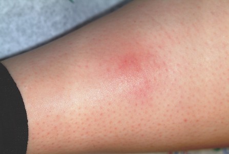
Erythema nodosum (EN; choice “c”) is the presumptive diagnosis in cases such as this one. The “erythemas” (multiforme, annulare centrifigum, etc) are all secondary reactions to antigenic triggers—such as herpes simplex virus, strep, or other organisms—and not primary conditions.
EN is a type of panniculitis that can be triggered by a number of conditions (eg, sarcoidosis), medicines (eg, penicillin), or organisms (eg, group A β streptococcus). It can also, as in this case, accompany inflammatory bowel disease, paralleling the flares and remissions of Crohn’s or ulcerative colitis and responding to treatment for those diseases. This is why part of the workup for EN includes evaluation for bowel disease.
The absence of breaks in the skin in such cases also speaks loudly for EN, since it involves only the adipose tissues and spares the skin. In this case, given the dearth of other suspicious items in the differential, a presumptive diagnosis of EN was quite reasonable.
Deep punch biopsy (choice “b”), which must include significant adipose tissue (5 mm or surgical wedge), can be crucial in questionable cases. It can show septal panniculitis (as opposed to lobular, which is more consistent with erythema induratum), as well as characteristic Miescher’s granulomas.
A chest radiograph (choice “d”), while not especially indicated in this case, is likewise an intelligent choice. Both tuberculosis and sarcoidosis can lead to radiographic changes and trigger EN.
If local infection were truly suspected as the direct cause of this patient’s lesions, an additional punch biopsy or two might serve to provide sufficient material for aerobic bacterial culture, acid-fast culture, and deep fungal culture—all of which are possible, though unlikely, explanations. Literally, the last thing we’d do is throw yet another antibiotic at this patient, who was in need of one simple thing: a correct diagnosis that would dictate proper treatment.
TREATMENT
A letter was dictated and sent to the gastroenterologist the patient was scheduled to see, to alert him as to our findings. If his workup
(H&P, stool cultures for salmonella, yersinia, and campylobacter, colonoscopy with biopsy) fails to pinpoint a particular trigger for her EN, additional labs will be needed. These would include antistreptolysin-O titer and tuberculosis and coccidioidomycosis skin testing, as well as actual biopsy of the lesions.
As of this writing, the patient’s GI appointment was pending, but the likely outcome will be the diagnosis of Crohn’s disease or ulcerative colitis. Treatment for either of these conditions will quiet down her EN. Often, however, EN is idiopathic and must be treated as a stand-alone diagnosis, with NSAIDs, antimalarials, or potassium iodide.