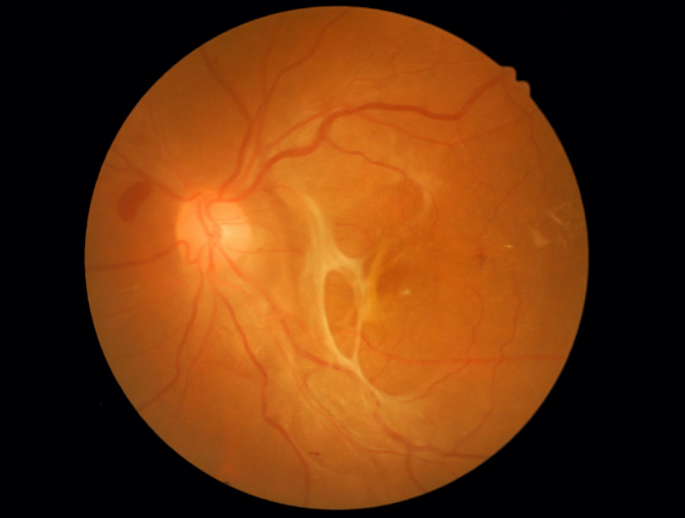
The retina is a thin layer of light-sensitive nerve tissue that lines the back of your eye. When light passes through the natural lens of the eye, it is focused on the retina. These specialized cells convert light into electrical impulses that are transmitted to the brain along your optic nerve. Your brain interprets these electrical signals and forms a visual image that you can identify.
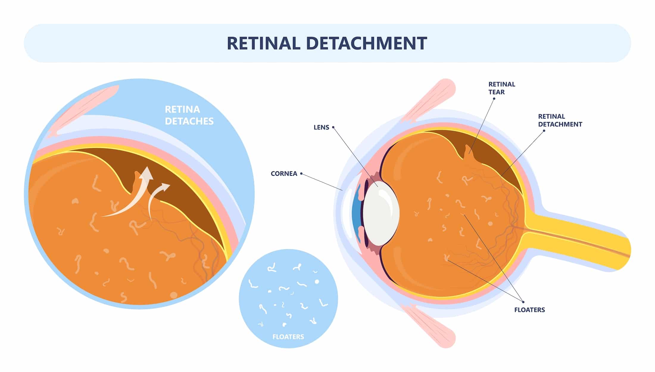
A retinal detachment is when the retina pulls away from its normal position at the back of the eye. When this occurs the blood vessels in the back of the eye are deprived of oxygen and nourishment. If a retinal detachment is left untreated, the retinal cells can die, and vision loss in the affected eye can become permanent.
Contact your eyecare specialist immediately if you are experiencing any of the following symptoms:
Diagnosing a retinal detachment requires a dilated exam with our specialists.
Treating a retinal detachment requires surgical intervention. These interventions include several techniques to repair the area that has become detached. Our specialists may opt to do laser pneumatic retinopexy, where a gas bubble will be placed in the eye to push the retina back in place. Surgical interventions may also include a vitrectomy to prevent further tugging on the retina. A vitrectomy is the removal of some or all of the vitreous from the middle of your eye; a gas bubble or silicone oil may be put in place of the vitreous while the retina heals.
Sources: American Academy of Ophthalmology, Medline Plus, National Eye Institute, and Krames