


Retinal Detachment
A retinal detachment means the retina has come off the back wall of the eye. A retinal detachment can be classified as one of three types: 1. Rhegmatogenous 2. Exudative 3. Tractional
Rhegmatogenous Retinal Detachment
The most common type of retinal detachment is a rhegmatogenous retinal detachment. When we are young, the vitreous gel inside the eye is firmly attached to the retina. As we age, the 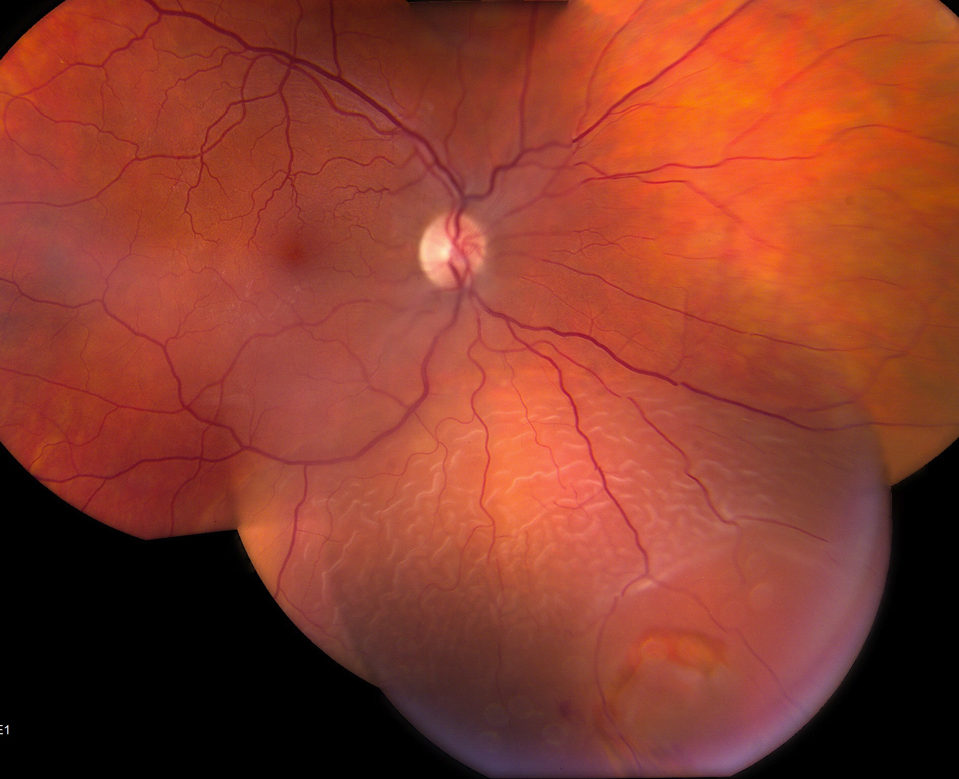 vitreous gel begins to liquefy and pull away from the retina. But sometimes the sticky vitreous gel can pull too hard and cause a tear in the retina. Once there is a retinal tear, the fluid in the middle of the eye can go through the tear and under the retina causing a retinal detachment. This type of retinal detachment usually occurs spontaneously and is related to aging, but sometimes trauma can cause it as well. The treatment for this type of retinal detachment varies depending on the clinical picture and may include laser, freezing therapy (cryotherapy), pneumatic retinopexy, scleral buckle, vitrectomy, or a combination of procedures.
Exudative Retinal Detachment
An exudative retinal detachment occurs when fluid builds up under the retina in the absence of any identifiable retinal tears. The most common causes of exudative retinal detachments are underlying inflammation or tumors. The treatment for this type of retinal detachment is to treat the underlying cause.
Traction Retinal Detachment
This type of retinal detachment occurs when scar tissue on the surface of the retina contracts and pulls the retina away from the eye wall. The most common cause of this type of retinal detachment is from severe proliferative diabetic retinopathy. Scar tissue growth after the repair of a retinal detachment can also cause a traction retinal detachment. The treatment of this type of retinal detachment depends on the clinical picture and may include laser, vitrectomy with membrane peeling, or scleral buckle with vitrectomy.