
CASE REPORT
A 23-year-old female presented complaining of sudden redness in her right eye, which began suddenly two days before. She describes mild discomfort, but no pain, or discharge. There was no headache, no photophobia, and no blurry vision.

There was no history of recent illness/ collagen vascular disease, general health was unremarkable, and there were no significant family histories. She did not recall any previous similar episodes.
Entering acuities were 6/6 OU, pupils equal, and reactions normal. Motilities were smooth and accurate, and no pain or tenderness upon eye movements. IOPs were 14mmHg.
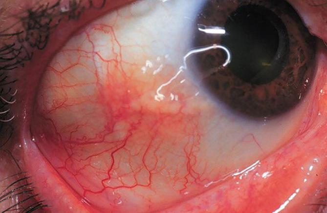
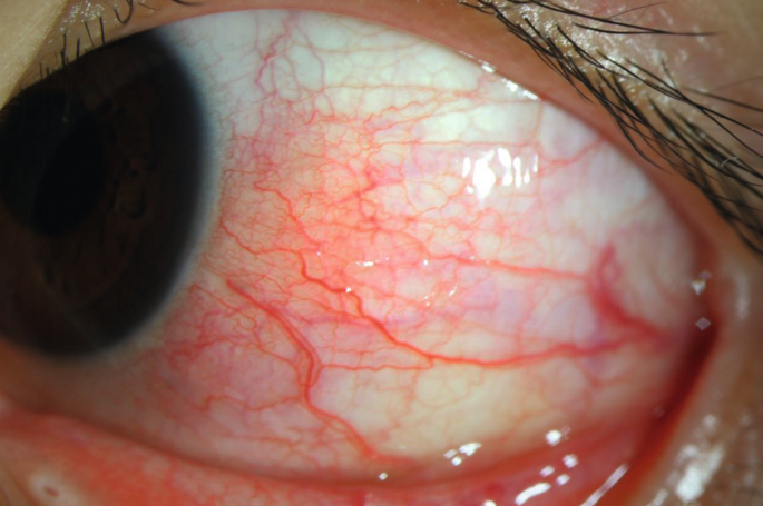
Slit-lamp examination revealed a quiet-looking left eye. Her right eye, however, showed a sectorial slightly elevated area of injection in her temporal/ bulbar conjunctiva. It looked bright red in color.
Closer inspection showed engorged episcleral vessels. Corneas were clear with no staining and no foreign body present. No anterior chamber reaction was seen. Lids/lashes were clear, with no sign of any blepharitis or trichiasis.
Lid eversion ruled out a foreign body under the lid. Van Herrick’s angles were wide open. Ophthalmoscopy (un-dilated) showed healthy retinas/maculae, cup-to-disc ratios of 0.3, and NRR healthy. A diagnosis of Simple Episcleritis was made.
Episcleritis entity
Episclera is a fibroelastic structure that lies between the superficial scleral stroma and Tenon’s capsules. Benign, self-limited inflammation of the episcleral tissues is known as episcleritis.
Episcleritis is a very common disease. Simple episcleritis is more common than nodular episcleritis. It usually affects young or middle-aged people and it is more common in women than in men.
Most cases are idiopathic but it can be associated with an underlying systemic inflammatory disease such as rheumatoid arthritis, systemic lupus erythematosus, Gout, Rosacea, inflammatory bowel disease, Crohn’s disease, collagen vascular disease, relapsing polychondritis.
It may be associated with some viral and bacterial diseases Herpes Zoster, Syphilis, and Tuberculosis.
Symptoms of Episcleritis
The presenting complaints are acute and gradual onset of redness, discomfort, photophobia, and foreign body sensation in one or both eyes.
Signs
Bright red or pink bulbar injection is present on slit lamp examination. Eyelid edema and conjunctival chemosis may also be present in some patients. There is no ocular discharge on examination.
Types of Episcleritis
There are two types of episcleritis:
- Simple episcleritis is an acute onset of redness either in a specific area (sectoral) or throughout the eye (diffuse). The acute inflammation peaks up in a few hours and then slowly resolves over in next 2-3 days.
- Nodular episcleritis is a slightly raised bump or nodule formation surrounded by dilated blood vessels and causes more discomfort. It is present usually in a specific area of the eye. In nodular episcleritis, there is a gradual onset of redness. After The onset, the inflammation slowly increases over the next few days and usually clears on its own but takes more time than simple episcleritis.
MANAGEMENT
Episcleritis is a self–limited condition and usually clears on its own without any treatment. But in case of recurrence or if the patient feels severe discomfort then the following treatment can be initiated.
· Medical treatment:
- NSAIDs typically ibuprofen is prescribed orally.
- Mild corticosteroids can be prescribed but the use of the steroids should be monitored because steroids may increase the chance of recurrence followed by a more intense attack. Patients may develop cataracts and glaucoma with the use of steroids.
- Treat the underlying systemic inflammatory disease causing it.
· Supportive Measures:
- Applying a cool compressor on your eyes. It helps in relieving inflammation quickly.
- Use artificial tears to relieve the discomfort.
- Wear sunglasses outside in case of photophobia.
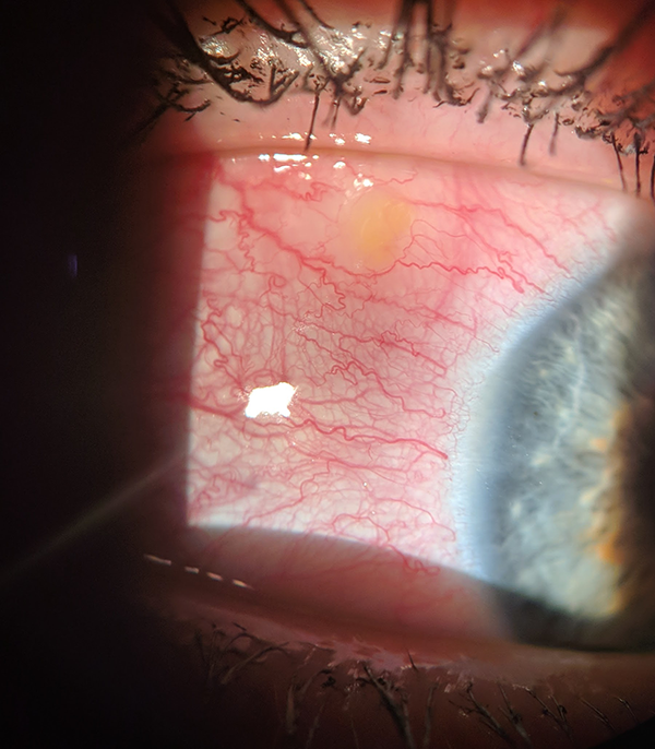
HOW TO TAKE SLIT-LAMP EXAM IMAGES WITH A SMARTPHONE?
Smartphone slit-lamp photography is the new advancement in the field of science and technology in which photographs of the desired slit-lamp finding can be taken with smartphones by using the slit-lamp adapters.
Slit-lamp Smartphone photography
REFERENCES
- Akpek EK, Uy HS, Christen W, Gurdal C, Foster CS. Severity of episcleritis and systemic disease association. Ophthalmology. 1999 Apr;106(4):729-31.
- Sainz de la Maza M, Molina N, Gonzalez-Gonzalez LA, Doctor PP, Tauber J, Foster CS. Clinical characteristics of a large cohort of patients with scleritis and episcleritis. Ophthalmology. 2012 Jan;119(1):43-50.
- Daniel Diaz J, Sobol EK, Gritz DC. Treatment and management of scleral disorders. Surv Ophthalmol. 2016 Nov-Dec;61(6):702-717.
- Watson PG, Hayreh SS. Scleritis and episcleritis. Br J Ophthalmol. 1976 Mar;60(3):163-91.
- Sainz de la Maza M, Jabbur NS, Foster CS. Severity of scleritis and episcleritis. Ophthalmology. 1994 Feb;101(2):389-96.
Slit-lamp Smartphone photography

RETINAL IMAGING BY YOUR SMARTPHONE