WO2000048550A2 - Device and method for regeneration and repair of cartilage lesions - Google Patents
Device and method for regeneration and repair of cartilage lesions Download PDFInfo
- Publication number
- WO2000048550A2 WO2000048550A2 PCT/US2000/003972 US0003972W WO0048550A2 WO 2000048550 A2 WO2000048550 A2 WO 2000048550A2 US 0003972 W US0003972 W US 0003972W WO 0048550 A2 WO0048550 A2 WO 0048550A2
- Authority
- WO
- WIPO (PCT)
- Prior art keywords
- proteins
- mixture
- bmp
- product
- cartilage
- Prior art date
Links
Classifications
-
- A—HUMAN NECESSITIES
- A61—MEDICAL OR VETERINARY SCIENCE; HYGIENE
- A61L—METHODS OR APPARATUS FOR STERILISING MATERIALS OR OBJECTS IN GENERAL; DISINFECTION, STERILISATION OR DEODORISATION OF AIR; CHEMICAL ASPECTS OF BANDAGES, DRESSINGS, ABSORBENT PADS OR SURGICAL ARTICLES; MATERIALS FOR BANDAGES, DRESSINGS, ABSORBENT PADS OR SURGICAL ARTICLES
- A61L27/00—Materials for grafts or prostheses or for coating grafts or prostheses
- A61L27/50—Materials characterised by their function or physical properties, e.g. injectable or lubricating compositions, shape-memory materials, surface modified materials
- A61L27/54—Biologically active materials, e.g. therapeutic substances
-
- A—HUMAN NECESSITIES
- A61—MEDICAL OR VETERINARY SCIENCE; HYGIENE
- A61F—FILTERS IMPLANTABLE INTO BLOOD VESSELS; PROSTHESES; DEVICES PROVIDING PATENCY TO, OR PREVENTING COLLAPSING OF, TUBULAR STRUCTURES OF THE BODY, e.g. STENTS; ORTHOPAEDIC, NURSING OR CONTRACEPTIVE DEVICES; FOMENTATION; TREATMENT OR PROTECTION OF EYES OR EARS; BANDAGES, DRESSINGS OR ABSORBENT PADS; FIRST-AID KITS
- A61F2/00—Filters implantable into blood vessels; Prostheses, i.e. artificial substitutes or replacements for parts of the body; Appliances for connecting them with the body; Devices providing patency to, or preventing collapsing of, tubular structures of the body, e.g. stents
- A61F2/02—Prostheses implantable into the body
- A61F2/30—Joints
- A61F2/30756—Cartilage endoprostheses
-
- A—HUMAN NECESSITIES
- A61—MEDICAL OR VETERINARY SCIENCE; HYGIENE
- A61L—METHODS OR APPARATUS FOR STERILISING MATERIALS OR OBJECTS IN GENERAL; DISINFECTION, STERILISATION OR DEODORISATION OF AIR; CHEMICAL ASPECTS OF BANDAGES, DRESSINGS, ABSORBENT PADS OR SURGICAL ARTICLES; MATERIALS FOR BANDAGES, DRESSINGS, ABSORBENT PADS OR SURGICAL ARTICLES
- A61L27/00—Materials for grafts or prostheses or for coating grafts or prostheses
- A61L27/14—Macromolecular materials
- A61L27/22—Polypeptides or derivatives thereof, e.g. degradation products
- A61L27/227—Other specific proteins or polypeptides not covered by A61L27/222, A61L27/225 or A61L27/24
-
- A—HUMAN NECESSITIES
- A61—MEDICAL OR VETERINARY SCIENCE; HYGIENE
- A61L—METHODS OR APPARATUS FOR STERILISING MATERIALS OR OBJECTS IN GENERAL; DISINFECTION, STERILISATION OR DEODORISATION OF AIR; CHEMICAL ASPECTS OF BANDAGES, DRESSINGS, ABSORBENT PADS OR SURGICAL ARTICLES; MATERIALS FOR BANDAGES, DRESSINGS, ABSORBENT PADS OR SURGICAL ARTICLES
- A61L27/00—Materials for grafts or prostheses or for coating grafts or prostheses
- A61L27/14—Macromolecular materials
- A61L27/22—Polypeptides or derivatives thereof, e.g. degradation products
- A61L27/24—Collagen
-
- A—HUMAN NECESSITIES
- A61—MEDICAL OR VETERINARY SCIENCE; HYGIENE
- A61L—METHODS OR APPARATUS FOR STERILISING MATERIALS OR OBJECTS IN GENERAL; DISINFECTION, STERILISATION OR DEODORISATION OF AIR; CHEMICAL ASPECTS OF BANDAGES, DRESSINGS, ABSORBENT PADS OR SURGICAL ARTICLES; MATERIALS FOR BANDAGES, DRESSINGS, ABSORBENT PADS OR SURGICAL ARTICLES
- A61L27/00—Materials for grafts or prostheses or for coating grafts or prostheses
- A61L27/14—Macromolecular materials
- A61L27/26—Mixtures of macromolecular compounds
-
- A—HUMAN NECESSITIES
- A61—MEDICAL OR VETERINARY SCIENCE; HYGIENE
- A61B—DIAGNOSIS; SURGERY; IDENTIFICATION
- A61B17/00—Surgical instruments, devices or methods, e.g. tourniquets
- A61B17/04—Surgical instruments, devices or methods, e.g. tourniquets for suturing wounds; Holders or packages for needles or suture materials
- A61B17/06—Needles ; Sutures; Needle-suture combinations; Holders or packages for needles or suture materials
- A61B17/06166—Sutures
-
- A—HUMAN NECESSITIES
- A61—MEDICAL OR VETERINARY SCIENCE; HYGIENE
- A61F—FILTERS IMPLANTABLE INTO BLOOD VESSELS; PROSTHESES; DEVICES PROVIDING PATENCY TO, OR PREVENTING COLLAPSING OF, TUBULAR STRUCTURES OF THE BODY, e.g. STENTS; ORTHOPAEDIC, NURSING OR CONTRACEPTIVE DEVICES; FOMENTATION; TREATMENT OR PROTECTION OF EYES OR EARS; BANDAGES, DRESSINGS OR ABSORBENT PADS; FIRST-AID KITS
- A61F2/00—Filters implantable into blood vessels; Prostheses, i.e. artificial substitutes or replacements for parts of the body; Appliances for connecting them with the body; Devices providing patency to, or preventing collapsing of, tubular structures of the body, e.g. stents
- A61F2/02—Prostheses implantable into the body
- A61F2/28—Bones
- A61F2002/2817—Bone stimulation by chemical reactions or by osteogenic or biological products for enhancing ossification, e.g. by bone morphogenetic or morphogenic proteins [BMP] or by transforming growth factors [TGF]
-
- A—HUMAN NECESSITIES
- A61—MEDICAL OR VETERINARY SCIENCE; HYGIENE
- A61F—FILTERS IMPLANTABLE INTO BLOOD VESSELS; PROSTHESES; DEVICES PROVIDING PATENCY TO, OR PREVENTING COLLAPSING OF, TUBULAR STRUCTURES OF THE BODY, e.g. STENTS; ORTHOPAEDIC, NURSING OR CONTRACEPTIVE DEVICES; FOMENTATION; TREATMENT OR PROTECTION OF EYES OR EARS; BANDAGES, DRESSINGS OR ABSORBENT PADS; FIRST-AID KITS
- A61F2/00—Filters implantable into blood vessels; Prostheses, i.e. artificial substitutes or replacements for parts of the body; Appliances for connecting them with the body; Devices providing patency to, or preventing collapsing of, tubular structures of the body, e.g. stents
- A61F2/02—Prostheses implantable into the body
- A61F2/30—Joints
- A61F2002/30001—Additional features of subject-matter classified in A61F2/28, A61F2/30 and subgroups thereof
- A61F2002/30003—Material related properties of the prosthesis or of a coating on the prosthesis
- A61F2002/3006—Properties of materials and coating materials
- A61F2002/30062—(bio)absorbable, biodegradable, bioerodable, (bio)resorbable, resorptive
-
- A—HUMAN NECESSITIES
- A61—MEDICAL OR VETERINARY SCIENCE; HYGIENE
- A61F—FILTERS IMPLANTABLE INTO BLOOD VESSELS; PROSTHESES; DEVICES PROVIDING PATENCY TO, OR PREVENTING COLLAPSING OF, TUBULAR STRUCTURES OF THE BODY, e.g. STENTS; ORTHOPAEDIC, NURSING OR CONTRACEPTIVE DEVICES; FOMENTATION; TREATMENT OR PROTECTION OF EYES OR EARS; BANDAGES, DRESSINGS OR ABSORBENT PADS; FIRST-AID KITS
- A61F2/00—Filters implantable into blood vessels; Prostheses, i.e. artificial substitutes or replacements for parts of the body; Appliances for connecting them with the body; Devices providing patency to, or preventing collapsing of, tubular structures of the body, e.g. stents
- A61F2/02—Prostheses implantable into the body
- A61F2/30—Joints
- A61F2002/30001—Additional features of subject-matter classified in A61F2/28, A61F2/30 and subgroups thereof
- A61F2002/30667—Features concerning an interaction with the environment or a particular use of the prosthesis
- A61F2002/30677—Means for introducing or releasing pharmaceutical products, e.g. antibiotics, into the body
-
- A—HUMAN NECESSITIES
- A61—MEDICAL OR VETERINARY SCIENCE; HYGIENE
- A61F—FILTERS IMPLANTABLE INTO BLOOD VESSELS; PROSTHESES; DEVICES PROVIDING PATENCY TO, OR PREVENTING COLLAPSING OF, TUBULAR STRUCTURES OF THE BODY, e.g. STENTS; ORTHOPAEDIC, NURSING OR CONTRACEPTIVE DEVICES; FOMENTATION; TREATMENT OR PROTECTION OF EYES OR EARS; BANDAGES, DRESSINGS OR ABSORBENT PADS; FIRST-AID KITS
- A61F2210/00—Particular material properties of prostheses classified in groups A61F2/00 - A61F2/26 or A61F2/82 or A61F9/00 or A61F11/00 or subgroups thereof
- A61F2210/0004—Particular material properties of prostheses classified in groups A61F2/00 - A61F2/26 or A61F2/82 or A61F9/00 or A61F11/00 or subgroups thereof bioabsorbable
-
- A—HUMAN NECESSITIES
- A61—MEDICAL OR VETERINARY SCIENCE; HYGIENE
- A61F—FILTERS IMPLANTABLE INTO BLOOD VESSELS; PROSTHESES; DEVICES PROVIDING PATENCY TO, OR PREVENTING COLLAPSING OF, TUBULAR STRUCTURES OF THE BODY, e.g. STENTS; ORTHOPAEDIC, NURSING OR CONTRACEPTIVE DEVICES; FOMENTATION; TREATMENT OR PROTECTION OF EYES OR EARS; BANDAGES, DRESSINGS OR ABSORBENT PADS; FIRST-AID KITS
- A61F2310/00—Prostheses classified in A61F2/28 or A61F2/30 - A61F2/44 being constructed from or coated with a particular material
- A61F2310/00005—The prosthesis being constructed from a particular material
- A61F2310/00365—Proteins; Polypeptides; Degradation products thereof
-
- A—HUMAN NECESSITIES
- A61—MEDICAL OR VETERINARY SCIENCE; HYGIENE
- A61L—METHODS OR APPARATUS FOR STERILISING MATERIALS OR OBJECTS IN GENERAL; DISINFECTION, STERILISATION OR DEODORISATION OF AIR; CHEMICAL ASPECTS OF BANDAGES, DRESSINGS, ABSORBENT PADS OR SURGICAL ARTICLES; MATERIALS FOR BANDAGES, DRESSINGS, ABSORBENT PADS OR SURGICAL ARTICLES
- A61L2300/00—Biologically active materials used in bandages, wound dressings, absorbent pads or medical devices
- A61L2300/20—Biologically active materials used in bandages, wound dressings, absorbent pads or medical devices containing or releasing organic materials
- A61L2300/252—Polypeptides, proteins, e.g. glycoproteins, lipoproteins, cytokines
-
- A—HUMAN NECESSITIES
- A61—MEDICAL OR VETERINARY SCIENCE; HYGIENE
- A61L—METHODS OR APPARATUS FOR STERILISING MATERIALS OR OBJECTS IN GENERAL; DISINFECTION, STERILISATION OR DEODORISATION OF AIR; CHEMICAL ASPECTS OF BANDAGES, DRESSINGS, ABSORBENT PADS OR SURGICAL ARTICLES; MATERIALS FOR BANDAGES, DRESSINGS, ABSORBENT PADS OR SURGICAL ARTICLES
- A61L2300/00—Biologically active materials used in bandages, wound dressings, absorbent pads or medical devices
- A61L2300/40—Biologically active materials used in bandages, wound dressings, absorbent pads or medical devices characterised by a specific therapeutic activity or mode of action
- A61L2300/412—Tissue-regenerating or healing or proliferative agents
- A61L2300/414—Growth factors
-
- A—HUMAN NECESSITIES
- A61—MEDICAL OR VETERINARY SCIENCE; HYGIENE
- A61L—METHODS OR APPARATUS FOR STERILISING MATERIALS OR OBJECTS IN GENERAL; DISINFECTION, STERILISATION OR DEODORISATION OF AIR; CHEMICAL ASPECTS OF BANDAGES, DRESSINGS, ABSORBENT PADS OR SURGICAL ARTICLES; MATERIALS FOR BANDAGES, DRESSINGS, ABSORBENT PADS OR SURGICAL ARTICLES
- A61L2300/00—Biologically active materials used in bandages, wound dressings, absorbent pads or medical devices
- A61L2300/40—Biologically active materials used in bandages, wound dressings, absorbent pads or medical devices characterised by a specific therapeutic activity or mode of action
- A61L2300/45—Mixtures of two or more drugs, e.g. synergistic mixtures
-
- A—HUMAN NECESSITIES
- A61—MEDICAL OR VETERINARY SCIENCE; HYGIENE
- A61L—METHODS OR APPARATUS FOR STERILISING MATERIALS OR OBJECTS IN GENERAL; DISINFECTION, STERILISATION OR DEODORISATION OF AIR; CHEMICAL ASPECTS OF BANDAGES, DRESSINGS, ABSORBENT PADS OR SURGICAL ARTICLES; MATERIALS FOR BANDAGES, DRESSINGS, ABSORBENT PADS OR SURGICAL ARTICLES
- A61L2430/00—Materials or treatment for tissue regeneration
- A61L2430/06—Materials or treatment for tissue regeneration for cartilage reconstruction, e.g. meniscus
Definitions
- the present invention relates to a cartilage regeneration and repair product that induces cell ingrowth into a bioresorbable material and cell differentiation into cartilage tissue, and to methods of using such a product to repair cartilage lesions.
- Articular cartilage an avascular tissue found at the ends of articulating bones, has limited natural capacity to heal.
- mesenchymal stem cells condense to form areas of high density and proceed through a series of developmental stages that ends in the mature chondrocyte.
- the final hyaline cartilage tissue contains only chondrocytes that are surrounded by a matrix composed of type II collagen, sulfated proteoglycans, and additional proteins.
- the matrix is heterogenous in structure and consists of three morphologically distinct zones: superficial, intermediate, and deep.
- Zones differ among collagen and proteoglycan distribution, calcification, orientation of collagen fibrils, and the positioning and alignment of chondrocytes (Archer etal., 1996, J. Anat. 189(l):23-35; Morrison etal., 1996, J. Anat. 189(1): 9-22; and Mow et al. , 1992, Biomaterials 13 (2) : 67-97) . These properties provide the unique mechanical and physical parameters to hyaline cartilage tissue.
- the meniscus a C-shaped cartilaginous tissue, performs several functions in the knee including load transmission from the femur to the tibia, stabilization in the anterior- posterior position during flexion, and joint lubrication. Damage to the meniscus results in reduced knee stability and knee locking.
- meniscectomies were performed which permitted immediate pain relief, but were subsequently found to induce the early onset of osteoarthritis (Fairbank, J. Bone Joint Surg. 30B: 664-670; Allen et al., 1984, J. Bone Joint Surg. 66B: 666-671; and Roos et al., 1998, Arth. Rheum. 41:687- 693). More recently, partial meniscectomies and repair of meniscal tears have been performed (Fig. 9A-D; Jackson, D., ed., 1995, Reconstructive Knee Surgery Master
- the proximal, concave surface of the meniscus contacts the femoral condyle and the distal, flat surface contacts the tibial plateaus.
- the outer one-third of the meniscus is highly vascularized and contains dense, enervated, connective tissue.
- the remaining memscus is semivascular or avascular, aneural tissue consisting of fibrochondrocytes surrounded by abundant extracellular matrix (McDevitt et al., Clin. Orthop. Rel. Res. 252:8-17).
- Fibrochondrocytes are distinctive in both appearance and function compared to undifferentiated fibroblasts. Fibroblasts are elongated cells containing many cellular processes and produce predominantly type I collagen.
- BMP Bone Morphogenetic Proteins
- TGF- ⁇ family members direct a cascade of events that includes chemotaxis, differentiation of pluripotential cells to the cartilage lineage, maturation of chondrocytes to the hypertrophic stage, mineralization of cartilage, replacement of cartilage with bone cells, and the formation of a calcified matrix (Reddi, 1997, Cytokine & Growth Factor Reviews 8.11-20). Although individual, recombinant BMPs can induce these events, the prevalence of multiple TGF- ⁇ family members in bone tissue underlies the complexity involved in natural osteogenesis.
- Bone Protein (Sulzer Orthopedics Biologies, Wheatridge, CO), also referred to herein as BP, is a naturally derived mixture of proteins isolated from demineralized bovine bones that has osteogenic activity in vitro and in vivo.
- BP induces endochondral bone formation or bone formation through a cartilage intermediate (Damien, C. et al., 1990, J. Biomed. Mater. Res. 24:639-654).
- BP in combination with calcium carbonate promotes bone formation in the body (Poser and Benedict, PCT Publication No. W095/13767).
- BP has been shown to promote differentiation to cartilage of murine embryonic mesenchymal stem cells (Atkinson et al., 1996, In
- Hunziker U.S. Patent Nos. 5,368,858 and 5,206,023 describes a cartilage repair composition consisting of a biodegradable matrix, a proliferation and/or chemotactic agent, and a transforming factor.
- a two-stage approach is used where each component has a specific function over time.
- a specific concentration of proliferation/chemotactic agent fills the defect with repair cells.
- a larger transforming factor concentration preferably provided in conjunction with a delivery system, transforms repair cells to chondrocytes.
- the second stage delivery of a high concentration of transforming factor in a delivery system i.e., liposomes
- Valee and King (U.S. Patent No.4,952,404) describe healing of injured, avascular meniscus tissue by release of the angiogenic factor, angiogenin, over at least 3 weeks.
- Amoczky et al. described a method using an autogenous fibrin clot to repair an avascular, circular lesion in canine menisci (Amoczky et al., 1988, J. Bone Joint
- Hashimoto et al. described a method using fibrin sealant with or without endothelial cell growth factor in avascular, circular meniscal defects in the canine model (Hashimoto et al., 1992, Am. J. Sports Med. 20:537-541).
- the growth factor added a modest benefit compared to healing with fibrin sealant alone and this additional effect was not observed until three months after treatment, indicating an indirect contribution of the growth factor.
- the defect was filled with hyaline cartilage-like cells, which are not typically present in normal meniscus tissue.
- BMP bone morphogenetic proteins
- TGF ⁇ transforming growth factor ⁇
- Urist described a substantially pure, but not recombinant, BMP combined with a biodegradable poly(lactic acid) polymer delivery system for bone repair (U.S. Patent No. 4,563,489). This system blends together equal quantities of BMP and poly(lactic acid) (PLA) powder (100 ⁇ g of each) and decreases the amount of BMP required to promote bone repair.
- PHA poly(lactic acid)
- WO 96/39170 disclose a two factor composition for inducing cartilaginous tissue formation using a cartilage formation-inducing protein and a cartilage maintenance-inducing protein.
- Specific recombinant cartilage inducing proteins are specified as BMP-13, MP-52 and BMP-12, and specific cartilage maintenance-inducing proteins are specified as BMP-9.
- BMP-9 is encapsulated in a resorbable polymer system and delivered to coincide with the presence of cartilage formation inducing protein(s).
- a chondrogenesis-inducing device consisting of a polyanhydride and polyorthoester, that delivers water soluble proteins derived from demineralized bone matrix, TGF ⁇ , epidermal growth factor (EGF), fibroblast growth factor (FGF) or platelet-derived growth factor (PDGF).
- EGF epidermal growth factor
- FGF fibroblast growth factor
- PDGF platelet-derived growth factor
- Bentz et al. (PCT Publication No. WO 92/09697) have described the use of a bone morphogenetic protein (BMP) with a TGF ⁇ protein for bone repair.
- BMP bone morphogenetic protein
- the ratio of BMP to TGF ⁇ in the mixture is in the range of 10: 1 to 1 : 10.
- the addition of TGF- ⁇ with either BMP-2 or BMP-3 results in increased osteoinductive activity and an increased ratio of cartilage to bone when compared to either factor alone (Bentz et al., Matrix 11:269-275 (1991);Ogawaetal.,J.5/o/. C ⁇ e w., 267(20):14233-7(1992);WO92/09697).
- this composition produced substantial bone in the rodent subcutaneous assay.
- Meniscal Augmentation Device that consists of biocompatible and bioresorbable fibers that acts as a scaffold for the ingrowth of meniscal fibrochondrocytes, supports normal meniscal loads, and has an outer surface that approximates the natural meniscus contour. After partial resection of the meniscus to the vascular zone, this device is implanted into the resulting segmental defect.
- This results have been described in both canines and humans (Stone et al., 1992, Am. J. Sports Med. 20:104-111; and Stone et al., 1997, J. Bone Joint Surg. 79:17701777).
- the Meniscus Augmentation Device, the research reports and patent disclosures described above, and current repair surgeries provide encouraging results in the area of cartilage repair, but are not satisfactory to induce repair of "non-repairable" avascular tears in which the repair tissue is meniscus tissue, and are not satisfactory to produce short patient rehabilitation times and regenerated meniscus tissue in the vascular zone. Furthermore, no reports have been described that demonstrate enhanced healing rates of
- the present invention relates to a product and method for repairing and/or regenerating cartilage lesions.
- the product and method of the present invention are useful for repairing a variety of cartilage lesions, including articular and mensical lesions, including vascular, semivascular and avascular lesions.
- the product and method of the present invention can be used to repair different sizes and shapes of cartilage lesions, including radial tears, bucket handle tears, and segmental defects.
- a first embodiment of the product of the present invention relates to a product for repair of cartilage lesions.
- Such a product includes: (a) a cartilage repair matrix; and, (b) a cartilage-inducing composition associated with the matrix for provision of a mixture of proteins.
- a cartilage-inducing composition includes a mixture of proteins which includes: transforming growth factor ⁇ 1 (TGF ⁇ l), bone morphogenetic protein (BMP)-2, BMP-3, and BMP-7.
- TGF ⁇ l transforming growth factor ⁇ 1
- BMP bone morphogenetic protein
- BMP-7 bone morphogenetic protein
- a cartilage-inducing composition in second embodiment of the product of the present invention, includes a mixture of proteins which includes (a) a bone-derived osteogenic or chondrogenic formulation; and, (b) an exogenous TGF ⁇ protein.
- the exogenous TGF ⁇ protein is present in an amount sufficient to increase cartilage induction by the composition over a level of cartilage induction by the bone-derived osteogenic or chondrogenic protein formulation in the absence of the exogenous TGF ⁇ protein.
- the exogenous TGF ⁇ protein is TGF ⁇ l.
- the ratio of TGF ⁇ l to all other proteins in the mixture of proteins is at least about 1:10, at least about 1:3, at least about 1:1, or at least about 10:1.
- a cartilage-inducing composition includes a mixture of proteins comprising: (a) a TGF ⁇ protein; and, (b) at least one bone morphogenetic protein (BMP), wherein the ratio of the TGF ⁇ protein to the BMP protein is greater than about 10: 1.
- the TGF ⁇ protein can be any TGF ⁇ protein, including TGF ⁇ l, TGF ⁇ 2, TGF ⁇ 3, TGF ⁇ 4, TGF ⁇ 5, or mixtures thereof.
- the TGF ⁇ protein is TGF ⁇ 1 or TGF ⁇ 2, with TGF ⁇ 1 being most preferred.
- the BMP protein can be any BMP protein, including, but not limited to, BMP-2, BMP-3, BMP-4, BMP-5, BMP-6, BMP-7, BMP-8, BMP-9, CDMP, and mixtures thereof.
- the mixture of proteins includes TGF ⁇ superfamily proteins: TGF ⁇ l, bone morphogenetic protein (BMP)-2, BMP-3, and BMP-7, wherein the TGF ⁇ superfamily proteins comprise from about 0.5% to about 99.99% of the mixture of proteins.
- the TGF ⁇ superfamily proteins comprise from about 0.5% to about 25% of the mixture of proteins; in another aspect, the TGF ⁇ superfamily proteins comprise from about 1% to about 10% of the mixture of proteins.
- the quantity of the TGF ⁇ 1 in the mixture is from about 0.01 % to about 75% of total proteins in the mixture; in another aspect, the quantity of the TGF ⁇ 1 in the mixture is from about 0.01% to about 50% of total proteins in the mixture; in another aspect, the quantity of the TGF ⁇ l in the mixture is from about 0.01% to about 25% of total proteins in the mixture; in another aspect, the quantity of the TGF ⁇ l in the mixture is from about 0.01% to about 10% of total proteins in the mixture; in another aspect, the quantity of the TGF ⁇ 1 in the mixture is from about 0.1% to about 1% of total proteins in the mixture; in another aspect, the quantity of the TGF ⁇ 1 in the mixture is from about 33% to about 99.99% of total proteins in the mixture; in another aspect, the quantity of the TGF ⁇ l in the mixture is from about 50% to about 99.99% of total proteins in the mixture. In one aspect of each of the above-referenced embodiments, the quantity of the quantity of the quantity of the quantity of the quantity of the mixture is from about 0.01%
- BMP-2 in the mixture is from about 0.1% to about 5% of total proteins in the mixture.
- the quantity of the BMP-3 in the mixture is from about 0.1% to about 5% of total proteins in the mixture.
- the quantity of the BMP-7 in the mixture is from about 0.1% to about 5% of total proteins in the mixture.
- the quantity of BMP-3 in the mixture is from about 0.1% to about 10% of total proteins in the mixture.
- the mixture of proteins further comprises a protein selected from the group consisting of TGF ⁇ 2, TGF ⁇ 3, BMP-4, BMP-5, BMP-6, BMP-8, BMP-9, and cartilage-derived morphogenetic protein (CDMP).
- a protein selected from the group consisting of TGF ⁇ 2, TGF ⁇ 3, BMP-4, BMP-5, BMP-6, BMP-8, BMP-9, and cartilage-derived morphogenetic protein (CDMP).
- the TGF ⁇ 2 comprises from about 0.5% to about 12% of the mixture of proteins; in another aspect, the TGF ⁇ 3 comprises from about 0.01% to about 15% of the mixture of proteins; in another aspect, the BMP-4 comprises from about 0.01% to about 1% of the mixture of proteins; in another aspect, the BMP-5 comprises from about 0.01% to about 1% of the mixture of proteins; in another aspect, the BMP-6 comprises from about 0.01% to about 1% of the mixture of proteins; in another aspect, the CDMP comprises from about 0.01% to about 1% of the mixture of proteins.
- the mixture of proteins further comprises at least one bone matrix protein.
- the bone matrix protein can include, but is not limited to, osteocalcin, osteonectin, bone sialoprotein (BSP), lysyloxidase, cathepsinL pre, osteopontin, matrix GLA protein (MGP), biglycan, decorin, proteoglycan-chondroitin sulfate III (PG-CS IQ), bone acidic glycoprotein (BAG-75), thrombospondin (TSP) and fibronectin.
- the bone matrix protein comprises from about 20% to about 98% of the mixture of proteins.
- the bone matrix proteins comprise: osteocalcin, osteonectin, bone sialoprotein (BSP), lysyloxidase, and cathepsin L pre.
- the bone matrix protein comprises from about 40% to about 98% of the mixture of proteins.
- the mixture of proteins further comprises at least one growth factor protein.
- the growth factor protein can include, but is not limited to, fibroblast growth factor-I (FGF-I), FGF-II, FGF-9, leukocyte inhibitory factor (LIF), insulin, insulin growth factor I (IGF-I), IGF-II, platelet- derived growth factor AA (PDGF-AA), PDGF-BB, PDGF-AB, stromal derived factor-2 (SDF-2), pituitary thyroid hormone (PTH), growth hormone, hepatocyte growth factor (HGF), epithelial growth factor (EGF), transforming growth factor- ⁇ (TGF ⁇ ) and hedgehog proteins.
- FGF-I fibroblast growth factor-I
- FGF-9 leukocyte inhibitory factor
- IGF-I insulin growth factor I
- IGF-II insulin growth factor I
- IGF-II platelet- derived growth factor AA
- PDGF-BB platelet- derived growth factor AA
- PDGF-AB stromal
- the growth factor protein comprises from about 0.01% to about 50% of the mixture of proteins. In one aspect, the growth factor protein comprises from about 0.05% to about 25% of the mixture of proteins; in another aspect, the growth factor protein comprises from about 0.1% to about 10% of the mixture of proteins.
- the growth factor protein is fibroblast growth factor-I (FGF-I). In this aspect, the FGF-I comprises from about 0.001% to about 10% of the mixture of proteins.
- the composition further comprises one or more serum proteins.
- the serum proteins can include, but are not limited to, albumin, transferrin, ⁇ 2-Hs GlycoP, IgG, ⁇ l- antitrypsin, ⁇ 2-microglobulin, Apo Al lipoprotein (LP) and Factor XUIb.
- the serum proteins are selected from the group consisting of albumin, transferrin, Apo Al
- the mixture of proteins comprises TGF ⁇ l, TGF ⁇ 2, TGF ⁇ 3, BMP-2, BMP-3, BMP-4, BMP-5, BMP-6, BMP-7, CDMP, FGF-I, osteocalcin, osteonectin, BSP, lysyloxidase, cathepsin L pre, albumin, transferrin, Apo Al LP and Factor XUIb.
- the mixture of proteins comprises Bone Protein (BP).
- the cartilage inducing composition has an identifying characteristic selected from the group consisting of an ability to induce cellular infiltration, an ability to induce cellular proliferation, an ability to induce angiogenesis, and an ability to induce cellular differentiation to type II collagen-producing chondrocytes.
- the cartilage-inducing composition is at a concentration of from about 0.5% to about 33% by weight of the product. In another aspect, the cartilage-inducing composition is at a concentration of from about 1% to about 20% by weight of the product.
- the mixture of proteins when used at a concentration of at least about 10 ⁇ g per 6.5-7.3 mg of bovine tendon collagen in a rat subcutaneous assay, induces a bone score of from about 1.0 to about 3.5, using a bone grading scale set forth in Table 8, and induces a cartilage score of at least about 1.2, using a cartilage grading scale set forth in Table 9.
- the composition when used at a concentration of at least about 10 ⁇ g per 6.5-7.3 mg of bovine tendon collagen in a rat subcutaneous assay, induces a bone score of less than about 2.0, using a bone grading scale set forth in Table 8, and induces a cartilage score of at least about 2.0, using a cartilage grading scale set forth in Table 9.
- the composition when used at a concentration of at least about 10 ⁇ g per 6.5-7.3 mg of bovine tendon collagen in a rat subcutaneous assay, induces a bone score of less than about 2.0, using a bone grading scale set forth in Table 8, and induces a cartilage score of at least about 2.5, using a cartilage grading scale set forth in Table 9.
- the composition when used at a concentration of at least about 10 ⁇ g per 6.5-7.3 mg of bovine tendon collagen in a rat subcutaneous assay, induces a bone score of less than about 2.0, using a bone grading scale set forth in Table 8, and induces a cartilage score of at least about 3.0, using a cartilage grading scale set forth in Table 9.
- the ratio of TGF ⁇ 1 to all other proteins in the mixture of proteins is at least about 1:10; in another aspect, the ratio of TGF ⁇ l to all other proteins in the mixture of proteins is at least about 1:3; in another aspect, the ratio of TGF ⁇ l to all other proteins in the mixture of proteins is at least about 1 : 1; in another aspect, the ratio of TGF ⁇ l to all other proteins in the mixture of proteins is at least about 10:1.
- the TGF ⁇ protein can be a recombinant TGF ⁇ protein, or can be purified from a bone-derived protein mixture.
- the ratio of the TGF ⁇ protein to the BMP protein is greater than about 100:1; in another aspect, the ratio of the TGF ⁇ protein to the BMP protein is greater than about 1000: 1; in another aspect, the ratio of the TGF ⁇ protein to the BMP protein is greater than about 10,000: 1.
- the TGF ⁇ protein is TGF ⁇ l.
- the BMP protein is selected from the group consisting of BMP-2, BMP-3, BMP-4, BMP-5, BMP-6, BMP-7, BMP-8, BMP-9 and CDMP.
- the product of the present invention can also be formulated to include: (a) a cartilage repair matrix; and (b) a cartilage-inducing composition associated with the matrix, which includes cells that have been cultured with the above-described mixture of chondrogenesis-enhancing proteins.
- the cartilage repair matrix of a shape and size that conforms to the cartilage defect such that the defect is repaired.
- the matrix can be configured as a sheet, which is most suitable for repairing cartilage tears, or the matrix can be configured to repair a segmental defect, which can include a tapered shape.
- the cartilage repair matrix can be formed of any suitable material, including synthetic polymeric material and ground substances.
- the matrix is bioresorbable.
- the matrix is porous.
- the cartilage-inducing composition can be associated with the matrix by any suitable method, including, but not limited to freeze-drying the composition onto a surface of said matrix and suspension within said cartilage repair matrix of a delivery formulation containing said composition. Additionally, the composition can be associated with the matrix ex vivo or in vivo.
- Another embodiment of the present invention relates to a method for repair of cartilage lesions, which includes the steps of implanting and fixing into a cartilage lesion a cartilage repair product of the present invention, as described above, including a cartilage repair product including an of the above-referenced embodiments of a cartilage- inducing composition.
- the method of the present invention can be used to enhance the rate and/or quality of repair of vascular cartilage tears and segmental defects, and can provide the ability to repair semivascular and avascular tears and segmental defects that, prior to the present invention, were typically considered to be irreparable.
- the product can additionally include a time controlled delivery formulation.
- the method of the present invention includes the use of two cartilage repair products to repair a segmental defect.
- the first product includes a cartilage repair matrix, which is configured as a sheet, is associated with the chondrogenesis-inducing composition as described above.
- the second product includes a cartilage repair matrix configured to replace cartilage removed from the segmental defect, which may or may not be associated with the chondrogenesis-inducing composition of the present invention.
- Fig. 1 A shows a meniscal radial tear.
- Fig. IB shows a conventional suture repair and resection of the meniscal radial tear illustrated in Fig. 1A.
- Fig. 1C shows a meniscal triple bucket handle tear.
- Fig. ID shows a conventional suture repair and resection of the meniscal triple bucket handle tear illustrated in Fig. lC.
- Fig. 2A illustrates implantation of a cartilage repair product of the present invention to a meniscal segmental lesion.
- Fig. 2B illustrates fixation of a cartilage repair product of the present invention to a meniscal segmental lesion.
- Fig. 3 A is a diagram illustrating a meniscus cross section having vascular, semivascular and avascular zones.
- Fig. 3B illustrates one approximate shape of a cartilage repair product of the present invention.
- Fig. 4 A is an illustration of a meniscus having a longitudinal tear in the avascular region as viewed from the femur towards the tibia.
- Fig. 4B is a diagram illustrating a cross section of the meniscus depicted in Fig.4A containing a cartilage repair product of the present invention.
- Fig. 5 is a line graph showing quantitation of Alcian Blue staining of ATDC5 micromass cultures.
- Fig. 6 is a bar graph showing quantitation of Alcian Blue staining of ATDC5 micromass cultures in Nutridoma-containing media at 7 and 14 days.
- Fig. 7 is a bar graph showing quantitation of Alcian Blue staining of ATDC5 micromass cultures containing HPLC fractions of proteins isolated from demineralized bovine bones.
- Fig. 8 is a diagram illustrating a cross-section view of a combination collagen meniscus implant (CMI) and sheet cartilage repair product of the present invention used to repair a meniscal defect.
- CMI collagen meniscus implant
- the present application generally relates to a product for repairing and/or regenerating cartilage lesions, and methods of repairing or regenerating cartilage lesions using such a product.
- the product and methods of the present invention are particularly useful for repairing defects (i.e., lesions) in articular cartilage (e.g., hyaline cartilage) and meniscal cartilage (e.g., fibrocartilage).
- defects i.e., lesions
- articular cartilage e.g., hyaline cartilage
- meniscal cartilage e.g., fibrocartilage
- the product and method of the present invention are effective for repairing both vascular and avascular meniscal cartilage lesions.
- the product and method of the present invention increase the rate of meniscus repair and induce more normal (i.e., endogenous- type) meniscal tissue than is commonly observed during the conventional repair practiced currently.
- the cartilage repair product of the present invention can also induce meniscus repair of avascular, "irreparable” tears and, furthermore, fill the defect with meniscus-like tissue.
- the product and method are useful for repairing and regenerating meniscal tissue which has been removed by partial or complete meniscectomy.
- the product and method of the present invention can enhance blood vessel formation, produce fibrochondrocytes, induce cellular infiltration into the product, induce cellular proliferation, and produce cellular and spatial organization to form a three-dimensional memscus tissue.
- the system used to provide one or two recombinant factors may not have been able to mimic the gradient complexity of the natural system to a satisfactory degree or to maintain a factor concentration for a time that is sufficient to allow a full and permanent differentiation of precursor cells to chondrocytes.
- the present inventors believe that the repair of certain defects, particularly large defects, requires the maintenance of a sufficient concentration of a particular complex mixture of repair factors at the site for a time sufficient to induce the proper formation of cartilage.
- Bone Protein (BP) and mixtures derived from BP are chondrogenic in avascular environments (e.g. articular cartilage and in vitro), with limited osteogenic activity.
- BP is chondrogenic in other, vascularized environments (lumbar spine, subcutaneous, etc.), it has significant osteogenic activity as well.
- Certain cartilage regeneration applications, such as the meniscus require cartilaginous repair in a vascular environment. Because the meniscus is vascularized, it is therefore expected that BP and other bone-inducing molecules will induce bone formation during repair despite also inducing cartilage formation, which is not desirable for clinical applications.
- the present invention describes the identification of a novel mixture of factors derived from bovine bone to induce cartilage without bone formation. The cartilage induction activity was observed in a permissive bone-forming environment: the vascularized, rodent subcutaneous model.
- this new mixture combines unexpectedly high concentrations of TGF ⁇ proteins with the cartilage-inducing compositions disclosed herein, including mixtures that also have osteoinductive properties, to produce this novel chondrogenic activity with significantly reduced, or eliminated, osteogenic activity.
- the combination of high concentration TGF ⁇ protein plus osteogenic and/or chondrogenic protein(s) to promote only cartilage formation in the absence of bone formation in a vascularized (permissive bone forming) environment has not been described.
- a product for repair of cartilage lesions includes: (a) a cartilage repair matrix; and, (b) a cartilage-inducing composition associated with the matrix for provision of a mixture of proteins, which can be referred to herein as chondrogenesis-enhancing proteins (described in detail below).
- chondrogenesis-enhancing proteins a cartilage-inducing composition associated with the matrix for provision of a mixture of proteins, which can be referred to herein as chondrogenesis-enhancing proteins (described in detail below).
- the phrase "cartilage- inducing composition” refers to a formulation which contains a mixture of different chondrogenesis-enhancing proteins and which enhances (i.e., augments, amplifies, improves, increases, or supplements) cartilage growth in vivo.
- the cartilage-inducing composition useful in the product of the present invention has an identifying characteristic which includes: an ability to induce cellular infiltration, an ability to induce cellular proliferation, an ability to induce angiogenesis, and/or an ability to induce cellular differentiation to type II collagen-producing chondrocytes, in vivo or under appropriate in vitro conditions.
- the cartilage-inducing composition of the present invention provides a mixture of proteins which includes proteins that have osteogenic- and/or chondrogenic-enhancing activities, particularly when combined into mixtures as described in detail herein.
- the term "enhancing", particularly with regard to enhancing chondrogenesis refers to any measure of augmenting, amplifying, improving, increasing, or supplementing a biological activity associated with chondrogenesis such that cartilage forms in a manner that more closely mimics the natural ontogeny of cartilage formation, as compared to cartilage formation that would occur in the absence of the product, or in the absence of the composition portion of the product.
- the term enhancing also means that endochondral maturation to mineralized cartilage and bone may be prevented or delayed.
- a mixture of chondrogenesis- enhancing proteins that are included in a chondrogenesis-inducing composition can be characterized as being capable, when cultured together with ATDC5 cells for seven days at a concentration of about 100 ng/ml or less, of inducing a statistically significant increase in A 59J in an Alcian Blue assay performed with the ATDC5 cells.
- the specific conditions associated with such an ATDC5/Alcian Blue assay are described in detail below. It is noted that although the mixture of chondrogenesis-enhancing proteins has the above- described characteristic, an individual chondrogenesis-enhancing protein, when isolated from the other proteins in the mixture, is not necessarily chondrogenic.
- a bone matrix protein such as osteocalcin is a chondrogenesis-enhancing protein according to the present invention, because when such protein is combined with other suitable proteins, such as combinations of TGF ⁇ superfamily proteins as described herein, the mixture of proteins is capable of inducing a significant increase in A 595 in an ATDC5 Alcian Blue assay. Osteocalcin is not, however, chondrogenic in the absence of such TGF ⁇ superfamily proteins.
- the chondrogenesis-enhancing proteins in the cartilage-inducing composition of the present invention typically include at least two different members of the TGF ⁇ superfamily proteins.
- the chondrogenesis-enhancing proteins include at least three different members of the TGF ⁇ superfamily proteins, and in alternative embodiments, at least four, five, six, seven, eight, nine, and most preferably ten different members of the TGF ⁇ superfamily proteins.
- a "TGF ⁇ superfamily protein” can be any protein of the art-recognized superfamily of extracellular signal transduction proteins that are structurally related to TGF ⁇ l-5.
- a TGF ⁇ superfamily protein suitable for use in the present invention includes, but is not limited to the following proteins: TGF ⁇ l, TGF ⁇ 2, TGF ⁇ 3, TGF ⁇ 4, TGF ⁇ 5, bone morphogenetic protein (BMP)-2, BMP-3, BMP-
- the chondrogenesis-enhancing proteins useful in the composition of the present invention include, but are not limited to: TGF ⁇ l, TGF ⁇ 2, TGF ⁇ 3, BMP-2, BMP-3, BMP-4, BMP-5, BMP-6, BMP-7, and/or CDMP (CDMP-1, CDMP-2, and/or CDMP-3).
- the cartilage-inducing composition of the present invention can include at least one bone matrix protein and/or at least one growth factor protein.
- the mixture of proteins includes at least one bone matrix protein and at least one growth factor protein.
- the chondrogenesis-enhancing proteins include, in increasing preference, at least two, three, four, and most preferably five different bone matrix proteins, and/or at least two growth factor proteins.
- bone matrix proteins are any of a group of proteins known in the art to be a component of or associated with the minute collagenous fibers and ground substances which form bone matrix.
- a bone matrix protein is not a member of the TGF ⁇ superfamily as described herein, nor a growth factor protein as described herein.
- Bone matrix proteins can include, but are not limited to, osteocalcin, osteonectin, bone sialoprotein (BSP), lysyloxidase, cathepsin L pre, osteopontin, matrix GLA protein (MGP), biglycan, decorin, proteoglycan-chondroitin sulfate III (PG-CS III), bone acidic glycoprotein (BAG-75), thrombospondin (TSP) and/or fibronectin.
- BSP bone sialoprotein
- MGP matrix GLA protein
- biglycan decorin
- proteoglycan-chondroitin sulfate III PG-CS III
- BAG-75 bone acidic glycoprotein
- TSP thrombospondin
- bone matrix proteins suitable for use with the product of the present invention include one or more of: osteocalcin, osteonectin, MGP, TSP, BSP, lysyloxidase and cathepsin L pre.
- the at least one bone matrix protein includes at least osteocalcin, osteonectin, BSP, lysyloxidase and cathepsin L pre.
- a particularly preferred bone matrix protein is MGP, and more preferred is osteonectin, and most preferred is TSP.
- growth factor proteins are any of a group of proteins characterized as an extracellular polypeptide signaling molecule that stimulates a cell to grow or proliferate. Such growth factors may also have other actions besides the induction of cell growth or proliferation.
- a growth factor is not a member of the TGF ⁇ superfamily as defined herein nor is it a bone matrix protein as defined herein.
- growth factor proteins suitable for use with the product of the present invention include one or more of: fibroblast growth factor I (FGF-I), FGF-II, FGF-9, leukocyte inhibitory factor (LIF), insulin, insulin growth factor I (IGF-I), IGF-II, platelet- derived growth factor AA (PDGF-AA), PDGF-BB, PDGF-AB, stromal derived factor-2 (SDF-2), pituitary thyroid hormone (PTH), growth hormone, hepatocyte growth factor
- HGF epithelial growth factor
- TGF ⁇ transforming growth factor- ⁇
- hedgehog proteins A most preferred growth factor protein for use with the product of the present invention is FGF-I.
- the mixture of proteins in the chondrogenesis-inducing composition can also include one or more serum proteins.
- serum proteins are any of a group of proteins that is a component of serum.
- a serum protein is not a member of the TGF ⁇ superfamily, a bone matrix protein or a growth factor, as described herein.
- chondrogenesis-inducing compositions include, in increasing preference, at least one, two, three, and most preferably four different serum proteins.
- Serum proteins suitable for use with the product of the present invention include one or more of albumin, transferrin, ⁇ 2-Hs GlycoP, IgG, ⁇ l-antitrypsin, ⁇ 2-microglobulin, Apo Al lipoprotein (LP) and Factor XTIIb.
- serum proteins suitable for use with the product of the present invention include one or more of albumin, transferrin, Apo Al LP and Factor XTHb.
- the relative proportions of the proteins in the mixture of proteins are any proportions which are sufficient for the mixture, at a concentration of 100 ng/ml or less, to induce a statistically significant increase in A 595 in an Alcian Blue assay performed with ATDC5 cells as described below.
- the percentage of TGF ⁇ superfamily members within the mixture ranges between about 0.1% to about 99.99% of the total mixture, and preferably between about 0.5% to about 99.99% of the total mixture, and more preferably between about 0.1% to about 50% of the total mixture, and more preferably between about 0.5% to about 50% of the total mixture, and more preferably, between about 0.5% and about 25%, and even more preferably, between about 1% and about 10% of the total mixture.
- the percentage of growth factors within the mixture ranges between about 0.01% to about 50% of the total mixture, and preferably, between about 0.05% and about 25%, and even more preferably, between about 0.1% and about 10% of the total mixture.
- the percentage of serum and bone matrix protein components ranges between about 20% to about 98%, and preferably between about 40% to about 98%, and even more preferably between about 80% to about 98%.
- the mixture of chondrogenesis-inducing proteins contains at least BMP-3,
- BMP-2 and TGF ⁇ l wherein the quantity of BMP-3 in the mixture is about 2-6 fold greater than the quantity of BMP-2 and about 10-30 fold greater than the quantity of TGF ⁇ l in the mixture.
- reference to percentages of proteins or ratios of proteins either to the mixture of proteins or to specific proteins is based on weight to weight (w/w).
- a cartilage-inducing composition includes a mixture of proteins which includes: transforming growth factor ⁇ 1 (TGF ⁇ 1), bone morphogenetic protein (BMP)-2, BMP-3, and BMP-7.
- TGF ⁇ 1 transforming growth factor ⁇ 1
- BMP-2 bone morphogenetic protein
- BMP-7 bone morphogenetic protein
- the quantity of the TGF ⁇ 1 in the mixture is typically from about 0.01% to about 99.99% of total proteins in the mixture.
- the quantity of the BMP-2 in the mixture is typically from about 0.01% to about 10% of total proteins in the mixture, or from about 0.01 to about 1%, or from about 0.01 to about 0.1%, or from about 0.1 to about 1%, or from about 0.1 to about 10% of total proteins.
- the quantity of the BMP-3 in the mixture is typically from about 0.1% to about 15% of total proteins in the mixture, or from about 0.1 to about 1%, or from about 0.01 to about 15%, or from about 0.01 to about 1%, or from about 0.01 to about 0.1%.
- the quantity of the BMP-7 in the mixture is typically from about 0.01% to about 10% of total proteins in the mixture, or from about 0.01 to about 1%, or from about 0.01 to about 0.1%, or from about 0.1 to about 1%, or from about 0.1 to about 10% of total proteins.
- amino acid and nucleic acid sequence for each of the above-referenced proteins are known in the art and can be publicly accessed, for example, through a database such as GenBank. Additionally, these proteins can be purified, if desired, from an appropriate source, such as demineralized bone. For example, high purity TGF ⁇ -1 can be isolated from bovine bone using methods disclosed by Seyedin (Ogawa et al., Meth. Enzymol,
- the mixture of proteins comprises TGF ⁇ superfamily proteins: TGF ⁇ l, bone morphogenetic protein (BMP)-2, BMP-3, and BMP-7, wherein the TGF ⁇ superfamily proteins comprise from about 0.5% to about 99.99% of the mixture of proteins.
- TGF ⁇ superfamily proteins can be present at a percentage of from about 0.5% to about 25% of the mixture of proteins, or from about 1% to about 10% of the mixture of proteins.
- mixtures of proteins according to the present invention that are capable of inducing significant chondrogenesis in vivo may have a relatively low percentage of TGF ⁇ proteins, and particularly TGF ⁇ l, relative to the total amount of protein in the mixture of proteins (e.g., from about 0.01 to about 1%).
- TGF ⁇ proteins and particularly TGF ⁇ l
- the present inventors have discovered that the use of unexpectedly high concentrations of a TGF ⁇ protein, and particularly, TGF ⁇ l, relative to the total mixture of proteins of the present invention, results in enhanced induction of chondrogenesis in vivo, with significantly reduced osteogenesis.
- a mixture of proteins in this embodiment of the present invention can therefore include a quantity of TGF ⁇ 1 which is from about 0.01% to about 99.99% of the total quantity of proteins in the mixture, with increasing TGF ⁇ l relative to the total amount of protein resulting in enhanced chondrogenesis.
- the quantity of TGF ⁇ l is from about 0.01% to about 75% of total proteins in the mixture, from about 0.01% to about 50% of total proteins in the mixture, from about 0.01% to about 25% of total proteins in the mixture, from about 0.01% to about 10% of total proteins in the mixture, or from about 0.1% to about 1% of total proteins in the mixture.
- the quantity of TGF ⁇ l is at least about 33% of total proteins in the mixture (up to a maximum of about 99.99%).
- the quantity of TGF ⁇ l is at least about 50% of total proteins in the mixture (up to a maximum of about 99.99%).
- the amount of TGF ⁇ 1 to be added to the mixture of proteins can be determined as a ratio.
- the ratio of TGF ⁇ 1 to all other proteins in the mixture of proteins is at least about 1:10.
- the ratio of TGF ⁇ l to all other proteins in the mixture of proteins is at least about 1 :3, and more preferably, at least about 1:1, and even more preferably, at least about 10:1.
- Examples of the use of TGF ⁇ l at a ratio of 1 : 10, 1:3 and 1 : 1 relative to the total quantity of protein in the mixture in vivo is demonstrated in Example 15.
- Example 15 demonstrates that unexpectedly high concentrations of TGF ⁇ protein relative to the total protein results in significantly enhanced chondrogenesis and significantly decreased osteogenesis.
- the optimal amount of TGF ⁇ 1 to be added for a given mixture of proteins, cartilage repair matrix, and in vivo environment can be determined, for example, by using a simple rat subcutaneous assay as described in detail in the examples section (e.g., see Example
- the amount of TGF ⁇ proteins such as TGF ⁇ l to be included in the mixture of proteins can be determined as an amount of TGF ⁇ protein in excess of one or the total of BMP proteins in the mixture.
- the amount of TGF ⁇ protein should be greater than 10X higher than the amount of BMP (one or a combination of BMPs in the mixture), but less than 100X higher than the amount of BMP in the mixture.
- the quantity of the BMP-2 in the mixture is typically from about 0.01% to about 10% of total proteins in the mixture, or from about 0.01 to about 1%, or from about 0.01 to about 0.1%, or from about 0.1 to about 1%, or from about 0.1 to about 10%, and in one embodiment, is present in a quantity of from about 0.1% to about 5% of total proteins in the mixture.
- the quantity of BMP-3 in the mixture is typically from about 0.01% to about 15% of total proteins in the mixture, or from about 0.01 to about 1%, or from about 0.01 to about 0.1%, or from about 0.1 to about 1%, or from about 0.1 to about 15%, and in one embodiment, is present in a quantity of from about 0.1% to about 10% of total proteins in the mixture, and in another embodiment, is present in a quantity of from about
- the quantity of BMP-7 in the mixture is typically from about 0.01% to about 10% of total proteins in the mixture, or from about 0.01 to about 1%, or from about 0.01 to about 0.1%, or from about 0.1 to about 1%, or from about 0.1 to about 10%, and in one embodiment, can be present in a quantity of from about 0.1% to about 5% of total proteins in the mixture.
- the mixture of proteins further includes one or more of the following TGF ⁇ superfamily proteins: TGF ⁇ 2, TGF ⁇ 3, BMP-4, BMP-5, BMP-6, BMP-8, BMP-9, and cartilage-derived morphogenetic protein (CDMP), which can include one or more of CDMP- 1 , CDMP-2 or CDMP-3.
- TGF ⁇ 2 in such a mixture is typically from about 0.5% to about 12% of the total mixture of proteins, although additional TGF ⁇ 2 can be added to enhance the chondrogenic activity of the mixture of proteins, if desired, up to as much as about 99.99% of the total mixture of proteins.
- the quantity of TGF ⁇ 3 in such a mixture is typically from about 0.01% to about 15% of the total mixture of proteins, although additional TGF ⁇ 3 can be added to enhance the chondrogenic activity of the mixture of proteins, if desired, up to as much as about 99.99% of the total mixture of proteins.
- the quantity of BMP-4 in such a mixture typically comprises from about 0.01% to about 1% of the total mixture of proteins, although additional BMP-4 can be added to enhance the chondrogenic activity of the mixture of proteins, if desired, up to as much as about 10% of the total mixture of proteins.
- the quantity of BMP-5 in the mixture of proteins is typically from about 0.01% to about 1% of the total mixture of proteins, although additional BMP-5 can be added to enhance the chondrogenic activity of the mixture of proteins, if desired, up to as much as about 10% of the total mixture of proteins.
- the quantity of BMP-6 in the mixture of proteins is typically from about 0.01% to about 1% of the total mixture of proteins, although additional BMP-6 can be added to enhance the chondrogenic activity of the mixture of proteins, if desired, up to as much as about 10% of the total mixture of proteins.
- the quantity of CDMP in the mixture of proteins is typically from about 0.01% to about 1% of the total mixture of proteins, although additional CDMP can be added to enhance the chondrogenic activity of the mixture of proteins, if desired, up to as much as about 10% of the total mixture of proteins.
- the mixture of proteins can additionally include at least one bone matrix protein. Bone matrix proteins are generally described above.
- bone matrix proteins for use in this mixture of proteins include, but are not limited to, osteocalcin, osteonectin, bone sialoprotein (BSP), lysyloxidase, cathepsin L pre, osteopontin, matrix GL A protein (MGP), biglycan, decorin, proteoglycan-chondroitin sulfate ⁇ i (PG-CS ffl), bone acidic glycoprotein (BAG-75), thrombospondin (TSP) and fibronectin. More preferably, bone matrix proteins suitable for use in this mixture of proteins include, but are not limited to, osteocalcin, osteonectin, bone sialoprotein (BSP), lysyloxidase, and cathepsin L pre. The bone matrix proteins are typically present in the mixture in a quantity from about 20% to about 98% of the total mixture of proteins. In one embodiment, the bone matrix proteins are present in the mixture in a quantity from about 40% to about 98% of the total mixture of proteins.
- the mixture of proteins can additionally include at least one growth factor protein.
- Growth factor proteins are generally described above.
- Preferred growth factor proteins for use in this mixture of proteins include, but are not limited to, fibroblast growth factor-I (FGF-I), FGF-II, FGF-9, leukocyte inhibitory factor (LIF), insulin, insulin growth factor I (IGF-I), IGF-II, platelet-derived growth factor AA (PDGF-AA), PDGF-BB, PDGF-AB, stromal derived factor-2 (SDF-2), pituitary thyroid hormone (PTH), growth hormone, hepatocyte growth factor (HGF), epithelial growth factor (EGF), transforming growth factor- ⁇ (TGF ⁇ ) and hedgehog proteins.
- FGF-I fibroblast growth factor-I
- FGF-9 leukocyte inhibitory factor
- IGF-I insulin growth factor I
- IGF-II insulin growth factor I
- IGF-II platelet-derived growth factor AA
- PDGF-BB platelet-
- a particularly preferred growth factor for use in this mixture of the present invention is FGF-I.
- the growth factor protein is present in the mixture of proteins at a quantity from about 0.01% to about 50% of the total mixture of proteins.
- the quantity of growth factor proteins in the mixture is from about 0.5% to about 25% of the total mixture of proteins; or from about 0.1% to about 10% of the total mixture of proteins.
- the quantity of FGF-I in the mixture of proteins is typically from about 0.001% to about 10% of the total mixture of proteins.
- the mixture of proteins can include one or more serum proteins.
- Serum proteins have been generally described above.
- serum proteins useful in this mixture include, but are not limited to, albumin, transferrin, ⁇ 2-Hs GlycoP, IgG, ⁇ l-antitrypsin, ⁇ 2-microglobulin, Apo Al lipoprotein (LP) and/or Factor X ⁇ ib. More preferably, serum proteins useful in this mixture include, but are not limited to, albumin, transferrin, Apo Al LP and/or Factor Xi ⁇ b.
- a mixture of proteins suitable for use in a chondrogenesis-inducing composition portion of a cartilage repair product ofthe present invention includes the following proteins: TGF ⁇ l, TGF ⁇ 2, TGF ⁇ 3, BMP-2, BMP-3, BMP-4, BMP-5, BMP-6, BMP-7, CDMP, FGF-I, osteocalcin, osteonectin, BSP, lysyloxidase, cathepsin L pre, albumin, transferrin, Apo Al LP and Factor XDIb.
- a suitable mixture of chondrogenesis-enhancing proteins includes the mixture of proteins referred to herein as Bone Protein (BP), which is defined herein as a partially-purified protein mixture from bovine long bones as described in Poser and Benedict, WO 95/13767, incorporated herein by reference in its entirety.
- the cartilage inducing composition has an identifying characteristic selected from the group consisting of an ability to induce cellular infiltration, an ability to induce cellular proliferation, an ability to induce angiogenesis, and an ability to induce cellular differentiation to type II collagen-producing chondrocytes.
- the mixture of proteins when used at a concentration of at least about 10 ⁇ g per 6.5-7.3 mg of bovine tendon collagen in a rat subcutaneous assay, induces a bone score of from about 1.0 to about 3.5, using a bone grading scale set forth in Table 8
- a cartilage-inducing composition includes a mixture of proteins which includes (a) a bone-derived osteogenic or chondrogenic formulation; and, (b) an exogenous TGF ⁇ protein.
- the exogenous TGF ⁇ protein is present in an amount sufficient to increase cartilage induction by the composition over a level of cartilage induction by the bone-derived osteogenic or chondrogenic protein formulation in the absence of the exogenous TGF ⁇ protein.
- a "bone-derived osteogenic or chondrogenic formulation” refers to any mixture of proteins containing a complex mixture of proteins which is isolated, or derived, (e.g., by at least one, and typically, multiple purification steps) from a starting material of bone, and which is osteogenic or chondrogenic in vivo.
- a bone-derived osteogenic or chondrogenic formulation contains at least one bone morphogenetic protein (BMP) and the ratio of exogenous TGF ⁇ to the BMP (or more than one BMP) in the mixture is greater than about 10 : 1.
- the BMP protein can include any BMP protein, including, but not limited to, BMP-2, BMP-3, BMP-4, BMP-5, BMP-6, BMP-7, BMP-8, BMP-9, CDMP, and mixtures thereof.
- the bone-derived formulation is capable of inducing bone and/or cartilage formation in an in vivo rat subcutaneous assay such as that described in the Examples section ofthe Rosen modified
- the bone-derived formulation is capable of inducing a bone score of at least about 1.0 when used at a concentration of at least about 10 ⁇ g per 6.5- 7.3 mg of bovine tendon collagen in a rat subcutaneous assay as set forth in Example 10 using a bone grading scale set forth in Table 8 (Example 10), and/or induces a cartilage score of at least about 1.0 under the same conditions, using a cartilage grading scale set forth in Table 9 (Example 10).
- the present inventors believe that given their discovery described herein ofthe enhanced chondrogenic effects on osteogenic or chondrogenic protein mixtures of significantly high levels of TGF ⁇ protein, it is hkely that even protein mixtures that have strong osteogenic activity and little or no chondrogenic activity in the absence of excess (exogenous) TGF ⁇ protein can be converted to chondrogenic mixtures by the addition of high dose TGF ⁇ protein, and particularly, TGF ⁇ l.
- Particularly preferred bone-derived osteogenic or chondrogenic formulations for use in this embodiment ofthe present invention include Bone Protein (BP) and subtractions and related derivatives thereof.
- BP Bone Protein
- the Examples Section describes examples of BP (Example 9), subtractions thereof (Example 12) and related derivatives thereof (Example 11).
- the starting material of bone can be a bone sample from any source, including, but not limited to, bovine bone and human bone.
- the bone can be processed by any of a number of methods known in the art for producing compositions which have osteogenic activity, alone or in combination with some level of chondrogenic activity (See for example, U.S. Patent No. 4,563,489 to Urist; U.S. Patent No. 5,629,009 to Laurencin et al.; PCT Publication No. WO 92/09697 to Bentz et al.; and Poser and Benedict, PCT Publication No. W095/13767).
- a "bone-derived osteogenic or chondrogenic formulation" is not used to refer to mixtures of one or more recombinant proteins, since recombinant proteins are not produced using bone as a starting material.
- one method for producing Bone Protein according to the present invention typically includes the steps of conducting anion exchange chromatography on a demineralized bone extract solution, a cation exchange procedure, and reverse phase HPLC procedure.
- an "exogenous TGF ⁇ protein” refers to a TGF ⁇ protein that is in substantially pure form and which is not a part ofthe bone-derived osteogenic or chondrogenic formulation of proteins (i.e., the exogenous TGF ⁇ protein was not isolated from with the bone-derived osteogenic or chondrogenic formulation of proteins).
- the exogenous TGF ⁇ protein is instead added to the formulation as an additional protein from a different source.
- the bone-derived osteogenic or chondrogenic formulation of proteins can contain TGF ⁇ proteins (i.e.,
- exogenous TGF ⁇ protein since such mixtures typically do contain at least TGF ⁇ l and TGF ⁇ 2.
- the second component in the composition of exogenous TGF ⁇ protein is intended as a means of increasing the total amount of TGF ⁇ protein in the composition beyond what is naturally found in bone and mixtures derived therefrom.
- the TGF ⁇ protein is provided in an amount that is sufficient to increase cartilage induction
- the exogenous TGF ⁇ protein can be recombinant TGF ⁇ protein or substantially purified TGF ⁇ protein from any suitable source, such as bone.
- Recombinant TGF ⁇ proteins are publicly available and TGF ⁇ proteins can be purified to high purity from bone, for example, using previously described methods (Ogawa et al., Meth. Enzymol, 198:317-327 (1991); Seyedin et al., PNAS, 82:2267-71 (1985).
- the exogenous TGF ⁇ protein can be any TGF ⁇ protein, including TGF ⁇ l, TGF ⁇ 2, TGF ⁇ 3, TGF ⁇ 4, TGF ⁇ 5, or mixtures thereof.
- the TGF ⁇ protein is TGF ⁇ l or TGF ⁇ 2, with TGF ⁇ l being most preferred.
- the ratio ofthe TGF ⁇ protein to the at least one BMP protein in the mixture is greater than about 10:1, and in one aspect, is greater than about 100: 1, and in another aspect, is greater than about 1000: 1, and in another aspect, is greater than about 10,000: 1.
- the ratio of TGF ⁇ protein to total BMP proteins in the mixture of proteins is greater than 10:1, and in another aspect, is greater than about 100:1, and in another aspect, is greater than about 1000:1, and in another aspect, is greater than about 10,000: 1. It is noted that the percentages of various components in a mixture of proteins will adjust according to the amount of excess TGF ⁇ added, and in some embodiments, including where very high concentrations of TGF ⁇ are added, the percentage of a BMP as a total of the composition, for example, may fall significantly below the more typical amount of BMP in the mixture.
- the mixture of proteins which includes both the bone-derived osteogenic or chondrogenic formulation of proteins and the exogenous TGF ⁇ protein, comprises TGF ⁇ superfamily proteins: TGF ⁇ l, bone morphogenetic protein (BMP)-2, BMP-3, and BMP-7, wherein the TGF ⁇ superfamily proteins comprise from about 0.5% to about 99.99% of the mixture of proteins.
- TGF ⁇ superfamily proteins TGF ⁇ l, bone morphogenetic protein (BMP)-2, BMP-3, and BMP-7, wherein the TGF ⁇ superfamily proteins comprise from about 0.5% to about 99.99% of the mixture of proteins.
- the TGF ⁇ superfamily proteins can be present at a percentage of from about 0.5% to about 25% of the mixture of proteins, or from about 1% to about 10% ofthe mixture of proteins.
- a TGF ⁇ protein can be provided by the bone-derived osteogenic or chondrogenic formulation and/or the exogenous source of TGF ⁇ . All other proteins are typically provided by the bone-derived osteogenic or chondrogenic formulation, although other exogenous proteins may be added to the mixture to further enhance the chondrogenic properties of the composition, if desired.
- a mixture of proteins in this embodiment ofthe present invention can therefore include a quantity of TGF ⁇ l which is from about 0.01% to about 99.99% ofthe total quantity of proteins in the mixture, with increasing TGF ⁇ 1 relative to the total amount of protein resulting in enhanced chondrogenesis.
- the quantity of TGF ⁇ l is from about 0.01% to about 75% of total proteins in the mixture, from about 0.01% to about 50% of total proteins in the mixture, from about 0.01% to about 25% of total proteins in the mixture, from about 0.01% to about 10% of total proteins in the mixture, or from about 0.1% to about 1% of total proteins in the mixture.
- the bone-derived osteogenic or chondrogenic formulation will contribute from about 0.01% to about 1% TGF ⁇ 1 protein to the mixture, with higher amounts being contributed by the exogenous TGF ⁇ l.
- the quantity of TGF ⁇ l is at least about 33% of total proteins in the mixture (up to a maximum of about 99.99%). In another preferred embodiment, the quantity of TGF ⁇ l is at least about 50% of total proteins in the mixture (up to a maximum of about 99.99%).
- the amount of TGF ⁇ 1 to be added to the mixture of proteins can be determined as a ratio.
- the ratio of TGF ⁇ l to all other proteins in the mixture of proteins is at least about 1:10 (with the ratio of TGF ⁇ protein to at least one BMP being greater than about 10:1) .
- the ratio of TGF ⁇ 1 to all other proteins in the mixture of proteins is at least about 1 :3, and more preferably, at least about 1:1, and even more preferably, at least about 10:1.
- the amount of TGF ⁇ proteins such as TGF ⁇ l to be included in the mixture of proteins can be determined as an amount of TGF ⁇ protein in excess of one or the total of BMP proteins in the mixture.
- the amount of TGF ⁇ protein should be greater than 10X higher than the amount of BMP (one or a combination of BMPs in the mixture), but less than 100X higher than the amount of BMP in the mixture.
- the quantity ofthe BMP-2 in the mixture is typically from about 0.1% to about 5% of total proteins in the mixture, or from about 0.01 to about 1%, or from about 0.01 to about 0.1 %, or from about 0.1 to about 1%, although the percentage of BMP-2 can be less than 0.01% of the total proteins when high concentrations of TGF ⁇ proteins are added (e.g., when the ratio of TGF ⁇ protein to BMP is greater than 10,000:1).
- the quantity of BMP-3 in the mixture is typically from about 0.1% to about 5% of total proteins in the mixture, or from about 0.01 to about 1%, or from about 0.01 to about 0.1 %, or from about 0.1 to about 1 %, although the percentage of BMP-3 can be less than
- the quantity of BMP-7 in the mixture is typically from about 0.1% to about 5% of total proteins in the mixture, or from about 0.01 to about 1%, or from about 0.01 to about 0.1%, or from about 0.1 to about 1%, although the percentage of BMP-7 can be less than 0.01% ofthe total proteins when high concentrations of TGF ⁇ proteins are added.
- the mixture of proteins further includes one or more of the following TGF ⁇ superfamily proteins: TGF ⁇ 2, TGF ⁇ 3, BMP-4, BMP-5, BMP-6, BMP-8, BMP-9, and cartilage-derived morphogenetic protein (CDMP), which can include one or more of CDMP- 1 , CDMP-2 or CDMP-3.
- TGF ⁇ 2 in the bone-derived osteogenic or chondrogenic formulation is typically from about 0.5% to about 12-15% ofthe total formulation, although additional TGF ⁇ 2 can be added as an exogenous TGF ⁇ protein to enhance the chondrogenic activity ofthe mixture of proteins, if desired, up to as much as about 99.99% ofthe total mixture of proteins.
- the quantity of TGF ⁇ 3 in the bone-derived osteogenic or chondrogenic formulation istypicallyfromabout0.01%to about 15% of the total formulation, although additional TGF ⁇ 3 can be added to enhance the chondrogenic activity ofthe mixture of proteins, if desired, up to as much as about 99.99% ofthe total mixture of proteins.
- the quantity of BMP-4 in the bone-derived osteogenic or chondrogenic formulation typically comprises from about 0.01% to about 1% ofthe total formulation, although additional BMP-4 can be added to enhance the chondrogenic activity ofthe mixture of proteins. In some embodiments, the percentage of BMP-4 can be less than 0.01% ofthe total proteins when high concentrations of TGF ⁇ proteins are added (e.g., when the ratio of TGF ⁇ to BMP is greater than 10,000: 1).
- the quantity of BMP-5 in the bone-derived osteogenic or chondrogenic formulation is typically from about 0.01% to about 1% of the total formulation, although additional BMP-5 can be added to enhance the chondrogenic activity ofthe mixture of proteins.
- the percentage of BMP-5 can be less than 0.01% ofthe total proteins when high concentrations of TGF ⁇ proteins are added (e.g., when the ratio of TGF ⁇ to BMP is greater than 10,000:1).
- the quantity of BMP-6 in the bone-derived osteogenic or chondrogenic formulation is typically from about 0.01% to about 1% of the total formulation, although additional BMP-6 can be added to enhance the chondrogenic activity of the mixture of proteins.
- the percentage of BMP-6 can be less than 0.01% ofthe total proteins when high concentrations of TGF ⁇ proteins are added (e.g., when the ratio of TGF ⁇ to BMP is greater than 10,000:1).
- the quantity of CDMP in the bone-derived osteogenic or chondrogenic formulation is typically from about 0.01% to about 1% of the total formulation, although additional CDMP can be added to enhance the chondrogenic activity ofthe mixture of proteins.
- the percentage of CDMP can be less than 0.01% ofthe total proteins when high concentrations of TGF ⁇ proteins are added (e.g., when the ratio of TGF ⁇ to BMP is greater than 10,000:1).
- the mixture of proteins can additionally include at least one bone matrix protein, typically provided by the bone-derived osteogenic or chondrogenic formulation.
- Bone matrix proteins are generally described above.
- Preferred bone matrix proteins for use in this mixture of proteins include, but are not limited to, osteocalcin, osteonectin, bone sialoprotein (BSP), lysyloxidase, cathepsin L pre, osteopontin, matrix GLA protein (MGP), biglycan, decorin, proteoglycan-chondroitin sulfate III (PG-CS III), bone acidic glycoprotein (BAG-75), thrombospondin (TSP) and fibronectin.
- bone matrix proteins suitable for use in this mixture of proteins include, but are not limited to, osteocalcin, osteonectin, bone sialoprotein (BSP), lysyloxidase, and cathepsin L pre.
- the bone matrix proteins are typically present in the mixture in a quantity from about 20% to about 98% ofthe total mixture of proteins. In one embodiment, the bone matrix proteins are present in the mixture in a quantity from about 40% to about 98% ofthe total mixture of proteins.
- the mixture of proteins can additionally include at least one growth factor protein.
- Growth factor proteins are generally described above.
- Preferred growth factor proteins for use in this mixture of proteins include, but are not limited to, fibroblast growth factor-I (FGF-I), FGF-II, FGF-9, leukocyte inhibitory factor (LIF), insulin, insulin growth factor I (IGF-I), IGF-II, platelet-derived growth factor AA (PDGF-AA), PDGF-BB, PDGF-AB, stromal derived factor-2 (SDF-2), pituitary thyroid hormone (PTH), growth hormone, hepatocyte growth factor (HGF), epithelial growth factor (EGF), transforming growth factor- ⁇ (TGF ⁇ ) and hedgehog proteins.
- FGF-I fibroblast growth factor-I
- FGF-9 leukocyte inhibitory factor
- IGF-I insulin growth factor I
- IGF-II insulin growth factor I
- IGF-II platelet-derived growth factor AA
- PDGF-BB platelet-
- a particularly preferred growth factor for use in this mixture of the present invention is FGF-I.
- the growth factor protein are present in the mixture of proteins at a quantity from about 0.01% to about 50% ofthe total mixture of proteins.
- the quantity of growth factor proteins in the mixture is from about 0.5% to about 25% ofthe total mixture of proteins; or from about 0.1% to about 10% of the total mixture of proteins.
- the quantity of FGF-I in the mixture of proteins is typically from about 0.001% to about 10% ofthe total mixture of proteins.
- the mixture of proteins can include one or more serum proteins.
- Serum proteins have been generally described above.
- serum proteins useful in this mixture include, but are not limited to, albumin, transferrin, ⁇ 2-Hs GlycoP, IgG, ⁇ l-antitrypsin, ⁇ 2-microglobulin, Apo Al lipoprotein (LP) and/or
- serum proteins useful in this mixture include, but are not limited to, albumin, transferrin, Apo Al LP and/or Factor Xlllb.
- a mixture of proteins suitable for use in a chondrogenesis-inducing composition portion of a cartilage repair product ofthe present invention includes the following proteins: TGF ⁇ l, TGF ⁇ 2, TGF ⁇ 3,
- a suitable mixture of chondrogenesis-enhancing proteins includes the mixture of proteins referred to herein as Bone Protein (BP), which is defined herein as a partially-purified protein mixture from bovine long bones as described in Poser and Benedict, WO 95/13767, incorporated herein by reference in its entirety.
- BP Bone Protein
- the cartilage inducing composition has an identifying characteristic selected from the group consisting of an ability to induce cellular infiltration, an ability to induce cellular proliferation, an ability to induce angiogenesis, and an ability to induce cellular differentiation to type II collagen-producing chondrocytes.
- the composition when used at a concentration of at least about 10 ⁇ gper ⁇ .5-7.3 mg ofbovine tendon collagen in a rat subcutaneous assay, induces a bone score of less than about 2.0, using a bone grading scale set forth in Table 8, and induces a cartilage score of at least about 2.0, using a cartilage grading scale set forth in Table 9.
- the composition when used at a concentration of at least about 10 ⁇ g per 6.5-7.3 mg of bovine tendon collagen in a rat subcutaneous assay, induces a bone score of less than about 2.0, using a bone grading scale set forth in Table 8, and induces a cartilage score of at least about 2.5, using a cartilage grading scale set forth in Table 9.
- the composition when used at a concentration of at least about 10 ⁇ g per 6.5-7.3 mg of bovine tendon collagen in a rat subcutaneous assay, induces a bone score of less than about 2.0, using a bone grading scale set forth in Table 8, and induces a cartilage score of at least about 3.0, using a cartilage grading scale set forth in Table 9.
- a cartilage-inducing composition includes a mixture of proteins comprising: (a) a TGF ⁇ protein; and, (b) at least one bone morphogenetic protein (BMP), wherein the ratio ofthe TGF ⁇ protein to the BMP protein is greater than about 10: 1.
- the TGF ⁇ protein can be any TGF ⁇ protein, including TGF ⁇ l, TGF ⁇ 2, TGF ⁇ 3, TGF ⁇ 4, TGF ⁇ 5, or mixtures thereof.
- the TGF ⁇ protein is TGF ⁇ 1 or TGF ⁇ 2, with TGF ⁇ 1 being most preferred.
- the BMP protein can be any BMP protein, including, but not limited to, BMP-2, BMP-3, BMP-4, BMP-5, BMP-6, BMP-7, BMP-8, BMP-9, CDMP, and mixtures thereof.
- the ratio ofthe TGF ⁇ protein to the at least one BMP protein in the mixture is greater than about 10:1, and in one aspect, is greater than about 100: 1, and in another aspect, is greater than about 1000: 1, and in another aspect, is greater than about 10,000:1.
- the ratio of TGF ⁇ protein to total BMP proteins in the mixture of proteins is greater than 10:1, and in another aspect, is greater than about 100:1, and in another aspect, is greater than about 1000:1, and in another aspect, is greater than about 10,000: 1.
- the percentages of various components in a mixture of proteins will adjust according to the amount of excess TGF ⁇ added, and in some embodiments, including where very high concentrations of TGF ⁇ are added, the percentage of a BMP as a total ofthe composition, for example, may fall significantly below the more typical amount of BMP in the mixture.
- the amount of TGF ⁇ proteins such as TGF ⁇ l to be included in the mixture of proteins can be determined as an amount of TGF ⁇ protein in excess of one or the total of BMP proteins in the mixture.
- the amount of TGF ⁇ protein should be greater than 10X higher than the amount of BMP (one or a combination of BMPs in the mixture), but less than 100X higher than the amount of BMP in the mixture.
- the TGF ⁇ protein and the at least one BMP protein can be provided as a recombinant protein, as a substantially pure protein, or as a component of a mixture of proteins, such as a component of a bone- derived osteogenic or chondrogenic formulation as described in the embodiment above.
- the mixture of proteins can include as few as one TGF ⁇ protein and one BMP protein
- the mixture of proteins comprises TGF ⁇ superfamily proteins: TGF ⁇ l, bone morphogenetic protein (BMP)-2, BMP-3, and BMP-7, wherein the TGF ⁇ superfamily proteins comprise from about 0.5% to about 99.99% of the mixture of proteins.
- the TGF ⁇ superfamily proteins can be present at a percentage of from about 0.5% to about 25% ofthe mixture of proteins, or from about 1% to about 10% ofthe mixture of proteins.
- a mixture of proteins in this embodiment ofthe present invention can include a quantity of TGF ⁇ l which is from about 0.01% to about 99.99% ofthe total quantity of proteins in the mixture, as long as the ratio of TGF ⁇ to at least one BMP protein in the mixture is greater than 10:1.
- the quantity of TGF ⁇ l is from about 0.01% to about 75% of total proteins in the mixture, from about 0.01% to about 50% of total proteins in the mixture, from about 0.01% to about 25% of total proteins in the mixture, from about 0.01% to about 10% of total proteins in the mixture, or from about 0.1% to about 1% of total proteins in the mixture.
- the quantity of TGF ⁇ l is at least about 33% of total proteins in the mixture (up to a maximum of about 99.99%).
- the quantity of TGF ⁇ 1 is at least about
- the quantity ofthe BMP-2 in the mixture is typically from about 0.1% to about 5% of total proteins in the mixture, or from about 0.01 to about 1%, or from about 0.01 to about 0.1%, or from about 0.1 to about 1%, although the percentage of BMP-2 can be less than 0.01% of the total proteins when high concentrations of TGF ⁇ proteins are added (e.g., when the ratio of TGF ⁇ protein to BMP is greater than 10,000:1).
- the quantity of BMP-3 in the mixture is typically from about 0.1% to about 5% of total proteins in the mixture, or from about 0.01 to about 1%, or from about 0.01 to about 0.1%, or from about 0.1 to about 1%, although the percentage of BMP-3 can be less than 0.01% ofthe total proteins when high concentrations of TGF ⁇ proteins are added.
- the quantity of BMP-7 in the mixture is typically from about 0.1% to about 5% of total proteins in the mixture, or from about 0.01 to about 1%, or from about 0.01 to about 0.1%, or from about 0.1 to about 1%, although the percentage of BMP-7 can be less than 0.01% ofthe total proteins when high concentrations of TGF ⁇ proteins are added.
- the mixture of proteins further includes one or more of the following TGF ⁇ superfamily proteins: TGF ⁇ 2,
- CDMP cartilage-derived morphogenetic protein
- the quantity of TGF ⁇ 2 in such a mixture is typically from about 0.5% to about 12% ofthe total mixture of proteins, although additional TGF ⁇ 2 can be added to enhance the chondrogenic activity of the mixture of proteins, if desired, up to as much as about
- the quantity of TGF ⁇ 3 in such a mixture is typically from about 0.01% to about 15% of the total mixture of proteins, although additional TGF ⁇ 3 can be added to enhance the chondrogenic activity ofthe mixture of proteins, if desired, up to as much as about 99.99% ofthe total mixture of proteins.
- the quantity of BMP-4 in the bone-derived osteogenic or chondrogenic formulation typically comprises from about 0.01% to about 1% ofthe total formulation, although additional BMP-4 can be added to enhance the chondrogenic activity ofthe mixture of proteins.
- the percentage of BMP-4 can be less than 0.01% ofthe total proteins when high concentrations of TGF ⁇ proteins are added (e.g., when the ratio of TGF ⁇ to BMP is greater than 10,000: 1).
- the quantity of BMP-5 in the bone-derived osteogenic or chondrogenic formulation is typically from about 0.01% to about 1% of the total formulation, although additional BMP-5 can be added to enhance the chondrogenic activity ofthe mixture of proteins.
- the percentage of BMP-5 can be less than 0.01% ofthe total proteins when high concentrations of TGF ⁇ proteins are added (e.g., when the ratio of TGF ⁇ to BMP is greater than 10,000:1).
- the quantity of BMP-6 in the bone-derived osteogenic or chondrogenic formulation is typically from about 0.01% to about 1% ofthe total formulation, although additional BMP-6 can be added to enhance the chondrogenic activity of the mixture of proteins. In some embodiments, the percentage of BMP-6 can be less than 0.01% ofthe total proteins when high concentrations of TGF ⁇ proteins are added (e.g., when the ratio of TGF ⁇ to BMP is greater than 10,000:1).
- the quantity of CDMP in the bone-derived osteogenic or chondrogenic formulation is typically from about 0.01% to about 1% of the total formulation, although additional CDMP can be added to enhance the chondrogenic activity ofthe mixture of proteins. In some embodiments, the percentage of CDMP can be less than 0.01% ofthe total proteins when high concentrations of TGF ⁇ proteins are added (e.g., when the ratio of TGF ⁇ to BMP is greater than 10,000: 1).
- the mixture of proteins can additionally include at least one bone matrix protein.
- Bone matrix proteins are generally described above.
- Preferred bone matrix proteins for use in this mixture of proteins include, but are not limited to, osteocalcin, osteonectin, bone sialoprotein (BSP), lysyloxidase, cathepsin L pre, osteopontin, matrix GL A protein (MGP), biglycan, decorin, proteoglycan-chondroitin sulfate III (PG-CS III), bone acidic glycoprotein (BAG-75), thrombospondin (TSP) and fibronectin.
- bone matrix proteins suitable for use in this mixture of proteins include, but are not limited to, osteocalcin, osteonectin, bone sialoprotein (BSP), lysyloxidase, and cathepsin L pre.
- the bone matrix proteins are typically present in the mixture in a quantity from about 20% to about 98% ofthe total mixture of proteins. In one embodiment, the bone matrix proteins are present in the mixture in a quantity from about 40% to about 98% ofthe total mixture of proteins.
- the mixture of proteins can additionally include at least one growth factor protein.
- Growth factor proteins are generally described above.
- Preferred growth factor proteins for use in this mixture of proteins include, but are not limited to, fibroblast growth factor-I (FGF-I), FGF-II, FGF-9, leukocyte inhibitory factor (LIF), insulin, insulin growth factor I (IGF-I), IGF-II, platelet-derived growth factor AA (PDGF-AA), PDGF-BB, PDGF-AB, stromal derived factor-2 (SDF-2), pituitary thyroid hormone (PTH), growth hormone, hepatocyte growth factor (HGF), epithelial growth factor (EGF), transforming growth factor- ⁇ (TGF ⁇ ) and hedgehog proteins.
- FGF-I fibroblast growth factor-I
- FGF-9 leukocyte inhibitory factor
- IGF-I insulin growth factor I
- IGF-II insulin growth factor I
- IGF-II platelet-derived growth factor AA
- PDGF-BB platelet-
- a particularly preferred growth factor for use in this mixture of the present invention is FGF-I.
- the growth factor protein are present in the mixture of proteins at a quantity from about 0.01% to about 50% ofthe total mixture of proteins.
- the quantity of growth factor proteins in the mixture is from about 0.5% to about 25% ofthe total mixture of proteins; or from about 0.1% to about 10% of the total mixture of proteins.
- the quantity of FGF-I in the mixture of proteins is typically from about 0.001% to about 10% ofthe total mixture of proteins.
- the mixture of proteins can include one or more serum proteins.
- Serum proteins have been generally described above.
- serum proteins useful in this mixture include, but are not limited to, albumin, transferrin, ⁇ 2-Hs GlycoP, IgG, ⁇ l-antitrypsin, ⁇ 2-microglobulin, Apo Al lipoprotein (LP) and/or Factor Xlllb. More preferably, serum proteins useful in this mixture include, but are not limited to, albumin, transferrin, Apo Al LP and or Factor XTIIb.
- a mixture of chondrogenesis- enhancing proteins suitable for use in a chondrogenesis-inducing composition portion of a cartilage repair product ofthe present invention includes the following proteins: TGF ⁇ 1 , TGF ⁇ 2, TGF ⁇ 3, BMP-2, BMP-3, BMP-4, BMP-5, BMP-6, BMP-7, CDMP, FGF-I, osteocalcin, osteonectin, MGP, BSP, lysyloxidase, and cathepsin L pre, wherein the ratio of TGF ⁇ l to at least one, and preferably all, BMP proteins (including CDMP) is greater than 10:1.
- a suitable mixture of chondrogenesis-enhancing proteins includes the following proteins: TGF ⁇ l, TGF ⁇ 2, TGF ⁇ 3, BMP-2, BMP-3, BMP- 4, BMP-5, BMP-6, BMP-7, CDMP, FGF-I, osteocalcin, osteonectin, BSP, lysyloxidase, cathepsin L pre, albumin, transferrin, Apo Al LP and Factor XTflb, wherein the ratio of TGF ⁇ l to at least one, and preferably all, BMP proteins (including CDMP) is greater than 10:1.
- a suitable mixture of chondrogenesis-enhancing proteins includes the mixture of proteins referred to herein as Bone Protein (BP; described above), wherein the ratio of TGF ⁇ 1 in the mixture to at least one, and preferably all, BMP proteins in the mixture, including in BP, is greater than 10:1.
- BP Bone Protein
- An example of a mixture of proteins which contained TGF ⁇ l and at least one BMP protein (as present in BP) at a ratio of greater than from about 10:1 to 10,000:1 is illustrated in Example 14.
- each of the chondrogenesis-enhancing proteins in the cartilage-inducing composition is provided by the composition either: (1) directly as a protein that is associated with the matrix, or (2) as a recombinant nucleic acid molecule associated with the matrix, such recombinant nucleic acid molecule encoding the protein and being operatively linked to a transcription control sequence such that the protein can be expressed under suitable conditions.
- a cartilage-inducing composition of the present invention can include proteins, recombinant nucleic acid molecules, or a combination of proteins and recombinant nucleic acid molecules, such composition providing the chondrogenesis- enhancing proteins described above.
- a chondrogenesis-enhancing protein can be obtained from its natural source, produced using recombinant DNA technology, or synthesized chemically.
- a chondrogenesis-enhancing protein can be a full- length protein (i.e., in its full-length, naturally occurring form), any homologue of such a protein, any fusion protein containing such a protein, or any mimetope of such a protein.
- the amino acid sequences for chondrogenesis-enhancing proteins disclosed herein, including the TGF ⁇ superfamily proteins described herein, as well as nucleic acid sequences encoding the same are known in the art and are publicly available, for example, from sequence databases such as GenBank. Such sequences can therefore be obtained and used to produce proteins and/or recombinant nucleic acid molecules of the present invention.
- a homologue is defined as a protein in which amino acids have been deleted (e.g., a truncated version of the protein, such as a peptide or fragment), inserted, inverted, substituted and/or derivatized (e.g., by glycosylation, phosphorylation, acetylation, myristoylation, prenylation, palmitation, amidation and/or addition of glycosylphosphatidyl inositol).
- a homologue of a chondrogenesis-enhancing protein is a protein having an amino acid sequence that is sufficiently similar to a naturally occurring chondrogenesis- enhancing protein amino acid sequence that the homologue has substantially the same or enhanced biological activity compared to the corresponding naturally occurring protein.
- a mimetope (also referred to as a synthetic mimic) of a chondrogenesis-enhancing protein refers to any compound that is able to mimic the activity of such a chondrogenesis-enhancing protein, often because the mimetope has a structure that mimics the chondrogenesis-enhancing protein.
- Mimetopes can be, but are not limited to: peptides that have been modified to decrease their susceptibility to degradation; anti-idiotypic and/or catalytic antibodies, or fragments thereof; non-proteinaceous immunogenic portions of an isolated protein (e.g., carbohydrate structures); and synthetic or natural organic molecules, including nucleic acids.
- mimetopes can be designed using computer-generated structures of naturally occurring chondrogenesis-enhancing protein.
- Mimetopes can also be obtained by generating random samples of molecules, such as oligonucleotides, peptides or other organic or inorganic molecules, and screening such samples by affinity chromatography techniques using the corresponding binding partner.
- a fusion protein is a protein that includes a chondrogenesis-enhancing protein-containing domain attached to one or more fusion segments.
- Suitable fusion segments for use with the present invention include, but are not limited to, segments that can: enhance a protein's stability; enhance the biological activity of a chondrogenesis-enhancing protein; and/or assist purification of a chondrogenesis- enhancing protein (e.g., by affinity chromatography).
- a suitable fusion segment can be a domain of any size that has the desired function (e.g., imparts increased stability, imparts enhanced biological activity to a protein, and/or simplifies purification of a protein).
- Fusion segments can be joined to amino and/or carboxyl termini ofthe chondrogenesis- enhancing protein-containing domain ofthe protein and can be susceptible to cleavage in order to enable straight-forward recovery of a chondrogenesis-enhancing protein.
- Fusion proteins are preferably produced by culturing a recombinant cell transformed with a fusion nucleic acid molecule that encodes a protein including the fusion segment attached to either the carboxyl and/or amino terminal end of a chondrogenesis-enhancing protein- containing domain.
- Preferred fusion segments include a metal binding domain (e.g., a poly-histidine segment); an immunoglobulin binding domain (e.g., Protein a; Protein G; T cell; B cell; Fc receptor or complement protein antibody-binding domains); a sugar binding domain (e.g., a maltose binding domain); and/or a "tag" domain (e.g., at least a portion of ⁇ -galactosidase, a strep tag peptide, other domains that can be purified using compounds that bind to the domain, such as monoclonal antibodies).
- a metal binding domain e.g., a poly-histidine segment
- an immunoglobulin binding domain e.g., Protein a; Protein G; T cell; B cell; Fc receptor or complement protein antibody-binding domains
- a sugar binding domain e.g., a maltose binding domain
- a "tag" domain e.g., at least a portion
- a mixture of chondrogenesis-enhancing proteins according to the present invention is capable, when cultured together with ATDC5 cells for seven days at a concentration of about 100 ng/ml or less, of inducing a statistically significant increase in A 595 in an Alcian Blue assay performed with the ATDC5 cells.
- a mixture of chondrogenesis-enhancing proteins is capable of inducing a significant increase in A ⁇ in an Alcian Blue assay performed with the ATDC5 cells when cultured under the above conditions at a concentration of about 50 ng/ml or less, and more preferably, about 25 ng/ml or less, and even more preferably, about 10 ng/ml or less.
- a statistically significant increase is defined as an increase in as compared to a control, in which the probability of such an increase being due to chance is p ⁇ 0.05, and more preferably, p ⁇ 0.001, and even more preferably, p ⁇ 0.005.
- an ATDC5 Alcian Blue assay is known in the art and is described, for example, in von Schroeder et al., 1994, Teratology 50:54-62.
- a mixture of chondrogenesis-enhancing proteins meets the requirements of being capable of inducing a significant increase in A 595 in an Alcian Blue assay performed with the ATDC5 cells when cultured with such cells at a given concentration (e.g., 100 ng/ml)
- a given concentration e.g. 100 ng/ml
- Murine ATDC5 cells were deposited by T. Atsumi (Deposit No. RCB0565) and are publicly available from the Riken Cell Bank, 3-1-1 Koyadai, Tsukuba Science City, 305 Japan.
- the ATDC5 cells are maintained in 100 X 20 mm standard tissue culture plates in Dulbecco's modified Eagle's medium (DMEM):Ham's F-12 (1:1) media that contains 5% fetal bovine serum, penicillin (50 U/ml), and streptomycin (50 mg/ml). Cultures are incubated in a humidified incubator at 37 °C and 5% CO 2 . Passages 3-8 can be used to assay the activity ofthe mixture of proteins to be evaluated.
- DMEM Dulbecco's modified Eagle's medium
- Ham's F-12 (1:1) media that contains 5% fetal bovine serum, penicillin (50 U/ml), and streptomycin (50 mg/ml). Cultures are incubated in a
- the micromass culture technique is performed as described in Atkinson et al., 1997, ibid., with minor alterations. Briefly, trypsinized cells are resuspended in the ATDC5 culture medium described above at a concentration of about 100,000 cells/25 ⁇ l. The 25 ⁇ l spot of cells is placed in the center of a 24 well polystyrene microtiter tissue culture dish. After 1.5 hours, 1 ml ofthe culture media described above is added to the dish.
- media containing various concentrations of the mixture of chondrogenesis-inducing proteins e.g., 100 ng/ml, 50 ng/ml, 25 ng/ml, 10 ng/ml), 5% FBS, 50 ⁇ g/ml ascorbic acid, and 10 mM ⁇ - glycerophosphate are added (Day 0), and the incubation is continued. This latter media is then replaced every 3-4 days (for a total of 2 more additions of BP). After incubation with the mixture of proteins to be tested, on Day 7, the culture media is removed and the cultures are washed three times with 1 ml of PBS.
- various concentrations of the mixture of chondrogenesis-inducing proteins e.g., 100 ng/ml, 50 ng/ml, 25 ng/ml, 10 ng/ml
- FBS 50 ⁇ g/ml ascorbic acid
- 10 mM ⁇ - glycerophosphate 10 mM ⁇ - g
- the cultures are then fixed with 10% neutral buffered formalin for 15 hours and washed twice with 0.5 N HC1. Cultures are stained for one hour at room temperature with a 0.5% AlcianBlue solution (pH 1.4). The stain is then removed and the cultures are washed with PBS to remove unbound stain. The blue stain is then extracted with guanidium HC1 (4M, pH 1.7) at 70 °C for 18 hours, followed by measurement of absorption at 595 nm. It is noted that those of skill in the art will be able to make minor modifications to the above protocol and obtain a similar outcome.
- Such modifications include seeding the bottom of a well with ATDC5 cells (i.e, about 25,000-50,000, not in micromass culture), using a serum substitute in the media, altering the concentration of serum, and/or omitting ascorbic acid and/or ⁇ -glycerophosphate from the media.
- Minor modifications to the Alcian Blue assay itself can include altering the pH ofthe Alcian Blue solution within the range from about pH 1 to about pH 1.4 and altering the concentration ofthe Alcian Blue solution within the range from about 0.05% to about 0.5%.
- one or more ofthe chondrogenesis-enhancing proteins in the cartilage-inducing composition can be provided by the composition as a recombinant nucleic acid molecule associated with the cartilage repair matrix, such recombinant nucleic acid molecule encoding a chondrogenesis-enhancing protein and being operatively linked to a transcription control sequence such that the protein can be expressed under suitable conditions.
- a recombinant nucleic acid molecule useful in the present invention can include an isolated natural gene encoding a chondrogenesis-enhancing protein or a homologue of such a gene, the latter of which is described in more detail below.
- a nucleic acid molecule useful in the present invention can include one or more regulatory regions, full-length or partial coding regions, or combinations thereof.
- an isolated nucleic acid molecule is a nucleic acid molecule that has been removed from its natural milieu (i.e., that has been subject to human manipulation) and can include DNA, RNA, or derivatives of either DNA or RNA. As such, "isolated” does not reflect the extent to which the nucleic acid molecule has been purified.
- An isolated nucleic acid molecule encoding a chondrogenesis- enhancing protein can be isolated from its natural source or produced using recombinant DNA technology (e.g., polymerase chain reaction (PCR) amplification, cloning) or chemical synthesis.
- PCR polymerase chain reaction
- Isolated nucleic acid molecules can include, for example, natural allelic variants and nucleic acid molecule homologues modified by nucleotide insertions, deletions, substitutions, and/or inversions in a manner such that the modifications do not substantially interfere with the nucleic acid molecule's ability to encode a chondrogenesis- enhancing protein of the present invention or to form stable hybrids under stringent conditions with natural gene isolates.
- An isolated nucleic acid molecule can include degeneracies. As used herein, nucleotide degeneracies refers to the phenomenon that one amino acid can be encoded by different nucleotide codons.
- nucleic acid sequence of a nucleic acid molecule that encodes a chondrogenesis-enhancing protein of the present invention can vary due to degeneracies.
- a nucleic acid molecule homologue can be produced using a number of methods known to those skilled in the art (see, for example, Sambrook et al., ibid.).
- nucleic acid molecules can be modified using a variety of techniques including, but not limited to, by classic mutagenesis and recombinant DNA techniques (e.g., site-directed mutagenesis, chemical treatment, restriction enzyme cleavage, ligation of nucleic acid fragments and/or PCR amplification), or synthesis of oligonucleotide mixtures and ligation of mixture groups to "build" a mixture of nucleic acid molecules and combinations thereof.
- Nucleic acid molecule homologues can be selected by hybridization with a naturally occurring gene or by screening for the function of a protein encoded by the naturally occurring nucleic acid molecule.
- nucleic acid molecule primarily refers to the physical nucleic acid molecule and the phrase “nucleic acid sequence” primarily refers to the sequence of nucleotides on the nucleic acid molecule, the two phrases can be used interchangeably, especially with respect to a nucleic acid molecule, or a nucleic acid sequence, being capable of encoding a chondrogenesis-enhancing protein.
- nucleic acid sequence encoding a naturally occurring chondrogenesis- enhancing protein allows one skilled in the art to, for example, (a) make copies of those nucleic acid molecules, and (b) obtain nucleic acid molecules including at least a portion of such nucleic acid molecules (e.g., nucleic acid molecules including full-length genes, full-length coding regions, regulatory control sequences, truncated coding regions).
- nucleic acid molecules can be obtained in a variety of ways including screening appropriate expression libraries with antibodies; traditional cloning techniques using oligonucleotide probes to screen appropriate libraries or DNA; and PCR amplification of appropriate libraries or DNA using oligonucleotide primers. Techniques to clone and amplify genes are disclosed, for example, in Sambrook et al., ibid.
- a nucleic acid molecule encoding a chondrogenesis-enhancing protein is operatively linked to one or more transcription control sequences to form a recombinant molecule.
- the phrase "operatively linked” refers to linking a nucleic acid molecule to a transcription control sequence in a manner such that the molecule is able to be expressed when transfected (i.e., transformed, transduced or transfected) into a host cell.
- Transcription control sequences are sequences which control the initiation, elongation, and termination of transcription. Particularly important transcription control sequences are those which control transcription initiation, such as promoter, enhancer, operator and repressor sequences.
- Suitable transcription control sequences include any transcription control sequence that can function in at least one ofthe recombinant cells useful in the product and method ofthe present invention. A variety of such transcription control sequences are known to those skilled in the art. Preferred transcription control sequences include those which function in mammalian, bacterial, insect cells, and preferably in mammalian cells.
- One or more recombinant nucleic acid molecules encoding a chondrogenesis- enhancing protein can be used to produce the protein.
- the protein is produced by expressing a recombinant nucleic acid molecule under conditions effective to produce the protein.
- a preferred method to produce an encoded protein is by transforming a host cell with one or more recombinant molecules to form a recombinant cell. Suitable host cells to transform include any mammalian cell that can be transformed. Host cells can be either untransfected cells or cells that are already transformed with at least one nucleic acid molecule. Host cells useful in the present invention can be any cell capable of producing a chondrogenesis-enhancing protein. In a preferred embodiment, the host cell itself is useful in enhancing chondrogenesis.
- a particularly preferred host cell includes a fibrochondrocyte, a chondrocyte, and a mesenchymal precursor cell.
- a host cell can be transformed with a recombinant nucleic acid molecule encoding a chondrogenesis-enhancing protein in vitro or in vivo. Transformation of a recombinant nucleic acid molecule into a cell in vitro can be accomplished by any method by which a nucleic acid molecule can be inserted into the cell. Transformation techniques include, but are not limited to, transfection, electroporation, microinjection, lipofection, adsorption, and protoplast fusion. The resulting recombinant cell can then be associated with the cartilage repair matrix ofthe present invention by any suitable method to provide the chondrogenesis-enhancing proteins.
- Recombinant nucleic acid molecules can be delivered in vivo and associated with the cartilage repair matrix in a variety of methods including, but not limited to, (a) administering a naked (i.e., not packaged in a viral coat or cellular membrane) nucleic acid molecule (e.g., as naked DNA or RNA molecules, such as is taught, for example in Wolff et al., 1990, Science 247, 1465-1468); (b) administering a nucleic acid molecule packaged as a recombinant virus or a recombinant cell (i.e., the nucleic acid molecule is delivered by a viral or cellular vehicle), whereby the virus or cell is associated with the cartilage repair matrix; or (c) administering a recombinant nucleic acid molecule associated with the cartilage repair matrix via a delivery vehicle such as a liposome or nanosphere delivery system described herein.
- a naked nucleic acid molecule e.g., as naked DNA or RNA molecules, such
- a recombinant nucleic acid molecule encoding a chondrogenesis-enhancing protein can be associated with the cartilage repair matrix as a recombinant virus particle.
- a recombinant virus includes a recombinant molecule that is packaged in a viral coat and that can be expressed in an animal after administration.
- the recombinant molecule is packaging-deficient.
- a number of recombinant virus particles can be used, including, but not limited to, those based on alphaviruses, poxviruses, adenoviruses, herpesviruses, and retroviruses.
- a recombinant virus When administered to an animal, a recombinant virus infects cells at the site of administration ofthe cartilage repair product and directs the production of a chondrogenesis-enhancing protein.
- Suitable liposomes for use as a delivery vehicle for a recombinant nucleic acid in vivo include any liposome.
- Preferred liposomes ofthe present invention include those liposomes commonly used in, for example, gene delivery methods known to those of skill in the art. Methods for preparation of liposomes and complexing nucleic acids with liposomes are well known in the art.
- a cartilage-inducing composition is associated with a cartilage repair matrix, such that the cartilage repair matrix serves, in one capacity, as a delivery vehicle for the composition to be delivered to the site of a cartilage lesion.
- Suitable methods for associating a cartilage-inducing composition containing chondrogenesis-enhancing proteins and/or recombinant nucleic acid molecules encoding such proteins with a cartilage repair matrix include any method which allows the proteins and/or recombinant nucleic acid molecules to be delivered to a site of cartilage repair together with a cartilage repair matrix such that the cartilage repair product is effective to repair and/or regenerate cartilage at the site.
- Such methods of association include, but are not limited to, suspension ofthe composition within the cartilage repair matrix, freeze- drying ofthe composition onto a surface ofthe matrix and suspension within the matrix of a carrier/delivery formulation containing the composition.
- the cartilage- inducing composition can be associated with the matrix prior to placement ofthe product into a cartilage lesion (i.e., the association ofthe composition with matrix occurs ex vivo) or alternatively, a cartilage repair matrix can first be implanted into a lesion, followed by association ofthe cartilage-inducing composition with the matrix, such as by injection into or on top ofthe matrix (i.e., the association ofthe composition with matrix occurs in vivo).
- a cartilage-inducing composition can contain additional delivery formulations or carriers which enhance the association ofthe composition with the matrix, which enhance the delivery ofthe composition to the appropriate cells and tissue at the site ofthe lesion, and which assist in controlling the release ofthe factors in the composition at the site of the lesion.
- Suitable delivery formulations include carriers, which, as used herein, include compounds that increase the half-life of a cartilage-inducing composition in the treated animal.
- Suitable carriers include, but are not limited to, polymeric controlled release vehicles, biodegradable implants, liposomes, bacteria, viruses, oils, cells, esters, and glycols.
- a controlled release formulation that is capable of slowly releasing a composition ofthe present invention into an animal.
- a controlled release formulation comprises a cartilage-inducing composition ofthe present invention in a controlled release vehicle.
- Suitable controlled release vehicles include, but are not limited to, biocompatible polymers, other polymeric matrices, capsules, microcapsules, microparticles, bolus preparations, osmotic pumps, diffusion devices, liposomes, lipospheres, and transdermal delivery systems.
- Other controlled release formulations ofthe present invention include liquids that, upon association with the matrix or upon administration to an animal, form a solid or a gel in situ.
- Such controlled release vehicles are preferably associated with the cartilage repair matrix by one of the above-described methods.
- Preferred controlled release formulations are biodegradable (i.e., bioerodible).
- a preferred controlled release formulation ofthe present invention is capable of releasing a composition ofthe present invention at the site of a cartilage lesion of a treated animal at a constant rate sufficient to attain therapeutic dose levels ofthe chondrogenesis- enhancing proteins provided by the composition to result in enhancement of chondrogenesis at the lesion.
- a particularly preferred controlled release vehicle according to the present invention is a nanosphere delivery vehicle.
- a nanosphere delivery vehicle according to the present invention includes the nanosphere delivery vehicle described in copending PCT Application No. PCT/EP
- such a delivery vehicle includes polymer particles having a size of less than 1000 nm and being loaded with between 0.001% and 17% by weight ofthe cartilage- inducing composition.
- the nanospheres have an in vitro analytically determined release rate profile with an initial burst of about 10% to about 20% ofthe total amount ofthe composition over a first 24 hour period, and a long time release rate of a least 0.1% per day during at least seven following days.
- a cartilage-inducing composition useful in the cartilage repair product of the present invention can also include one or more pharmaceutically acceptable excipients.
- a pharmaceutically acceptable excipient refers to any substance suitable for associating a cartilage-inducing composition with a cartilage repair matrix and maintaining and delivering the components of the composition (e.g., proteins and/or recombinant nucleic acid molecules) to the appropriate cells at a suitable in vivo site (i.e., a cartilage lesion).
- Preferred pharmaceutically acceptable excipients are capable of maintaining a nucleic acid molecule in a form that, upon arrival of the nucleic acid molecule at the delivery site, the nucleic acid molecule is capable of expressing a chondrogenesis-enhancing protein either by being expressed by a recombinant cell or by entering a host cell at the site ofthe lesion and being expressed by the cell.
- a suitable pharmaceutically acceptable excipient is capable of maintaining a protein in a form that, upon arrival ofthe protein at the delivery site, the protein is biologically active such that chondrogenesis at the site is enhanced.
- Examples of pharmaceutically acceptable excipients include, but are not limited to water, phosphate buffered saline, Ringer's solution, dextrose solution, serum-containing solutions, Hank's solution, other aqueous physiologically balanced solutions, oils, esters and glycols.
- Aqueous carriers can contain suitable auxiliary substances required to approximate the physiological conditions ofthe recipient, for example, by enhancing chemical stability and isotonicity.
- Particularly preferred excipients include non-ionic diluents, with a preferred non-ionic buffer being 5% dextrose in water (DW5).
- Suitable auxiliary substances include, for example, sodium acetate, sodium chloride, sodium lactate, potassium chloride, calcium chloride, and other substances used to produce phosphate buffer, Tris buffer, and bicarbonate buffer.
- Auxiliary substances can also include preservatives, such as thimerosal, — or o-cresol, formalin and benzol alcohol.
- Cartilage-inducing compositions ofthe present invention can be sterilized by conventional methods and/or lyophilized.
- a cartilage-inducing composition is present in the cartilage repair product ofthe present invention at a concentration that is effective to induce, at the site of a cartilage lesion, one or more of: cellular infiltration, cellular proliferation, angiogenesis, and cellular differentiation to type II collagen-producing chondrocytes.
- a cartilage- inducing composition is present in the cartilage repair product ofthe present invention at a concentration that is effective to induce cartilage repair and/or regeneration at the site of a cartilage lesion.
- the cartilage-inducing composition is typically provided at a concentration of from about 0.5% to about 33% by weight ofthe cartilage repair product.
- the cartilage-inducing composition is provided at a concentration of from about 1% to about 20% by weight of the cartilage repair product.
- an appropriate concentration of a nucleic acid molecule expressing one chondrogenesis-enhancing protein is an amount which results in at least about 1 pg of protein expressed per mg of total tissue protein at the site of delivery per ⁇ g of nucleic acid delivered, and more preferably, an amount which results in at least about 10 pg of protein expressed per mg of total tissue protein per ⁇ g of nucleic acid delivered; and even more preferably, at least about 50 pg of protein expressed per mg of total tissue protein per ⁇ g of nucleic acid delivered; and most preferably, at least about 100 pg of protein expressed per mg of total tissue protein per ⁇ g of nucleic acid delivered.
- a cartilage-inducing composition can also contain a factor that non-covalently attaches to one or more of any ofthe chondrogenesis-enhancing proteins or recombinant nucleic acid molecules in the composition and thus, modify the release rate ofthe factor.
- factors include, but are not limited to, any ground substance or other polymeric substance.
- a ground substance is defined as the non-living matrix of connective tissue, which includes natural polymers and proteoglycans. Natural polymers include, but are not limited to collagen, elastin, reticulin and analogs thereof.
- Proteoglycans include, but are not Umited to any glycosaminoglycan-containing molecules, and include chondroitin sulfate, dermatan sulphate, heparan sulphate, keratan sulphate and hyaluronan.
- Preferred ground substances include, but are not Umited to, type I collagen, type II coUagen, type III collagen, type IV collagen and hyaluronic acid.
- Preferred other polymeric substances include, but are not limited to, poly(lactic acid) and poly(glycolic acid).
- the cartilage-inducing composition can include one or more types of ceUs which are provided to further enhance chondrogenesis at the site of the cartilage lesion.
- Such cells include, but are not limited to, fibrochondrocytes, chondrocytes, mesenchymal precursors, and any other cell that can serve as a chondrocyte precursor.
- Such cells can be associated with the composition and the matrix by any ofthe methods described above.
- at least some ofthe cells are transformed with a recombinant nucleic acid molecule encoding a chondrogenesis-enhancing protein to form a recombinant ceU.
- the cartilage repair product ofthe present invention also includes a cartilage repair matrix.
- the cartilage repair matrix is the component ofthe cartilage repair device which provides a vehicle for delivery of the cartilage-inducing composition to the site of a cartilage lesion and a suitable scaffold upon which cartilage repair and regeneration can occur.
- the cartilage repair matrix is bioresorbable.
- a cartilage repair matrix can be formed of any material that is suitable for in vivo use, and which provides the above-described characteristics of a cartilage repair matrix for use with a cartilage-inducing composition ofthe present invention.
- the matrix can be formed of materials which include, but are not limited to, synthetic polymers and/or a ground substance.
- Preferred ground substances include natural polymers and proteoglycans.
- Natural polymers include, but are not Umited to collagen, elastin, reticulin and analogs thereof.
- Proteoglycans include, but are not Umited to, any glycosaminoglycan-containing molecules.
- glycosaminoglycans include chondroitin sulfate, dermatan sulphate, heparan sulphate, keratan sulphate and hyaluronan.
- Other preferred ground substances include, but are not limited to, type I coUagen, type II collagen, type HI collagen, type IV collagen and hyaluronic acid.
- Preferred synthetic polymers include poly(lactic acid) and poly(glycoUc acid).
- the cartilage repair matrix includes coUagen.
- the matrix contains from about 20% to about 100% collagen by dry weight ofthe matrix, and more preferably, from about 50% to about 100% collagen by dry weight of the matrix, and even more preferably, from about 75% to about 100% collagen by dry weight of the matrix.
- a suitable cartilage repair matrix includes coUagen from bovine tendon.
- a cartilage repair matrix suitable for use in the present invention can include a material as described above which is in any suitable form for use in repairing a cartilage lesion, including a sponge, a membrane, a film or a gel.
- a suitable cartilage repair matrix includes autograft tissue, allograft tissue and/or xenograft tissue.
- a cartilage repair product ofthe present invention is useful for repairing a variety of defects in cartilage, including both tears and segmental defects in both vascular and avascular cartilage tissue.
- the product is particularly useful for repairing defects in hyaUne (e.g., articular) and/or fibrocartilage (e.g., meniscal).
- hyaUne e.g., articular
- fibrocartilage e.g., meniscal
- Examples of various types of cartilage tears and segmental defects for which the cartilage repair product of the present invention can be used are illustrated in Figs. 1-4. Briefly, Fig. 1 shows a meniscal radial tear (Fig. 1 A); a meniscal triple bucket handle tear (Fig. 1C); and a longitudinal tear in the avascular area of a meniscus (Fig. 4 A) .
- FIG. 3 A additionally schematically illustrates a cross section ofthe meniscus, which includes vascular, semi- vascular and avascular regions for which, prior to the present invention, only tears in the vascular region were repairable.
- a cartilage repair matrix suitable for use in a cartilage repair product ofthe present invention is of a shape and size sufficient to conform to a specific defect in the cartilage ofthe patient to be treated.
- the cartilage repair matrix when used in the repair of a cartilage defect, achieves a geometry at the defect site that is suitable to provide a therapeutic benefit to the patient.
- a therapeutic benefit can be any improvement in a patient's health and well being that is related to a correction of the cartilage defect, and preferably, the therapeutic benefit includes the repair ofthe defect such that the natural configuration ofthe cartilage is at least partially restored.
- the cartilage repair matrix is typically configured as a sheet.
- the sheet is preferably of a shape and size suitable for insertion into the tear and to cover the entire tear surface.
- a sheet type matrix is schematically illustrated in Fig. 3B.
- the use of such a matrix to repair an avascular longitudinal tear is schematically illustrated in Fig. 4B.
- the matrix provides an immediate mechanical repair ofthe tear, a surface for interacting the cartilage-inducing composition with the natural cartilage tissue, and a scaffold upon which chondrogenesis can occur.
- the matrix preferably imparts upon the product a mechanical stab ⁇ ity sufficient to allow the product to be anchored into the lesion.
- a tear in meniscal vascular cartilage as shown in Figs. 1 A and 1C was repaired by suture repair and resection as illustrated in Fig. IB and ID, respectively. Additionally, prior to the present invention, if the tear occurred in avascular cartilage tissue (or in semivascular tissue as shown in Fig. 3 A), the tear would have been considered "irreparable”.
- the cartilage repair product ofthe present invention advantageously allows for the repair of tears in both avascular and vascular cartilage and when used in vascular cartilage, the product enhances the rate and quality of the repair as compared to previously used products and methods, such as shown in Figs. IB and ID.
- the matrix preferably has a thickness of from about 0.1 mm to about 3 mm, and more preferably, from about 0.5 mm to about 2 mm.
- the thickness can, of course, be varied depending on the configuration ofthe tear which is to be repaired.
- the matrix can be prepared by applying an aqueous dispersion of matrix material into a mold, for example, wherein the mold sets the appropriate thickness for the sheet. Such a method is described in Example 4.
- the matrix is prepared from an aqueous dispersion of from about 0.2% to about 4% collagen by weight, and more preferably, the matrix is prepared from an aqueous dispersion of from about 0.5% to about 3% collagen by weight.
- the cartilage repair matrix is typically configured to achieve a suitable geometry that repairs the defect, and includes matrices which are configured to replace damaged cartilage which has been removed.
- repair of a segmental defect or indeed, any defect which occurred in the avascular region of meniscal tissue typically involved the removal ofthe damaged tissue, such as by partial or complete excision of the meniscus (i.e., a meniscectomy). Excision was then sometimes followed by a replacement prosthetic memscus, but until the present invention, such methods were unable to regenerate endogenous-type cartilage in the avascular region ofthe meniscus.
- the cartilage repair matrix useful for segmental cartilage defects is preferably of a shape and size suitable for providing an immediate mechanical repair of the defect, a surface for interacting the cartilage-inducing composition with the natural cartilage tissue, and a scaffold upon which chondrogenesis can occur. Additionally, the matrix preferably imparts upon the product a mechanical stability sufficient to allow the product to be anchored into the lesion.
- a cartilage repair matrix suitable for use for repairing segmental defects in the cartilage repair product of the present invention is described in detail in U.S. Patent 5,681,353 to Li et al. which is incorporated herein by reference in its entirety. Other preferred cartilage repair matrices are described in the Examples section.
- a cartilage repair matrix used to repair a segmental defect in meniscal cartilage contains a porous ground substance composite which includes collagen and has the shape and mechanical characteristics suitable to repair a meniscus lesion.
- a cartilage repair matrix suitable for use in the repair of segmental defects has a tapered shape.
- Such a matrix typically varies in thickness from about 0.5 mm to about 3 mm at its thinnest region to from about 4 mm to about 10 mm at its thickest region.
- Such a matrix typically has a density of from about 0.07 to about 0.5 grams matrix per cm 3 , and more preferably, from about 0.1 to about 0.25 grams matrix per cm 3 , wherein g/cm 3 represents the number of grams in a cubic centimeter of matrix.
- Figs. 2A and 2B illustrate a cartilage repair matrix configured to repair a segmental defect in a meniscus.
- the cartilage repair matrix ofthe present invention is porous, which enhances the abiUty of the matrix to serve as a delivery vehicle for the cartilage-inducing composition and particularly, as a scaffold for chondrogenesis, such as by allowing for the ingrowth of ceUs into the matrix.
- the pore size is sufficient to maintain the desired mechanical strength of the matrix, while allowing sufficient ingrowth of cells for regeneration of cartilage at the lesion.
- the porosity ofthe matrix can vary depending on the configuration ofthe matrix, but typically, the matrix has a pore size of from about 10 ⁇ m to about 500 ⁇ m. When the matrix is configured to repair a tear defect, the pore size is typically from about 10 ⁇ m to about 100 ⁇ m. When the matrix is configured to repair a segmental defect, the pore size is typically from about 50 ⁇ m to about 500 ⁇ m.
- cartilage repair matrix When the cartilage repair matrix is configured as a sheet, it is preferably not cross- linked.
- the matrix can be cross-linked, such as by artificial cross-linking methods, although such cross-linking is not required, a cartilage repair matrix can be cross-linked by any suitable agent which includes, but is not limited to, formaldehyde, glutaraldehyde, dimethyl suberimidate, carbodiimides, multi-functional epoxides, succinimidyls, Genipin, poly(glycidyl ether), diisocyanates, acyl azide, ultraviolet irradiation, dehydrothermal treatment, tris(hydroxymethyl) phosphine, ascorbate-copper, glucose-lysine and photo- oxidizers.
- a cartilage repair matrix is cross-linked with an aldehyde.
- Another embodiment ofthe present invention relates to a product for the repair of cartilage lesions which includes: (a) a cartilage repair matrix as described in detail above; and, (b) a cartilage inducing composition associated with the matrix which includes ceUs that have been cultured with a mixture ofthe chondrogenesis-enhancing proteins as previously described herein.
- the cartilage-inducing composition can be any ofthe above- described cartilage-inducing compositions useful in a product ofthe present invention.
- the cells to be cultured with the mixture of proteins are cells which are involved in chondrogenesis, and include, but are not limited to, fibrochondrocytes, chondrocytes, mesenchymal precursors, and any other cell that can serve as a chondrocyte precursor.
- Such cells are preferably cultured in vitro prior to their association with a cartilage repair matrix, under conditions effective to allow the cells to interact with the proteins and initiate chondrogenesis by the cells.
- Effective culture conditions include, but are not limited to, effective media, bioreactor, temperature, pH and oxygen conditions that permit interaction ofthe proteins and cells and initiation of chondrogenesis processes by the cells.
- An effective, medium refers to any medium in which a cell is cultured provide such a result.
- Such medium typically comprises an aqueous medium having assimilable carbon, nitrogen and phosphate sources, and appropriate salts, minerals, metals and other nutrients, such as vitamins.
- CeUs can be cultured in conventional fermentation bioreactors, shake flasks, test tubes, microtiter dishes, and petri plates. Culturing can be carried out at a temperature, pH and oxygen content appropriate for a the cell. Such culturing conditions are within the expertise of one of ordinary skill in the art.
- a cartilage repair matrix is cultured in vitro together with the ceUs and mixture of proteins prior to implantation into a cartilage lesion in vivo.
- the cells that have been cultured with the mixture of proteins can be associated with the cartilage repair matrix in conjunction with additional chondrogenesis-enhancing proteins and/or recombinant nucleic acid molecules encoding such proteins as described above.
- a product for the repair of vascular and avascular meniscus tears includes: (a) a cartilage repair matrix comprising collagen and configured as a sheet; and, (b) a cartilage-inducing composition associated with the matrix.
- the cartilage-inducing composition can be any ofthe above-described cartilage-inducing compositions useful in a product ofthe present invention.
- Each ofthe chondrogenesis-enhancing proteins is provided as a protein or by a recombinant nucleic acid molecule encoding the protein operatively linked to a transcription control sequence.
- the cartilage repair matrix is formed of a collagen sponge.
- the product can additionally include a time controlled delivery formulation as described in detail above.
- Yet another embodiment ofthe present invention relates to a method for repair of cartilage lesions.
- the method includes the steps of implanting and fixing into a cartilage lesion a product which includes: (a) a cartilage repair matrix; and (b) a cartilage-inducing composition associated with the matrix.
- the cartilage-inducing composition can be any ofthe above-described embodiments of a cartilage-inducing composition suitable for use with a product of the present invention.
- a cartilage-inducing composition useful in the method ofthe present invention includes a mixture of proteins including: transforming growth factor ⁇ l (TGF ⁇ l), bone morphogenetic protein (BMP)- 2, BMP-3, and BMP-7, wherein the quantity ofthe TGF ⁇ l in the mixture is from about 0.01% to about 99.99% oftotal proteins in the mixture; wherein the quantity of the BMP- 2 in the mixture is from about 0.01% to about 10% of total proteins in the mixture; wherein the quantity ofthe BMP-3 in the mixture is from about 0.1% to about 15% of total proteins in the mixture; and, wherein the quantity ofthe BMP-7 in the mixture is from about 0.01% to about 10% oftotal proteins in the mixture.
- TGF ⁇ l transforming growth factor ⁇ l
- BMP bone morphogenetic protein
- BMP-7 bone morphogenetic protein
- the ratio of TGF ⁇ l to all other proteins in the mixture of proteins is at least about 1:10, and more preferably, at least about 1:3, and more preferably, at least about 1:1, and even more preferably, at least about 10:1, weight for weight (w/w).
- the cartilage-inducing composition suitable for use in the method of the present invention includes a mixture of proteins including: (a) a bone- derived osteogenic or chondrogenic formulation of proteins; and, (b) an exogenous TGF ⁇ protein, wherein the exogenous TGF ⁇ protein is present in an amount sufficient to increase cartilage induction by the composition over a level of cartilage induction by the bone-derived osteogenic or chondrogenic protein formulation in the absence of the exogenous TGF ⁇ protein.
- a cartilage-inducing composition includes a mixture of proteins including: (a) a bone- derived osteogenic or chondrogenic formulation of proteins; and, (b) an exogenous TGF ⁇ protein, wherein the exogenous TGF ⁇ protein is present in an amount sufficient to increase cartilage induction by the composition over a level of cartilage induction by the bone-derived osteogenic or chondrogenic protein formulation in the absence of the exogenous TGF ⁇ protein.
- the TGF ⁇ protein is TGF ⁇ l
- the ratio of TGF ⁇ l to all other proteins in the mixture of proteins is at least about 1:10, and more preferably, at least about 1 :3, and more preferably, at least about 1:1, and even more preferably, at least about 10:1, weight for weight (w/w).
- the cartilage-inducing composition suitable for use in the method of the present invention includes a mixture of proteins including: (a) a TGF ⁇ protein; and, (b) at least one bone morphogenetic protein (BMP), wherein the ratio ofthe TGF ⁇ protein to the BMP protein is greater than about 10:1.
- BMP bone morphogenetic protein
- the TGF ⁇ protein is TGF ⁇ 1
- the ratio of TGF ⁇ 1 to the BMP protein is greater than about 100: 1 , and more preferably, greater than about 1000:1, and even more preferably, greater than about 10,000:1, weight for weight (w/w).
- the method ofthe present invention is useful for repairing any ofthe cartilage lesions described above, including both articular and meniscal cartilage lesions, and both avascular and vascular defects.
- the step of implanting is performed using surgical techniques known in the art, and typically involves inserting the repair product directly into the tear when the matrix is configured as a sheet, and involves a more complex process of removing damaged tissue and implanting the repair product when the matrix is configured to repair a segmental defect.
- the step of fixing can include attaching the product to the cartilage at the site of the lesion by any means suitable for attaching a matrix as described herein to cartilage or tissue surrounding the cartilage in vivo.
- Such a means for attaching can include, but is not limited to application of bioresorbable sutures, application of non-resorbable sutures, press-fitting, application of arrows, application of nails, or application of a T-fix suture anchor device.
- Examples 5 and 7-9 describe the method ofthe present invention when the cartilage repair matrix is configured as a sheet and used to repair meniscal and articular cartilage.
- Example 6 describes the method ofthe present invention which is used to repair meniscal and articular cartUage when the repair matrix is configured to repair a segmental defect.
- Figs. 2 A and 2B Ulustrate the implantation and fixation of a cartilage repair product into a segmental defect in meniscal cartilage.
- Figs. 4A and 4B schematically illustrate the repair of a longitudinal tear in avascular meniscal tissue using a cartilage repair product configured as a sheet.
- a cartilage lesion which is a segmental defect is repaired by using two cartilage repair products of the present invention.
- a segmental defect and preferably a meniscal segmental defect, is repaired by trimming damaged cartilage tissue away to form a suitable interface for implantation ofthe repair devices.
- a first cartUage repair product having a matrix configured as a sheet is implanted and fixed along the defect (e.g., along the meniscal rim when the defect is in vascular cartilage and this cartilage has been removed).
- a second cartilage repair matrix configured to replace the segmental defect, or a cartilage repair product having a matrix configured to replace the segmental defect is implanted and fixed to the first product configured as a sheet.
- the sheet provides an interface in which cells can quickly infiltrate and react with the cartilage repair composition.
- the cartilage repair product configured as a sheet contains a cartUage repair composition as described herein, and the cartilage repair product configured to repair the segmental defect may or may not be associated with the cartilage repair composition, as deemed necessary by the surgeon.
- Fig. 8 schematically illustrates the concurrent use of both a cartilage repair product configured as a sheet and a cartilage repair product configured to repair a segmental defect.
- Another embodiment ofthe method ofthe present invention relates to a method for repair of avascular meniscus lesions.
- the method includes the steps of implanting and fixing into a cartilage lesion of the avascular region of a meniscus a cartUage repair product as described herein, wherein the cartilage repair matrix is configured as a sheet, and wherein the cartilage repair product further includes a time controlled delivery formulation which is associated with the matrix in conjunction with the cartilage-inducing composition.
- Another embodiment of the method of the present invention is a method for enhanced repair of vascular meniscus lesions.
- the method includes the steps of implanting and fixing into a cartilage lesion in the vascular region of a meniscus a cartUage repair product as described herein, wherein the cartilage repair matrix includes collagen and is configured as a sheet.
- the present inventors have discovered that the use ofthe cartilage repair product ofthe present invention to repair vascular lesions measurably enhances the rate of repair ofthe vascular lesion as compared to the rate of repair of a meniscus lesion repaired in the absence ofthe product.
- a measurable enhancement ofthe rate of repair is any measurable improvement in the time between Day 0 ofthe repair (i.e., the day the product is implanted into the patient) and the day on which it is determined that suitable cartilage tissue growth has occurred at the lesion as compared to a vascular lesion that is repaired in the absence ofthe product ofthe present invention.
- Suitable cartilage tissue growth is defined as an initial indication of enhanced blood vessel formation, production of fibrochondrocytes, induction of cellular infiltration into the product, induction of cellular proUferation, and production of ceUular and spatial organization to form a three-dimensional tissue that more nearly represents endogenous cartilage tissue from the site ofthe lesion.
- the product ofthe present invention measurably enhances the rate of repair of a vascular cartilage lesion, as compared to a vascular lesion that is repaired in the absence ofthe product ofthe present invention, by at least 25%, and more preferably, by at least about 50%, and more preferably by at least about 100%.
- the use of the cartilage repair product of the present invention to repair vascular cartilage lesions results in a measurable enhancement of the quality of repair ofthe vascular lesion as compared to the quality of repair of a lesion repaired in the absence ofthe product.
- a measurable enhancement in the quality of repair ofthe vascular lesion is defined as any measurable improvement in quaUty of cartilage formation at the site ofthe lesion, with an improvement being defined as development of a more normal cartilage tissue, which can be indicated by enhanced blood vessel formation, production of fibrochondrocytes, induction of cellular infiltration into the product, induction of cellular proliferation, and production of cellular and spatial organization to form a three- dimensional tissue that more nearly represents endogenous cartilage tissue from the site ofthe lesion.
- the quality of repair of cartilage tissue according to the present invention can be evaluated as follows.
- the quality of meniscal cartilage repair (i.e., fibrocartilage cartilage) can be evaluated by histological analysis ofthe tissue at the repair site on a scale of 0 to 4 based on the following parameters: I. Histological cartilage staining in the defect, II. Cellular infiltration into the collagen implant, and III. Integration into the endogenous meniscus.
- a measurable enhancement in the quality of repair of a meniscal lesion is defined as a higher score in at least one ofthe parameters defined above as I, II and III, with a "4" being the highest score in each parameter, than a lesion repaired in the absence ofthe product ofthe present invention. More preferably, a measurable enhancement in the quality of repair of a meniscal lesion is defined as a score of at least 1, and more preferably at least 2 and more preferably at least 3, and most preferably at least 4 in at least one of the parameters defined above as I, II and/or III, as compared to a lesion repaired in the absence ofthe product ofthe present invention.
- the quality of osteochondral repair can be evaluated by histological analysis ofthe tissue at the repair site on a scale of 0 to 4 based on the following parameters: : I. defect filling with bone, II. thickness ofthe repaired cartilage, and III. repaired cartilage integration with the endogenous cartilage.
- a measurable enhancement in the quality of repair of an osteochondral lesion is defined as a higher score in at least one ofthe parameters defined above as I, II and III, with "4" being the highest score, than a lesion repaired in the absence of the product of the present invention. More preferably, a measurable enhancement in the quality of repair of an osteochondral lesion is defined as a score of at least 1, and more preferably at least 2 and more preferably at least 3, and most preferably at least 4 in at least one ofthe parameters defined above as I, II or III, as compared to a lesion repaired in the absence ofthe product ofthe present invention.
- the grading scale utilized for bone-inductive activity in the rodent subcutaneous assay which was developed by Sulzer Biologies, Inc., is shown in Example 10, Table 8.
- Such grading scales can also be used to optimize compositions for use in the present invention and to evaluate the predicted in vivo efficacy of such compositions.
- a or “an” entity refers to one or more of that entity; for example, a protein refers to one or more proteins, or to at least one protein.
- a protein refers to one or more proteins, or to at least one protein.
- the terms “a” (or “an”), “one or more” and “at least one” can be used interchangeably herein.
- the terms “comprising”, “including”, and “having” can be used interchangeably.
- Example 1 The following example demonstrates that a naturally derived mixture of proteins isolated from demineralized bovine bones (BP) induces spheroid formation and chondrogenesis in vitro in the mesenchymal precursor cell lines, 10T1/2 and C 2 C 12 .
- BP demineralized bovine bones
- Murine C3H/10T1/2 embryonic mesenchymal stem cells and C 2 C 12 adult myoblast cells (ATCC No. CRL-1772; derived from leg muscle) were obtained from the American Type Tissue Collection. 10T1/2 and C 2 C 12 cells were proUferated in the presence of 10% and 15% FB S, respectively. Micromass cultures were performed as follows. Briefly, trypsinized ceUs were resuspended in media containing FBS at a concentration of 10 7 cells/ml, and 10 ⁇ l of cells were placed in the center of a 24 well microtiter tissue culture dish.
- the myosin F 1.652 antibody was purchased from the Iowa Hybridoma Bank.
- spheroids were dehydrated and fixed for 20 minutes in absolute methanol at 4° C. Fixed sections were infiltrated and polymerized using the glycol methacrylate embedding technique. The polymerized plugs were then sectioned at 5 mm thickness using a JB-4 Sorvall microtome. Sections were mounted on silate-coated slides and stained with 0.2% Azure II at pH 1.
- spheroids were snap-frozen in a 100% isopentane/dry ice solution, sectioned at 5 ⁇ m thickness using aReichert-Jung cryostat, and mounted on silane-coated slides. Frozen sections were then fixed in 1% paraformaldehyde for 20 min. rinsed in 0.05 M Tris-Cl (pH 7.4), blocked with 1% BSA for 20 min. at room temperature, and incubated with either goat or mouse primary antibody for 1 hour at room temperature. After rinsing, the sections were blocked with 10% normal rabbit serum for 20 min. at room temperature.
- the sections were then treated with 1 :2,000 biotinylated, rabbit anti-goat IgG followed by incubation with a 1 : 100 streptavidin-conjugated alkaline phosphatase. Each incubation was for 30 min. at room temperature.
- the mouse-antibody treated slides were incubated with an unlabeled rabbit anti-mouse (rat absorbed) antibody and then incubated with an alkaline phosphatase anti-alkaline phosphatase antibody. Each incubation was for 30 minutes at room temperature. The reaction was visualized with an alkaline phosphatase substrate, New Fuchsin.
- Chondrogenesis is indicated by positive staining of sulfated proteoglycans with Azure at pH 1, morphology of a rounded cell type encompassed by a territorial matrix, and the presence of type II collagen.
- the collagen quantity was subjectively graded in duplicate samples with a '-' representing no stain detected and increasing '+'s reflecting increasing amounts and intensity of stain. Samples lacking the primary antibody received a '-' score.
- Table 2 shows that BP (500 ng/ml) induces chondrogenic markers in 10T1/2 cells over a period of 28 days.
- Table 3 shows that in C 2 C 12 cells, BP (1000 ng/ml) inhibits myosin production (an indicator of muscle) and induces type II collagen production (an indicator of cartilage) after three days in culture.
- Example 2 The following example demonstrates that a naturally derived mixture of proteins isolated from demineraUzed bovine bones (BP) inhibits myogenesis in a dose dependent manner in C 2 C 12 ceUs.
- BP demineraUzed bovine bones
- BP concentrations (0, 10, 20, 60, 100, 400, 1000, or 3000 ng/ml) were tested for the effect on C 2 C 12 myotube formation. As shown in Table 4, a BP concentration of 10 ng/ml produced no morphological differences when compared to cultures lacking BP.
- BP substantially decreased the number of myotubes.
- the following example demonstrates that a naturally derived mixture of proteins isolated from demineraUzed bovine bones (BP) quantitatively induces chondrogenesis of ATDC5 cells in a dose dependent manner.
- BP demineraUzed bovine bones
- ATDC5 cells/25 ⁇ l media (DMEM:Ham'sF- 12 (1:1); 5% FBS; 50 U/ml penicillin; 50 mg/ml streptomycin) were seeded in triplicate to a 24 well plate (micromass culture). ATDC5 cells were deposited by T. Atusmi and are publicly available as Deposit No. RCB0565 from the Riken CeU Bank, 3-1-1 Koyadai, Tsukuba Science City, 305 Japan. After 1 V2 hour, 1 ml ofthe media was added.
- an Alcian Blue staining method was used as previously described (von Schroeder et al, 1993, ibid.) with minor modifications. Briefly, after incubation with BP as described above, the culture media was removed and the cultures were washed three times with 1 ml of PBS. The cultures were then fixed with 10% neutral buffered formalin for 15 hours and washed twice with 0.5 N HC1. Cultures were stained for 1 hour at room temperature with a 0.5% Alcian Blue solution (pH 1.4). The stain was removed and the cultures were washed with PBS to remove the unbound stain. The blue stain was then extracted with guanidium HC1 (4M, pH 1.7) at 70 °C for
- BP also stimulates chondrogenesis in cultures containing a serum substitute
- the following example describes a procedure for preparation of a cartilage repair product ofthe present invention, configured as a sheet.
- Bovine tendon type I collagen obtained from ReGen Biologies, Inc., was placed in one syringe. The appropriate volume of 10 mM acetic acid was placed in another syringe. For sponges that contain BP, the appropriate dose of BP was placed in the syringe containing 10 mM acetic acid. The syringes were coupled, and the contents of each syringe were mixed to produce a 2% collagen (w/w) slurry. After an overnight incubation, the preparation was placed in molds of appropriate thickness, frozen at -20 °C for more than 4 hours, and lyophUized until dry. Approximate implant dimensions for the sheet are length: 15.5 mm; width: 4.8 mm; and thickness: 1.2 mm.
- the following example describes the procedure for surgical implantation of a cartilage repair product ofthe present invention which is configured as a sheet.
- a cartilage repair product ofthe present invention which is configured as a sheet.
- Adult castrated male Capra hircus goats weighing 50-70 pounds were used in this study. After pre-anesthesia administration, the goats were anesthetized with an inhalation anesthetic (IsoFlurane). The hind limbs of each goat were clipped, scrubbed, and draped in preparation for the surgical procedure. An incision was made over the medial aspect ofthe knee (stifle) joint. The sartorial fascia was dissected to expose the medial collateral ligament (MCL). A wedge shaped femoral bone block centered on the MCL was created with an osciUating saw and osteotome.
- IsoFlurane inhalation anesthetic
- the block was then drilled and tapped for later reattachment using a 3.5 mm bicortical screw.
- the bone block was then elevated to expose the surface of the medial meniscus leaving the coronary ligaments intact.
- a longitudinal tear was placed in the central, avascular portion of the meniscus using a scalpel.
- Four treatments were tested in the defect as follows. Group I: nothing was placed in the defect, but the defect was repaired using 1-0 non-absorbable sutures and a horizontal mattress technique (i.e., a conventional repair method); Group II: a collagen sheet was sutured into the defect using the method of Group I; and, Group III: a collagen sheet containing 35 ⁇ g BP was sutured into the defect using the method of Group I.
- the sutures were tied outside the capsulate.
- the subcutaneous tissues were then closed with absorbable suture and subcuticular 3-0 prolene skin closure.
- the goats were placed in holding pens and then placed in an outdoor facUity with unrestricted activity.
- the following example describes the procedure for surgical implantation of a cartilage repair product of the present invention which is configured to replace a segmental meniscal cartilage defect. If a meniscal repair can not be accomplished (i.e., such as by using a product of the present invention configured as a sheet) due to the severity ofthe tear or poor quality ofthe tissue, then preparation ofthe meniscal rim is undertaken by removing the torn portions ofthe cartilaginous tissue.
- a cartUage repair product ofthe present invention can be configured to replace the segmental meniscal cartilage defect, thereby serving to both regenerate and repair memscal cartilage.
- the surgical procedure, in the absence ofthe cartUage repair product ofthe present invention has been previously described by Stone and Rosenberg (cites).
- the following procedure describes a modification ofthe procedure of Stone and Rosenberg, incorporating the use of a cartilage repair product ofthe present invention. Briefly, the torn, fragmented pieces of native meniscal cartilage are removed, and the attachment sites for meniscal horns are anatomically placed or the natural peripheral rim and horns are preserved. Using the surgical techniques described by Stone and Rosenberg, a cartilage repair matrix configured as a collagen meniscus implant (CMI; having the shape and mechanical characteristics ofthe meniscus), which is associated with a cartilage repair composition according to the present invention, is implanted and fixed to the memscal rim.
- CMI collagen meniscus implant
- a cartilage repair product of the present invention which is configured as a sheet can be fixed to the meniscus rim using the same surgical procedures as described for the CMI product above.
- This combination use of the cartilage repair product ofthe present invention is illustrated schematically in Fig. 8.
- Use of such a sheet optimizes the integration between the meniscal rim and the CMI by providing a thin, porous collagen containing the cartilage repair composition.
- the sheet provides an interface in which ceUs can quickly infiltrate and react with the cartilage repair composition.
- the cartilage repair product configured as a sheet contains the cartilage repair composition ofthe present invention, and the cartilage repair product configured as a CMI may or may not be associated with the cartilage repair composition.
- a cartilage repair product of the present invention enhances and induces meniscus regeneration in both vascular and avascular meniscal tissue in vivo.
- One hind leg was operated on in 1-2 year old female sheep.
- the surgical approach was to make an incision in the skin and subcutaneous tissues from the distal fourth of the femur distally to the proximal fourth of the tibia.
- a bone block that contained the origin ofthe medial coUateral Ugament was then removed from the femoral condyle.
- the tibia was abducted and externally rotated.
- Using a 3 mm dermal biopsy punch (Miltex) two defects were created in the vascular/avascular zone ofthe medial meniscus.
- CMI Collagen Meniscus Implant
- Example 8 The following example demonstrates that a cartilage repair product ofthe present invention enhances and induces articular cartilage repair in vivo.
- a 500 ⁇ m slab of the defect area was embedded in glycol methacrylate after fixation in cold methanol and stained with azure B at pH 1 and 4.5.
- An adjacent 500 ⁇ m slab was fixed and decalcified with EDTA in 4% paraformaldehyde and embedded in paraffin.
- Serial sections were digested with chondroitinase ABC and stained with hemotoxilin and eosin.
- bone protein is a complex mixture of proteins which includes at least: TGF ⁇ l, TGF ⁇ 2, TGF ⁇ 3, BMP-2, BMP-3, BMP-4, BMP-5, BMP-6, BMP-7, CDMP, FGF-I, osteocalcin, osteonectin, BSP, lysyloxidase, cathepsin L pre, albumin, transferrin, Apo Al LP and Factor XTIIb.
- the present inventors used standard techniques and reagents available in the art to identify these proteins within bone protem (See for example, "Current Protocols in Protein Science", Ed. JE CoUgan et al.; 1995-1998, John Wiley and Sons, Inc.; "Protein Purification: Principles and Practice” Scopes and Verlas; 1982).
- the proteins which have been identified as present in BP by at least one of these assays are identified in Table 6.
- intracellular proteins identified in Bone Protein include intracellular proteins: Dynein associated protein, protamine II, histone-like protein, L6 (ribosomal protein), and L32 (ribosomal protein).
- Other serum proteins that have been identified in BP include ⁇ 2 microglobulin.
- Other extracellular matrix (bone matrix) proteins that have been identified include Frizzled related protein. All such proteins can be included, if desired, in a composition ofthe present invention.
- TGF ⁇ 1, TGF ⁇ 2, BMP-3, or BMP-7 was utilized: (lane volume sample/lane volume standard TGF ⁇ l) X (quantity of standard TGF ⁇ l loaded).
- Anti- bovine TGF ⁇ antibody was used. This method is a specific estimator ofthe amount of TGF ⁇ l hi the samples and a gross estimator ofthe quantities of TGF ⁇ 2, BMP-3 and BMP-7 in the mixtures. Only gross estimates of TGF ⁇ 2, BMP-3 and BMP-7 can be made, because bovine standards and bovine antibodies for each of these three proteins are not avaUable (human antibodies were used for these proteins, and quantities were estimated based on the TGF ⁇ l standard). The low and high quantity estimates for each ofthe four proteins over all of the compositions tested is shown in Table 7. For these compositions, the ratio (w/w) of TGF ⁇ l to all other proteins in the mixture is from about 1:1000 to about 1:100.
- TGF ⁇ -1 have identical amino acid sequences and, therefore, that bovine TGF ⁇ -1 and rhTGF ⁇ -1 should have identical activities.
- high purity TGF ⁇ -1 can be isolated from bovine bone using methods disclosed by Seyedin (Ogawa et al., Meth. Enzymol, 198:317-327 (1991); Seyedin et al., PNAS, 82:2267-71 (1985)).
- BP bone protein
- protein mixtures derived from the complex mixture of proteins in BP is superior to the cartilage-inductive activity of individual recombinant protem components, as determined using a standard in vivo rodent subcutaneous assay.
- a matrix is prepared from a suitable material.
- the material was type I collagen
- a dispersion of 4% w/w collagen was prepared using bovine type I collagen and 1% glacial acetic acid.
- Collagen discs/sponges were formed in molds, and then frozen and lyophUized.
- the discs were loaded with the composition (e.g., bone protein, a subset of bone protein, a growth factor) by incubation ofthe disc with the composition at room temperature for about 30 minutes, foUowed by freezing and lyophilization of the discs.
- the discs were implanted into Long-Evans rats in subcutaneous positions.
- Mineralized tissue at the periphery is generally not woven, but a mature band containing lamellar bone.
- Mature bone is associated with continuous osteoblast surfaces in at least 50% of bony area.
- Osteoid contains active osteoblasts and a visible osteoid matrix.
- Bone marrow as evidenced by granulocytes, hemopoietic cords and sinusoids is common.
- Mature bone contains osteocytes in organized patterns.
- Mature bone contains wide dark staining (in TBO stain) osteoid.
- Osteoid seams are continuous with few breaks; very tick with osteoblasts that may be flattened.
- Bone marrow contains hemopoietic cords packed with cells, granulocytes, sinusoids and adipocytes.
- Trabecular bone in marrow is resorbing and may appear as focal areas with little branching.
- Osteoclastic resorption is occurring on outer edge of mature bone (presence of osteoclasts and/or Howship's lacunae).
- Explant center may contain mature woven bone or be infarcted and largely acellular.
- BP administered at a dose of 10 ⁇ g on a collagen sponge, routinely scores between 1.5 and 2.2 for cartilage, and between 2.0 and 2.5 for bone.
- recombinant human BMP-2 gave the results shown in Table 10, revealing that BMP-2 has both a lower bone and cartUage score as compared to BP.
- E TGF ⁇ -1 and -2 were initially identified by the ability to stimulate chondrogenesis in vitro.
- TGF ⁇ 1-5 and growth factors such as FGF and PDGF, do not induce bone or cartilage in the rat subcutaneous assay, such as the in vivo rodent ectopic assay (e.g., both TGF ⁇ -1 and -2 are unable to initiate cartilage or bone formation; only fibrous tissue is observed).
- the assay was performed using Sulzer Biologies, Inc. rat subcutaneous assay and grading system as described in section (A) above.
- Table 11 shows that recombinant human TGF ⁇ 1 does not induce bone or cartilage in this assay.
- Bone Protein (BP) administered at a dose of 10 ⁇ g, routinely scores between 1.5 and 2.2 for cartilage, and between 2.0 and 2.5 for bone (see examples below).
- Example 11 The following example demonstrates that another mixture of proteins derived from a modified purification process for BP that contains BMP-2, BMP-3, BMP-7, and TGF ⁇ -
- BP is normally purified using anion exchange (AX), cation exchange (CX) and high performance liquid chromatography (HPLC) steps (PCT Publication No. W095/13767, incorporated herein by reference in its entirety).
- AX anion exchange
- CX cation exchange
- HPLC high performance liquid chromatography
- proteins were eluted from the AX column at pH 9.0, 9.5, and 10.0.
- the samples were then purified across the CX and HPLC columns as described previously
- the TGF ⁇ -1 quantity increases much more than the increase for BMP-2, -3, and -7, and the pH 10 fraction shows the greatest cartilage score, indicating a specific role for TGF ⁇ 1 in cartilage induction within the mixture of proteins.
- the HPLC purified fractions ( 10 ⁇ g) were tested in the rat subcutaneous model, the results of which are shown in Table 12.
- the foUowing example demonstrates that a composition purified from BP and including BMP-2, BMP-3, BMP-7 and TGF ⁇ -1, contains components required for cartilage formation.
- proteins within BP were separated on a hydroxyapatite column.
- the void material contained proteins that eluted with ⁇ 120 mM KPO 4 buffer.
- the pH gradient ranged from 6.0-7.4.
- 10 ⁇ g of each fraction was added to collagen sponges and the rodent subcutaneous assay was performed as described in Example 10 above.
- an equivalent quantity of each fraction was loaded onto a protein gel and a Western blot was performed with antibodies against BMP-2, BMP-3, BMP-7, and TGF ⁇ -1 (Table 13).
- a (-) indicates no signal detected and a (+) indicates that a signal was detected.
- Example 13 The following example demonstrates that increasing amounts of high levels of a pure source of TGF ⁇ l, when added exogenously to a complex mixture of osteogenic/chondrogenic proteins, leads to the progressive loss of bone and mineralized cartilage formation, and the progressive formation of non-mineralized cartilage in vivo.
- Bone Protein (BP) naturally contains TGF ⁇ l, in addition to a variety of other proteins, as described in various examples above. Examples 11-12, however, indicated that TGF ⁇ l may be particularly important for the chondrogenic abUities of the osteogenic/chondrogenic mixture. Therefore, the present inventors sought to determine whether an exogenous source of substantially pure TGF ⁇ 1 (i.e., recombinant or purified), when provided in a high concentration relative to the amount of other osteogenic/chondrogenic proteins in a mixture such as BP, would influence the chondrogenic induction capabilities ofthe composition as a whole.
- substantially pure TGF ⁇ 1 i.e., recombinant or purified
- recombinant human TGF ⁇ l (rhTGF ⁇ l) was added in increasing amounts (0, 1 ⁇ g, 3.5 ⁇ g, 10 ⁇ g) to 10 ⁇ g of Bone Protein (BP).
- BP Bone Protein
- the mixture was then placed on collagen sponges and the rodent subcutaneous assay was performed as described above in Example 10.
- the results ofthe different ratios of rhTGF ⁇ 1 to BP for cartUage and bone induction are shown below in Table 15. It is noted that since the quantity of individual BMP proteins in BP ranges from about 0.01% to about 9% (minimum and maximum quantities), the effective ratio of TGF ⁇ l to at least one BMP in the mixture ranges from greater than 10: 1 to greater than 1000:1.
- Table 15 shows that the osteogenic and chondrogenic activity previously demonstrated herein for BP can be altered to progressively decrease bone production and increase cartUage production by the addition of increasing amounts of exogenous TGF ⁇ 1.
- the composition is primarily chrondrogenic, with very little osteogenic activity observed.
- the following example demonstrates that different cartilage repair matrices combined with BP induce cartilage formation in vivo according to the present invention.
- a This example demonstrates the use of a poly lactic acid:polyglycolic acid material that contains BP for bone and cartilage formation.
- a 60% (v/v) solution of polylactic acid: polyglycolic acid (PLGA) (50:50) inN- Methyl PyrriUdone was made. Collagen (6 mg) was pressed in a Delrin mold. The PLGA (140 mg) was added and mixed. BP (100 ⁇ g) contained in a 10 mM HC1 solution (100 microliters) was added and all components were mixed together to form a solid disc. This mixture was pressed into a mold and incubated at room temperature for one hour and then implanted into rats in the rat subcutaneous assay as described in section 5. Histology was performed after one month of implantation. Table 16 shows that BP combined with the PLGA matrix induces cartilage formation in this in vivo model.
- the material was injectable using #18 and #20 gauge needles, but not with #22 or #25 gauge needles.
- a 100 microliter volume contained 35 ⁇ gBP.
- Table 20 demonstrates that 3% acidic collagen matrices in the form of a gel and combined with BP induce cartilage in this in vivo system.
Abstract
Description


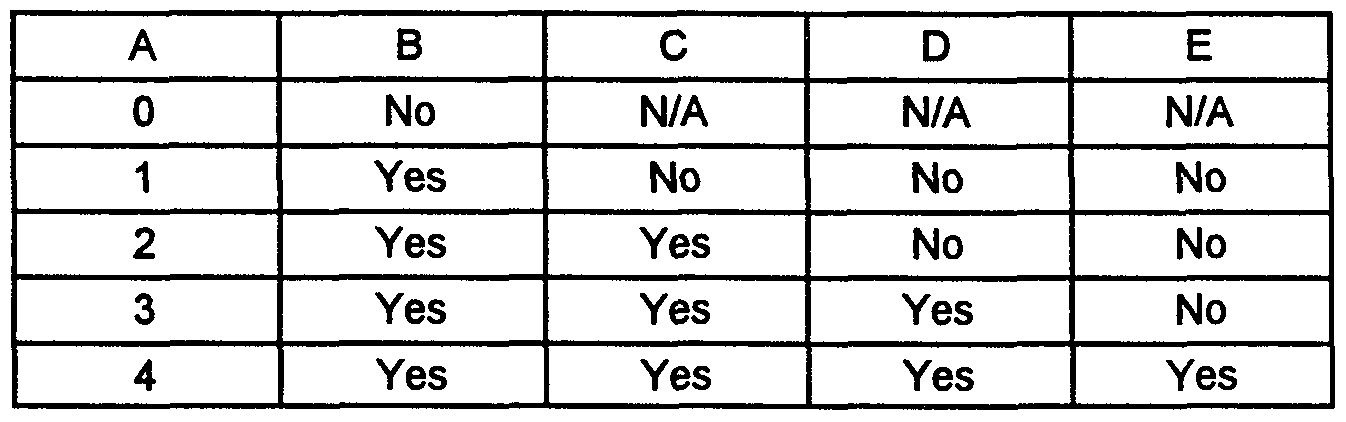


Claims
Priority Applications (4)
| Application Number | Priority Date | Filing Date | Title |
|---|---|---|---|
| AU36999/00A AU3699900A (en) | 1999-02-16 | 2000-02-16 | Device and method for regeneration and repair of cartilage lesions |
| JP2000599344A JP2002537022A (en) | 1999-02-16 | 2000-02-16 | Apparatus and method for regenerating and repairing cartilage lesion |
| EP00915782A EP1161201A4 (en) | 1999-02-16 | 2000-02-16 | Device and method for regeneration and repair of cartilage lesions |
| CA002362600A CA2362600A1 (en) | 1999-02-16 | 2000-02-16 | Device and method for regeneration and repair of cartilage lesions |
Applications Claiming Priority (2)
| Application Number | Priority Date | Filing Date | Title |
|---|---|---|---|
| US09/250,370 US6514514B1 (en) | 1997-08-14 | 1999-02-16 | Device and method for regeneration and repair of cartilage lesions |
| US09/250,370 | 1999-02-16 |
Publications (2)
| Publication Number | Publication Date |
|---|---|
| WO2000048550A2 true WO2000048550A2 (en) | 2000-08-24 |
| WO2000048550A3 WO2000048550A3 (en) | 2000-12-14 |
Family
ID=22947449
Family Applications (1)
| Application Number | Title | Priority Date | Filing Date |
|---|---|---|---|
| PCT/US2000/003972 WO2000048550A2 (en) | 1999-02-16 | 2000-02-16 | Device and method for regeneration and repair of cartilage lesions |
Country Status (5)
| Country | Link |
|---|---|
| EP (1) | EP1161201A4 (en) |
| JP (1) | JP2002537022A (en) |
| AU (1) | AU3699900A (en) |
| CA (1) | CA2362600A1 (en) |
| WO (1) | WO2000048550A2 (en) |
Cited By (19)
| Publication number | Priority date | Publication date | Assignee | Title |
|---|---|---|---|---|
| EP1084719A1 (en) * | 1999-09-17 | 2001-03-21 | Depuy Orthopaedics, Inc. | Bone sialoprotein-based compositions for enhancing connective tissue repair |
| EP1261365A1 (en) * | 2000-03-09 | 2002-12-04 | Sulzer Biologics, Inc. | Product and method for biological anchoring of connective tissue to bone |
| WO2006032915A3 (en) * | 2004-09-24 | 2006-06-01 | Univ Bristol | Cell bandage |
| US7148209B2 (en) | 2000-06-29 | 2006-12-12 | Ecole Polytechnique | Composition and method for the repair and regeneration of cartilage and other tissues |
| JP2007105547A (en) * | 2001-01-30 | 2007-04-26 | Orthogene Inc | Compositions and methods for treatment and repair of defects or lesions in articular cartilage using synovial-derived tissue or cells |
| US8545569B2 (en) | 2001-05-25 | 2013-10-01 | Conformis, Inc. | Patient selectable knee arthroplasty devices |
| US9055953B2 (en) | 2001-05-25 | 2015-06-16 | Conformis, Inc. | Methods and compositions for articular repair |
| US9066728B2 (en) | 2001-05-25 | 2015-06-30 | Conformis, Inc. | Surgical tools facilitating increased accuracy, speed and simplicity in performing joint arthroplasty |
| US9072531B2 (en) | 2001-05-25 | 2015-07-07 | Conformis, Inc. | Patient selectable joint arthroplasty devices and surgical tools |
| US9089342B2 (en) | 2009-02-24 | 2015-07-28 | Microport Orthopedics Holdings Inc. | Patient specific surgical guide locator and mount |
| US9592125B2 (en) | 2006-12-22 | 2017-03-14 | Laboratoire Medidom S.A. | In situ system for intra-articular chondral and osseous tissue repair |
| US9603711B2 (en) | 2001-05-25 | 2017-03-28 | Conformis, Inc. | Patient-adapted and improved articular implants, designs and related guide tools |
| US9642632B2 (en) | 2009-02-24 | 2017-05-09 | Microport Orthopedics Holdings Inc. | Orthopedic surgical guide |
| US9649117B2 (en) | 2009-02-24 | 2017-05-16 | Microport Orthopedics Holdings, Inc. | Orthopedic surgical guide |
| US9675471B2 (en) | 2012-06-11 | 2017-06-13 | Conformis, Inc. | Devices, techniques and methods for assessing joint spacing, balancing soft tissues and obtaining desired kinematics for joint implant components |
| US9700971B2 (en) | 2001-05-25 | 2017-07-11 | Conformis, Inc. | Implant device and method for manufacture |
| US9700420B2 (en) | 2008-03-05 | 2017-07-11 | Conformis, Inc. | Implants for altering wear patterns of articular surfaces |
| US9775680B2 (en) | 2001-05-25 | 2017-10-03 | Conformis, Inc. | Patient-adapted and improved articular implants, designs and related guide tools |
| US10085839B2 (en) | 2004-01-05 | 2018-10-02 | Conformis, Inc. | Patient-specific and patient-engineered orthopedic implants |
Families Citing this family (23)
| Publication number | Priority date | Publication date | Assignee | Title |
|---|---|---|---|---|
| US7468075B2 (en) | 2001-05-25 | 2008-12-23 | Conformis, Inc. | Methods and compositions for articular repair |
| US8083745B2 (en) | 2001-05-25 | 2011-12-27 | Conformis, Inc. | Surgical tools for arthroplasty |
| US8882847B2 (en) | 2001-05-25 | 2014-11-11 | Conformis, Inc. | Patient selectable knee joint arthroplasty devices |
| US8234097B2 (en) | 2001-05-25 | 2012-07-31 | Conformis, Inc. | Automated systems for manufacturing patient-specific orthopedic implants and instrumentation |
| US8556983B2 (en) | 2001-05-25 | 2013-10-15 | Conformis, Inc. | Patient-adapted and improved orthopedic implants, designs and related tools |
| ATE220564T1 (en) | 1997-08-14 | 2002-08-15 | Sulzer Innotec Ag | COMPOSITION AND DEVICE FOR REPAIRING CARTILAGE TISSUE IN VIVO CONSISTING OF NANOCAPSULES WITH OSTEOINDUCTIVE AND/OR CHONDROINDUCTIVE FACTORS |
| US7239908B1 (en) | 1998-09-14 | 2007-07-03 | The Board Of Trustees Of The Leland Stanford Junior University | Assessing the condition of a joint and devising treatment |
| AU772012B2 (en) | 1998-09-14 | 2004-04-08 | Board Of Trustees Of The Leland Stanford Junior University | Assessing the condition of a joint and preventing damage |
| ES2491866T3 (en) | 1999-11-15 | 2014-09-08 | Piramal Healthcare (Canada) Limited | Temperature-controlled, pH-dependent, self-gelling aqueous biopolymer solution |
| EP2036495A1 (en) | 2000-09-14 | 2009-03-18 | The Board of Trustees of The Leland Stanford Junior University | Assessing condition of a joint and cartilage loss |
| US8439926B2 (en) | 2001-05-25 | 2013-05-14 | Conformis, Inc. | Patient selectable joint arthroplasty devices and surgical tools |
| CN1780594A (en) | 2002-11-07 | 2006-05-31 | 康复米斯公司 | Methods for determining meniscal size and shape and for devising treatment |
| US7074412B2 (en) * | 2003-01-30 | 2006-07-11 | The University Of Zurich | Pharmaceutical composition |
| EP1462126A1 (en) * | 2003-03-28 | 2004-09-29 | BIOPHARM GESELLSCHAFT ZUR BIOTECHNOLOGISCHEN ENTWICKLUNG VON PHARMAKA mbH | Improved Osteoinductive Materials |
| US7217294B2 (en) * | 2003-08-20 | 2007-05-15 | Histogenics Corp. | Acellular matrix implants for treatment of articular cartilage, bone or osteochondral defects and injuries and method for use thereof |
| US8657881B2 (en) * | 2004-04-20 | 2014-02-25 | Depuy Mitek, Llc | Meniscal repair scaffold |
| US8623026B2 (en) | 2006-02-06 | 2014-01-07 | Conformis, Inc. | Patient selectable joint arthroplasty devices and surgical tools incorporating anatomical relief |
| WO2008157412A2 (en) | 2007-06-13 | 2008-12-24 | Conformis, Inc. | Surgical cutting guide |
| AU2009246474B2 (en) | 2008-05-12 | 2015-04-16 | Conformis, Inc. | Devices and methods for treatment of facet and other joints |
| EP2419035B1 (en) | 2009-04-16 | 2017-07-05 | ConforMIS, Inc. | Patient-specific joint arthroplasty methods for ligament repair |
| JP2011041472A (en) * | 2009-08-19 | 2011-03-03 | Tokai Univ | Spheroid for cell transplantation treatment composed of cell mixture and method for preparing the same |
| EP2754419B1 (en) | 2011-02-15 | 2024-02-07 | ConforMIS, Inc. | Patient-adapted and improved orthopedic implants |
| US9486226B2 (en) | 2012-04-18 | 2016-11-08 | Conformis, Inc. | Tibial guides, tools, and techniques for resecting the tibial plateau |
Citations (4)
| Publication number | Priority date | Publication date | Assignee | Title |
|---|---|---|---|---|
| WO1992009697A1 (en) * | 1990-11-30 | 1992-06-11 | Celtrix Laboratories, Inc. | USE OF A BONE MORPHOGENETIC PROTEIN IN SYNERGISTIC COMBINATION WITH TGF-β FOR BONE REPAIR |
| US5206023A (en) * | 1991-01-31 | 1993-04-27 | Robert F. Shaw | Method and compositions for the treatment and repair of defects or lesions in cartilage |
| US5681353A (en) * | 1987-07-20 | 1997-10-28 | Regen Biologics, Inc. | Meniscal augmentation device |
| EP0896825A1 (en) * | 1997-08-14 | 1999-02-17 | Sulzer Innotec Ag | Composition and device for in vivo cartilage repair comprising nanocapsules with osteoinductive and/or chondroinductive factors |
Family Cites Families (1)
| Publication number | Priority date | Publication date | Assignee | Title |
|---|---|---|---|---|
| US5563124A (en) * | 1991-04-22 | 1996-10-08 | Intermedics Orthopedics/ Denver, Inc. | Osteogenic product and process |
-
2000
- 2000-02-16 WO PCT/US2000/003972 patent/WO2000048550A2/en active Application Filing
- 2000-02-16 JP JP2000599344A patent/JP2002537022A/en active Pending
- 2000-02-16 EP EP00915782A patent/EP1161201A4/en not_active Withdrawn
- 2000-02-16 CA CA002362600A patent/CA2362600A1/en not_active Abandoned
- 2000-02-16 AU AU36999/00A patent/AU3699900A/en not_active Abandoned
Patent Citations (4)
| Publication number | Priority date | Publication date | Assignee | Title |
|---|---|---|---|---|
| US5681353A (en) * | 1987-07-20 | 1997-10-28 | Regen Biologics, Inc. | Meniscal augmentation device |
| WO1992009697A1 (en) * | 1990-11-30 | 1992-06-11 | Celtrix Laboratories, Inc. | USE OF A BONE MORPHOGENETIC PROTEIN IN SYNERGISTIC COMBINATION WITH TGF-β FOR BONE REPAIR |
| US5206023A (en) * | 1991-01-31 | 1993-04-27 | Robert F. Shaw | Method and compositions for the treatment and repair of defects or lesions in cartilage |
| EP0896825A1 (en) * | 1997-08-14 | 1999-02-17 | Sulzer Innotec Ag | Composition and device for in vivo cartilage repair comprising nanocapsules with osteoinductive and/or chondroinductive factors |
Non-Patent Citations (1)
| Title |
|---|
| See also references of EP1161201A2 * |
Cited By (45)
| Publication number | Priority date | Publication date | Assignee | Title |
|---|---|---|---|---|
| US6458763B1 (en) | 1999-09-17 | 2002-10-01 | Depuy Orthopeadics | Bone sialoprotein-based compositions for enhancing connective tissue repair |
| EP1084719A1 (en) * | 1999-09-17 | 2001-03-21 | Depuy Orthopaedics, Inc. | Bone sialoprotein-based compositions for enhancing connective tissue repair |
| EP1261365A1 (en) * | 2000-03-09 | 2002-12-04 | Sulzer Biologics, Inc. | Product and method for biological anchoring of connective tissue to bone |
| EP1261365A4 (en) * | 2000-03-09 | 2003-06-25 | Sulzer Biolog Inc | Product and method for biological anchoring of connective tissue to bone |
| EP1704866A2 (en) * | 2000-03-09 | 2006-09-27 | Zimmer Orthobiologics, Inc. | TGFbeta superfamily proteins for biological anchoring of connective tissue to bone |
| EP1704866A3 (en) * | 2000-03-09 | 2006-10-04 | Zimmer Orthobiologics, Inc. | TGFbeta superfamily proteins for biological anchoring of connective tissue to bone |
| US7148209B2 (en) | 2000-06-29 | 2006-12-12 | Ecole Polytechnique | Composition and method for the repair and regeneration of cartilage and other tissues |
| US7575743B2 (en) | 2001-01-30 | 2009-08-18 | Orthogene, Inc. | Compositions and methods for the treatment and repair of defects or lesions in articular cartilage using synovial-derived tissue or cells |
| JP2007105547A (en) * | 2001-01-30 | 2007-04-26 | Orthogene Inc | Compositions and methods for treatment and repair of defects or lesions in articular cartilage using synovial-derived tissue or cells |
| US9055953B2 (en) | 2001-05-25 | 2015-06-16 | Conformis, Inc. | Methods and compositions for articular repair |
| US9603711B2 (en) | 2001-05-25 | 2017-03-28 | Conformis, Inc. | Patient-adapted and improved articular implants, designs and related guide tools |
| US8545569B2 (en) | 2001-05-25 | 2013-10-01 | Conformis, Inc. | Patient selectable knee arthroplasty devices |
| US9700971B2 (en) | 2001-05-25 | 2017-07-11 | Conformis, Inc. | Implant device and method for manufacture |
| US9066728B2 (en) | 2001-05-25 | 2015-06-30 | Conformis, Inc. | Surgical tools facilitating increased accuracy, speed and simplicity in performing joint arthroplasty |
| US9072531B2 (en) | 2001-05-25 | 2015-07-07 | Conformis, Inc. | Patient selectable joint arthroplasty devices and surgical tools |
| US9084617B2 (en) | 2001-05-25 | 2015-07-21 | Conformis, Inc. | Patient selectable joint arthroplasty devices and surgical tools |
| US9775680B2 (en) | 2001-05-25 | 2017-10-03 | Conformis, Inc. | Patient-adapted and improved articular implants, designs and related guide tools |
| US9877790B2 (en) | 2001-05-25 | 2018-01-30 | Conformis, Inc. | Tibial implant and systems with variable slope |
| US10085839B2 (en) | 2004-01-05 | 2018-10-02 | Conformis, Inc. | Patient-specific and patient-engineered orthopedic implants |
| AU2005286271B2 (en) * | 2004-09-24 | 2011-11-24 | The University Of Bristol | Cell bandage |
| US9539364B2 (en) | 2004-09-24 | 2017-01-10 | The University Of Bristol | Cell bandage |
| WO2006032915A3 (en) * | 2004-09-24 | 2006-06-01 | Univ Bristol | Cell bandage |
| US10549012B2 (en) | 2004-09-24 | 2020-02-04 | The University Of Bristol | Cell bandage |
| US9592125B2 (en) | 2006-12-22 | 2017-03-14 | Laboratoire Medidom S.A. | In situ system for intra-articular chondral and osseous tissue repair |
| US9700420B2 (en) | 2008-03-05 | 2017-07-11 | Conformis, Inc. | Implants for altering wear patterns of articular surfaces |
| US9675365B2 (en) | 2009-02-24 | 2017-06-13 | Microport Orthopedics Holdings Inc. | System and method for anterior approach for installing tibial stem |
| US9089342B2 (en) | 2009-02-24 | 2015-07-28 | Microport Orthopedics Holdings Inc. | Patient specific surgical guide locator and mount |
| US9649117B2 (en) | 2009-02-24 | 2017-05-16 | Microport Orthopedics Holdings, Inc. | Orthopedic surgical guide |
| US9883870B2 (en) | 2009-02-24 | 2018-02-06 | Microport Orthopedics Holdings Inc. | Method for forming a patient specific surgical guide mount |
| US9901353B2 (en) | 2009-02-24 | 2018-02-27 | Microport Holdings Inc. | Patient specific surgical guide locator and mount |
| US9949747B2 (en) | 2009-02-24 | 2018-04-24 | Microport Orthopedics Holdings, Inc. | Systems and methods for installing an orthopedic implant |
| US10039557B2 (en) | 2009-02-24 | 2018-08-07 | Micorport Orthopedics Holdings, Inc. | Orthopedic surgical guide |
| US9642632B2 (en) | 2009-02-24 | 2017-05-09 | Microport Orthopedics Holdings Inc. | Orthopedic surgical guide |
| US10512476B2 (en) | 2009-02-24 | 2019-12-24 | Microport Orthopedics Holdings, Inc. | Orthopedic surgical guide |
| US11911046B2 (en) | 2009-02-24 | 2024-02-27 | Microport Orthopedics Holdings, Inc. | Patient specific surgical guide locator and mount |
| US10646238B2 (en) | 2009-02-24 | 2020-05-12 | Microport Orthopedics Holdings, Inc. | Systems and methods for installing an orthopedic implant |
| US10660654B2 (en) | 2009-02-24 | 2020-05-26 | Microport Orthopedics Holdings Inc. | Method for forming a patient specific surgical guide mount |
| US10973536B2 (en) | 2009-02-24 | 2021-04-13 | Microport Orthopedics Holdings, Inc. | Orthopedic surgical guide |
| US11154305B2 (en) | 2009-02-24 | 2021-10-26 | Microport Orthopedics Holdings Inc. | Patient specific surgical guide locator and mount |
| US11389177B2 (en) | 2009-02-24 | 2022-07-19 | Microport Orthopedics Holdings Inc. | Method for forming a patient specific surgical guide mount |
| US11464527B2 (en) | 2009-02-24 | 2022-10-11 | Microport Orthopedics Holdings Inc. | Systems and methods for installing an orthopedic implant |
| US11534186B2 (en) | 2009-02-24 | 2022-12-27 | Microport Orthopedics Holdings Inc. | Orthopedic surgical guide |
| US11779356B2 (en) | 2009-02-24 | 2023-10-10 | Microport Orthopedics Holdings, Inc. | Orthopedic surgical guide |
| US11779347B2 (en) | 2009-02-24 | 2023-10-10 | Microport Orthopedics Holdings Inc. | System for forming a patient specific surgical guide mount |
| US9675471B2 (en) | 2012-06-11 | 2017-06-13 | Conformis, Inc. | Devices, techniques and methods for assessing joint spacing, balancing soft tissues and obtaining desired kinematics for joint implant components |
Also Published As
| Publication number | Publication date |
|---|---|
| CA2362600A1 (en) | 2000-08-24 |
| AU3699900A (en) | 2000-09-04 |
| EP1161201A2 (en) | 2001-12-12 |
| WO2000048550A3 (en) | 2000-12-14 |
| EP1161201A4 (en) | 2006-07-19 |
| JP2002537022A (en) | 2002-11-05 |
Similar Documents
| Publication | Publication Date | Title |
|---|---|---|
| US6511958B1 (en) | Compositions for regeneration and repair of cartilage lesions | |
| USRE41286E1 (en) | Compositions for regeneration and repair of cartilage lesions | |
| EP1161201A2 (en) | Device and method for regeneration and repair of cartilage lesions | |
| Kokubo et al. | Bone regeneration by recombinant human bone morphogenetic protein-2 and a novel biodegradable carrier in a rabbit ulnar defect model | |
| Jansen et al. | Growth factor-loaded scaffolds for bone engineering | |
| EP0968012B1 (en) | Osteogenic devices and methods of use thereof for repair of bone | |
| Kroese-Deutman et al. | Bone inductive properties of rhBMP-2 loaded porous calcium phosphate cement implants inserted at an ectopic site in rabbits | |
| JP5623764B2 (en) | Repair of pharynx, trachea and other fibrocartilage tissues | |
| US8372805B1 (en) | Osteogenic devices and methods of use thereof for repair of endochondral bone, osteochondral and chondral defects | |
| USRE46402E1 (en) | Bioactive scaffolds | |
| Wozney et al. | Protein-based tissue engineering in bone and cartilage repair | |
| EP1261365B1 (en) | Product for biological anchoring of connective tissue to bone | |
| JP2009101174A (en) | POROUS GRANULE OF beta-TRICALCIUM PHOSPHATE, AND METHOD FOR PRODUCING THE SAME | |
| WO2005033272A2 (en) | In vivo synthesis of connective tissues | |
| Brekke | A rationale for delivery of osteoinductive proteins | |
| Takahashi | Material design of biodegradable cell | |
| Takahashi | MATERIAL DESIGN OF BIODEGRADABLE CELL SCAFFOLDS FOR CONTROLLED RELEASE OF BONE MORPHOGENETIC PROTEIN-2 AND THE BONE REGENERATION POTENTIAL | |
| Johnson et al. | Perspectives on Growth Factors, Bone Graft Substitutes and Fracture Healing | |
| Kroese-Deutman | Closing capacity of bone defects, scaffolds, porosity and growth factors updated. |
Legal Events
| Date | Code | Title | Description |
|---|---|---|---|
| AK | Designated states |
Kind code of ref document: A2 Designated state(s): AE AL AM AT AU AZ BA BB BG BR BY CA CH CN CR CU CZ DE DK DM EE ES FI GB GD GE GH GM HR HU ID IL IN IS JP KE KG KP KR KZ LC LK LR LS LT LU LV MA MD MG MK MN MW MX NO NZ PL PT RO RU SD SE SG SI SK SL TJ TM TR TT TZ UA UG UZ VN YU ZA ZW |
|
| AL | Designated countries for regional patents |
Kind code of ref document: A2 Designated state(s): GH GM KE LS MW SD SL SZ TZ UG ZW AM AZ BY KG KZ MD RU TJ TM AT BE CH CY DE DK ES FI FR GB GR IE IT LU MC NL PT SE BF BJ CF CG CI CM GA GN GW ML MR NE SN TD TG |
|
| 121 | Ep: the epo has been informed by wipo that ep was designated in this application | ||
| DFPE | Request for preliminary examination filed prior to expiration of 19th month from priority date (pct application filed before 20040101) | ||
| AK | Designated states |
Kind code of ref document: A3 Designated state(s): AE AL AM AT AU AZ BA BB BG BR BY CA CH CN CR CU CZ DE DK DM EE ES FI GB GD GE GH GM HR HU ID IL IN IS JP KE KG KP KR KZ LC LK LR LS LT LU LV MA MD MG MK MN MW MX NO NZ PL PT RO RU SD SE SG SI SK SL TJ TM TR TT TZ UA UG UZ VN YU ZA ZW |
|
| AL | Designated countries for regional patents |
Kind code of ref document: A3 Designated state(s): GH GM KE LS MW SD SL SZ TZ UG ZW AM AZ BY KG KZ MD RU TJ TM AT BE CH CY DE DK ES FI FR GB GR IE IT LU MC NL PT SE BF BJ CF CG CI CM GA GN GW ML MR NE SN TD TG |
|
| ENP | Entry into the national phase in: |
Ref country code: CA Ref document number: 2362600 Kind code of ref document: A Format of ref document f/p: F Ref document number: 2362600 Country of ref document: CA |
|
| ENP | Entry into the national phase in: |
Ref country code: JP Ref document number: 2000 599344 Kind code of ref document: A Format of ref document f/p: F |
|
| WWE | Wipo information: entry into national phase |
Ref document number: 2000915782 Country of ref document: EP |
|
| WWP | Wipo information: published in national office |
Ref document number: 2000915782 Country of ref document: EP |
|
| REG | Reference to national code |
Ref country code: DE Ref legal event code: 8642 |