WO1999010494A2 - Agonist antibodies to the thrombopoietin receptor, and their therapeutic uses - Google Patents
Agonist antibodies to the thrombopoietin receptor, and their therapeutic uses Download PDFInfo
- Publication number
- WO1999010494A2 WO1999010494A2 PCT/US1998/017364 US9817364W WO9910494A2 WO 1999010494 A2 WO1999010494 A2 WO 1999010494A2 US 9817364 W US9817364 W US 9817364W WO 9910494 A2 WO9910494 A2 WO 9910494A2
- Authority
- WO
- WIPO (PCT)
- Prior art keywords
- seq
- antibody
- receptor
- mpl
- antibodies
- Prior art date
Links
- 239000000556 agonist Substances 0.000 title claims abstract description 72
- 102000005763 Thrombopoietin Receptors Human genes 0.000 title claims description 8
- 108010070774 Thrombopoietin Receptors Proteins 0.000 title claims description 8
- 230000001225 therapeutic effect Effects 0.000 title description 14
- 210000003593 megakaryocyte Anatomy 0.000 claims abstract description 47
- 230000004069 differentiation Effects 0.000 claims abstract description 26
- 210000004027 cell Anatomy 0.000 claims description 231
- 238000000034 method Methods 0.000 claims description 101
- 102000005962 receptors Human genes 0.000 claims description 90
- 108020003175 receptors Proteins 0.000 claims description 90
- 241000282414 Homo sapiens Species 0.000 claims description 77
- 239000012634 fragment Substances 0.000 claims description 68
- 230000027455 binding Effects 0.000 claims description 64
- 239000013598 vector Substances 0.000 claims description 52
- 150000007523 nucleic acids Chemical class 0.000 claims description 43
- 108020004707 nucleic acids Proteins 0.000 claims description 35
- 102000039446 nucleic acids Human genes 0.000 claims description 35
- 125000003275 alpha amino acid group Chemical group 0.000 claims description 31
- 239000000203 mixture Substances 0.000 claims description 25
- 238000004519 manufacturing process Methods 0.000 claims description 24
- 230000035755 proliferation Effects 0.000 claims description 24
- 230000012010 growth Effects 0.000 claims description 20
- 102000003675 cytokine receptors Human genes 0.000 claims description 18
- 108010057085 cytokine receptors Proteins 0.000 claims description 18
- 230000001965 increasing effect Effects 0.000 claims description 17
- 230000004927 fusion Effects 0.000 claims description 15
- 230000003213 activating effect Effects 0.000 claims description 7
- 108020001507 fusion proteins Proteins 0.000 claims description 6
- 102000037865 fusion proteins Human genes 0.000 claims description 6
- 101800000592 Capsid protein 3 Proteins 0.000 claims description 5
- 125000002924 primary amino group Chemical group [H]N([H])* 0.000 claims description 5
- 230000004936 stimulating effect Effects 0.000 claims description 5
- 101710132601 Capsid protein Proteins 0.000 claims description 4
- 101710094648 Coat protein Proteins 0.000 claims description 4
- 102100021181 Golgi phosphoprotein 3 Human genes 0.000 claims description 4
- 101710125418 Major capsid protein Proteins 0.000 claims description 4
- 108091008553 MuSK receptors Proteins 0.000 claims description 4
- 101710141454 Nucleoprotein Proteins 0.000 claims description 4
- 101710083689 Probable capsid protein Proteins 0.000 claims description 4
- 102000004278 Receptor Protein-Tyrosine Kinases Human genes 0.000 claims description 4
- 108090000873 Receptor Protein-Tyrosine Kinases Proteins 0.000 claims description 4
- 238000002823 phage display Methods 0.000 claims description 4
- 108010001267 Protein Subunits Proteins 0.000 claims description 2
- 239000003937 drug carrier Substances 0.000 claims description 2
- 239000011159 matrix material Substances 0.000 claims description 2
- 230000002232 neuromuscular Effects 0.000 claims description 2
- 101100295738 Gallus gallus COR3 gene Proteins 0.000 claims 3
- 101100459905 Saccharomyces cerevisiae (strain ATCC 204508 / S288c) NCP1 gene Proteins 0.000 claims 3
- 101150046305 cpr-1 gene Proteins 0.000 claims 3
- 101150047265 COR2 gene Proteins 0.000 claims 2
- 102100035966 DnaJ homolog subfamily A member 2 Human genes 0.000 claims 2
- 102100037584 FAST kinase domain-containing protein 4 Human genes 0.000 claims 2
- 101000931210 Homo sapiens DnaJ homolog subfamily A member 2 Proteins 0.000 claims 2
- 101001028251 Homo sapiens FAST kinase domain-containing protein 4 Proteins 0.000 claims 2
- 101100467189 Saccharomyces cerevisiae (strain ATCC 204508 / S288c) QCR2 gene Proteins 0.000 claims 2
- 230000035800 maturation Effects 0.000 abstract description 16
- 206010043554 thrombocytopenia Diseases 0.000 abstract description 12
- 230000010076 replication Effects 0.000 abstract description 9
- 210000002361 Megakaryocyte Progenitor Cell Anatomy 0.000 abstract description 7
- 150000001875 compounds Chemical class 0.000 abstract description 7
- 210000000601 blood cell Anatomy 0.000 abstract 1
- 108010041111 Thrombopoietin Proteins 0.000 description 168
- 102000036693 Thrombopoietin Human genes 0.000 description 167
- 108090000623 proteins and genes Proteins 0.000 description 114
- 108020004414 DNA Proteins 0.000 description 97
- 235000018102 proteins Nutrition 0.000 description 72
- 102000004169 proteins and genes Human genes 0.000 description 72
- 230000000694 effects Effects 0.000 description 66
- 108090000765 processed proteins & peptides Proteins 0.000 description 52
- 210000004369 blood Anatomy 0.000 description 48
- 239000008280 blood Substances 0.000 description 48
- 230000014509 gene expression Effects 0.000 description 48
- 102000004196 processed proteins & peptides Human genes 0.000 description 47
- 241000402754 Erythranthe moschata Species 0.000 description 46
- 229920001184 polypeptide Polymers 0.000 description 46
- 239000002953 phosphate buffered saline Substances 0.000 description 40
- 239000000427 antigen Substances 0.000 description 38
- LOKCTEFSRHRXRJ-UHFFFAOYSA-I dipotassium trisodium dihydrogen phosphate hydrogen phosphate dichloride Chemical compound P(=O)(O)(O)[O-].[K+].P(=O)(O)([O-])[O-].[Na+].[Na+].[Cl-].[K+].[Cl-].[Na+] LOKCTEFSRHRXRJ-UHFFFAOYSA-I 0.000 description 38
- 101710190051 Muscle, skeletal receptor tyrosine protein kinase Proteins 0.000 description 37
- 102100038168 Muscle, skeletal receptor tyrosine-protein kinase Human genes 0.000 description 37
- 108091007433 antigens Proteins 0.000 description 37
- 102000036639 antigens Human genes 0.000 description 37
- 238000003556 assay Methods 0.000 description 34
- 239000003446 ligand Substances 0.000 description 31
- 239000013604 expression vector Substances 0.000 description 25
- 239000013612 plasmid Substances 0.000 description 25
- 241000699670 Mus sp. Species 0.000 description 23
- 230000004071 biological effect Effects 0.000 description 23
- 239000013615 primer Substances 0.000 description 23
- 238000002965 ELISA Methods 0.000 description 22
- 240000004808 Saccharomyces cerevisiae Species 0.000 description 22
- 235000014680 Saccharomyces cerevisiae Nutrition 0.000 description 22
- 108010047041 Complementarity Determining Regions Proteins 0.000 description 21
- 235000001014 amino acid Nutrition 0.000 description 21
- 102000003951 Erythropoietin Human genes 0.000 description 20
- 108090000394 Erythropoietin Proteins 0.000 description 20
- 229940105423 erythropoietin Drugs 0.000 description 20
- OXCMYAYHXIHQOA-UHFFFAOYSA-N potassium;[2-butyl-5-chloro-3-[[4-[2-(1,2,4-triaza-3-azanidacyclopenta-1,4-dien-5-yl)phenyl]phenyl]methyl]imidazol-4-yl]methanol Chemical compound [K+].CCCCC1=NC(Cl)=C(CO)N1CC1=CC=C(C=2C(=CC=CC=2)C2=N[N-]N=N2)C=C1 OXCMYAYHXIHQOA-UHFFFAOYSA-N 0.000 description 20
- 108060003951 Immunoglobulin Proteins 0.000 description 19
- 108010076504 Protein Sorting Signals Proteins 0.000 description 19
- 230000003394 haemopoietic effect Effects 0.000 description 19
- 102000018358 immunoglobulin Human genes 0.000 description 19
- 238000000746 purification Methods 0.000 description 19
- FAPWRFPIFSIZLT-UHFFFAOYSA-M Sodium chloride Chemical compound [Na+].[Cl-] FAPWRFPIFSIZLT-UHFFFAOYSA-M 0.000 description 18
- 125000000539 amino acid group Chemical group 0.000 description 18
- 150000001413 amino acids Chemical class 0.000 description 18
- 239000000872 buffer Substances 0.000 description 18
- RAXXELZNTBOGNW-UHFFFAOYSA-N imidazole Natural products C1=CNC=N1 RAXXELZNTBOGNW-UHFFFAOYSA-N 0.000 description 18
- 238000011534 incubation Methods 0.000 description 18
- 108091008146 restriction endonucleases Proteins 0.000 description 18
- 102000004127 Cytokines Human genes 0.000 description 17
- 108090000695 Cytokines Proteins 0.000 description 17
- 102100039064 Interleukin-3 Human genes 0.000 description 17
- 241001529936 Murinae Species 0.000 description 17
- 229940024606 amino acid Drugs 0.000 description 17
- 108091003079 Bovine Serum Albumin Proteins 0.000 description 16
- 108010002386 Interleukin-3 Proteins 0.000 description 16
- 108091028043 Nucleic acid sequence Proteins 0.000 description 16
- 241001465754 Metazoa Species 0.000 description 15
- 108020000715 acetylcholine receptors Proteins 0.000 description 15
- 102000034337 acetylcholine receptors Human genes 0.000 description 15
- 239000002609 medium Substances 0.000 description 15
- 101150079978 AGRN gene Proteins 0.000 description 14
- 108700019743 Agrin Proteins 0.000 description 14
- 102100040026 Agrin Human genes 0.000 description 14
- 102000004190 Enzymes Human genes 0.000 description 14
- 108090000790 Enzymes Proteins 0.000 description 14
- 238000010367 cloning Methods 0.000 description 14
- 229940088598 enzyme Drugs 0.000 description 14
- 239000012091 fetal bovine serum Substances 0.000 description 14
- 238000001727 in vivo Methods 0.000 description 14
- 210000002381 plasma Anatomy 0.000 description 14
- 238000011084 recovery Methods 0.000 description 14
- 238000013518 transcription Methods 0.000 description 14
- 230000035897 transcription Effects 0.000 description 14
- 229920002684 Sepharose Polymers 0.000 description 13
- 238000004458 analytical method Methods 0.000 description 13
- 230000015572 biosynthetic process Effects 0.000 description 13
- 239000003623 enhancer Substances 0.000 description 13
- 238000002360 preparation method Methods 0.000 description 13
- 210000002966 serum Anatomy 0.000 description 13
- 102100039620 Granulocyte-macrophage colony-stimulating factor Human genes 0.000 description 12
- 230000004913 activation Effects 0.000 description 12
- 239000002299 complementary DNA Substances 0.000 description 12
- 238000000338 in vitro Methods 0.000 description 12
- 239000000463 material Substances 0.000 description 12
- 239000000523 sample Substances 0.000 description 12
- 238000006467 substitution reaction Methods 0.000 description 12
- 239000006228 supernatant Substances 0.000 description 12
- 241000588724 Escherichia coli Species 0.000 description 11
- 108010017213 Granulocyte-Macrophage Colony-Stimulating Factor Proteins 0.000 description 11
- OUYCCCASQSFEME-QMMMGPOBSA-N L-tyrosine Chemical group OC(=O)[C@@H](N)CC1=CC=C(O)C=C1 OUYCCCASQSFEME-QMMMGPOBSA-N 0.000 description 11
- 230000029087 digestion Effects 0.000 description 11
- 229940072221 immunoglobulins Drugs 0.000 description 11
- 210000004962 mammalian cell Anatomy 0.000 description 11
- 210000000715 neuromuscular junction Anatomy 0.000 description 11
- 230000026731 phosphorylation Effects 0.000 description 11
- 238000006366 phosphorylation reaction Methods 0.000 description 11
- 230000001361 thrombopoietic effect Effects 0.000 description 11
- 241000196324 Embryophyta Species 0.000 description 10
- WQZGKKKJIJFFOK-GASJEMHNSA-N Glucose Natural products OC[C@H]1OC(O)[C@H](O)[C@@H](O)[C@@H]1O WQZGKKKJIJFFOK-GASJEMHNSA-N 0.000 description 10
- 108010017080 Granulocyte Colony-Stimulating Factor Proteins 0.000 description 10
- 102000004269 Granulocyte Colony-Stimulating Factor Human genes 0.000 description 10
- 241000829100 Macaca mulatta polyomavirus 1 Species 0.000 description 10
- FPPNZSSZRUTDAP-UWFZAAFLSA-N carbenicillin Chemical compound N([C@H]1[C@H]2SC([C@@H](N2C1=O)C(O)=O)(C)C)C(=O)C(C(O)=O)C1=CC=CC=C1 FPPNZSSZRUTDAP-UWFZAAFLSA-N 0.000 description 10
- 229960003669 carbenicillin Drugs 0.000 description 10
- 238000001516 cell proliferation assay Methods 0.000 description 10
- 238000010276 construction Methods 0.000 description 10
- 239000008103 glucose Substances 0.000 description 10
- 230000013595 glycosylation Effects 0.000 description 10
- 238000006206 glycosylation reaction Methods 0.000 description 10
- -1 however Proteins 0.000 description 10
- 230000001939 inductive effect Effects 0.000 description 10
- 238000003780 insertion Methods 0.000 description 10
- 230000037431 insertion Effects 0.000 description 10
- YBYRMVIVWMBXKQ-UHFFFAOYSA-N phenylmethanesulfonyl fluoride Chemical compound FS(=O)(=O)CC1=CC=CC=C1 YBYRMVIVWMBXKQ-UHFFFAOYSA-N 0.000 description 10
- 210000000130 stem cell Anatomy 0.000 description 10
- 241000699666 Mus <mouse, genus> Species 0.000 description 9
- 108010022394 Threonine synthase Proteins 0.000 description 9
- 239000002253 acid Substances 0.000 description 9
- 238000007792 addition Methods 0.000 description 9
- 210000004899 c-terminal region Anatomy 0.000 description 9
- 102000004419 dihydrofolate reductase Human genes 0.000 description 9
- 230000006870 function Effects 0.000 description 9
- 239000011780 sodium chloride Substances 0.000 description 9
- 230000000638 stimulation Effects 0.000 description 9
- 230000004083 survival effect Effects 0.000 description 9
- OUYCCCASQSFEME-UHFFFAOYSA-N tyrosine Natural products OC(=O)C(N)CC1=CC=C(O)C=C1 OUYCCCASQSFEME-UHFFFAOYSA-N 0.000 description 9
- 241000894006 Bacteria Species 0.000 description 8
- 241000282693 Cercopithecidae Species 0.000 description 8
- 108010021625 Immunoglobulin Fragments Proteins 0.000 description 8
- 102000008394 Immunoglobulin Fragments Human genes 0.000 description 8
- 108090001005 Interleukin-6 Proteins 0.000 description 8
- 102000004889 Interleukin-6 Human genes 0.000 description 8
- 108090000581 Leukemia inhibitory factor Proteins 0.000 description 8
- 102000004058 Leukemia inhibitory factor Human genes 0.000 description 8
- 108091034117 Oligonucleotide Proteins 0.000 description 8
- 239000007983 Tris buffer Substances 0.000 description 8
- 230000003321 amplification Effects 0.000 description 8
- 238000005119 centrifugation Methods 0.000 description 8
- 238000006243 chemical reaction Methods 0.000 description 8
- 239000003636 conditioned culture medium Substances 0.000 description 8
- NOESYZHRGYRDHS-UHFFFAOYSA-N insulin Chemical compound N1C(=O)C(NC(=O)C(CCC(N)=O)NC(=O)C(CCC(O)=O)NC(=O)C(C(C)C)NC(=O)C(NC(=O)CN)C(C)CC)CSSCC(C(NC(CO)C(=O)NC(CC(C)C)C(=O)NC(CC=2C=CC(O)=CC=2)C(=O)NC(CCC(N)=O)C(=O)NC(CC(C)C)C(=O)NC(CCC(O)=O)C(=O)NC(CC(N)=O)C(=O)NC(CC=2C=CC(O)=CC=2)C(=O)NC(CSSCC(NC(=O)C(C(C)C)NC(=O)C(CC(C)C)NC(=O)C(CC=2C=CC(O)=CC=2)NC(=O)C(CC(C)C)NC(=O)C(C)NC(=O)C(CCC(O)=O)NC(=O)C(C(C)C)NC(=O)C(CC(C)C)NC(=O)C(CC=2NC=NC=2)NC(=O)C(CO)NC(=O)CNC2=O)C(=O)NCC(=O)NC(CCC(O)=O)C(=O)NC(CCCNC(N)=N)C(=O)NCC(=O)NC(CC=3C=CC=CC=3)C(=O)NC(CC=3C=CC=CC=3)C(=O)NC(CC=3C=CC(O)=CC=3)C(=O)NC(C(C)O)C(=O)N3C(CCC3)C(=O)NC(CCCCN)C(=O)NC(C)C(O)=O)C(=O)NC(CC(N)=O)C(O)=O)=O)NC(=O)C(C(C)CC)NC(=O)C(CO)NC(=O)C(C(C)O)NC(=O)C1CSSCC2NC(=O)C(CC(C)C)NC(=O)C(NC(=O)C(CCC(N)=O)NC(=O)C(CC(N)=O)NC(=O)C(NC(=O)C(N)CC=1C=CC=CC=1)C(C)C)CC1=CN=CN1 NOESYZHRGYRDHS-UHFFFAOYSA-N 0.000 description 8
- 230000004048 modification Effects 0.000 description 8
- 238000012986 modification Methods 0.000 description 8
- 238000003199 nucleic acid amplification method Methods 0.000 description 8
- 125000003729 nucleotide group Chemical group 0.000 description 8
- 230000008569 process Effects 0.000 description 8
- 230000001105 regulatory effect Effects 0.000 description 8
- 239000000243 solution Substances 0.000 description 8
- 210000001519 tissue Anatomy 0.000 description 8
- 238000005406 washing Methods 0.000 description 8
- 108091032973 (ribonucleotides)n+m Proteins 0.000 description 7
- LFQSCWFLJHTTHZ-UHFFFAOYSA-N Ethanol Chemical compound CCO LFQSCWFLJHTTHZ-UHFFFAOYSA-N 0.000 description 7
- 102100020880 Kit ligand Human genes 0.000 description 7
- 229920001213 Polysorbate 20 Polymers 0.000 description 7
- 201000003710 autoimmune thrombocytopenic purpura Diseases 0.000 description 7
- 230000001580 bacterial effect Effects 0.000 description 7
- 238000002512 chemotherapy Methods 0.000 description 7
- 230000002950 deficient Effects 0.000 description 7
- 230000001419 dependent effect Effects 0.000 description 7
- 229940079593 drug Drugs 0.000 description 7
- 239000003814 drug Substances 0.000 description 7
- 239000000499 gel Substances 0.000 description 7
- 210000003958 hematopoietic stem cell Anatomy 0.000 description 7
- 210000003292 kidney cell Anatomy 0.000 description 7
- 238000011813 knockout mouse model Methods 0.000 description 7
- 239000003550 marker Substances 0.000 description 7
- 125000001360 methionine group Chemical group N[C@@H](CCSC)C(=O)* 0.000 description 7
- 210000002569 neuron Anatomy 0.000 description 7
- 239000002773 nucleotide Substances 0.000 description 7
- 238000004091 panning Methods 0.000 description 7
- 239000000256 polyoxyethylene sorbitan monolaurate Substances 0.000 description 7
- 235000010486 polyoxyethylene sorbitan monolaurate Nutrition 0.000 description 7
- 241000894007 species Species 0.000 description 7
- 238000003786 synthesis reaction Methods 0.000 description 7
- MTCFGRXMJLQNBG-REOHCLBHSA-N (2S)-2-Amino-3-hydroxypropansäure Chemical compound OC[C@H](N)C(O)=O MTCFGRXMJLQNBG-REOHCLBHSA-N 0.000 description 6
- 108091026890 Coding region Proteins 0.000 description 6
- 108010071942 Colony-Stimulating Factors Proteins 0.000 description 6
- 239000006144 Dulbecco’s modified Eagle's medium Substances 0.000 description 6
- 108010075944 Erythropoietin Receptors Proteins 0.000 description 6
- 102100036509 Erythropoietin receptor Human genes 0.000 description 6
- 208000028622 Immune thrombocytopenia Diseases 0.000 description 6
- XUJNEKJLAYXESH-REOHCLBHSA-N L-Cysteine Chemical compound SC[C@H](N)C(O)=O XUJNEKJLAYXESH-REOHCLBHSA-N 0.000 description 6
- VMHLLURERBWHNL-UHFFFAOYSA-M Sodium acetate Chemical compound [Na+].CC([O-])=O VMHLLURERBWHNL-UHFFFAOYSA-M 0.000 description 6
- 108010039445 Stem Cell Factor Proteins 0.000 description 6
- 241000251539 Vertebrata <Metazoa> Species 0.000 description 6
- 241000700605 Viruses Species 0.000 description 6
- 239000011543 agarose gel Substances 0.000 description 6
- 239000005557 antagonist Substances 0.000 description 6
- 239000011324 bead Substances 0.000 description 6
- 150000001720 carbohydrates Chemical group 0.000 description 6
- 230000000295 complement effect Effects 0.000 description 6
- 238000005516 engineering process Methods 0.000 description 6
- 230000000925 erythroid effect Effects 0.000 description 6
- BRZYSWJRSDMWLG-CAXSIQPQSA-N geneticin Natural products O1C[C@@](O)(C)[C@H](NC)[C@@H](O)[C@H]1O[C@@H]1[C@@H](O)[C@H](O[C@@H]2[C@@H]([C@@H](O)[C@H](O)[C@@H](C(C)O)O2)N)[C@@H](N)C[C@H]1N BRZYSWJRSDMWLG-CAXSIQPQSA-N 0.000 description 6
- 239000003102 growth factor Substances 0.000 description 6
- 230000006698 induction Effects 0.000 description 6
- 238000002347 injection Methods 0.000 description 6
- 239000007924 injection Substances 0.000 description 6
- 230000003834 intracellular effect Effects 0.000 description 6
- 238000002955 isolation Methods 0.000 description 6
- 230000003343 megakaryocytopoietic effect Effects 0.000 description 6
- 239000012528 membrane Substances 0.000 description 6
- 230000003039 myelosuppressive effect Effects 0.000 description 6
- 230000003582 thrombocytopenic effect Effects 0.000 description 6
- 238000001890 transfection Methods 0.000 description 6
- 230000009466 transformation Effects 0.000 description 6
- XLYOFNOQVPJJNP-UHFFFAOYSA-N water Substances O XLYOFNOQVPJJNP-UHFFFAOYSA-N 0.000 description 6
- 102000002260 Alkaline Phosphatase Human genes 0.000 description 5
- 108020004774 Alkaline Phosphatase Proteins 0.000 description 5
- 102000007644 Colony-Stimulating Factors Human genes 0.000 description 5
- 108010051696 Growth Hormone Proteins 0.000 description 5
- 206010021245 Idiopathic thrombocytopenic purpura Diseases 0.000 description 5
- 102000000589 Interleukin-1 Human genes 0.000 description 5
- 108010002352 Interleukin-1 Proteins 0.000 description 5
- WHUUTDBJXJRKMK-VKHMYHEASA-N L-glutamic acid Chemical compound OC(=O)[C@@H](N)CCC(O)=O WHUUTDBJXJRKMK-VKHMYHEASA-N 0.000 description 5
- AYFVYJQAPQTCCC-GBXIJSLDSA-N L-threonine Chemical compound C[C@@H](O)[C@H](N)C(O)=O AYFVYJQAPQTCCC-GBXIJSLDSA-N 0.000 description 5
- KDXKERNSBIXSRK-UHFFFAOYSA-N Lysine Chemical group NCCCCC(N)C(O)=O KDXKERNSBIXSRK-UHFFFAOYSA-N 0.000 description 5
- 241000124008 Mammalia Species 0.000 description 5
- 108091007491 NSP3 Papain-like protease domains Proteins 0.000 description 5
- 229930193140 Neomycin Natural products 0.000 description 5
- MTCFGRXMJLQNBG-UHFFFAOYSA-N Serine Natural products OCC(N)C(O)=O MTCFGRXMJLQNBG-UHFFFAOYSA-N 0.000 description 5
- 102100038803 Somatotropin Human genes 0.000 description 5
- ZMANZCXQSJIPKH-UHFFFAOYSA-N Triethylamine Chemical compound CCN(CC)CC ZMANZCXQSJIPKH-UHFFFAOYSA-N 0.000 description 5
- 230000033228 biological regulation Effects 0.000 description 5
- 108700010039 chimeric receptor Proteins 0.000 description 5
- 229940047120 colony stimulating factors Drugs 0.000 description 5
- 235000018417 cysteine Nutrition 0.000 description 5
- 150000001945 cysteines Chemical class 0.000 description 5
- 238000012217 deletion Methods 0.000 description 5
- 230000037430 deletion Effects 0.000 description 5
- 208000037265 diseases, disorders, signs and symptoms Diseases 0.000 description 5
- 238000010494 dissociation reaction Methods 0.000 description 5
- 230000005593 dissociations Effects 0.000 description 5
- 210000003743 erythrocyte Anatomy 0.000 description 5
- 239000000122 growth hormone Substances 0.000 description 5
- 230000001900 immune effect Effects 0.000 description 5
- 239000006194 liquid suspension Substances 0.000 description 5
- 108020004999 messenger RNA Proteins 0.000 description 5
- 235000013336 milk Nutrition 0.000 description 5
- 239000008267 milk Substances 0.000 description 5
- 210000004080 milk Anatomy 0.000 description 5
- 230000035772 mutation Effects 0.000 description 5
- 229960004927 neomycin Drugs 0.000 description 5
- 239000008188 pellet Substances 0.000 description 5
- 229920000136 polysorbate Polymers 0.000 description 5
- 238000012216 screening Methods 0.000 description 5
- 229940104230 thymidine Drugs 0.000 description 5
- 230000010474 transient expression Effects 0.000 description 5
- 238000011144 upstream manufacturing Methods 0.000 description 5
- 230000003612 virological effect Effects 0.000 description 5
- HZAXFHJVJLSVMW-UHFFFAOYSA-N 2-Aminoethan-1-ol Chemical compound NCCO HZAXFHJVJLSVMW-UHFFFAOYSA-N 0.000 description 4
- QTBSBXVTEAMEQO-UHFFFAOYSA-N Acetic acid Chemical compound CC(O)=O QTBSBXVTEAMEQO-UHFFFAOYSA-N 0.000 description 4
- 229920000936 Agarose Polymers 0.000 description 4
- 108010005939 Ciliary Neurotrophic Factor Proteins 0.000 description 4
- 102100031614 Ciliary neurotrophic factor Human genes 0.000 description 4
- 241000699802 Cricetulus griseus Species 0.000 description 4
- RTZKZFJDLAIYFH-UHFFFAOYSA-N Diethyl ether Chemical compound CCOCC RTZKZFJDLAIYFH-UHFFFAOYSA-N 0.000 description 4
- 108700028146 Genetic Enhancer Elements Proteins 0.000 description 4
- DHMQDGOQFOQNFH-UHFFFAOYSA-N Glycine Chemical compound NCC(O)=O DHMQDGOQFOQNFH-UHFFFAOYSA-N 0.000 description 4
- 102000003886 Glycoproteins Human genes 0.000 description 4
- 108090000288 Glycoproteins Proteins 0.000 description 4
- 108010092372 Granulocyte-Macrophage Colony-Stimulating Factor Receptors Proteins 0.000 description 4
- 102000016355 Granulocyte-Macrophage Colony-Stimulating Factor Receptors Human genes 0.000 description 4
- 241000282412 Homo Species 0.000 description 4
- 101000799461 Homo sapiens Thrombopoietin Proteins 0.000 description 4
- 102000004877 Insulin Human genes 0.000 description 4
- 108090001061 Insulin Proteins 0.000 description 4
- 108010002350 Interleukin-2 Proteins 0.000 description 4
- 102000000588 Interleukin-2 Human genes 0.000 description 4
- 108010002616 Interleukin-5 Proteins 0.000 description 4
- 102100039897 Interleukin-5 Human genes 0.000 description 4
- 108010002586 Interleukin-7 Proteins 0.000 description 4
- 102100021592 Interleukin-7 Human genes 0.000 description 4
- 108010002335 Interleukin-9 Proteins 0.000 description 4
- 102000000585 Interleukin-9 Human genes 0.000 description 4
- KDXKERNSBIXSRK-YFKPBYRVSA-N L-lysine Chemical group NCCCC[C@H](N)C(O)=O KDXKERNSBIXSRK-YFKPBYRVSA-N 0.000 description 4
- TWRXJAOTZQYOKJ-UHFFFAOYSA-L Magnesium chloride Chemical compound [Mg+2].[Cl-].[Cl-] TWRXJAOTZQYOKJ-UHFFFAOYSA-L 0.000 description 4
- 206010028980 Neoplasm Diseases 0.000 description 4
- 102000004140 Oncostatin M Human genes 0.000 description 4
- 108090000630 Oncostatin M Proteins 0.000 description 4
- 241000283973 Oryctolagus cuniculus Species 0.000 description 4
- 229910019142 PO4 Inorganic materials 0.000 description 4
- 108091000080 Phosphotransferase Proteins 0.000 description 4
- 108010029485 Protein Isoforms Proteins 0.000 description 4
- 102000001708 Protein Isoforms Human genes 0.000 description 4
- 102000004022 Protein-Tyrosine Kinases Human genes 0.000 description 4
- 108090000412 Protein-Tyrosine Kinases Proteins 0.000 description 4
- 241000700584 Simplexvirus Species 0.000 description 4
- 108010090804 Streptavidin Proteins 0.000 description 4
- 239000004473 Threonine Substances 0.000 description 4
- JLCPHMBAVCMARE-UHFFFAOYSA-N [3-[[3-[[3-[[3-[[3-[[3-[[3-[[3-[[3-[[3-[[3-[[5-(2-amino-6-oxo-1H-purin-9-yl)-3-[[3-[[3-[[3-[[3-[[3-[[5-(2-amino-6-oxo-1H-purin-9-yl)-3-[[5-(2-amino-6-oxo-1H-purin-9-yl)-3-hydroxyoxolan-2-yl]methoxy-hydroxyphosphoryl]oxyoxolan-2-yl]methoxy-hydroxyphosphoryl]oxy-5-(5-methyl-2,4-dioxopyrimidin-1-yl)oxolan-2-yl]methoxy-hydroxyphosphoryl]oxy-5-(6-aminopurin-9-yl)oxolan-2-yl]methoxy-hydroxyphosphoryl]oxy-5-(6-aminopurin-9-yl)oxolan-2-yl]methoxy-hydroxyphosphoryl]oxy-5-(6-aminopurin-9-yl)oxolan-2-yl]methoxy-hydroxyphosphoryl]oxy-5-(6-aminopurin-9-yl)oxolan-2-yl]methoxy-hydroxyphosphoryl]oxyoxolan-2-yl]methoxy-hydroxyphosphoryl]oxy-5-(5-methyl-2,4-dioxopyrimidin-1-yl)oxolan-2-yl]methoxy-hydroxyphosphoryl]oxy-5-(4-amino-2-oxopyrimidin-1-yl)oxolan-2-yl]methoxy-hydroxyphosphoryl]oxy-5-(5-methyl-2,4-dioxopyrimidin-1-yl)oxolan-2-yl]methoxy-hydroxyphosphoryl]oxy-5-(5-methyl-2,4-dioxopyrimidin-1-yl)oxolan-2-yl]methoxy-hydroxyphosphoryl]oxy-5-(6-aminopurin-9-yl)oxolan-2-yl]methoxy-hydroxyphosphoryl]oxy-5-(6-aminopurin-9-yl)oxolan-2-yl]methoxy-hydroxyphosphoryl]oxy-5-(4-amino-2-oxopyrimidin-1-yl)oxolan-2-yl]methoxy-hydroxyphosphoryl]oxy-5-(4-amino-2-oxopyrimidin-1-yl)oxolan-2-yl]methoxy-hydroxyphosphoryl]oxy-5-(4-amino-2-oxopyrimidin-1-yl)oxolan-2-yl]methoxy-hydroxyphosphoryl]oxy-5-(6-aminopurin-9-yl)oxolan-2-yl]methoxy-hydroxyphosphoryl]oxy-5-(4-amino-2-oxopyrimidin-1-yl)oxolan-2-yl]methyl [5-(6-aminopurin-9-yl)-2-(hydroxymethyl)oxolan-3-yl] hydrogen phosphate Polymers Cc1cn(C2CC(OP(O)(=O)OCC3OC(CC3OP(O)(=O)OCC3OC(CC3O)n3cnc4c3nc(N)[nH]c4=O)n3cnc4c3nc(N)[nH]c4=O)C(COP(O)(=O)OC3CC(OC3COP(O)(=O)OC3CC(OC3COP(O)(=O)OC3CC(OC3COP(O)(=O)OC3CC(OC3COP(O)(=O)OC3CC(OC3COP(O)(=O)OC3CC(OC3COP(O)(=O)OC3CC(OC3COP(O)(=O)OC3CC(OC3COP(O)(=O)OC3CC(OC3COP(O)(=O)OC3CC(OC3COP(O)(=O)OC3CC(OC3COP(O)(=O)OC3CC(OC3COP(O)(=O)OC3CC(OC3COP(O)(=O)OC3CC(OC3COP(O)(=O)OC3CC(OC3COP(O)(=O)OC3CC(OC3COP(O)(=O)OC3CC(OC3CO)n3cnc4c(N)ncnc34)n3ccc(N)nc3=O)n3cnc4c(N)ncnc34)n3ccc(N)nc3=O)n3ccc(N)nc3=O)n3ccc(N)nc3=O)n3cnc4c(N)ncnc34)n3cnc4c(N)ncnc34)n3cc(C)c(=O)[nH]c3=O)n3cc(C)c(=O)[nH]c3=O)n3ccc(N)nc3=O)n3cc(C)c(=O)[nH]c3=O)n3cnc4c3nc(N)[nH]c4=O)n3cnc4c(N)ncnc34)n3cnc4c(N)ncnc34)n3cnc4c(N)ncnc34)n3cnc4c(N)ncnc34)O2)c(=O)[nH]c1=O JLCPHMBAVCMARE-UHFFFAOYSA-N 0.000 description 4
- 230000002776 aggregation Effects 0.000 description 4
- 238000004220 aggregation Methods 0.000 description 4
- 230000004075 alteration Effects 0.000 description 4
- 230000006907 apoptotic process Effects 0.000 description 4
- 210000001185 bone marrow Anatomy 0.000 description 4
- 238000004113 cell culture Methods 0.000 description 4
- 230000008859 change Effects 0.000 description 4
- 239000003795 chemical substances by application Substances 0.000 description 4
- 210000004978 chinese hamster ovary cell Anatomy 0.000 description 4
- 238000003776 cleavage reaction Methods 0.000 description 4
- 238000012258 culturing Methods 0.000 description 4
- 238000011161 development Methods 0.000 description 4
- 230000018109 developmental process Effects 0.000 description 4
- 238000010790 dilution Methods 0.000 description 4
- 239000012895 dilution Substances 0.000 description 4
- 239000000539 dimer Substances 0.000 description 4
- 239000012636 effector Substances 0.000 description 4
- 230000010437 erythropoiesis Effects 0.000 description 4
- 210000003527 eukaryotic cell Anatomy 0.000 description 4
- 239000011521 glass Substances 0.000 description 4
- 229940088597 hormone Drugs 0.000 description 4
- 239000005556 hormone Substances 0.000 description 4
- 229940125396 insulin Drugs 0.000 description 4
- 230000000670 limiting effect Effects 0.000 description 4
- 238000005259 measurement Methods 0.000 description 4
- 230000001404 mediated effect Effects 0.000 description 4
- 244000005700 microbiome Species 0.000 description 4
- 238000002703 mutagenesis Methods 0.000 description 4
- 231100000350 mutagenesis Toxicity 0.000 description 4
- 230000001400 myeloablative effect Effects 0.000 description 4
- 210000001672 ovary Anatomy 0.000 description 4
- 235000021317 phosphate Nutrition 0.000 description 4
- 102000020233 phosphotransferase Human genes 0.000 description 4
- 229920000642 polymer Polymers 0.000 description 4
- 239000000047 product Substances 0.000 description 4
- 239000011347 resin Substances 0.000 description 4
- 229920005989 resin Polymers 0.000 description 4
- 238000004007 reversed phase HPLC Methods 0.000 description 4
- 238000012552 review Methods 0.000 description 4
- 230000007017 scission Effects 0.000 description 4
- 230000003248 secreting effect Effects 0.000 description 4
- 230000028327 secretion Effects 0.000 description 4
- 238000000926 separation method Methods 0.000 description 4
- UCSJYZPVAKXKNQ-HZYVHMACSA-N streptomycin Chemical compound CN[C@H]1[C@H](O)[C@@H](O)[C@H](CO)O[C@H]1O[C@@H]1[C@](C=O)(O)[C@H](C)O[C@H]1O[C@@H]1[C@@H](NC(N)=N)[C@H](O)[C@@H](NC(N)=N)[C@H](O)[C@H]1O UCSJYZPVAKXKNQ-HZYVHMACSA-N 0.000 description 4
- 238000002560 therapeutic procedure Methods 0.000 description 4
- YBJHBAHKTGYVGT-ZKWXMUAHSA-N (+)-Biotin Chemical compound N1C(=O)N[C@@H]2[C@H](CCCCC(=O)O)SC[C@@H]21 YBJHBAHKTGYVGT-ZKWXMUAHSA-N 0.000 description 3
- UZOVYGYOLBIAJR-UHFFFAOYSA-N 4-isocyanato-4'-methyldiphenylmethane Chemical compound C1=CC(C)=CC=C1CC1=CC=C(N=C=O)C=C1 UZOVYGYOLBIAJR-UHFFFAOYSA-N 0.000 description 3
- 241000589155 Agrobacterium tumefaciens Species 0.000 description 3
- 102100022717 Atypical chemokine receptor 1 Human genes 0.000 description 3
- 241000193830 Bacillus <bacterium> Species 0.000 description 3
- 102000000844 Cell Surface Receptors Human genes 0.000 description 3
- 108010001857 Cell Surface Receptors Proteins 0.000 description 3
- IAZDPXIOMUYVGZ-UHFFFAOYSA-N Dimethylsulphoxide Chemical compound CS(C)=O IAZDPXIOMUYVGZ-UHFFFAOYSA-N 0.000 description 3
- KCXVZYZYPLLWCC-UHFFFAOYSA-N EDTA Chemical compound OC(=O)CN(CC(O)=O)CCN(CC(O)=O)CC(O)=O KCXVZYZYPLLWCC-UHFFFAOYSA-N 0.000 description 3
- 241000724791 Filamentous phage Species 0.000 description 3
- 238000012413 Fluorescence activated cell sorting analysis Methods 0.000 description 3
- 241000233866 Fungi Species 0.000 description 3
- WHUUTDBJXJRKMK-UHFFFAOYSA-N Glutamic acid Natural products OC(=O)C(N)CCC(O)=O WHUUTDBJXJRKMK-UHFFFAOYSA-N 0.000 description 3
- 241000238631 Hexapoda Species 0.000 description 3
- 101000678879 Homo sapiens Atypical chemokine receptor 1 Proteins 0.000 description 3
- 108010054477 Immunoglobulin Fab Fragments Proteins 0.000 description 3
- 102000001706 Immunoglobulin Fab Fragments Human genes 0.000 description 3
- 108090000723 Insulin-Like Growth Factor I Proteins 0.000 description 3
- 108090000978 Interleukin-4 Proteins 0.000 description 3
- 102000004388 Interleukin-4 Human genes 0.000 description 3
- 102000015696 Interleukins Human genes 0.000 description 3
- 108010063738 Interleukins Proteins 0.000 description 3
- CKLJMWTZIZZHCS-REOHCLBHSA-N L-aspartic acid Chemical compound OC(=O)[C@@H](N)CC(O)=O CKLJMWTZIZZHCS-REOHCLBHSA-N 0.000 description 3
- ROHFNLRQFUQHCH-YFKPBYRVSA-N L-leucine Chemical compound CC(C)C[C@H](N)C(O)=O ROHFNLRQFUQHCH-YFKPBYRVSA-N 0.000 description 3
- FBOZXECLQNJBKD-ZDUSSCGKSA-N L-methotrexate Chemical compound C=1N=C2N=C(N)N=C(N)C2=NC=1CN(C)C1=CC=C(C(=O)N[C@@H](CCC(O)=O)C(O)=O)C=C1 FBOZXECLQNJBKD-ZDUSSCGKSA-N 0.000 description 3
- QIVBCDIJIAJPQS-VIFPVBQESA-N L-tryptophane Chemical compound C1=CC=C2C(C[C@H](N)C(O)=O)=CNC2=C1 QIVBCDIJIAJPQS-VIFPVBQESA-N 0.000 description 3
- 241000282560 Macaca mulatta Species 0.000 description 3
- 108010046938 Macrophage Colony-Stimulating Factor Proteins 0.000 description 3
- 102000007651 Macrophage Colony-Stimulating Factor Human genes 0.000 description 3
- 101710135898 Myc proto-oncogene protein Proteins 0.000 description 3
- 102100038895 Myc proto-oncogene protein Human genes 0.000 description 3
- 241001028048 Nicola Species 0.000 description 3
- 230000004989 O-glycosylation Effects 0.000 description 3
- 239000002202 Polyethylene glycol Substances 0.000 description 3
- 239000012980 RPMI-1640 medium Substances 0.000 description 3
- 239000006146 Roswell Park Memorial Institute medium Substances 0.000 description 3
- AYFVYJQAPQTCCC-UHFFFAOYSA-N Threonine Natural products CC(O)C(N)C(O)=O AYFVYJQAPQTCCC-UHFFFAOYSA-N 0.000 description 3
- 208000007536 Thrombosis Diseases 0.000 description 3
- IQFYYKKMVGJFEH-XLPZGREQSA-N Thymidine Chemical compound O=C1NC(=O)C(C)=CN1[C@@H]1O[C@H](CO)[C@@H](O)C1 IQFYYKKMVGJFEH-XLPZGREQSA-N 0.000 description 3
- 101710150448 Transcriptional regulator Myc Proteins 0.000 description 3
- QIVBCDIJIAJPQS-UHFFFAOYSA-N Tryptophan Natural products C1=CC=C2C(CC(N)C(O)=O)=CNC2=C1 QIVBCDIJIAJPQS-UHFFFAOYSA-N 0.000 description 3
- 238000002835 absorbance Methods 0.000 description 3
- 239000000654 additive Substances 0.000 description 3
- 229910052921 ammonium sulfate Inorganic materials 0.000 description 3
- 239000003242 anti bacterial agent Substances 0.000 description 3
- 229940088710 antibiotic agent Drugs 0.000 description 3
- 239000012131 assay buffer Substances 0.000 description 3
- WQZGKKKJIJFFOK-VFUOTHLCSA-N beta-D-glucose Chemical compound OC[C@H]1O[C@@H](O)[C@H](O)[C@@H](O)[C@@H]1O WQZGKKKJIJFFOK-VFUOTHLCSA-N 0.000 description 3
- 210000002960 bfu-e Anatomy 0.000 description 3
- 201000011510 cancer Diseases 0.000 description 3
- 239000001913 cellulose Substances 0.000 description 3
- 229920002678 cellulose Polymers 0.000 description 3
- 238000004587 chromatography analysis Methods 0.000 description 3
- 239000000356 contaminant Substances 0.000 description 3
- 229920001577 copolymer Polymers 0.000 description 3
- 125000000151 cysteine group Chemical group N[C@@H](CS)C(=O)* 0.000 description 3
- 230000003247 decreasing effect Effects 0.000 description 3
- 201000010099 disease Diseases 0.000 description 3
- 238000004520 electroporation Methods 0.000 description 3
- 238000010828 elution Methods 0.000 description 3
- 238000001976 enzyme digestion Methods 0.000 description 3
- 238000009472 formulation Methods 0.000 description 3
- 208000018706 hematopoietic system disease Diseases 0.000 description 3
- 230000001976 improved effect Effects 0.000 description 3
- 238000010348 incorporation Methods 0.000 description 3
- 238000001802 infusion Methods 0.000 description 3
- BPHPUYQFMNQIOC-NXRLNHOXSA-N isopropyl beta-D-thiogalactopyranoside Chemical compound CC(C)S[C@@H]1O[C@H](CO)[C@H](O)[C@H](O)[C@H]1O BPHPUYQFMNQIOC-NXRLNHOXSA-N 0.000 description 3
- 229930027917 kanamycin Natural products 0.000 description 3
- 229960000318 kanamycin Drugs 0.000 description 3
- SBUJHOSQTJFQJX-NOAMYHISSA-N kanamycin Chemical compound O[C@@H]1[C@@H](O)[C@H](O)[C@@H](CN)O[C@@H]1O[C@H]1[C@H](O)[C@@H](O[C@@H]2[C@@H]([C@@H](N)[C@H](O)[C@@H](CO)O2)O)[C@H](N)C[C@@H]1N SBUJHOSQTJFQJX-NOAMYHISSA-N 0.000 description 3
- 229930182823 kanamycin A Natural products 0.000 description 3
- 210000000265 leukocyte Anatomy 0.000 description 3
- 229960000485 methotrexate Drugs 0.000 description 3
- 210000003205 muscle Anatomy 0.000 description 3
- 230000007935 neutral effect Effects 0.000 description 3
- 235000015097 nutrients Nutrition 0.000 description 3
- 230000005298 paramagnetic effect Effects 0.000 description 3
- 239000002245 particle Substances 0.000 description 3
- 239000000137 peptide hydrolase inhibitor Substances 0.000 description 3
- 239000012071 phase Substances 0.000 description 3
- NBIIXXVUZAFLBC-UHFFFAOYSA-K phosphate Chemical compound [O-]P([O-])([O-])=O NBIIXXVUZAFLBC-UHFFFAOYSA-K 0.000 description 3
- 239000010452 phosphate Substances 0.000 description 3
- 210000004623 platelet-rich plasma Anatomy 0.000 description 3
- 229920002401 polyacrylamide Polymers 0.000 description 3
- 229920001223 polyethylene glycol Polymers 0.000 description 3
- 238000001556 precipitation Methods 0.000 description 3
- 230000001737 promoting effect Effects 0.000 description 3
- 238000001742 protein purification Methods 0.000 description 3
- 230000002829 reductive effect Effects 0.000 description 3
- 230000003362 replicative effect Effects 0.000 description 3
- 238000011160 research Methods 0.000 description 3
- 230000004044 response Effects 0.000 description 3
- 230000011664 signaling Effects 0.000 description 3
- 238000002415 sodium dodecyl sulfate polyacrylamide gel electrophoresis Methods 0.000 description 3
- 238000010561 standard procedure Methods 0.000 description 3
- 238000007801 sublethal irradiation Methods 0.000 description 3
- 239000000126 substance Substances 0.000 description 3
- 239000000725 suspension Substances 0.000 description 3
- 238000013268 sustained release Methods 0.000 description 3
- 239000012730 sustained-release form Substances 0.000 description 3
- 235000013619 trace mineral Nutrition 0.000 description 3
- 239000011573 trace mineral Substances 0.000 description 3
- 230000002463 transducing effect Effects 0.000 description 3
- 238000012546 transfer Methods 0.000 description 3
- 241000701161 unidentified adenovirus Species 0.000 description 3
- 230000003827 upregulation Effects 0.000 description 3
- 238000001262 western blot Methods 0.000 description 3
- NFGXHKASABOEEW-UHFFFAOYSA-N 1-methylethyl 11-methoxy-3,7,11-trimethyl-2,4-dodecadienoate Chemical compound COC(C)(C)CCCC(C)CC=CC(C)=CC(=O)OC(C)C NFGXHKASABOEEW-UHFFFAOYSA-N 0.000 description 2
- GZCWLCBFPRFLKL-UHFFFAOYSA-N 1-prop-2-ynoxypropan-2-ol Chemical compound CC(O)COCC#C GZCWLCBFPRFLKL-UHFFFAOYSA-N 0.000 description 2
- BFSVOASYOCHEOV-UHFFFAOYSA-N 2-diethylaminoethanol Chemical compound CCN(CC)CCO BFSVOASYOCHEOV-UHFFFAOYSA-N 0.000 description 2
- OSJPPGNTCRNQQC-UWTATZPHSA-N 3-phospho-D-glyceric acid Chemical compound OC(=O)[C@H](O)COP(O)(O)=O OSJPPGNTCRNQQC-UWTATZPHSA-N 0.000 description 2
- QTBSBXVTEAMEQO-UHFFFAOYSA-M Acetate Chemical compound CC([O-])=O QTBSBXVTEAMEQO-UHFFFAOYSA-M 0.000 description 2
- 102000013563 Acid Phosphatase Human genes 0.000 description 2
- 108010051457 Acid Phosphatase Proteins 0.000 description 2
- 239000004475 Arginine Substances 0.000 description 2
- CIWBSHSKHKDKBQ-JLAZNSOCSA-N Ascorbic acid Chemical compound OC[C@H](O)[C@H]1OC(=O)C(O)=C1O CIWBSHSKHKDKBQ-JLAZNSOCSA-N 0.000 description 2
- IJGRMHOSHXDMSA-UHFFFAOYSA-N Atomic nitrogen Chemical compound N#N IJGRMHOSHXDMSA-UHFFFAOYSA-N 0.000 description 2
- 241000304886 Bacilli Species 0.000 description 2
- 241000701822 Bovine papillomavirus Species 0.000 description 2
- OYPRJOBELJOOCE-UHFFFAOYSA-N Calcium Chemical compound [Ca] OYPRJOBELJOOCE-UHFFFAOYSA-N 0.000 description 2
- BVKZGUZCCUSVTD-UHFFFAOYSA-L Carbonate Chemical compound [O-]C([O-])=O BVKZGUZCCUSVTD-UHFFFAOYSA-L 0.000 description 2
- HEDRZPFGACZZDS-UHFFFAOYSA-N Chloroform Chemical compound ClC(Cl)Cl HEDRZPFGACZZDS-UHFFFAOYSA-N 0.000 description 2
- 102000008186 Collagen Human genes 0.000 description 2
- 108010035532 Collagen Proteins 0.000 description 2
- 108010003384 Colony-Stimulating Factor Receptors Proteins 0.000 description 2
- 102000004626 Colony-Stimulating Factor Receptors Human genes 0.000 description 2
- 206010010356 Congenital anomaly Diseases 0.000 description 2
- 108091035707 Consensus sequence Proteins 0.000 description 2
- 241000701022 Cytomegalovirus Species 0.000 description 2
- SRBFZHDQGSBBOR-IOVATXLUSA-N D-xylopyranose Chemical compound O[C@@H]1COC(O)[C@H](O)[C@H]1O SRBFZHDQGSBBOR-IOVATXLUSA-N 0.000 description 2
- 101150074155 DHFR gene Proteins 0.000 description 2
- 108010017826 DNA Polymerase I Proteins 0.000 description 2
- 102000004594 DNA Polymerase I Human genes 0.000 description 2
- 230000004544 DNA amplification Effects 0.000 description 2
- 238000001712 DNA sequencing Methods 0.000 description 2
- BWGNESOTFCXPMA-UHFFFAOYSA-N Dihydrogen disulfide Chemical compound SS BWGNESOTFCXPMA-UHFFFAOYSA-N 0.000 description 2
- 241000255925 Diptera Species 0.000 description 2
- 102100031780 Endonuclease Human genes 0.000 description 2
- 108010042407 Endonucleases Proteins 0.000 description 2
- YQYJSBFKSSDGFO-UHFFFAOYSA-N Epihygromycin Natural products OC1C(O)C(C(=O)C)OC1OC(C(=C1)O)=CC=C1C=C(C)C(=O)NC1C(O)C(O)C2OCOC2C1O YQYJSBFKSSDGFO-UHFFFAOYSA-N 0.000 description 2
- 241000206602 Eukaryota Species 0.000 description 2
- 102000012673 Follicle Stimulating Hormone Human genes 0.000 description 2
- 108010079345 Follicle Stimulating Hormone Proteins 0.000 description 2
- 102100031132 Glucose-6-phosphate isomerase Human genes 0.000 description 2
- 108010070600 Glucose-6-phosphate isomerase Proteins 0.000 description 2
- 108010054017 Granulocyte Colony-Stimulating Factor Receptors Proteins 0.000 description 2
- 102100039622 Granulocyte colony-stimulating factor receptor Human genes 0.000 description 2
- 102100031573 Hematopoietic progenitor cell antigen CD34 Human genes 0.000 description 2
- 101100220044 Homo sapiens CD34 gene Proteins 0.000 description 2
- 101000777663 Homo sapiens Hematopoietic progenitor cell antigen CD34 Proteins 0.000 description 2
- 101000694103 Homo sapiens Thyroid peroxidase Proteins 0.000 description 2
- 101001023770 Homo sapiens Transcription factor NF-E2 45 kDa subunit Proteins 0.000 description 2
- 108010001336 Horseradish Peroxidase Proteins 0.000 description 2
- 108010000521 Human Growth Hormone Proteins 0.000 description 2
- 102000002265 Human Growth Hormone Human genes 0.000 description 2
- 239000000854 Human Growth Hormone Substances 0.000 description 2
- 102000014150 Interferons Human genes 0.000 description 2
- 108010050904 Interferons Proteins 0.000 description 2
- 108010038452 Interleukin-3 Receptors Proteins 0.000 description 2
- 102000010786 Interleukin-5 Receptors Human genes 0.000 description 2
- 108010038484 Interleukin-5 Receptors Proteins 0.000 description 2
- 108090001007 Interleukin-8 Proteins 0.000 description 2
- 102000004890 Interleukin-8 Human genes 0.000 description 2
- ROHFNLRQFUQHCH-UHFFFAOYSA-N Leucine Natural products CC(C)CC(N)C(O)=O ROHFNLRQFUQHCH-UHFFFAOYSA-N 0.000 description 2
- 102000004083 Lymphotoxin-alpha Human genes 0.000 description 2
- 108090000542 Lymphotoxin-alpha Proteins 0.000 description 2
- 201000003793 Myelodysplastic syndrome Diseases 0.000 description 2
- NQTADLQHYWFPDB-UHFFFAOYSA-N N-Hydroxysuccinimide Chemical compound ON1C(=O)CCC1=O NQTADLQHYWFPDB-UHFFFAOYSA-N 0.000 description 2
- 239000000020 Nitrocellulose Substances 0.000 description 2
- 238000000636 Northern blotting Methods 0.000 description 2
- 239000004677 Nylon Substances 0.000 description 2
- 238000012408 PCR amplification Methods 0.000 description 2
- 229930182555 Penicillin Natural products 0.000 description 2
- JGSARLDLIJGVTE-MBNYWOFBSA-N Penicillin G Chemical compound N([C@H]1[C@H]2SC([C@@H](N2C1=O)C(O)=O)(C)C)C(=O)CC1=CC=CC=C1 JGSARLDLIJGVTE-MBNYWOFBSA-N 0.000 description 2
- ISWSIDIOOBJBQZ-UHFFFAOYSA-N Phenol Chemical compound OC1=CC=CC=C1 ISWSIDIOOBJBQZ-UHFFFAOYSA-N 0.000 description 2
- 241000276498 Pollachius virens Species 0.000 description 2
- 241000985694 Polypodiopsida Species 0.000 description 2
- ONIBWKKTOPOVIA-UHFFFAOYSA-N Proline Natural products OC(=O)C1CCCN1 ONIBWKKTOPOVIA-UHFFFAOYSA-N 0.000 description 2
- 229940124158 Protease/peptidase inhibitor Drugs 0.000 description 2
- 101100084022 Pseudomonas aeruginosa (strain ATCC 15692 / DSM 22644 / CIP 104116 / JCM 14847 / LMG 12228 / 1C / PRS 101 / PAO1) lapA gene Proteins 0.000 description 2
- 108020004511 Recombinant DNA Proteins 0.000 description 2
- 108020005091 Replication Origin Proteins 0.000 description 2
- 229920005654 Sephadex Polymers 0.000 description 2
- 239000012507 Sephadex™ Substances 0.000 description 2
- PXIPVTKHYLBLMZ-UHFFFAOYSA-N Sodium azide Chemical compound [Na+].[N-]=[N+]=[N-] PXIPVTKHYLBLMZ-UHFFFAOYSA-N 0.000 description 2
- 238000002105 Southern blotting Methods 0.000 description 2
- 241000256251 Spodoptera frugiperda Species 0.000 description 2
- 239000004098 Tetracycline Substances 0.000 description 2
- 208000031981 Thrombocytopenic Idiopathic Purpura Diseases 0.000 description 2
- 102000006601 Thymidine Kinase Human genes 0.000 description 2
- 108020004440 Thymidine kinase Proteins 0.000 description 2
- 102000011923 Thyrotropin Human genes 0.000 description 2
- 108010061174 Thyrotropin Proteins 0.000 description 2
- 101710120037 Toxin CcdB Proteins 0.000 description 2
- 102100035412 Transcription factor NF-E2 45 kDa subunit Human genes 0.000 description 2
- 102000004338 Transferrin Human genes 0.000 description 2
- 108090000901 Transferrin Proteins 0.000 description 2
- 108010009583 Transforming Growth Factors Proteins 0.000 description 2
- 102000009618 Transforming Growth Factors Human genes 0.000 description 2
- 108060008682 Tumor Necrosis Factor Proteins 0.000 description 2
- 244000000188 Vaccinium ovalifolium Species 0.000 description 2
- 241001105470 Valenzuela Species 0.000 description 2
- 230000002378 acidificating effect Effects 0.000 description 2
- 150000007513 acids Chemical class 0.000 description 2
- 230000009471 action Effects 0.000 description 2
- 230000006978 adaptation Effects 0.000 description 2
- 230000000996 additive effect Effects 0.000 description 2
- OIRDTQYFTABQOQ-KQYNXXCUSA-N adenosine Chemical compound C1=NC=2C(N)=NC=NC=2N1[C@@H]1O[C@H](CO)[C@@H](O)[C@H]1O OIRDTQYFTABQOQ-KQYNXXCUSA-N 0.000 description 2
- 238000001042 affinity chromatography Methods 0.000 description 2
- 230000009824 affinity maturation Effects 0.000 description 2
- 101150115889 al gene Proteins 0.000 description 2
- 238000012867 alanine scanning Methods 0.000 description 2
- WQZGKKKJIJFFOK-PHYPRBDBSA-N alpha-D-galactose Chemical compound OC[C@H]1O[C@H](O)[C@H](O)[C@@H](O)[C@H]1O WQZGKKKJIJFFOK-PHYPRBDBSA-N 0.000 description 2
- 229960000723 ampicillin Drugs 0.000 description 2
- AVKUERGKIZMTKX-NJBDSQKTSA-N ampicillin Chemical compound C1([C@@H](N)C(=O)N[C@H]2[C@H]3SC([C@@H](N3C2=O)C(O)=O)(C)C)=CC=CC=C1 AVKUERGKIZMTKX-NJBDSQKTSA-N 0.000 description 2
- 239000003963 antioxidant agent Substances 0.000 description 2
- 235000006708 antioxidants Nutrition 0.000 description 2
- ODKSFYDXXFIFQN-UHFFFAOYSA-N arginine Natural products OC(=O)C(N)CCCNC(N)=N ODKSFYDXXFIFQN-UHFFFAOYSA-N 0.000 description 2
- 235000009697 arginine Nutrition 0.000 description 2
- 125000003118 aryl group Chemical group 0.000 description 2
- VSRXQHXAPYXROS-UHFFFAOYSA-N azanide;cyclobutane-1,1-dicarboxylic acid;platinum(2+) Chemical compound [NH2-].[NH2-].[Pt+2].OC(=O)C1(C(O)=O)CCC1 VSRXQHXAPYXROS-UHFFFAOYSA-N 0.000 description 2
- 230000008901 benefit Effects 0.000 description 2
- 230000000903 blocking effect Effects 0.000 description 2
- 125000000484 butyl group Chemical group [H]C([*])([H])C([H])([H])C([H])([H])C([H])([H])[H] 0.000 description 2
- 239000011575 calcium Substances 0.000 description 2
- 229910052791 calcium Inorganic materials 0.000 description 2
- 235000014633 carbohydrates Nutrition 0.000 description 2
- 229960004562 carboplatin Drugs 0.000 description 2
- 125000003178 carboxy group Chemical group [H]OC(*)=O 0.000 description 2
- 230000010261 cell growth Effects 0.000 description 2
- 239000013592 cell lysate Substances 0.000 description 2
- 210000002421 cell wall Anatomy 0.000 description 2
- 230000001413 cellular effect Effects 0.000 description 2
- YZIYKJHYYHPJIB-UUPCJSQJSA-N chlorhexidine gluconate Chemical compound OC[C@@H](O)[C@@H](O)[C@H](O)[C@@H](O)C(O)=O.OC[C@@H](O)[C@@H](O)[C@H](O)[C@@H](O)C(O)=O.C1=CC(Cl)=CC=C1NC(=N)NC(=N)NCCCCCCNC(=N)NC(=N)NC1=CC=C(Cl)C=C1 YZIYKJHYYHPJIB-UUPCJSQJSA-N 0.000 description 2
- HVYWMOMLDIMFJA-DPAQBDIFSA-N cholesterol Chemical compound C1C=C2C[C@@H](O)CC[C@]2(C)[C@@H]2[C@@H]1[C@@H]1CC[C@H]([C@H](C)CCCC(C)C)[C@@]1(C)CC2 HVYWMOMLDIMFJA-DPAQBDIFSA-N 0.000 description 2
- 210000000349 chromosome Anatomy 0.000 description 2
- 229920001436 collagen Polymers 0.000 description 2
- 238000012875 competitive assay Methods 0.000 description 2
- 230000002860 competitive effect Effects 0.000 description 2
- 238000004132 cross linking Methods 0.000 description 2
- 239000013078 crystal Substances 0.000 description 2
- 230000006378 damage Effects 0.000 description 2
- 230000007423 decrease Effects 0.000 description 2
- 230000002638 denervation Effects 0.000 description 2
- 230000030609 dephosphorylation Effects 0.000 description 2
- 238000006209 dephosphorylation reaction Methods 0.000 description 2
- 208000035475 disorder Diseases 0.000 description 2
- 238000009826 distribution Methods 0.000 description 2
- 230000002708 enhancing effect Effects 0.000 description 2
- 230000002255 enzymatic effect Effects 0.000 description 2
- 238000009585 enzyme analysis Methods 0.000 description 2
- 210000003013 erythroid precursor cell Anatomy 0.000 description 2
- 238000012869 ethanol precipitation Methods 0.000 description 2
- 239000000284 extract Substances 0.000 description 2
- 229940028334 follicle stimulating hormone Drugs 0.000 description 2
- 229930182830 galactose Natural products 0.000 description 2
- 229960002989 glutamic acid Drugs 0.000 description 2
- 102000006602 glyceraldehyde-3-phosphate dehydrogenase Human genes 0.000 description 2
- 108020004445 glyceraldehyde-3-phosphate dehydrogenase Proteins 0.000 description 2
- 230000036541 health Effects 0.000 description 2
- 230000011132 hemopoiesis Effects 0.000 description 2
- 102000053400 human TPO Human genes 0.000 description 2
- 210000004408 hybridoma Anatomy 0.000 description 2
- 239000000017 hydrogel Substances 0.000 description 2
- 238000006460 hydrolysis reaction Methods 0.000 description 2
- 230000002209 hydrophobic effect Effects 0.000 description 2
- 238000004191 hydrophobic interaction chromatography Methods 0.000 description 2
- 208000026278 immune system disease Diseases 0.000 description 2
- 238000011532 immunohistochemical staining Methods 0.000 description 2
- 238000011065 in-situ storage Methods 0.000 description 2
- 230000005764 inhibitory process Effects 0.000 description 2
- 229940047122 interleukins Drugs 0.000 description 2
- 238000001990 intravenous administration Methods 0.000 description 2
- 210000003734 kidney Anatomy 0.000 description 2
- 208000032839 leukemia Diseases 0.000 description 2
- 150000002632 lipids Chemical class 0.000 description 2
- 239000002502 liposome Substances 0.000 description 2
- 210000005229 liver cell Anatomy 0.000 description 2
- 229910001629 magnesium chloride Inorganic materials 0.000 description 2
- 238000012423 maintenance Methods 0.000 description 2
- 230000001785 maturational effect Effects 0.000 description 2
- 230000007246 mechanism Effects 0.000 description 2
- HPNSFSBZBAHARI-UHFFFAOYSA-N micophenolic acid Natural products OC1=C(CC=C(C)CCC(O)=O)C(OC)=C(C)C2=C1C(=O)OC2 HPNSFSBZBAHARI-UHFFFAOYSA-N 0.000 description 2
- 238000002156 mixing Methods 0.000 description 2
- 238000010369 molecular cloning Methods 0.000 description 2
- 201000006938 muscular dystrophy Diseases 0.000 description 2
- 229960000951 mycophenolic acid Drugs 0.000 description 2
- HPNSFSBZBAHARI-RUDMXATFSA-N mycophenolic acid Chemical compound OC1=C(C\C=C(/C)CCC(O)=O)C(OC)=C(C)C2=C1C(=O)OC2 HPNSFSBZBAHARI-RUDMXATFSA-N 0.000 description 2
- 229920001220 nitrocellulos Polymers 0.000 description 2
- 229920001778 nylon Polymers 0.000 description 2
- 229940049954 penicillin Drugs 0.000 description 2
- 239000000546 pharmaceutical excipient Substances 0.000 description 2
- 125000001997 phenyl group Chemical group [H]C1=C([H])C([H])=C(*)C([H])=C1[H] 0.000 description 2
- 101150009573 phoA gene Proteins 0.000 description 2
- 239000008363 phosphate buffer Substances 0.000 description 2
- 230000008488 polyadenylation Effects 0.000 description 2
- 238000003752 polymerase chain reaction Methods 0.000 description 2
- 239000000843 powder Substances 0.000 description 2
- 238000012545 processing Methods 0.000 description 2
- 230000009696 proliferative response Effects 0.000 description 2
- 230000017854 proteolysis Effects 0.000 description 2
- 230000006337 proteolytic cleavage Effects 0.000 description 2
- 238000002708 random mutagenesis Methods 0.000 description 2
- 238000010188 recombinant method Methods 0.000 description 2
- 230000002441 reversible effect Effects 0.000 description 2
- PYWVYCXTNDRMGF-UHFFFAOYSA-N rhodamine B Chemical compound [Cl-].C=12C=CC(=[N+](CC)CC)C=C2OC2=CC(N(CC)CC)=CC=C2C=1C1=CC=CC=C1C(O)=O PYWVYCXTNDRMGF-UHFFFAOYSA-N 0.000 description 2
- 150000003839 salts Chemical class 0.000 description 2
- 238000012163 sequencing technique Methods 0.000 description 2
- 238000013207 serial dilution Methods 0.000 description 2
- 239000004017 serum-free culture medium Substances 0.000 description 2
- 230000019491 signal transduction Effects 0.000 description 2
- 239000007787 solid Substances 0.000 description 2
- 210000000952 spleen Anatomy 0.000 description 2
- 229960005322 streptomycin Drugs 0.000 description 2
- 239000000758 substrate Substances 0.000 description 2
- 239000013589 supplement Substances 0.000 description 2
- 230000000946 synaptic effect Effects 0.000 description 2
- 229960002180 tetracycline Drugs 0.000 description 2
- 229930101283 tetracycline Natural products 0.000 description 2
- 235000019364 tetracycline Nutrition 0.000 description 2
- 150000003522 tetracyclines Chemical class 0.000 description 2
- 125000003396 thiol group Chemical group [H]S* 0.000 description 2
- 201000003067 thrombocytopenia due to platelet alloimmunization Diseases 0.000 description 2
- 230000003022 thrombocytopoietic effect Effects 0.000 description 2
- 230000001732 thrombotic effect Effects 0.000 description 2
- 230000005030 transcription termination Effects 0.000 description 2
- 239000012581 transferrin Substances 0.000 description 2
- 238000000844 transformation Methods 0.000 description 2
- 238000013519 translation Methods 0.000 description 2
- 238000000108 ultra-filtration Methods 0.000 description 2
- 241001515965 unidentified phage Species 0.000 description 2
- 239000003981 vehicle Substances 0.000 description 2
- DGVVWUTYPXICAM-UHFFFAOYSA-N β‐Mercaptoethanol Chemical compound OCCS DGVVWUTYPXICAM-UHFFFAOYSA-N 0.000 description 2
- YMXHPSHLTSZXKH-RVBZMBCESA-N (2,5-dioxopyrrolidin-1-yl) 5-[(3as,4s,6ar)-2-oxo-1,3,3a,4,6,6a-hexahydrothieno[3,4-d]imidazol-4-yl]pentanoate Chemical compound C([C@H]1[C@H]2NC(=O)N[C@H]2CS1)CCCC(=O)ON1C(=O)CCC1=O YMXHPSHLTSZXKH-RVBZMBCESA-N 0.000 description 1
- UVGHPGOONBRLCX-UHFFFAOYSA-N (2,5-dioxopyrrolidin-1-yl) 6-[5-(2-oxo-1,3,3a,4,6,6a-hexahydrothieno[3,4-d]imidazol-4-yl)pentanoylamino]hexanoate Chemical compound S1CC2NC(=O)NC2C1CCCCC(=O)NCCCCCC(=O)ON1C(=O)CCC1=O UVGHPGOONBRLCX-UHFFFAOYSA-N 0.000 description 1
- DIGQNXIGRZPYDK-WKSCXVIASA-N (2R)-6-amino-2-[[2-[[(2S)-2-[[2-[[(2R)-2-[[(2S)-2-[[(2R,3S)-2-[[2-[[(2S)-2-[[2-[[(2S)-2-[[(2S)-2-[[(2R)-2-[[(2S,3S)-2-[[(2R)-2-[[(2S)-2-[[(2S)-2-[[(2S)-2-[[2-[[(2S)-2-[[(2R)-2-[[2-[[2-[[2-[(2-amino-1-hydroxyethylidene)amino]-3-carboxy-1-hydroxypropylidene]amino]-1-hydroxy-3-sulfanylpropylidene]amino]-1-hydroxyethylidene]amino]-1-hydroxy-3-sulfanylpropylidene]amino]-1,3-dihydroxypropylidene]amino]-1-hydroxyethylidene]amino]-1-hydroxypropylidene]amino]-1,3-dihydroxypropylidene]amino]-1,3-dihydroxypropylidene]amino]-1-hydroxy-3-sulfanylpropylidene]amino]-1,3-dihydroxybutylidene]amino]-1-hydroxy-3-sulfanylpropylidene]amino]-1-hydroxypropylidene]amino]-1,3-dihydroxypropylidene]amino]-1-hydroxyethylidene]amino]-1,5-dihydroxy-5-iminopentylidene]amino]-1-hydroxy-3-sulfanylpropylidene]amino]-1,3-dihydroxybutylidene]amino]-1-hydroxy-3-sulfanylpropylidene]amino]-1,3-dihydroxypropylidene]amino]-1-hydroxyethylidene]amino]-1-hydroxy-3-sulfanylpropylidene]amino]-1-hydroxyethylidene]amino]hexanoic acid Chemical compound C[C@@H]([C@@H](C(=N[C@@H](CS)C(=N[C@@H](C)C(=N[C@@H](CO)C(=NCC(=N[C@@H](CCC(=N)O)C(=NC(CS)C(=N[C@H]([C@H](C)O)C(=N[C@H](CS)C(=N[C@H](CO)C(=NCC(=N[C@H](CS)C(=NCC(=N[C@H](CCCCN)C(=O)O)O)O)O)O)O)O)O)O)O)O)O)O)O)N=C([C@H](CS)N=C([C@H](CO)N=C([C@H](CO)N=C([C@H](C)N=C(CN=C([C@H](CO)N=C([C@H](CS)N=C(CN=C(C(CS)N=C(C(CC(=O)O)N=C(CN)O)O)O)O)O)O)O)O)O)O)O)O DIGQNXIGRZPYDK-WKSCXVIASA-N 0.000 description 1
- XMQUEQJCYRFIQS-YFKPBYRVSA-N (2s)-2-amino-5-ethoxy-5-oxopentanoic acid Chemical compound CCOC(=O)CC[C@H](N)C(O)=O XMQUEQJCYRFIQS-YFKPBYRVSA-N 0.000 description 1
- KYBXNPIASYUWLN-WUCPZUCCSA-N (2s)-5-hydroxypyrrolidine-2-carboxylic acid Chemical compound OC1CC[C@@H](C(O)=O)N1 KYBXNPIASYUWLN-WUCPZUCCSA-N 0.000 description 1
- OWEGMIWEEQEYGQ-UHFFFAOYSA-N 100676-05-9 Natural products OC1C(O)C(O)C(CO)OC1OCC1C(O)C(O)C(O)C(OC2C(OC(O)C(O)C2O)CO)O1 OWEGMIWEEQEYGQ-UHFFFAOYSA-N 0.000 description 1
- FZWBNHMXJMCXLU-UHFFFAOYSA-N 2,3,4,5-tetrahydroxy-6-[3,4,5-trihydroxy-6-[[3,4,5-trihydroxy-6-(hydroxymethyl)oxan-2-yl]oxymethyl]oxan-2-yl]oxyhexanal Chemical compound OC1C(O)C(O)C(CO)OC1OCC1C(O)C(O)C(O)C(OCC(O)C(O)C(O)C(O)C=O)O1 FZWBNHMXJMCXLU-UHFFFAOYSA-N 0.000 description 1
- JKMHFZQWWAIEOD-UHFFFAOYSA-N 2-[4-(2-hydroxyethyl)piperazin-1-yl]ethanesulfonic acid Chemical compound OCC[NH+]1CCN(CCS([O-])(=O)=O)CC1 JKMHFZQWWAIEOD-UHFFFAOYSA-N 0.000 description 1
- QKNYBSVHEMOAJP-UHFFFAOYSA-N 2-amino-2-(hydroxymethyl)propane-1,3-diol;hydron;chloride Chemical compound Cl.OCC(N)(CO)CO QKNYBSVHEMOAJP-UHFFFAOYSA-N 0.000 description 1
- 125000000979 2-amino-2-oxoethyl group Chemical group [H]C([*])([H])C(=O)N([H])[H] 0.000 description 1
- XBBVURRQGJPTHH-UHFFFAOYSA-N 2-hydroxyacetic acid;2-hydroxypropanoic acid Chemical compound OCC(O)=O.CC(O)C(O)=O XBBVURRQGJPTHH-UHFFFAOYSA-N 0.000 description 1
- NKDFYOWSKOHCCO-YPVLXUMRSA-N 20-hydroxyecdysone Chemical compound C1[C@@H](O)[C@@H](O)C[C@]2(C)[C@@H](CC[C@@]3([C@@H]([C@@](C)(O)[C@H](O)CCC(C)(O)C)CC[C@]33O)C)C3=CC(=O)[C@@H]21 NKDFYOWSKOHCCO-YPVLXUMRSA-N 0.000 description 1
- XMIIGOLPHOKFCH-UHFFFAOYSA-N 3-phenylpropionic acid Chemical compound OC(=O)CCC1=CC=CC=C1 XMIIGOLPHOKFCH-UHFFFAOYSA-N 0.000 description 1
- ZBQCCTCQUCOXBO-UHFFFAOYSA-N 4-(4-aminophenyl)-2,2,6,6-tetramethylcyclohex-3-en-1-amine Chemical compound CC1(C)C(N)C(C)(C)CC(C=2C=CC(N)=CC=2)=C1 ZBQCCTCQUCOXBO-UHFFFAOYSA-N 0.000 description 1
- QFVHZQCOUORWEI-UHFFFAOYSA-N 4-[(4-anilino-5-sulfonaphthalen-1-yl)diazenyl]-5-hydroxynaphthalene-2,7-disulfonic acid Chemical compound C=12C(O)=CC(S(O)(=O)=O)=CC2=CC(S(O)(=O)=O)=CC=1N=NC(C1=CC=CC(=C11)S(O)(=O)=O)=CC=C1NC1=CC=CC=C1 QFVHZQCOUORWEI-UHFFFAOYSA-N 0.000 description 1
- FWMNVWWHGCHHJJ-SKKKGAJSSA-N 4-amino-1-[(2r)-6-amino-2-[[(2r)-2-[[(2r)-2-[[(2r)-2-amino-3-phenylpropanoyl]amino]-3-phenylpropanoyl]amino]-4-methylpentanoyl]amino]hexanoyl]piperidine-4-carboxylic acid Chemical compound C([C@H](C(=O)N[C@H](CC(C)C)C(=O)N[C@H](CCCCN)C(=O)N1CCC(N)(CC1)C(O)=O)NC(=O)[C@H](N)CC=1C=CC=CC=1)C1=CC=CC=C1 FWMNVWWHGCHHJJ-SKKKGAJSSA-N 0.000 description 1
- 229940117976 5-hydroxylysine Drugs 0.000 description 1
- GOJJWDOZNKBUSR-UHFFFAOYSA-N 7-sulfamoyloxyheptyl sulfamate Chemical compound NS(=O)(=O)OCCCCCCCOS(N)(=O)=O GOJJWDOZNKBUSR-UHFFFAOYSA-N 0.000 description 1
- HRPVXLWXLXDGHG-UHFFFAOYSA-N Acrylamide Chemical compound NC(=O)C=C HRPVXLWXLXDGHG-UHFFFAOYSA-N 0.000 description 1
- 108010059616 Activins Proteins 0.000 description 1
- 241000256118 Aedes aegypti Species 0.000 description 1
- 241000256173 Aedes albopictus Species 0.000 description 1
- 229920001817 Agar Polymers 0.000 description 1
- 108010088751 Albumins Proteins 0.000 description 1
- 102000009027 Albumins Human genes 0.000 description 1
- 101710187573 Alcohol dehydrogenase 2 Proteins 0.000 description 1
- 101710133776 Alcohol dehydrogenase class-3 Proteins 0.000 description 1
- 108700028369 Alleles Proteins 0.000 description 1
- 102400000310 Alpha-dystroglycan Human genes 0.000 description 1
- 108020000948 Antisense Oligonucleotides Proteins 0.000 description 1
- 208000032467 Aplastic anaemia Diseases 0.000 description 1
- 101100107610 Arabidopsis thaliana ABCF4 gene Proteins 0.000 description 1
- 244000105624 Arachis hypogaea Species 0.000 description 1
- 241000228212 Aspergillus Species 0.000 description 1
- 241001203868 Autographa californica Species 0.000 description 1
- 241000713842 Avian sarcoma virus Species 0.000 description 1
- 108090001008 Avidin Proteins 0.000 description 1
- 108700003860 Bacterial Genes Proteins 0.000 description 1
- DWRXFEITVBNRMK-UHFFFAOYSA-N Beta-D-1-Arabinofuranosylthymine Natural products O=C1NC(=O)C(C)=CN1C1C(O)C(O)C(CO)O1 DWRXFEITVBNRMK-UHFFFAOYSA-N 0.000 description 1
- 241000255789 Bombyx mori Species 0.000 description 1
- 241000409811 Bombyx mori nucleopolyhedrovirus Species 0.000 description 1
- 206010065553 Bone marrow failure Diseases 0.000 description 1
- 101001011741 Bos taurus Insulin Proteins 0.000 description 1
- 239000002126 C01EB10 - Adenosine Substances 0.000 description 1
- UXVMQQNJUSDDNG-UHFFFAOYSA-L Calcium chloride Chemical compound [Cl-].[Cl-].[Ca+2] UXVMQQNJUSDDNG-UHFFFAOYSA-L 0.000 description 1
- 241000222120 Candida <Saccharomycetales> Species 0.000 description 1
- 241000222122 Candida albicans Species 0.000 description 1
- 241000282465 Canis Species 0.000 description 1
- 241000283707 Capra Species 0.000 description 1
- 241000282552 Chlorocebus aethiops Species 0.000 description 1
- 102100021809 Chorionic somatomammotropin hormone 1 Human genes 0.000 description 1
- 108020004638 Circular DNA Proteins 0.000 description 1
- 108091062157 Cis-regulatory element Proteins 0.000 description 1
- KRKNYBCHXYNGOX-UHFFFAOYSA-K Citrate Chemical compound [O-]C(=O)CC(O)(CC([O-])=O)C([O-])=O KRKNYBCHXYNGOX-UHFFFAOYSA-K 0.000 description 1
- 108091033380 Coding strand Proteins 0.000 description 1
- 108020004705 Codon Proteins 0.000 description 1
- 108020004635 Complementary DNA Proteins 0.000 description 1
- 229920000742 Cotton Polymers 0.000 description 1
- 241000699800 Cricetinae Species 0.000 description 1
- PKNIZMPLMSKROD-BIIVOSGPSA-N Cys-Ala-Pro Chemical compound C[C@@H](C(=O)N1CCC[C@@H]1C(=O)O)NC(=O)[C@H](CS)N PKNIZMPLMSKROD-BIIVOSGPSA-N 0.000 description 1
- KSMSFCBQBQPFAD-GUBZILKMSA-N Cys-Pro-Pro Chemical compound SC[C@H](N)C(=O)N1CCC[C@H]1C(=O)N1[C@H](C(O)=O)CCC1 KSMSFCBQBQPFAD-GUBZILKMSA-N 0.000 description 1
- FBPFZTCFMRRESA-FSIIMWSLSA-N D-Glucitol Natural products OC[C@H](O)[C@H](O)[C@@H](O)[C@H](O)CO FBPFZTCFMRRESA-FSIIMWSLSA-N 0.000 description 1
- FBPFZTCFMRRESA-KVTDHHQDSA-N D-Mannitol Chemical compound OC[C@@H](O)[C@@H](O)[C@H](O)[C@H](O)CO FBPFZTCFMRRESA-KVTDHHQDSA-N 0.000 description 1
- QNAYBMKLOCPYGJ-UWTATZPHSA-N D-alanine Chemical compound C[C@@H](N)C(O)=O QNAYBMKLOCPYGJ-UWTATZPHSA-N 0.000 description 1
- QNAYBMKLOCPYGJ-UHFFFAOYSA-N D-alpha-Ala Natural products CC([NH3+])C([O-])=O QNAYBMKLOCPYGJ-UHFFFAOYSA-N 0.000 description 1
- FBPFZTCFMRRESA-JGWLITMVSA-N D-glucitol Chemical compound OC[C@H](O)[C@@H](O)[C@H](O)[C@H](O)CO FBPFZTCFMRRESA-JGWLITMVSA-N 0.000 description 1
- WQZGKKKJIJFFOK-QTVWNMPRSA-N D-mannopyranose Chemical compound OC[C@H]1OC(O)[C@@H](O)[C@@H](O)[C@@H]1O WQZGKKKJIJFFOK-QTVWNMPRSA-N 0.000 description 1
- XUIIKFGFIJCVMT-GFCCVEGCSA-N D-thyroxine Chemical compound IC1=CC(C[C@@H](N)C(O)=O)=CC(I)=C1OC1=CC(I)=C(O)C(I)=C1 XUIIKFGFIJCVMT-GFCCVEGCSA-N 0.000 description 1
- 102000012410 DNA Ligases Human genes 0.000 description 1
- 108010061982 DNA Ligases Proteins 0.000 description 1
- 239000003155 DNA primer Substances 0.000 description 1
- 108010014303 DNA-directed DNA polymerase Proteins 0.000 description 1
- 102000016928 DNA-directed DNA polymerase Human genes 0.000 description 1
- 229920002307 Dextran Polymers 0.000 description 1
- 239000004375 Dextrin Substances 0.000 description 1
- 229920001353 Dextrin Polymers 0.000 description 1
- 108090000204 Dipeptidase 1 Proteins 0.000 description 1
- 241000255601 Drosophila melanogaster Species 0.000 description 1
- 206010059866 Drug resistance Diseases 0.000 description 1
- 108010071885 Dystroglycans Proteins 0.000 description 1
- 238000012286 ELISA Assay Methods 0.000 description 1
- LVGKNOAMLMIIKO-UHFFFAOYSA-N Elaidinsaeure-aethylester Natural products CCCCCCCCC=CCCCCCCCC(=O)OCC LVGKNOAMLMIIKO-UHFFFAOYSA-N 0.000 description 1
- 101710146739 Enterotoxin Proteins 0.000 description 1
- 102400001368 Epidermal growth factor Human genes 0.000 description 1
- 101800003838 Epidermal growth factor Proteins 0.000 description 1
- 241000283074 Equus asinus Species 0.000 description 1
- 241001646716 Escherichia coli K-12 Species 0.000 description 1
- 241000701959 Escherichia virus Lambda Species 0.000 description 1
- 241001524679 Escherichia virus M13 Species 0.000 description 1
- 108010037362 Extracellular Matrix Proteins Proteins 0.000 description 1
- 102000010834 Extracellular Matrix Proteins Human genes 0.000 description 1
- 108050001049 Extracellular proteins Proteins 0.000 description 1
- 108010074860 Factor Xa Proteins 0.000 description 1
- 108010003471 Fetal Proteins Proteins 0.000 description 1
- 102000004641 Fetal Proteins Human genes 0.000 description 1
- 102000018233 Fibroblast Growth Factor Human genes 0.000 description 1
- 108050007372 Fibroblast Growth Factor Proteins 0.000 description 1
- 241000700662 Fowlpox virus Species 0.000 description 1
- 241000287828 Gallus gallus Species 0.000 description 1
- 108010010803 Gelatin Proteins 0.000 description 1
- 108700023863 Gene Components Proteins 0.000 description 1
- 108700039691 Genetic Promoter Regions Proteins 0.000 description 1
- 229930182566 Gentamicin Natural products 0.000 description 1
- CEAZRRDELHUEMR-URQXQFDESA-N Gentamicin Chemical compound O1[C@H](C(C)NC)CC[C@@H](N)[C@H]1O[C@H]1[C@H](O)[C@@H](O[C@@H]2[C@@H]([C@@H](NC)[C@@](C)(O)CO2)O)[C@H](N)C[C@@H]1N CEAZRRDELHUEMR-URQXQFDESA-N 0.000 description 1
- 108010073178 Glucan 1,4-alpha-Glucosidase Proteins 0.000 description 1
- 102100022624 Glucoamylase Human genes 0.000 description 1
- 239000004471 Glycine Substances 0.000 description 1
- 244000068988 Glycine max Species 0.000 description 1
- 235000010469 Glycine max Nutrition 0.000 description 1
- 102000006771 Gonadotropins Human genes 0.000 description 1
- 108010086677 Gonadotropins Proteins 0.000 description 1
- 241000219146 Gossypium Species 0.000 description 1
- 102100020948 Growth hormone receptor Human genes 0.000 description 1
- HVLSXIKZNLPZJJ-TXZCQADKSA-N HA peptide Chemical compound C([C@@H](C(=O)N[C@@H](CC(O)=O)C(=O)N[C@@H](C(C)C)C(=O)N1[C@@H](CCC1)C(=O)N[C@@H](CC(O)=O)C(=O)N[C@@H](CC=1C=CC(O)=CC=1)C(=O)N[C@@H](C)C(O)=O)NC(=O)[C@H]1N(CCC1)C(=O)[C@@H](N)CC=1C=CC(O)=CC=1)C1=CC=C(O)C=C1 HVLSXIKZNLPZJJ-TXZCQADKSA-N 0.000 description 1
- 239000007995 HEPES buffer Substances 0.000 description 1
- 229910004373 HOAc Inorganic materials 0.000 description 1
- 101100082540 Haemophilus influenzae (strain ATCC 51907 / DSM 11121 / KW20 / Rd) pcp gene Proteins 0.000 description 1
- 239000012981 Hank's balanced salt solution Substances 0.000 description 1
- 102000012428 Hematopoietic Cell Growth Factors Human genes 0.000 description 1
- 108010022580 Hematopoietic Cell Growth Factors Proteins 0.000 description 1
- 208000032843 Hemorrhage Diseases 0.000 description 1
- 241000700721 Hepatitis B virus Species 0.000 description 1
- 208000009889 Herpes Simplex Diseases 0.000 description 1
- 102000005548 Hexokinase Human genes 0.000 description 1
- 108700040460 Hexokinases Proteins 0.000 description 1
- 101001078143 Homo sapiens Integrin alpha-IIb Proteins 0.000 description 1
- 101001002657 Homo sapiens Interleukin-2 Proteins 0.000 description 1
- 101000799466 Homo sapiens Thrombopoietin receptor Proteins 0.000 description 1
- 241000701109 Human adenovirus 2 Species 0.000 description 1
- 241000701024 Human betaherpesvirus 5 Species 0.000 description 1
- VEXZGXHMUGYJMC-UHFFFAOYSA-N Hydrochloric acid Chemical compound Cl VEXZGXHMUGYJMC-UHFFFAOYSA-N 0.000 description 1
- 101150088952 IGF1 gene Proteins 0.000 description 1
- 208000001911 Idiopathic aplastic anemia Diseases 0.000 description 1
- DGAQECJNVWCQMB-PUAWFVPOSA-M Ilexoside XXIX Chemical compound C[C@@H]1CC[C@@]2(CC[C@@]3(C(=CC[C@H]4[C@]3(CC[C@@H]5[C@@]4(CC[C@@H](C5(C)C)OS(=O)(=O)[O-])C)C)[C@@H]2[C@]1(C)O)C)C(=O)O[C@H]6[C@@H]([C@H]([C@@H]([C@H](O6)CO)O)O)O.[Na+] DGAQECJNVWCQMB-PUAWFVPOSA-M 0.000 description 1
- 102000009786 Immunoglobulin Constant Regions Human genes 0.000 description 1
- 108010009817 Immunoglobulin Constant Regions Proteins 0.000 description 1
- 102000012745 Immunoglobulin Subunits Human genes 0.000 description 1
- 108010079585 Immunoglobulin Subunits Proteins 0.000 description 1
- 108010067060 Immunoglobulin Variable Region Proteins 0.000 description 1
- 102000017727 Immunoglobulin Variable Region Human genes 0.000 description 1
- 102100026818 Inhibin beta E chain Human genes 0.000 description 1
- 108090001117 Insulin-Like Growth Factor II Proteins 0.000 description 1
- 102000014429 Insulin-like growth factor Human genes 0.000 description 1
- 102100037852 Insulin-like growth factor I Human genes 0.000 description 1
- 102100025947 Insulin-like growth factor II Human genes 0.000 description 1
- 102100025306 Integrin alpha-IIb Human genes 0.000 description 1
- 102000003996 Interferon-beta Human genes 0.000 description 1
- 108090000467 Interferon-beta Proteins 0.000 description 1
- 102000008070 Interferon-gamma Human genes 0.000 description 1
- 108010074328 Interferon-gamma Proteins 0.000 description 1
- 108090000177 Interleukin-11 Proteins 0.000 description 1
- 102000003815 Interleukin-11 Human genes 0.000 description 1
- 102000010790 Interleukin-3 Receptors Human genes 0.000 description 1
- 239000007760 Iscove's Modified Dulbecco's Medium Substances 0.000 description 1
- 241000235649 Kluyveromyces Species 0.000 description 1
- 241000235058 Komagataella pastoris Species 0.000 description 1
- ONIBWKKTOPOVIA-BYPYZUCNSA-N L-Proline Chemical compound OC(=O)[C@@H]1CCCN1 ONIBWKKTOPOVIA-BYPYZUCNSA-N 0.000 description 1
- QNAYBMKLOCPYGJ-REOHCLBHSA-N L-alanine Chemical compound C[C@H](N)C(O)=O QNAYBMKLOCPYGJ-REOHCLBHSA-N 0.000 description 1
- 125000000998 L-alanino group Chemical group [H]N([*])[C@](C([H])([H])[H])([H])C(=O)O[H] 0.000 description 1
- ODKSFYDXXFIFQN-BYPYZUCNSA-P L-argininium(2+) Chemical compound NC(=[NH2+])NCCC[C@H]([NH3+])C(O)=O ODKSFYDXXFIFQN-BYPYZUCNSA-P 0.000 description 1
- AGPKZVBTJJNPAG-WHFBIAKZSA-N L-isoleucine Chemical compound CC[C@H](C)[C@H](N)C(O)=O AGPKZVBTJJNPAG-WHFBIAKZSA-N 0.000 description 1
- LRQKBLKVPFOOQJ-YFKPBYRVSA-N L-norleucine Chemical compound CCCC[C@H]([NH3+])C([O-])=O LRQKBLKVPFOOQJ-YFKPBYRVSA-N 0.000 description 1
- 125000000174 L-prolyl group Chemical group [H]N1C([H])([H])C([H])([H])C([H])([H])[C@@]1([H])C(*)=O 0.000 description 1
- GUBGYTABKSRVRQ-QKKXKWKRSA-N Lactose Natural products OC[C@H]1O[C@@H](O[C@H]2[C@H](O)[C@@H](O)C(O)O[C@@H]2CO)[C@H](O)[C@@H](O)[C@H]1O GUBGYTABKSRVRQ-QKKXKWKRSA-N 0.000 description 1
- 108090001090 Lectins Proteins 0.000 description 1
- 102000004856 Lectins Human genes 0.000 description 1
- 235000014647 Lens culinaris subsp culinaris Nutrition 0.000 description 1
- 244000043158 Lens esculenta Species 0.000 description 1
- 235000007688 Lycopersicon esculentum Nutrition 0.000 description 1
- 102000008072 Lymphokines Human genes 0.000 description 1
- 108010074338 Lymphokines Proteins 0.000 description 1
- 239000004472 Lysine Substances 0.000 description 1
- FYYHWMGAXLPEAU-UHFFFAOYSA-N Magnesium Chemical compound [Mg] FYYHWMGAXLPEAU-UHFFFAOYSA-N 0.000 description 1
- GUBGYTABKSRVRQ-PICCSMPSSA-N Maltose Natural products O[C@@H]1[C@@H](O)[C@H](O)[C@@H](CO)O[C@@H]1O[C@@H]1[C@@H](CO)OC(O)[C@H](O)[C@H]1O GUBGYTABKSRVRQ-PICCSMPSSA-N 0.000 description 1
- 229930195725 Mannitol Natural products 0.000 description 1
- 208000035490 Megakaryoblastic Acute Leukemia Diseases 0.000 description 1
- 102000003792 Metallothionein Human genes 0.000 description 1
- 108090000157 Metallothionein Proteins 0.000 description 1
- 102000013967 Monokines Human genes 0.000 description 1
- 108010050619 Monokines Proteins 0.000 description 1
- 101100278853 Mus musculus Dhfr gene Proteins 0.000 description 1
- 101100345589 Mus musculus Mical1 gene Proteins 0.000 description 1
- VXOKDLACQICQFA-UHFFFAOYSA-N N-Desethylamiodarone Chemical compound CCCCC=1OC2=CC=CC=C2C=1C(=O)C1=CC(I)=C(OCCNCC)C(I)=C1 VXOKDLACQICQFA-UHFFFAOYSA-N 0.000 description 1
- 230000004988 N-glycosylation Effects 0.000 description 1
- 108010038349 NF-E2 Transcription Factor Proteins 0.000 description 1
- 102000010731 NF-E2 Transcription Factor Human genes 0.000 description 1
- 108010025020 Nerve Growth Factor Proteins 0.000 description 1
- 102000007072 Nerve Growth Factors Human genes 0.000 description 1
- 241000221960 Neurospora Species 0.000 description 1
- 241000221961 Neurospora crassa Species 0.000 description 1
- 244000061176 Nicotiana tabacum Species 0.000 description 1
- 235000002637 Nicotiana tabacum Nutrition 0.000 description 1
- 235000010676 Ocimum basilicum Nutrition 0.000 description 1
- 240000007926 Ocimum gratissimum Species 0.000 description 1
- 108020005187 Oligonucleotide Probes Proteins 0.000 description 1
- 108700020796 Oncogene Proteins 0.000 description 1
- 101710160107 Outer membrane protein A Proteins 0.000 description 1
- 108010067372 Pancreatic elastase Proteins 0.000 description 1
- 102000016387 Pancreatic elastase Human genes 0.000 description 1
- 108090000526 Papain Proteins 0.000 description 1
- 229930040373 Paraformaldehyde Natural products 0.000 description 1
- 102000003982 Parathyroid hormone Human genes 0.000 description 1
- 108090000445 Parathyroid hormone Proteins 0.000 description 1
- 235000019483 Peanut oil Nutrition 0.000 description 1
- 108010087702 Penicillinase Proteins 0.000 description 1
- 241000228143 Penicillium Species 0.000 description 1
- 102000057297 Pepsin A Human genes 0.000 description 1
- 108090000284 Pepsin A Proteins 0.000 description 1
- 108091005804 Peptidases Proteins 0.000 description 1
- 102000035195 Peptidases Human genes 0.000 description 1
- 102000015731 Peptide Hormones Human genes 0.000 description 1
- 108010038988 Peptide Hormones Proteins 0.000 description 1
- 102000003992 Peroxidases Human genes 0.000 description 1
- 240000007377 Petunia x hybrida Species 0.000 description 1
- 108010002747 Pfu DNA polymerase Proteins 0.000 description 1
- 102000001105 Phosphofructokinases Human genes 0.000 description 1
- 108010069341 Phosphofructokinases Proteins 0.000 description 1
- 102000011755 Phosphoglycerate Kinase Human genes 0.000 description 1
- 102000012288 Phosphopyruvate Hydratase Human genes 0.000 description 1
- 108010022181 Phosphopyruvate Hydratase Proteins 0.000 description 1
- 102000004160 Phosphoric Monoester Hydrolases Human genes 0.000 description 1
- 108090000608 Phosphoric Monoester Hydrolases Proteins 0.000 description 1
- NBIIXXVUZAFLBC-UHFFFAOYSA-N Phosphoric acid Chemical compound OP(O)(O)=O NBIIXXVUZAFLBC-UHFFFAOYSA-N 0.000 description 1
- 108010003044 Placental Lactogen Proteins 0.000 description 1
- 239000000381 Placental Lactogen Substances 0.000 description 1
- 206010035226 Plasma cell myeloma Diseases 0.000 description 1
- 108010039918 Polylysine Proteins 0.000 description 1
- 241001505332 Polyomavirus sp. Species 0.000 description 1
- 208000020584 Polyploidy Diseases 0.000 description 1
- 102100033237 Pro-epidermal growth factor Human genes 0.000 description 1
- 102100024819 Prolactin Human genes 0.000 description 1
- 108010057464 Prolactin Proteins 0.000 description 1
- 239000004365 Protease Substances 0.000 description 1
- 102000001253 Protein Kinase Human genes 0.000 description 1
- 102000002067 Protein Subunits Human genes 0.000 description 1
- 241000589516 Pseudomonas Species 0.000 description 1
- 108010011939 Pyruvate Decarboxylase Proteins 0.000 description 1
- 102000013009 Pyruvate Kinase Human genes 0.000 description 1
- 108020005115 Pyruvate Kinase Proteins 0.000 description 1
- 102000004879 Racemases and epimerases Human genes 0.000 description 1
- 108090001066 Racemases and epimerases Proteins 0.000 description 1
- 241000700159 Rattus Species 0.000 description 1
- 241000700157 Rattus norvegicus Species 0.000 description 1
- 101000959590 Rattus norvegicus Agrin Proteins 0.000 description 1
- 108090000103 Relaxin Proteins 0.000 description 1
- 102000003743 Relaxin Human genes 0.000 description 1
- 241000714474 Rous sarcoma virus Species 0.000 description 1
- 101100068078 Saccharomyces cerevisiae (strain ATCC 204508 / S288c) GCN4 gene Proteins 0.000 description 1
- 241000293869 Salmonella enterica subsp. enterica serovar Typhimurium Species 0.000 description 1
- 241000235347 Schizosaccharomyces pombe Species 0.000 description 1
- 241000607720 Serratia Species 0.000 description 1
- 108010071390 Serum Albumin Proteins 0.000 description 1
- 102000007562 Serum Albumin Human genes 0.000 description 1
- 244000000231 Sesamum indicum Species 0.000 description 1
- VYPSYNLAJGMNEJ-UHFFFAOYSA-N Silicium dioxide Chemical compound O=[Si]=O VYPSYNLAJGMNEJ-UHFFFAOYSA-N 0.000 description 1
- BQCADISMDOOEFD-UHFFFAOYSA-N Silver Chemical compound [Ag] BQCADISMDOOEFD-UHFFFAOYSA-N 0.000 description 1
- 240000003768 Solanum lycopersicum Species 0.000 description 1
- 244000061456 Solanum tuberosum Species 0.000 description 1
- 235000002595 Solanum tuberosum Nutrition 0.000 description 1
- 102000013275 Somatomedins Human genes 0.000 description 1
- 108010068542 Somatotropin Receptors Proteins 0.000 description 1
- 108091081024 Start codon Proteins 0.000 description 1
- 229930006000 Sucrose Natural products 0.000 description 1
- CZMRCDWAGMRECN-UGDNZRGBSA-N Sucrose Chemical compound O[C@H]1[C@H](O)[C@@H](CO)O[C@@]1(CO)O[C@@H]1[C@H](O)[C@@H](O)[C@H](O)[C@@H](CO)O1 CZMRCDWAGMRECN-UGDNZRGBSA-N 0.000 description 1
- 241000255588 Tephritidae Species 0.000 description 1
- 101001099217 Thermotoga maritima (strain ATCC 43589 / DSM 3109 / JCM 10099 / NBRC 100826 / MSB8) Triosephosphate isomerase Proteins 0.000 description 1
- 108090000190 Thrombin Proteins 0.000 description 1
- 241001149964 Tolypocladium Species 0.000 description 1
- 241000223259 Trichoderma Species 0.000 description 1
- 102000005924 Triose-Phosphate Isomerase Human genes 0.000 description 1
- 108700015934 Triose-phosphate isomerases Proteins 0.000 description 1
- 239000013504 Triton X-100 Substances 0.000 description 1
- 229920004890 Triton X-100 Polymers 0.000 description 1
- 102100040247 Tumor necrosis factor Human genes 0.000 description 1
- 206010054094 Tumour necrosis Diseases 0.000 description 1
- 108091023045 Untranslated Region Proteins 0.000 description 1
- KZSNJWFQEVHDMF-UHFFFAOYSA-N Valine Chemical compound CC(C)C(N)C(O)=O KZSNJWFQEVHDMF-UHFFFAOYSA-N 0.000 description 1
- 108010073929 Vascular Endothelial Growth Factor A Proteins 0.000 description 1
- 102000005789 Vascular Endothelial Growth Factors Human genes 0.000 description 1
- 108010019530 Vascular Endothelial Growth Factors Proteins 0.000 description 1
- 108020005202 Viral DNA Proteins 0.000 description 1
- IXKSXJFAGXLQOQ-XISFHERQSA-N WHWLQLKPGQPMY Chemical compound C([C@@H](C(=O)N[C@@H](CC=1C2=CC=CC=C2NC=1)C(=O)N[C@@H](CC(C)C)C(=O)N[C@@H](CCC(N)=O)C(=O)N[C@@H](CC(C)C)C(=O)N1CCC[C@H]1C(=O)NCC(=O)N[C@@H](CCC(N)=O)C(=O)N[C@@H](CC(O)=O)C(=O)N1CCC[C@H]1C(=O)N[C@@H](CCSC)C(=O)N[C@@H](CC=1C=CC(O)=CC=1)C(O)=O)NC(=O)[C@@H](N)CC=1C2=CC=CC=C2NC=1)C1=CNC=N1 IXKSXJFAGXLQOQ-XISFHERQSA-N 0.000 description 1
- 241000235013 Yarrowia Species 0.000 description 1
- 240000008042 Zea mays Species 0.000 description 1
- 235000005824 Zea mays ssp. parviglumis Nutrition 0.000 description 1
- 235000002017 Zea mays subsp mays Nutrition 0.000 description 1
- UCTLHLZWKJIXJI-LXIBVNSESA-N [(3s,8r,9s,10r,13s,14s)-17-chloro-16-formyl-10,13-dimethyl-2,3,4,7,8,9,11,12,14,15-decahydro-1h-cyclopenta[a]phenanthren-3-yl] acetate Chemical compound C([C@@H]12)C[C@]3(C)C(Cl)=C(C=O)C[C@H]3[C@@H]1CC=C1[C@]2(C)CC[C@H](OC(=O)C)C1 UCTLHLZWKJIXJI-LXIBVNSESA-N 0.000 description 1
- 230000001594 aberrant effect Effects 0.000 description 1
- 230000001133 acceleration Effects 0.000 description 1
- 239000003929 acidic solution Substances 0.000 description 1
- 239000011149 active material Substances 0.000 description 1
- 239000000488 activin Substances 0.000 description 1
- 208000013593 acute megakaryoblastic leukemia Diseases 0.000 description 1
- 208000020700 acute megakaryocytic leukemia Diseases 0.000 description 1
- 229960005305 adenosine Drugs 0.000 description 1
- 239000000443 aerosol Substances 0.000 description 1
- 239000008272 agar Substances 0.000 description 1
- 238000000246 agarose gel electrophoresis Methods 0.000 description 1
- 230000001270 agonistic effect Effects 0.000 description 1
- 235000004279 alanine Nutrition 0.000 description 1
- 125000001931 aliphatic group Chemical group 0.000 description 1
- 230000000735 allogeneic effect Effects 0.000 description 1
- 125000003277 amino group Chemical group 0.000 description 1
- 229940126575 aminoglycoside Drugs 0.000 description 1
- BFNBIHQBYMNNAN-UHFFFAOYSA-N ammonium sulfate Chemical compound N.N.OS(O)(=O)=O BFNBIHQBYMNNAN-UHFFFAOYSA-N 0.000 description 1
- 238000012870 ammonium sulfate precipitation Methods 0.000 description 1
- 235000011130 ammonium sulphate Nutrition 0.000 description 1
- 230000000702 anti-platelet effect Effects 0.000 description 1
- 239000003146 anticoagulant agent Substances 0.000 description 1
- 230000000890 antigenic effect Effects 0.000 description 1
- 239000000074 antisense oligonucleotide Substances 0.000 description 1
- 238000012230 antisense oligonucleotides Methods 0.000 description 1
- 108010054176 apotransferrin Proteins 0.000 description 1
- 238000013459 approach Methods 0.000 description 1
- 239000012223 aqueous fraction Substances 0.000 description 1
- PYMYPHUHKUWMLA-UHFFFAOYSA-N arabinose Natural products OCC(O)C(O)C(O)C=O PYMYPHUHKUWMLA-UHFFFAOYSA-N 0.000 description 1
- 229960005070 ascorbic acid Drugs 0.000 description 1
- 235000010323 ascorbic acid Nutrition 0.000 description 1
- 239000011668 ascorbic acid Substances 0.000 description 1
- 125000000613 asparagine group Chemical group N[C@@H](CC(N)=O)C(=O)* 0.000 description 1
- 235000003704 aspartic acid Nutrition 0.000 description 1
- 238000002820 assay format Methods 0.000 description 1
- 210000003719 b-lymphocyte Anatomy 0.000 description 1
- 230000004888 barrier function Effects 0.000 description 1
- 230000009286 beneficial effect Effects 0.000 description 1
- SRBFZHDQGSBBOR-UHFFFAOYSA-N beta-D-Pyranose-Lyxose Natural products OC1COC(O)C(O)C1O SRBFZHDQGSBBOR-UHFFFAOYSA-N 0.000 description 1
- 108010051210 beta-Fructofuranosidase Proteins 0.000 description 1
- IQFYYKKMVGJFEH-UHFFFAOYSA-N beta-L-thymidine Natural products O=C1NC(=O)C(C)=CN1C1OC(CO)C(O)C1 IQFYYKKMVGJFEH-UHFFFAOYSA-N 0.000 description 1
- OQFSQFPPLPISGP-UHFFFAOYSA-N beta-carboxyaspartic acid Natural products OC(=O)C(N)C(C(O)=O)C(O)=O OQFSQFPPLPISGP-UHFFFAOYSA-N 0.000 description 1
- 102000006635 beta-lactamase Human genes 0.000 description 1
- GUBGYTABKSRVRQ-QUYVBRFLSA-N beta-maltose Chemical compound OC[C@H]1O[C@H](O[C@H]2[C@H](O)[C@@H](O)[C@H](O)O[C@@H]2CO)[C@H](O)[C@@H](O)[C@@H]1O GUBGYTABKSRVRQ-QUYVBRFLSA-N 0.000 description 1
- 230000003115 biocidal effect Effects 0.000 description 1
- 229920001222 biopolymer Polymers 0.000 description 1
- 229960002685 biotin Drugs 0.000 description 1
- 235000020958 biotin Nutrition 0.000 description 1
- 239000011616 biotin Substances 0.000 description 1
- 230000006287 biotinylation Effects 0.000 description 1
- 238000007413 biotinylation Methods 0.000 description 1
- 230000000740 bleeding effect Effects 0.000 description 1
- 239000002981 blocking agent Substances 0.000 description 1
- 210000001124 body fluid Anatomy 0.000 description 1
- 239000010839 body fluid Substances 0.000 description 1
- 230000037396 body weight Effects 0.000 description 1
- 238000006664 bond formation reaction Methods 0.000 description 1
- 108010006025 bovine growth hormone Proteins 0.000 description 1
- IXIBAKNTJSCKJM-BUBXBXGNSA-N bovine insulin Chemical compound C([C@@H](C(=O)N[C@@H](CC(C)C)C(=O)N[C@H]1CSSC[C@H]2C(=O)N[C@@H](C)C(=O)N[C@@H](CO)C(=O)N[C@H](C(=O)N[C@H](C(N[C@@H](CO)C(=O)N[C@@H](CC(C)C)C(=O)N[C@@H](CC=3C=CC(O)=CC=3)C(=O)N[C@@H](CCC(N)=O)C(=O)N[C@@H](CC(C)C)C(=O)N[C@@H](CCC(O)=O)C(=O)N[C@@H](CC(N)=O)C(=O)N[C@@H](CC=3C=CC(O)=CC=3)C(=O)N[C@@H](CSSC[C@H](NC(=O)[C@H](C(C)C)NC(=O)[C@H](CC(C)C)NC(=O)[C@H](CC=3C=CC(O)=CC=3)NC(=O)[C@H](CC(C)C)NC(=O)[C@H](C)NC(=O)[C@H](CCC(O)=O)NC(=O)[C@H](C(C)C)NC(=O)[C@H](CC(C)C)NC(=O)[C@H](CC=3NC=NC=3)NC(=O)[C@H](CO)NC(=O)CNC1=O)C(=O)NCC(=O)N[C@@H](CCC(O)=O)C(=O)N[C@@H](CCCNC(N)=N)C(=O)NCC(=O)N[C@@H](CC=1C=CC=CC=1)C(=O)N[C@@H](CC=1C=CC=CC=1)C(=O)N[C@@H](CC=1C=CC(O)=CC=1)C(=O)N[C@@H]([C@@H](C)O)C(=O)N1[C@@H](CCC1)C(=O)N[C@@H](CCCCN)C(=O)N[C@@H](C)C(O)=O)C(=O)N[C@@H](CC(N)=O)C(O)=O)=O)CSSC[C@@H](C(N2)=O)NC(=O)[C@H](CCC(N)=O)NC(=O)[C@H](CCC(O)=O)NC(=O)[C@H](C(C)C)NC(=O)[C@@H](NC(=O)CN)[C@@H](C)CC)C(C)C)NC(=O)[C@H](CCC(N)=O)NC(=O)[C@H](CC(N)=O)NC(=O)[C@@H](NC(=O)[C@@H](N)CC=1C=CC=CC=1)C(C)C)C1=CN=CN1 IXIBAKNTJSCKJM-BUBXBXGNSA-N 0.000 description 1
- 229940098773 bovine serum albumin Drugs 0.000 description 1
- 239000007853 buffer solution Substances 0.000 description 1
- DQXBYHZEEUGOBF-UHFFFAOYSA-N but-3-enoic acid;ethene Chemical compound C=C.OC(=O)CC=C DQXBYHZEEUGOBF-UHFFFAOYSA-N 0.000 description 1
- 239000001110 calcium chloride Substances 0.000 description 1
- 229910001628 calcium chloride Inorganic materials 0.000 description 1
- 239000001506 calcium phosphate Substances 0.000 description 1
- 229910000389 calcium phosphate Inorganic materials 0.000 description 1
- 235000011010 calcium phosphates Nutrition 0.000 description 1
- 244000309466 calf Species 0.000 description 1
- 125000002057 carboxymethyl group Chemical group [H]OC(=O)C([H])([H])[*] 0.000 description 1
- 239000000969 carrier Substances 0.000 description 1
- 238000012219 cassette mutagenesis Methods 0.000 description 1
- 230000015556 catabolic process Effects 0.000 description 1
- 230000003197 catalytic effect Effects 0.000 description 1
- 230000024245 cell differentiation Effects 0.000 description 1
- 230000004663 cell proliferation Effects 0.000 description 1
- 239000006285 cell suspension Substances 0.000 description 1
- 108091092328 cellular RNA Proteins 0.000 description 1
- 230000007541 cellular toxicity Effects 0.000 description 1
- 208000019065 cervical carcinoma Diseases 0.000 description 1
- 239000003610 charcoal Substances 0.000 description 1
- 239000013522 chelant Substances 0.000 description 1
- 239000002738 chelating agent Substances 0.000 description 1
- 238000012412 chemical coupling Methods 0.000 description 1
- 230000007073 chemical hydrolysis Effects 0.000 description 1
- 239000007795 chemical reaction product Substances 0.000 description 1
- 239000003153 chemical reaction reagent Substances 0.000 description 1
- 235000012000 cholesterol Nutrition 0.000 description 1
- 238000011098 chromatofocusing Methods 0.000 description 1
- 239000013611 chromosomal DNA Substances 0.000 description 1
- 230000002759 chromosomal effect Effects 0.000 description 1
- 230000014107 chromosome localization Effects 0.000 description 1
- 230000001684 chronic effect Effects 0.000 description 1
- 230000002281 colonystimulating effect Effects 0.000 description 1
- 230000001143 conditioned effect Effects 0.000 description 1
- 230000001276 controlling effect Effects 0.000 description 1
- 235000005822 corn Nutrition 0.000 description 1
- 235000012343 cottonseed oil Nutrition 0.000 description 1
- 239000002385 cottonseed oil Substances 0.000 description 1
- 230000008878 coupling Effects 0.000 description 1
- 238000010168 coupling process Methods 0.000 description 1
- 238000005859 coupling reaction Methods 0.000 description 1
- 238000005520 cutting process Methods 0.000 description 1
- ATDGTVJJHBUTRL-UHFFFAOYSA-N cyanogen bromide Chemical compound BrC#N ATDGTVJJHBUTRL-UHFFFAOYSA-N 0.000 description 1
- XUJNEKJLAYXESH-UHFFFAOYSA-N cysteine Natural products SCC(N)C(O)=O XUJNEKJLAYXESH-UHFFFAOYSA-N 0.000 description 1
- 230000003436 cytoskeletal effect Effects 0.000 description 1
- 230000001086 cytosolic effect Effects 0.000 description 1
- 238000013499 data model Methods 0.000 description 1
- 230000034994 death Effects 0.000 description 1
- 230000007812 deficiency Effects 0.000 description 1
- 238000006731 degradation reaction Methods 0.000 description 1
- 230000003413 degradative effect Effects 0.000 description 1
- 230000018044 dehydration Effects 0.000 description 1
- 238000006297 dehydration reaction Methods 0.000 description 1
- 239000003405 delayed action preparation Substances 0.000 description 1
- YSMODUONRAFBET-UHFFFAOYSA-N delta-DL-hydroxylysine Natural products NCC(O)CCC(N)C(O)=O YSMODUONRAFBET-UHFFFAOYSA-N 0.000 description 1
- 238000004925 denaturation Methods 0.000 description 1
- 230000036425 denaturation Effects 0.000 description 1
- 238000001514 detection method Methods 0.000 description 1
- 235000019425 dextrin Nutrition 0.000 description 1
- 235000005911 diet Nutrition 0.000 description 1
- 230000037213 diet Effects 0.000 description 1
- 239000003085 diluting agent Substances 0.000 description 1
- 230000000447 dimerizing effect Effects 0.000 description 1
- 229940042399 direct acting antivirals protease inhibitors Drugs 0.000 description 1
- 150000002016 disaccharides Chemical class 0.000 description 1
- 208000009190 disseminated intravascular coagulation Diseases 0.000 description 1
- 238000004090 dissolution Methods 0.000 description 1
- 230000000001 effect on platelet aggregation Effects 0.000 description 1
- 238000001962 electrophoresis Methods 0.000 description 1
- 230000003028 elevating effect Effects 0.000 description 1
- 239000012149 elution buffer Substances 0.000 description 1
- 239000006274 endogenous ligand Substances 0.000 description 1
- 239000000147 enterotoxin Substances 0.000 description 1
- 231100000655 enterotoxin Toxicity 0.000 description 1
- 230000007071 enzymatic hydrolysis Effects 0.000 description 1
- 238000006047 enzymatic hydrolysis reaction Methods 0.000 description 1
- 229940116977 epidermal growth factor Drugs 0.000 description 1
- 239000006167 equilibration buffer Substances 0.000 description 1
- YSMODUONRAFBET-UHNVWZDZSA-N erythro-5-hydroxy-L-lysine Chemical compound NC[C@H](O)CC[C@H](N)C(O)=O YSMODUONRAFBET-UHNVWZDZSA-N 0.000 description 1
- NTNZTEQNFHNYBC-UHFFFAOYSA-N ethyl 2-aminoacetate Chemical compound CCOC(=O)CN NTNZTEQNFHNYBC-UHFFFAOYSA-N 0.000 description 1
- LVGKNOAMLMIIKO-QXMHVHEDSA-N ethyl oleate Chemical compound CCCCCCCC\C=C/CCCCCCCC(=O)OCC LVGKNOAMLMIIKO-QXMHVHEDSA-N 0.000 description 1
- 229940093471 ethyl oleate Drugs 0.000 description 1
- 239000005038 ethylene vinyl acetate Substances 0.000 description 1
- 230000007717 exclusion Effects 0.000 description 1
- 238000002474 experimental method Methods 0.000 description 1
- 210000002744 extracellular matrix Anatomy 0.000 description 1
- 238000000605 extraction Methods 0.000 description 1
- 238000000855 fermentation Methods 0.000 description 1
- 230000004151 fermentation Effects 0.000 description 1
- 210000004700 fetal blood Anatomy 0.000 description 1
- 239000012894 fetal calf serum Substances 0.000 description 1
- 210000002950 fibroblast Anatomy 0.000 description 1
- 229940126864 fibroblast growth factor Drugs 0.000 description 1
- 238000001914 filtration Methods 0.000 description 1
- 238000000684 flow cytometry Methods 0.000 description 1
- MHMNJMPURVTYEJ-UHFFFAOYSA-N fluorescein-5-isothiocyanate Chemical compound O1C(=O)C2=CC(N=C=S)=CC=C2C21C1=CC=C(O)C=C1OC1=CC(O)=CC=C21 MHMNJMPURVTYEJ-UHFFFAOYSA-N 0.000 description 1
- 238000013467 fragmentation Methods 0.000 description 1
- 238000006062 fragmentation reaction Methods 0.000 description 1
- 238000004108 freeze drying Methods 0.000 description 1
- 230000005714 functional activity Effects 0.000 description 1
- 238000001502 gel electrophoresis Methods 0.000 description 1
- 238000001641 gel filtration chromatography Methods 0.000 description 1
- 239000008273 gelatin Substances 0.000 description 1
- 229920000159 gelatin Polymers 0.000 description 1
- 235000019322 gelatine Nutrition 0.000 description 1
- 235000011852 gelatine desserts Nutrition 0.000 description 1
- 238000010353 genetic engineering Methods 0.000 description 1
- 210000004602 germ cell Anatomy 0.000 description 1
- 108060003196 globin Proteins 0.000 description 1
- 102000018146 globin Human genes 0.000 description 1
- 235000013922 glutamic acid Nutrition 0.000 description 1
- 239000004220 glutamic acid Substances 0.000 description 1
- ZDXPYRJPNDTMRX-UHFFFAOYSA-N glutamine Natural products OC(=O)C(N)CCC(N)=O ZDXPYRJPNDTMRX-UHFFFAOYSA-N 0.000 description 1
- 230000002414 glycolytic effect Effects 0.000 description 1
- 239000002622 gonadotropin Substances 0.000 description 1
- 230000005484 gravity Effects 0.000 description 1
- 238000003306 harvesting Methods 0.000 description 1
- 108010067006 heat stable toxin (E coli) Proteins 0.000 description 1
- 230000002607 hemopoietic effect Effects 0.000 description 1
- 230000002440 hepatic effect Effects 0.000 description 1
- 206010073071 hepatocellular carcinoma Diseases 0.000 description 1
- 239000000833 heterodimer Substances 0.000 description 1
- 238000002744 homologous recombination Methods 0.000 description 1
- 230000006801 homologous recombination Effects 0.000 description 1
- 102000054764 human MPL Human genes 0.000 description 1
- 235000003642 hunger Nutrition 0.000 description 1
- 238000009396 hybridization Methods 0.000 description 1
- 230000007062 hydrolysis Effects 0.000 description 1
- 229920001477 hydrophilic polymer Polymers 0.000 description 1
- 229920001600 hydrophobic polymer Polymers 0.000 description 1
- 125000002349 hydroxyamino group Chemical group [H]ON([H])[*] 0.000 description 1
- 230000003100 immobilizing effect Effects 0.000 description 1
- 238000003018 immunoassay Methods 0.000 description 1
- 230000005847 immunogenicity Effects 0.000 description 1
- 230000016784 immunoglobulin production Effects 0.000 description 1
- 238000001114 immunoprecipitation Methods 0.000 description 1
- 230000001771 impaired effect Effects 0.000 description 1
- 230000006872 improvement Effects 0.000 description 1
- 239000012535 impurity Substances 0.000 description 1
- 238000007901 in situ hybridization Methods 0.000 description 1
- 208000015181 infectious disease Diseases 0.000 description 1
- 230000002401 inhibitory effect Effects 0.000 description 1
- 150000002484 inorganic compounds Chemical class 0.000 description 1
- 229910010272 inorganic material Inorganic materials 0.000 description 1
- 230000003993 interaction Effects 0.000 description 1
- 238000012482 interaction analysis Methods 0.000 description 1
- 229940079322 interferon Drugs 0.000 description 1
- 229940047124 interferons Drugs 0.000 description 1
- 239000000543 intermediate Substances 0.000 description 1
- 230000000968 intestinal effect Effects 0.000 description 1
- 238000001361 intraarterial administration Methods 0.000 description 1
- 238000007918 intramuscular administration Methods 0.000 description 1
- 238000007912 intraperitoneal administration Methods 0.000 description 1
- 238000007913 intrathecal administration Methods 0.000 description 1
- 239000001573 invertase Substances 0.000 description 1
- 235000011073 invertase Nutrition 0.000 description 1
- 238000005342 ion exchange Methods 0.000 description 1
- 238000011005 laboratory method Methods 0.000 description 1
- 101150066555 lacZ gene Proteins 0.000 description 1
- 239000008101 lactose Substances 0.000 description 1
- 239000002523 lectin Substances 0.000 description 1
- 230000003902 lesion Effects 0.000 description 1
- 231100000518 lethal Toxicity 0.000 description 1
- 230000001665 lethal effect Effects 0.000 description 1
- RGLRXNKKBLIBQS-XNHQSDQCSA-N leuprolide acetate Chemical compound CC(O)=O.CCNC(=O)[C@@H]1CCCN1C(=O)[C@H](CCCNC(N)=N)NC(=O)[C@H](CC(C)C)NC(=O)[C@@H](CC(C)C)NC(=O)[C@@H](NC(=O)[C@H](CO)NC(=O)[C@H](CC=1C2=CC=CC=C2NC=1)NC(=O)[C@H](CC=1N=CNC=1)NC(=O)[C@H]1NC(=O)CC1)CC1=CC=C(O)C=C1 RGLRXNKKBLIBQS-XNHQSDQCSA-N 0.000 description 1
- 239000007788 liquid Substances 0.000 description 1
- 238000009630 liquid culture Methods 0.000 description 1
- 235000015250 liver sausages Nutrition 0.000 description 1
- 230000004807 localization Effects 0.000 description 1
- 101150074251 lpp gene Proteins 0.000 description 1
- 210000004072 lung Anatomy 0.000 description 1
- 210000005265 lung cell Anatomy 0.000 description 1
- 239000006166 lysate Substances 0.000 description 1
- 239000011777 magnesium Substances 0.000 description 1
- 229910052749 magnesium Inorganic materials 0.000 description 1
- 239000006249 magnetic particle Substances 0.000 description 1
- 230000014759 maintenance of location Effects 0.000 description 1
- 210000001161 mammalian embryo Anatomy 0.000 description 1
- 239000000594 mannitol Substances 0.000 description 1
- 235000010355 mannitol Nutrition 0.000 description 1
- 238000002483 medication Methods 0.000 description 1
- 230000004060 metabolic process Effects 0.000 description 1
- 229910052751 metal Inorganic materials 0.000 description 1
- 239000002184 metal Substances 0.000 description 1
- MYWUZJCMWCOHBA-VIFPVBQESA-N methamphetamine Chemical compound CN[C@@H](C)CC1=CC=CC=C1 MYWUZJCMWCOHBA-VIFPVBQESA-N 0.000 description 1
- KNWQLFOXPQZGPX-UHFFFAOYSA-N methanesulfonyl fluoride Chemical compound CS(F)(=O)=O KNWQLFOXPQZGPX-UHFFFAOYSA-N 0.000 description 1
- 239000003094 microcapsule Substances 0.000 description 1
- 239000004005 microsphere Substances 0.000 description 1
- 239000003607 modifier Substances 0.000 description 1
- 239000003068 molecular probe Substances 0.000 description 1
- 239000002808 molecular sieve Substances 0.000 description 1
- 210000005087 mononuclear cell Anatomy 0.000 description 1
- 150000002772 monosaccharides Chemical class 0.000 description 1
- 210000002161 motor neuron Anatomy 0.000 description 1
- 201000000050 myeloid neoplasm Diseases 0.000 description 1
- 231100000052 myelotoxic Toxicity 0.000 description 1
- 230000002556 myelotoxic effect Effects 0.000 description 1
- 210000003098 myoblast Anatomy 0.000 description 1
- 210000001087 myotubule Anatomy 0.000 description 1
- 210000004898 n-terminal fragment Anatomy 0.000 description 1
- 230000006654 negative regulation of apoptotic process Effects 0.000 description 1
- 210000005036 nerve Anatomy 0.000 description 1
- 210000000440 neutrophil Anatomy 0.000 description 1
- 229910052757 nitrogen Inorganic materials 0.000 description 1
- 239000002736 nonionic surfactant Substances 0.000 description 1
- 231100000252 nontoxic Toxicity 0.000 description 1
- 230000003000 nontoxic effect Effects 0.000 description 1
- 108010058731 nopaline synthase Proteins 0.000 description 1
- 238000011330 nucleic acid test Methods 0.000 description 1
- 239000002777 nucleoside Substances 0.000 description 1
- 125000003835 nucleoside group Chemical group 0.000 description 1
- 210000004940 nucleus Anatomy 0.000 description 1
- HEGSGKPQLMEBJL-RKQHYHRCSA-N octyl beta-D-glucopyranoside Chemical compound CCCCCCCCO[C@@H]1O[C@H](CO)[C@@H](O)[C@H](O)[C@H]1O HEGSGKPQLMEBJL-RKQHYHRCSA-N 0.000 description 1
- 239000002751 oligonucleotide probe Substances 0.000 description 1
- 230000002138 osteoinductive effect Effects 0.000 description 1
- 229940043515 other immunoglobulins in atc Drugs 0.000 description 1
- 230000001590 oxidative effect Effects 0.000 description 1
- 229940055729 papain Drugs 0.000 description 1
- 235000019834 papain Nutrition 0.000 description 1
- 229920002866 paraformaldehyde Polymers 0.000 description 1
- 239000000199 parathyroid hormone Substances 0.000 description 1
- 229960001319 parathyroid hormone Drugs 0.000 description 1
- 239000000312 peanut oil Substances 0.000 description 1
- 229950009506 penicillinase Drugs 0.000 description 1
- 229940111202 pepsin Drugs 0.000 description 1
- 239000000813 peptide hormone Substances 0.000 description 1
- 238000010647 peptide synthesis reaction Methods 0.000 description 1
- 230000002093 peripheral effect Effects 0.000 description 1
- 210000001322 periplasm Anatomy 0.000 description 1
- 108040007629 peroxidase activity proteins Proteins 0.000 description 1
- 235000020030 perry Nutrition 0.000 description 1
- 239000008194 pharmaceutical composition Substances 0.000 description 1
- 239000000825 pharmaceutical preparation Substances 0.000 description 1
- 230000000144 pharmacologic effect Effects 0.000 description 1
- 238000002205 phenol-chloroform extraction Methods 0.000 description 1
- 150000008300 phosphoramidites Chemical class 0.000 description 1
- 235000011007 phosphoric acid Nutrition 0.000 description 1
- 150000003013 phosphoric acid derivatives Chemical class 0.000 description 1
- OJMIONKXNSYLSR-UHFFFAOYSA-N phosphorous acid Chemical compound OP(O)O OJMIONKXNSYLSR-UHFFFAOYSA-N 0.000 description 1
- 230000001766 physiological effect Effects 0.000 description 1
- 238000013492 plasmid preparation Methods 0.000 description 1
- 238000007747 plating Methods 0.000 description 1
- 229920001983 poloxamer Polymers 0.000 description 1
- 229920000724 poly(L-arginine) polymer Polymers 0.000 description 1
- 229920001200 poly(ethylene-vinyl acetate) Polymers 0.000 description 1
- 229920000747 poly(lactic acid) Polymers 0.000 description 1
- 238000002264 polyacrylamide gel electrophoresis Methods 0.000 description 1
- 108010054442 polyalanine Proteins 0.000 description 1
- 108010011110 polyarginine Proteins 0.000 description 1
- 229920000728 polyester Polymers 0.000 description 1
- 229920002704 polyhistidine Polymers 0.000 description 1
- 229920002338 polyhydroxyethylmethacrylate Polymers 0.000 description 1
- 229920000656 polylysine Polymers 0.000 description 1
- 108091033319 polynucleotide Proteins 0.000 description 1
- 102000040430 polynucleotide Human genes 0.000 description 1
- 239000002157 polynucleotide Substances 0.000 description 1
- 229920001451 polypropylene glycol Polymers 0.000 description 1
- 230000017363 positive regulation of growth Effects 0.000 description 1
- 230000004481 post-translational protein modification Effects 0.000 description 1
- 230000001242 postsynaptic effect Effects 0.000 description 1
- 239000002244 precipitate Substances 0.000 description 1
- 239000002243 precursor Substances 0.000 description 1
- 239000003755 preservative agent Substances 0.000 description 1
- 230000037452 priming Effects 0.000 description 1
- 239000013587 production medium Substances 0.000 description 1
- 210000001236 prokaryotic cell Anatomy 0.000 description 1
- 229940097325 prolactin Drugs 0.000 description 1
- 230000002062 proliferating effect Effects 0.000 description 1
- 230000000644 propagated effect Effects 0.000 description 1
- 108010087851 prorelaxin Proteins 0.000 description 1
- 238000000159 protein binding assay Methods 0.000 description 1
- 230000006916 protein interaction Effects 0.000 description 1
- 108060006633 protein kinase Proteins 0.000 description 1
- 230000029983 protein stabilization Effects 0.000 description 1
- 229940024999 proteolytic enzymes for treatment of wounds and ulcers Drugs 0.000 description 1
- 230000003331 prothrombotic effect Effects 0.000 description 1
- 210000001938 protoplast Anatomy 0.000 description 1
- 230000005180 public health Effects 0.000 description 1
- 230000005855 radiation Effects 0.000 description 1
- 239000011535 reaction buffer Substances 0.000 description 1
- 230000009467 reduction Effects 0.000 description 1
- 230000022532 regulation of transcription, DNA-dependent Effects 0.000 description 1
- 239000007320 rich medium Substances 0.000 description 1
- 238000005185 salting out Methods 0.000 description 1
- 239000006152 selective media Substances 0.000 description 1
- 230000035945 sensitivity Effects 0.000 description 1
- 230000009919 sequestration Effects 0.000 description 1
- 210000000717 sertoli cell Anatomy 0.000 description 1
- 239000008159 sesame oil Substances 0.000 description 1
- 235000011803 sesame oil Nutrition 0.000 description 1
- 230000007781 signaling event Effects 0.000 description 1
- 239000000741 silica gel Substances 0.000 description 1
- 229910002027 silica gel Inorganic materials 0.000 description 1
- 229910052709 silver Inorganic materials 0.000 description 1
- 239000004332 silver Substances 0.000 description 1
- 238000002741 site-directed mutagenesis Methods 0.000 description 1
- 238000001542 size-exclusion chromatography Methods 0.000 description 1
- 239000011734 sodium Substances 0.000 description 1
- 229910052708 sodium Inorganic materials 0.000 description 1
- 239000001632 sodium acetate Substances 0.000 description 1
- 235000017281 sodium acetate Nutrition 0.000 description 1
- 239000007974 sodium acetate buffer Substances 0.000 description 1
- URGAHOPLAPQHLN-UHFFFAOYSA-N sodium aluminosilicate Chemical compound [Na+].[Al+3].[O-][Si]([O-])=O.[O-][Si]([O-])=O URGAHOPLAPQHLN-UHFFFAOYSA-N 0.000 description 1
- 239000012064 sodium phosphate buffer Substances 0.000 description 1
- 239000007790 solid phase Substances 0.000 description 1
- 239000000600 sorbitol Substances 0.000 description 1
- 238000009987 spinning Methods 0.000 description 1
- 230000006641 stabilisation Effects 0.000 description 1
- 238000011105 stabilization Methods 0.000 description 1
- 239000003381 stabilizer Substances 0.000 description 1
- 230000000087 stabilizing effect Effects 0.000 description 1
- 238000010186 staining Methods 0.000 description 1
- 230000037351 starvation Effects 0.000 description 1
- 238000011146 sterile filtration Methods 0.000 description 1
- 239000011550 stock solution Substances 0.000 description 1
- 238000003860 storage Methods 0.000 description 1
- 210000002536 stromal cell Anatomy 0.000 description 1
- 239000005720 sucrose Substances 0.000 description 1
- 235000000346 sugar Nutrition 0.000 description 1
- 150000005846 sugar alcohols Chemical class 0.000 description 1
- 150000008163 sugars Chemical class 0.000 description 1
- 230000008093 supporting effect Effects 0.000 description 1
- 230000001629 suppression Effects 0.000 description 1
- 238000002198 surface plasmon resonance spectroscopy Methods 0.000 description 1
- 238000004114 suspension culture Methods 0.000 description 1
- 208000024891 symptom Diseases 0.000 description 1
- 230000009044 synergistic interaction Effects 0.000 description 1
- 238000010189 synthetic method Methods 0.000 description 1
- 238000012360 testing method Methods 0.000 description 1
- 238000009210 therapy by ultrasound Methods 0.000 description 1
- 125000000341 threoninyl group Chemical group [H]OC([H])(C([H])([H])[H])C([H])(N([H])[H])C(*)=O 0.000 description 1
- 229960004072 thrombin Drugs 0.000 description 1
- 230000035921 thrombopoiesis Effects 0.000 description 1
- 229940126460 thrombopoietin receptor agonist Drugs 0.000 description 1
- 229940034208 thyroxine Drugs 0.000 description 1
- XUIIKFGFIJCVMT-UHFFFAOYSA-N thyroxine-binding globulin Natural products IC1=CC(CC([NH3+])C([O-])=O)=CC(I)=C1OC1=CC(I)=C(O)C(I)=C1 XUIIKFGFIJCVMT-UHFFFAOYSA-N 0.000 description 1
- 231100000419 toxicity Toxicity 0.000 description 1
- 230000001988 toxicity Effects 0.000 description 1
- 239000003053 toxin Substances 0.000 description 1
- 231100000765 toxin Toxicity 0.000 description 1
- 108700012359 toxins Proteins 0.000 description 1
- 230000002103 transcriptional effect Effects 0.000 description 1
- 230000001131 transforming effect Effects 0.000 description 1
- 238000003146 transient transfection Methods 0.000 description 1
- 238000002054 transplantation Methods 0.000 description 1
- QORWJWZARLRLPR-UHFFFAOYSA-H tricalcium bis(phosphate) Chemical compound [Ca+2].[Ca+2].[Ca+2].[O-]P([O-])([O-])=O.[O-]P([O-])([O-])=O QORWJWZARLRLPR-UHFFFAOYSA-H 0.000 description 1
- 102000003390 tumor necrosis factor Human genes 0.000 description 1
- 108010087967 type I signal peptidase Proteins 0.000 description 1
- 241001430294 unidentified retrovirus Species 0.000 description 1
- 210000002700 urine Anatomy 0.000 description 1
- VBEQCZHXXJYVRD-GACYYNSASA-N uroanthelone Chemical compound C([C@@H](C(=O)N[C@H](C(=O)N[C@@H](CS)C(=O)N[C@@H](CC(N)=O)C(=O)N[C@@H](CS)C(=O)N[C@H](C(=O)N[C@@H]([C@@H](C)CC)C(=O)NCC(=O)N[C@@H](CC=1C=CC(O)=CC=1)C(=O)N[C@@H](CO)C(=O)NCC(=O)N[C@@H](CC(O)=O)C(=O)N[C@@H](CCCNC(N)=N)C(=O)N[C@@H](CS)C(=O)N[C@@H](CCC(N)=O)C(=O)N[C@@H]([C@@H](C)O)C(=O)N[C@@H](CCCNC(N)=N)C(=O)N[C@@H](CC(O)=O)C(=O)N[C@@H](CC(C)C)C(=O)N[C@@H](CCCNC(N)=N)C(=O)N[C@@H](CC=1C2=CC=CC=C2NC=1)C(=O)N[C@@H](CC=1C2=CC=CC=C2NC=1)C(=O)N[C@@H](CCC(O)=O)C(=O)N[C@@H](CC(C)C)C(=O)N[C@@H](CCCNC(N)=N)C(O)=O)C(C)C)[C@@H](C)O)NC(=O)[C@H](CO)NC(=O)[C@H](CC(O)=O)NC(=O)[C@H](CC(C)C)NC(=O)[C@H](CO)NC(=O)[C@H](CCC(O)=O)NC(=O)[C@@H](NC(=O)[C@H](CC=1NC=NC=1)NC(=O)[C@H](CCSC)NC(=O)[C@H](CS)NC(=O)[C@@H](NC(=O)CNC(=O)CNC(=O)[C@H](CC(N)=O)NC(=O)[C@H](CC(C)C)NC(=O)[C@H](CS)NC(=O)[C@H](CC=1C=CC(O)=CC=1)NC(=O)CNC(=O)[C@H](CC(O)=O)NC(=O)[C@H](CC=1C=CC(O)=CC=1)NC(=O)[C@H](CO)NC(=O)[C@H](CO)NC(=O)[C@H]1N(CCC1)C(=O)[C@H](CS)NC(=O)CNC(=O)[C@H]1N(CCC1)C(=O)[C@H](CC=1C=CC(O)=CC=1)NC(=O)[C@H](CO)NC(=O)[C@@H](N)CC(N)=O)C(C)C)[C@@H](C)CC)C1=CC=C(O)C=C1 VBEQCZHXXJYVRD-GACYYNSASA-N 0.000 description 1
- 235000015112 vegetable and seed oil Nutrition 0.000 description 1
- 239000008158 vegetable oil Substances 0.000 description 1
- 210000005253 yeast cell Anatomy 0.000 description 1
Classifications
-
- C—CHEMISTRY; METALLURGY
- C40—COMBINATORIAL TECHNOLOGY
- C40B—COMBINATORIAL CHEMISTRY; LIBRARIES, e.g. CHEMICAL LIBRARIES
- C40B40/00—Libraries per se, e.g. arrays, mixtures
- C40B40/02—Libraries contained in or displayed by microorganisms, e.g. bacteria or animal cells; Libraries contained in or displayed by vectors, e.g. plasmids; Libraries containing only microorganisms or vectors
-
- A—HUMAN NECESSITIES
- A61—MEDICAL OR VETERINARY SCIENCE; HYGIENE
- A61P—SPECIFIC THERAPEUTIC ACTIVITY OF CHEMICAL COMPOUNDS OR MEDICINAL PREPARATIONS
- A61P37/00—Drugs for immunological or allergic disorders
- A61P37/02—Immunomodulators
-
- A—HUMAN NECESSITIES
- A61—MEDICAL OR VETERINARY SCIENCE; HYGIENE
- A61P—SPECIFIC THERAPEUTIC ACTIVITY OF CHEMICAL COMPOUNDS OR MEDICINAL PREPARATIONS
- A61P7/00—Drugs for disorders of the blood or the extracellular fluid
-
- A—HUMAN NECESSITIES
- A61—MEDICAL OR VETERINARY SCIENCE; HYGIENE
- A61P—SPECIFIC THERAPEUTIC ACTIVITY OF CHEMICAL COMPOUNDS OR MEDICINAL PREPARATIONS
- A61P7/00—Drugs for disorders of the blood or the extracellular fluid
- A61P7/04—Antihaemorrhagics; Procoagulants; Haemostatic agents; Antifibrinolytic agents
-
- A—HUMAN NECESSITIES
- A61—MEDICAL OR VETERINARY SCIENCE; HYGIENE
- A61P—SPECIFIC THERAPEUTIC ACTIVITY OF CHEMICAL COMPOUNDS OR MEDICINAL PREPARATIONS
- A61P7/00—Drugs for disorders of the blood or the extracellular fluid
- A61P7/06—Antianaemics
-
- C—CHEMISTRY; METALLURGY
- C07—ORGANIC CHEMISTRY
- C07K—PEPTIDES
- C07K16/00—Immunoglobulins [IGs], e.g. monoclonal or polyclonal antibodies
- C07K16/18—Immunoglobulins [IGs], e.g. monoclonal or polyclonal antibodies against material from animals or humans
- C07K16/28—Immunoglobulins [IGs], e.g. monoclonal or polyclonal antibodies against material from animals or humans against receptors, cell surface antigens or cell surface determinants
- C07K16/286—Immunoglobulins [IGs], e.g. monoclonal or polyclonal antibodies against material from animals or humans against receptors, cell surface antigens or cell surface determinants against neuromediator receptors, e.g. serotonin receptor, dopamine receptor
-
- C—CHEMISTRY; METALLURGY
- C12—BIOCHEMISTRY; BEER; SPIRITS; WINE; VINEGAR; MICROBIOLOGY; ENZYMOLOGY; MUTATION OR GENETIC ENGINEERING
- C12N—MICROORGANISMS OR ENZYMES; COMPOSITIONS THEREOF; PROPAGATING, PRESERVING, OR MAINTAINING MICROORGANISMS; MUTATION OR GENETIC ENGINEERING; CULTURE MEDIA
- C12N15/00—Mutation or genetic engineering; DNA or RNA concerning genetic engineering, vectors, e.g. plasmids, or their isolation, preparation or purification; Use of hosts therefor
- C12N15/09—Recombinant DNA-technology
- C12N15/10—Processes for the isolation, preparation or purification of DNA or RNA
- C12N15/1034—Isolating an individual clone by screening libraries
- C12N15/1037—Screening libraries presented on the surface of microorganisms, e.g. phage display, E. coli display
-
- A—HUMAN NECESSITIES
- A61—MEDICAL OR VETERINARY SCIENCE; HYGIENE
- A61K—PREPARATIONS FOR MEDICAL, DENTAL OR TOILETRY PURPOSES
- A61K38/00—Medicinal preparations containing peptides
Definitions
- This invention relates to the recombinant synthesis and purification of protein antibodies that influence survival, proliferation, differentiation or maturation of hematopoietic cells, especially platelet progenitor cells and to antibodies that influence the growth and differentiation of cells expressing a protein kinase receptor.
- This invention also relates to the cloning and expression of nucleic acids encoding antibody ligands (thrombopoietin receptor agonist antibodies) capable of binding to and activating a thrombopoietin receptor such as c-mpl, a member of the cytokine receptor superfamily.
- This invention further relates to the use of these antibodies alone or in combination with other cytokines to treat immune or hematopoietic disorders including thrombocytopenia and to uses in assays.
- TPO thrombopoietin
- c-Mpl The expression of c-Mpl was found to be restricted to progenitor cells, megakaryocytes and platelets, and c-Mpl antisense oligonucleotides selectively inhibited in vitro megakaryocytopoiesis (M. Methia et al, Blood 82:1395 (1993)). From this it was postulated that c-Mpl played a critical role in regulating megakaryocytopoiesis and that its putative ligand may be the long sought TPO (M. Methia et al, supra).
- TPO The Mpl ligand is currently referred to as either TPO or as megakaryocyte growth and differentiation factor (MGDF).
- MGDF megakaryocyte growth and differentiation factor
- TPO Human TPO consists of 332 amino acids that can be divided into 2 domains; an amino terminal domain of 153 amino acids showing 23% identity (50% similarity) to erythropoietin (EPO) and a unique 181 amino acid C-terminal domain that is highly glycosylated ((F. de Sauvage et al, supra; S. Lok et al, supra; TD. Bartley et al, supra).
- the EPO-like domain of TPO contains 4 cysteines, 3 of which are conserved with EPO. The first and last and the two middle cysteines form two disulfide bridges, respectively, which are both required for activity (T. Kato et al, Blood 86 (suppl 1):365 (1995)).
- the carboxy terminal domain of TPO contains 6 N-linked and 18 O-linked glycosylated sites and is rich in proline, serine and threonine (M. Eng et al, supra). The function of this domain remains to be elucidated. However, because of its high degree of glycosylation this region may act to stabilize and increase the half life of circulating TPO. This is supported by the observation that rTPO 153 has a half life of 1.5 hours compared to 18-24 hours for full length glycosylated rTPO (GR. Thomas et al, Stem Cells 14(suppl 1) (1996).
- TPO The two domains of TPO are separated by a potential dibasic proteolytic cleavage site that is conserved among the various species examined. Processing at this site could be responsible for releasing the C-terminal region from the EPO domain in vivo. The physiological relevance of this potential cleavage site is unclear at this time. Whether TPO circulates as an intact full length molecule or as a truncated form is also equivocal. When aplastic porcine plasma was subjected to gel filtration chromatography, TPO activity present in this plasma resolved with a Mr. of -150,000 ((F. de Sauvage et al, supra). Purified full length rTPO also resolves at this Mr., whereas the truncated forms resolve with Mr.
- TPO ELISAs that selectively detect either full length or truncated TPO it has also been shown that full length TPO is the predominant form in the plasma of marrow transplant patients (YG. Meng et al, Blood 86(suppl. 1):313 (1995)).
- rTPO appears to act directly on hematopoietic progenitors to induce megakaryocyte differentiation, it also acts synergistically and additively with early and late acting hematopoietic factors
- IL- 1 1 1 1 kit ligand (KL) or EPO act synergistically and IL-3 and IL-6 act additively with rTPO to stimulate proliferation of megakaryocyte progenitors
- KL kit ligand
- EPO act synergistically and IL-3 and IL-6 act additively with rTPO to stimulate proliferation of megakaryocyte progenitors
- IL-3 and IL-6 effects are additive to rTPO
- KL acts synergistically with rTPO
- None of the cytokines mentioned above affect the megakaryocyte maturational activity of rTPO
- rTPO has been shown to enhance erythroid burst (BFU-E) formation in human CD34+ colony assays ( M Kobayashi et al , Blood 86 2494 (1995), T Papayannopoulou et al , Blood 87 1833 (1996))
- BFU-E erythroid burst
- the burst promoting activity of rTPO is comparable to GM-CSF and KL and increases both the number and size of BFU-E colonies (M Kobayashi et al , supra)
- rTPO also stimulates CFU-E development, indicating that TPO acts on both early and late erythroid progenitors ( M Kobayashi et al , supra, T Papayannopoulou et al , supra ) In the absence of EPO
- TPO specific ELISAs and cell proliferation assays have now confirmed that TPO levels increase and decrease inversely with platelet mass (JL Nichol et al , supra, EVB Emmons et al , Blood 87 4068 (1996), H Oh et al , Blood 87 4918 ( 1996), M Chang et al , Blood 86(suppl 1) 368 (1995)) Unlike erythropoietin, however, TPO does not appear to be regulated at the transc ⁇ ptional level, but rather by platelet mass This was initially proposed de Gab ⁇ ele and Pennington (G de Gab ⁇ ele et al , Br J Haematol 13 202 (1967), G de Gab ⁇ ele et al , Br J Haematol 13
- hematopoietic cells are tightly regulated by factors that positively or negatively modulate plunpotential stem cell proliferation and multihneage differentiation These effects are mediated through the high-affinity binding of extracellular protein factors (ligands) to specific cell surface receptors
- ligands extracellular protein factors
- These cell surface receptors share considerable homology and are generally classified as members of the cytokine receptor superfamily Members of the superfamily include receptors for IL-2 (b and g chains) (Hatakeyama et al , Science, 244 551-556 (1989), Takeshita et al , Science, 257 379-382 (1991)), IL- 3 (Itoh et al , Science, 247 324-328 (1990), Gorman et al , Proc Natl Acad Sci USA, 87 5459-5463 (1990), Kitamura et al , Cell, 66 1 165-1174 (1991a), Kitamura et al Proc Natl Acad Sci USA, 88 50
- the first class comprises single chain receptors, such as erythropoietin receptor (EPO-R) or granulocyte colony stimulating factor receptor (G-CSF-R), which bind ligand with high affinity via the extracellular domain and also generate an intracellular signal
- a second class of receptors, so called a-subunits includes ⁇ nterleukm-6 receptor (IL6-R), granulocyte-macrophage colony stimulating factor receptor (GM-CSF-R), ⁇ nterleuk ⁇ n-3 receptor (IL3-Ra) and other members of the cytokine receptor superfamily These a-subunits bind ligand with low affinity but cannot transduce an intracellular signal
- a high affinity receptor capable of signaling is generated by a heterodimer between an a-subunit and a member of a third class of cytokine receptors,
- mpl is a member of the cytokine receptor superfamily comes from sequence homology (Gearing, EMBO J , 8 3667-3676 (1988), Bazan, Proc Natl Acad Sci USA, 87 6834-6938 (1990), Davis et al Science, 253 59-63 (1991 ) and Vigon et al Proc Natl Acad Sci USA, 89 5640-5644 (1992)) and its ability to transduce proliferative signals
- mpl may belong to the low affinity ligand binding class of cytokine receptors
- Activation of certain hematopoietic receptors is believed to cause one or more effects including, stimulation of proliferation, stimulation of differentiation, stimulation of growth and inhibition of apoptosis (Libol et al Proc Natl Acad Sci 248 378 (1993) Activation of hematopoietic receptors upon ligand binding may be due to dimenzation of two or more copies of the receptor
- agonist antibodies may also activate receptors by crosshnking or otherwise causing dimenzation of a receptor
- Such antibodies are useful for the same indications as the natural ligand and may have advantageous properties such as a longer half-life
- An example of a monoclonal antibody to a cytokine receptor that activates the erythropoietin receptor (EPO-R) is described in WO 96/03438 (published 8 February 1996) These agonist antibodies to EPO-R are about 3-4 orders of magnitude weaker in activity based on weight than the natural EPO
- the objects of the invention are achieved by providing an antibody or fragment thereof that activates a hematopoietic growth factor superfamily receptor having a biological activity within 2 orders of magnitude ( 100), preferably within one order of magnitude (10), of the natural ligand on a weight basis
- the antibody activates the thrombopoietin (TPO) receptor
- TPO thrombopoietin
- This antibody referred to as an agonist antibody, activates a thrombopoietin receptor which preferably comprises a mammalian c-mpl, more preferably human c-mpl
- the antibody will be a full length antibody such as an IgG antibody
- Suitable representative fragment agonist antibodies include Fv, ScFv, Fab, F(ab') 2 fragments, as well as diabodies and linear antibodies These fragments may be fused to other sequences including, for example, the F" or Fc region of an antibody, a "leucine zipper" or other sequences including pegylated sequences
- DNA (SEQ ID NO: 25) (SEQ ID NO: 31) (SEQ ID NO: 33) protein (SEQ ID NO: 26) (SEQ ID NO: 32) (SEQ ID NO: 34)
- DNA (SEQ ID NO: 35) (SEQ ID NO: 21) (SEQ ID NO: 23) protein (SEQ ID NO: 20) (SEQ ID NO: 22) (SEQ ID NO: 24)
- DNA (SEQ ID NO: 48) (SEQ ID NO: 50) (SEQ ID No: 52) protein (SEQ ID NO: 49) (SEQ ID NO: 51) (SEQ ID No: 53)
- c-mpl agonist antibodies of this invention include those that activate platelets in a manner similar to TPO or in a manner similar to ADP, collagen and the like.
- the c-mpl agonist antibodies of this invention do not activate platelets.
- the c-mpl agonist antibodies of this invention are used in a manner similar to TPO.
- substantially pure single chain antibodies are provided which bind to and act as agonist or antagonist antibodies to a cytokine receptor or to a kinase receptor.
- the invention also provides a method of obtaining these antibodies, in particular a method of screening a library of phage displayed antibodies, preferably human single chain antibodies.
- Fig. 1 shows examples of single chain antibody (scFv) fragments denominated 10F6, 5E5, 10D10,
- 12B5, 12D5 and 12E10 having sequences for CDRs and framework regions.
- Fig. 2 illustrates a method for the construction of a phage library containing single-chain antibodies fused to a coat protein of a phage.
- Fig. 3 shows a single-chain antibody displayed as a fusion protein on coat protein 3 of a filamentous phage.
- Fig. 4 illustrates a method of selecting scFv in a phage library by one or more binding selection cycles.
- Fig. 5 illustrates a method of panning high affinity phage using biotinylated antigen and streptavidin coated paramagnetic beads.
- Fig. 6 shows a process for identifying c-mpl binding phage using a phage ELISA method.
- Fig. 7 illustrates DNA fingerprinting of clones to determine diversity by BstNI restriction enzyme analysis.
- Fig. 8A-C show a typical BstNI analysis on a 3% agarose gel; see Example 2.
- Fig. 9 shows the results of agonist antibodies relative to TPO in the KIRA-ELISA assay.
- Fig. 10A-F show the results of TPO-antibody competitive binding assays for HU-03 cells. See Example 1.
- Fig 1 1 shows activity for MuSK agonist antibodies of Example 5
- agonist and “agonistic” when used herein refer to or describe a molecule which is capable of, directly or indirectly, substantially inducing, promoting or enhancing cytokine biological activity or cytokine receptor activation
- agonist ant ⁇ bod ⁇ es are antibodies or fragments thereof that possess the property of binding to a cytokine superfamily receptor and causing the receptor to transduce a survival, proliferation, mamration and/or differentiation signal Included within the definition of transducing a survival signal is a signal which modulates cell survival or death by apoptosis
- the agonist antibodies of this invention will be capable of inducing or causing survival, proliferation, mamration or differentiation at a concentration equal to or not less than 2 orders of magnitude ( 100-fold) below that of the natural in vivo ligand on a weight basis
- Activate a receptor is used interchangeably with transduce a growth, survival, proliferation, maturation and/or differentiation signal
- Activate platelets means to stimulate platelets to make them more likely to aggregate by comparison to unactivated platelets
- ADP and collagen are substances known to activate platelets
- affinity matured antibodies are antibodies that have had their binding affinity and/or biological activity increased by altering the type or location of one or more residues in the variable region
- An example of alteration is a mutation which may be in either a CDR or a framework region
- An affinity matured antibody will typically have its binding affinity increased above that of the isolated or natural antibody or fragment thereof by from 2 to 500 fold
- Preferred affinity matured antibodies will have nanomolar or even picomolar affinities to the receptor antigen
- Affinity matured antibodies are produced by procedures known in the art Marks, J D et al Bio/Technology 10 779-783 ( 1992) describes affinity maturation by VH and VL domain shuffling Random mutagenesis of CDR and/or framework residues is described by, Barbas, C F et al Proc Nat Acad Sci, USA 91 3809-3813 (1994), Schier, R et al Gene 169 147-155 (1995), Yelton, D E et al J Immunol 155 1994-2004 (1995
- Cytokine is a generic term for proteins released by one cell population which act on another cell as intercellular mediators
- cytokines are lymphokines, monokines, and traditional polypeptide hormones Included among the cytokines are growth hormone, insulin-like growth factors, human growth hormone, N-methionyl human growth hormone, bovine growth hormone, parathyroid hormone, thyroxine, insulin, proinsuhn, relaxin, prorelaxin, glycoprotein hormones such as follicle stimulating hormone (FSH), thyroid stimulating hormone (TSH), and leutinizmg hormone (LH), hematopoietic growth factor, hepatic growth factor, fibroblast growth factor, prolactm, placental lactogen, tumor necrosis factor-a (TNF-a and TNF- b) mullenan-inhibiting substance, mouse gonadotropin-associated peptide, mhibin, activin, vascular endothelial growth factor, mtegrin.
- FSH
- nerve growth factors such as NGF-b, platelet-growth factor, transforming growth factors (TGFs) such as TGF-a and TGF-b, insulin-like growth factor-I and -II, erythropoietin (EPO), osteoinductive factors, interferons such as interferon-a, -b, and -g, colony stimulating factors (CSFs) such as acrophage-CSF (M-CSF), granulocyte-macrophage-CSF (GM-CSF), and granulocyte-CSF (G-CSF), thrombopoietin (TPO) mterieukins (IL's) such as IL-1, IL-la, IL-2, IL-3, IL-4, IL-5, IL-6, IL-7, IL-8, IL-9, IL- 1 1, IL-12 and other polypeptide factors including LIF, SCF, and kit- gand As used herein the foregoing terms are meant to include proteins
- Thrombocytopenia in humans is defined as a platelet count below 150 X l ⁇ " per liter of blood
- Thrombopoietic activity is defined as biological activity that consists of accelerating the proliferation, differentiation and/or maturation of megakaryocytes or megakaryocyte precursors into the platelet producing form of these cells This activity may be measured in various assays including an in vivo mouse platelet rebound synthesis assay, induction of platelet cell surface antigen assay as measured by an anti-platelet immunoassay (anti-GPII D HIa) f° r a human leukemia megakaryoblastic cell line (CMK), and induction of polyploidization in a megakaryoblastic cell line (DAMI)
- a "thrombopoietin receptor” is a mammalian polypeptide receptor which, when activated by a ligand binding thereto, includes, causes or otherwise gives rise to "thrombopoietic activity" in a cell or mammal,
- Control sequences when referring to expression means DNA sequences necessary for the expression of an operably linked coding sequence in a particular host organism
- the control sequences that are suitable for prokaryotes include a promoter, optionally an operator sequence, a nbosome binding site, and possibly, other as yet poorly understood sequences
- Eukaryotic cells are known to utilize promoters, polyadenylation signals, and enhancers
- operably linked when referring to nucleic acids means that the nucleic acids are placed in a functional relationship with another nucleic acid sequence
- DNA for a presequence or secretory leader is operably linked to DNA for a polypeptide if it is expressed as a preprotem that participates in the secretion of the polypeptide
- a promoter or enhancer is operably linked to a coding sequence if it affects the transcription of the sequence
- a nbosome binding site is operably linked to a coding sequence if it is positioned so as to facilitate translation
- "operably linked” means that the DNA sequences being linked are contiguous and, in the case of a secretory leader, contiguous and in reading phase
- enhancers do not have to be contiguous Linking is accomplished by gation at convenient restriction sites If such sites do not exist, the synthetic oligonucleotide adapters or linkers are used in accord with conventional practice
- Exogenous when referring to an element means a nucleic acid sequence that is foreign to the cell, or homologous to the cell but in a position within the host cell nucleic acid in which the element is ordinarily not found
- Cell Cell
- cell line cell line
- cell culture are used interchangeably herein and such designations include all progeny of a cell or cell line
- terms like “transformants” and “transformed cells” include the primary subject cell and cultures derived therefrom without regard for the number of transfers It is also understood that all progeny may not be precisely identical in DNA content, due to deliberate or inadvertent mutations Mutant progeny that have the same function or biological activity as screened for in the originally transformed cell are included Where distinct designations are intended, it will be clear from the context
- Plasmids are autonomously replicating circular DNA molecules possessing independent origins of replication and are designated herein by a lower case “p” preceded and/or followed by capital letters and/or numbers
- the starting plasmids herein are either commercially available, publicly available on an unrestricted basis, or can be constructed from such available plasmids in accordance with published procedures
- other equivalent plasmids are known in the art and will be apparent to the ordinary artisan
- restriction enzyme digestion when referring to DNA means catalytic cleavage of internal phosphodiester bonds of DNA with an enzyme that acts only at certain locations or sites in the DNA sequence Such enzymes are called “restriction endonucleases” Each restriction endonuclease recognizes a specific DNA sequence called a “restriction site” that exhibits two-fold symmetry
- Restriction enzymes commonly are designated by abbreviations composed of a capital letter followed by other letters representing the microorganism from which each restriction enzyme originally was obtained and then a number designating the particular enzyme In general, about 1 ⁇ g of plasmid or DNA fragment is used with about 1 -2 units of enzyme m about 20 ⁇ l of buffer solution Appropriate buffers and substrate amounts for particular restriction enzymes are specified by the manufacturer Incubation of about 1 hour at 37°C is ordinarily used, but may vary in accordance with the
- Recovery or “isolation” of a given fragment of DNA from a restriction digest means separation of the digest on polyacrylamide or agarose gel by electrophoresis, identification of the fragment of interest by comparison of its mobility versus that of marker DNA fragments of known molecular weight removal of the gel section containing the desired fragment, and separation of the gel from DNA This procedure is known generally For example, see Lawn et al , Nucleic Acids Res 9 6103-61 14 (1981 ), and Goeddel et al , Nucleic Acids Res 8 4057 (1980)
- Southern analysis is a method by which the presence of DNA sequences in a restriction endonuclease digest of DNA or DNA-containing composition is confirmed by hybridization to a known, labeled oligonucleotide or DNA fragment
- Southern analysis typically involves electrophoretic separation of DNA digests on agarose gels, denaturation of the DNA after electrophoretic separation, and transfer of the DNA to nitrocellulose, nylon, or another suitable membrane support for analysis with a radiolabeled, biotinylated, or enzyme-labeled probe as described in sections 9 37-9 52 of Sambrook et al , supra
- Northern analysis or “Northern blotting” is a method used to identify RNA sequences that hybridize to a known probe such as an oligonucleotide, DNA fragment, cDNA or fragment thereof or RNA fragment
- the probe is labeled with a radioisotope such as ⁇ ?, or by biotinylation, or with an enzyme
- “Ligation” is the process of forming phosphodiester bonds between two nucleic acid fragments
- the ends of the fragments must be compatible with each other In some cases, the ends will be directly compatible after endonuclease digestion However, it may be necessary first to convert the staggered ends commonly produced after endonuclease digestion to blunt ends to make them compatible for ligation
- the DNA is treated in a suitable buffer for at least 15 minutes at 15°C with about 10 units of the Klenow fragment of DNA polymerase I or T4 DNA polymerase in the presence of the four deoxy ⁇ bonucleotide t ⁇ phosphates
- the DNA is then purified by phenol-chloroform extraction and ethanol precipitation
- the DNA fragments that are to be hgated together are put in solution in about equimolar amounts
- the solution will also contain ATP, hgase buffer, and a gase such as T4 DNA ligase at about 10 units per 0.5 ⁇ g of DNA.
- the vector is first linearized by digestion with the appropriate restriction endonuclease(s).
- the linearized fragment is then treated with bacterial alkaline phosphatase or calf intestinal phosphatase to prevent self-ligation during the ligation step.
- Preparation of DNA from cells means isolating the plasmid DNA from a culture of the host cells. Commonly used methods for DNA preparation are the large- and small-scale plasmid preparations described in sections 1.25-1.33 of Sambrook et al., supra. After preparation of the DNA, it can be purified by methods well known in the art such as that described in section 1.40 of Sambrook et al., supra.
- Oligonucleotides are short-length, single- or double-stranded polydeoxynucleotides that are chemically synthesized by known methods (such as phosphotriester, phosphite, or phosphoramidite chemistry, using solid-phase techniques such as described in EP 266,032 published 4 May 1988, or via deoxynucleoside H-phosphonate intermediates as described by Froehler et al, Nucl. Acids Res., 14:5399-5407 (1986)). Further methods include the polymerase chain reaction defined below and other autoprimer methods and oligonucleotide syntheses on solid supports. All of these methods are described in Engels et al., Agnew. Chem. Int.
- PCR Polymerase chain reaction
- sequence information from the ends of the region of interest or beyond needs to be available, such that oligonucleotide primers can be designed; these primers will be identical or similar in sequence to opposite strands of the template to be amplified.
- the 5' terminal nucleotides of the two primers may coincide with the ends of the amplified material.
- PCR can be used to amplify specific RNA sequences, specific DNA sequences from total genomic DNA, and cDNA transcribed from total cellular RNA, bacteriophage or plasmid sequences, etc. See generally Mullis et al., Cold Spring Harbor Symp. Quant. Biol., 51 :263 (1987); Erlich, ed., PCR Technology, (Stockton Press, NY, 1989).
- PCR is considered to be one, but not the only, example of a nucleic acid polymerase reaction method for amplifying a nucleic acid test sample comprising the use of a known nucleic acid as a primer and a nucleic acid polymerase to amplify or generate a specific piece of nucleic acid.
- “Native antibodies and immunoglobulins” are usually heterotetrameric glycoproteins of about 150,000 daltons, composed of two identical light (L) chains and two identical heavy (H) chains. Each light chain is linked to a heavy chain by one covalent disulfide bond, while the number of disulfide linkages varies between the heavy chains of different immunoglobulin isotypes. Each heavy and light chain also has regularly spaced intrachain disulfide bridges. Each heavy chain has at one end a variable domain (V i) followed by a number of constant domains.
- V i variable domain
- Each light chain has a variable domain at one and (V j J and a constant domain at its other end; the constant domain of the light chain is aligned with the first constant domain of the heavy chain, and the light chain variable domain is aligned with the variable domain of the heavy chain.
- Particular amino acid residues are believed to form an interface between the light and heavy chain variable domains (Clothia et al, J. Mol. Biol, 186:651-663 (1985); Novotny and Haber, Proc. Natl. Acad. Sci. USA, 82:4592-4596 (1985)).
- variable refers to the fact that certain portions of the variable domains differ extensively in sequence among antibodies and are used in the binding and specificity of each particular antibody for its particular antigen. However, the variability is not evenly distributed through the variable domains of antibodies. It is concentrated in three segments called complementarity determining regions (CDRs) or hypervariable regions both in the light chain and the heavy chain variable domains. The more highly conserved portions of variable domains are called the framework (FR).
- CDRs complementarity determining regions
- FR framework
- the variable domains of native heavy and light chains each comprise four FR regions, largely adopting a b-sheet configuration, connected by three CDRs, which form loops connecting, and in some cases forming part of, the b-sheet structure.
- the CDRs in each chain are held together in close proximity by the FR regions and, with the CDRs from the other chain, contribute to the formation of the antigen binding site of antibodies (see Kabat et al, Sequences of Proteins of
- the constant domains are not involved directly in binding an antibody to an antigen, but exhibit various effector functions, such as participation of the antibody in antibody-dependent cellular toxicity.
- Papain digestion of antibodies produces two identical antigen binding fragments, called “Fab” fragments, each with a single antigen binding site, and a residual “Fc” fragment, whose name reflects its ability to crystallize readily.
- Pepsin treatment yields an F(ab')2 fragment that has two antigen combining sites and is still capable of cross-linking antigen.
- Fv is the minimum antibody fragment which contains a complete antigen recognition and binding site. This region consists of a dimer of one heavy and one light chain variable domain in tight, non-covalent association. It is in this configuration that the three CDRs of each variable domain interact to define an antigen binding site on the surface of the V ⁇ -V j ⁇ dimer.
- the six CDRs confer antigen binding specificity to the antibody.
- a single variable domain or half of an Fv comprising only three CDRs specific for an antigen has the ability to recognize and bind antigen, although at a lower affinity than the entire binding site.
- the Fab fragment also contains the constant domain of the light chain and the first constant domain
- Fab fragments differ from Fab fragments by the addition of a few residues at the carboxy terminus of the heavy chain CHI domain including one or more cysteines from the antibody hinge region.
- Fab'-SH is the designation herein for Fab' in which the cysteine residue(s) of the constant domains bear a free thiol group.
- F(ab')2 antibody fragments originally were produced as pairs of Fab' fragments which have hinge cysteines between them. Other, chemical couplings of antibody fragments are also known.
- the "light chains" of antibodies (immunoglobulins) from any vertebrate species can be assigned to one of two clearly distinct types, called kappa and lambda (1), based on the amino acid sequences of their constant domains.
- immunoglobulins can be assigned to different classes. There are five major classes of immunoglobulins: IgA, IgD, IgE, IgG and IgM, and several of these may be further divided into subclasses (isotypes), e.g., IgG-1, IgG-2, IgG-3, and IgG- 4; IgA-1 and IgA-2.
- the heavy chain constant domains that correspond to the different classes of immunoglobulins are called alpha, delta, epsilon, gamma, and ⁇ , respectively.
- the subunit structures and three- dimensional configurations of different classes of immunoglobulins are well known.
- antibody is used in the broadest sense and specifically covers single monoclonal antibodies (including agonist and antagonist antibodies), antibody compositions with polyepitopic specificity, as well as antibody fragments (e.g., Fab, F(ab')2, scFv and Fv), so long as they exhibit the desired biological activity.
- the term "monoclonal antibody” as used herein refers to an antibody obtained from a population of substantially homogeneous antibodies, i.e., the individual antibodies comprising the population are identical except for possible naturally occurring mutations that may be present in minor amounts. Monoclonal antibodies are highly specific, being directed against a single antigenic site. Furthermore, in contrast to conventional (polyclonal) antibody preparations which typically include different antibodies directed against different determinants (epitopes), each monoclonal antibody is directed against a single determinant on the antigen. In addition to their specificity, the monoclonal antibodies are advantageous in that they are synthesized by the hybridoma culture, uncontaminated by other immunoglobulins.
- the modifier "monoclonal” indicates the character of the antibody as being obtained from a substantially homogeneous population of antibodies, and is not to be construed as requiring production of the antibody by any particular method.
- the monoclonal antibodies to be used in accordance with the present invention may be made by the hybridoma method first described by Kohler & Milstein, Nature, 256:495 (1975), or may be made by recombinant DNA methods (see, e.g., U.S. Patent No. 4,816,567 (Cabilly et al.)).
- the monoclonal antibodies herein specifically include "chimeric" antibodies (immunoglobulins) in which a portion of the heavy and/or light chain is identical with or homologous to corresponding sequences in antibodies derived from a particular species or belonging to a particular antibody class or subclass, while the remainder of the chain(s) is identical with or homologous to corresponding sequences in antibodies derived from another species or belonging to another antibody class or subclass, as well as fragments of such antibodies, so long as they exhibit the desired biological activity, e.g. binding to and activating mpl (U.S. Patent No. 4,816,567 (Cabilly et al); and Morrison et al., Proc. Natl. Acad. Sci. USA, 81 :6851-6855 (1984)).
- chimeric antibodies immunoglobulins in which a portion of the heavy and/or light chain is identical with or homologous to corresponding sequences in antibodies derived from a particular species or belonging to a particular antibody class or sub
- Humanized forms of non-human (e.g., murine) antibodies are chimeric immunoglobulins, immunoglobulin chains or fragments thereof (such as Fv, Fab, Fab', F(ab')2 or other antigen-binding subsequences of antibodies) which contain minimal sequence derived from non-human immunoglobulin.
- humanized antibodies are human immunoglobulins (recipient antibody) in which residues from a complementary determining region (CDR) of the recipient are replaced by residues from a CDR of a non- human species (donor antibody) such as mouse, rat or rabbit having the desired specificity, affinity and capacity.
- CDR complementary determining region
- humanized antibody may comprise residues which are found neither in the recipient antibody nor in the imported CDR or framework sequences. These modifications are made to further refine and optimize antibody performance.
- the humanized antibody will comprise substantially all of at least one, and typically two, variable domains, in which all or substantially all of the CDR regions correspond to those of a non-human immunoglobulin and all or substantially all of the FR regions are those of a human immunoglobulin consensus sequence.
- the humanized antibody optimally also will comprise at least a portion of an immunoglobulin constant region (Fc), typically that of a human immunoglobulin.
- Fc immunoglobulin constant region
- Single-chain Fv or “sFv” antibody fragments comprise the V H and V L domains of antibody, wherein these domains are present in a single polypeptide chain.
- the Fv polypeptide further comprises a polypeptide linker between the V H and V L domains which enables the sFv to form the desired structure for antigen binding.
- diabodies refers to small antibody fragments with two antigen-binding sites, which fragments comprise a heavy chain variable domain (V H ) connected to a light chain variable domain (V L ) in the same polypeptide chain (V H and V L ).
- V H heavy chain variable domain
- V L light chain variable domain
- linear antibodies when used throughout this application refers to the antibodies described in Zapata et al. Protein Eng. 8(10): 1057-1062 (1995). Briefly, these antibodies comprise a pair of tandem Fd segments (V H -C H 1-V H -C H 1) which form a pair of antigen binding regions. Linear antibodies can be bispecific or monospecific.
- a “variant” antibody refers herein to a molecule which differs in amino acid sequence from a "parent” antibody amino acid sequence by virtue of addition, deletion and/or substitution of one or more amino acid residue(s) in the parent antibody sequence.
- the variant comprises one or more amino acid substitution(s) in one or more hypervariable region(s) of the parent antibody.
- the variant may comprise at least one, e.g. from about one to about ten, and preferably from about two to about five, substitutions in one or more hypervariable regions of the parent antibody.
- the variant will have an amino acid sequence having at least 75% amino acid sequence identity with the parent antibody heavy or light chain variable domain sequences, more preferably at least 80%, more preferably at least 85%, more preferably at least 90%, and most preferably at least 95%.
- Identity or homology with respect to this sequence is defined herein as the percentage of amino acid residues in the candidate sequence that are identical with the parent antibody residues, after aligning the sequences and introducing gaps, if necessary, to achieve the maximum percent sequence identity. See Fig. 1. None of N-terminal, C-terminal, or internal extensions, deletions, or insertions into the antibody sequence shall be construed as affecting sequence identity or homology.
- the variant retains the ability to bind the receptor and preferably has properties which are superior to those of the parent antibody.
- the variant may have a stronger binding affinity, enhanced ability to activate the receptor, etc.
- To analyze such properties one should compare a Fab form of the variant to a Fab form of the parent antibody or a full length form of the variant to a full length form of the parent antibody, for example, since it has been found that the format of the antibody impacts its activity in the biological activity assays disclosed herein.
- the variant antibody of particular interest herein is one which displays at least about 10 fold, preferably at least about 20 fold, and most preferably at least about 50 fold, enhancement in biological activity when compared to the parent antibody.
- the "parent” antibody herein is one which is encoded by an ammo acid sequence used for the preparation of the variant
- the parent antibody has a human framework region and has human antibody constant reg ⁇ on(s)
- the parent antibody may be a humanized or human antibody
- an “isolated” antibody is one which has been identified and separated and/or recovered from a component of its natural environment Contaminant components of its natural environment are materials which would interfere with diagnostic or therapeutic uses for the antibody, and may include enzymes, hormones, and other proteinaceous or nonprotemaceous solutes
- the antibody will be purified (1) to greater than 95% by weight of antibody as determined by the Lowry method, and most preferably more than 99% by weight, (2) to a degree sufficient to obtain at least 15 residues of N-terminal or internal ammo acid sequence by use of a spinning cup sequenator, or (3) to homogeneity by SDS-PAGE under reducing or nonreducing conditions using Coomassie blue or, preferably, silver stain Isolated antibody includes the antibody in situ within recombinant cells since at least one component of the antibody's natural environment will not be present Ordinarily, however, isolated antibody will be prepared by at least one purification step
- epitope tag polypeptide has enough residues to provide an epitope against which an antibody thereagainst can be made, yet is short enough such that it does not interfere with activity of the antibody
- the epitope tag preferably is sufficiently unique so that the antibody thereagainst does not substantially cross-react with other epitopes
- Suitable tag polypeptides generally have at least 6 amino acid residues and usually between about 8-
- ammo acid residues examples include the flu HA tag polypeptide and its antibody 12CA5 (Field et al Mol Cell Biol 8 2159-2165 (1988)), the c-myc tag and the 8F9, 3C7,
- gD Herpes Simplex virus glycoprotein D
- the epitope tag is a "salvage receptor binding epitope"
- the term "salvage receptor binding epitope” refers to an epitope of the Fc region of an IgG molecule (e g , IgG j , IgG , IgG 3 , or IgG 4 ) that is responsible for increasing the in vivo serum half-life of the IgG molecule
- mpl ligand mpl ligand polypeptide
- ML cytokine receptor superfamily
- TPO thrombopoietin
- An exemplary biological property is the ability to stimulate the incorporation of labeled nucleotides (e g H-thymidine) into the DNA of IL-3 dependent Ba/F3 cells transfected with human mpl
- Another exemplary biological property is the ability to stimulate the incorporation of S into circulating platelets in a mouse platelet rebound assay
- a polypeptide isolated from a mpl ligand source such as aplastic porcine plasma described herein or from another source, such as another animal species, including humans, or prepared by recombinant or synthetic methods Examples include TPO(332) and rhTP0 332 Also included in this definition is the thrombopo
- the TPO of this invention preferably has at least 70% overall sequence identity with the amino acid sequence of the highly purified substantially homogeneous porcine mpl ligand polypeptide and at least 80% sequence identity with the "EPO-domain" of the porcine mpl ligand polypeptide.
- the TPO of this invention may be a mature human mpl ligand (hML), or a variant or post-transcriptionally modified form thereof or a protein having about 80% sequence identity with mature human mpl ligand.
- the TPO may be a fragment, especially an amino-terminus or "EPO-domain" fragment, of the mature human mpl ligand.
- the amino terminus fragment retains substantially all of the human ML sequence between the first and fourth cysteine residues but may contain substantial additions, deletions or substitutions outside that region.
- fragment polypeptide may be represented by the formula:
- hTPO(7-151) represents the human TPO (hML) amino acid sequence from Cys through
- X represents the amino group of Cys or one or more of the ammo-terminus amino acid residue(s) of the mature TPO or amino acid residue extensions thereto such as Met, Lys, Tyr or amino acid substitutions thereof such as arginine to lysine or leader sequences containing, for example, proteolytic cleavage sites (e.g. Factor Xa or thrombin); and Y represents the carboxy terminal group of Cys or one or more carboxy-terminus amino acid residue(s) of the mature TPO or extensions thereto.
- proteolytic cleavage sites e.g. Factor Xa or thrombin
- a "TPO fragment” means a portion of a naturally occurring mature full length mpl ligand or TPO sequence having one or more amino acid residues or carbohydrate units deleted.
- the deleted amino acid residue(s) may occur anywhere in the peptide including at either the N-terminal or C-terminal end or internally, so long as the fragment shares at least one biological property in common with mpl ligand.
- Mpl ligand fragments typically will have a consecutive sequence of at least 10, 15, 20, 25, 30 or 40 amino acid residues that are identical to the sequences of the mpl ligand isolated from a mammal including the ligand isolated from aplastic porcine plasma or the human or murine ligand, especially the EPO-domain thereof.
- Representative examples of N-terminal fragments are TPO(153), hML 153 or TPO(Met " ' 1-153).
- TPO isoform(s) and TPO sequence isoform(s) or the term “derivatives” in association with TPO, etc. as used herein means a biologically active material as defined below having less than 100% sequence identity with the TPO isolated from recombinant cell culture, aplastic porcine plasma or the human mpl ligand.
- a biologically active mpl ligand or TPO isoform will have an amino acid sequence having at least about 70% amino acid sequence identity with the mpl ligand/TPO isolated from aplastic porcine plasma or the mature murine, human mpl ligand or fragments thereof, preferably at least about 75%, more preferably at least about 80%, still more preferably at least about 85%, even more preferably at least about 90%, and most preferably at least about 95%.
- TPO "analogues” include covalent modification of TPO or mpl ligand by linking the TPO polypeptide to one of a variety of nonproteinaceous polymers, e.g. polyethylene glycol, polypropylene glycol, or polyoxyalkylenes, in the manner set forth in U.S. Patent Nos. 4,640,835; 4,496,689; 4,301,144; 4,670,417; 4,791,192 or 4,179,337.
- TPO polypeptides covalently linked to the forgoing polymers are referred to herein as pegylated TPO.
- TPO polypeptides of this invention include mpl ligand sequence variants and chimeras.
- preferred mpl ligand sequence variants and chimeras are biologically active mpl ligand variants that have an amino acid sequence having at least 90% amino acid sequence identity with the human mpl ligand and most preferably at least 95%.
- An exemplary preferred mpl ligand variant is a N-terminal domain hML variant (referred to as the "EPO-domain" because of its sequence homology to erythropoietin).
- the preferred hML EPO-domain comprises about the first 153 amino acid residues of mature hML and is referred to as hML 153.
- An optionally preferred hML sequence variant comprises one in which one or more of the basic or dibasic amino acid residue(s) in the C-terminal domain is substituted with a non-basic amino acid residue(s) (e.g., hydrophobic, neutral, acidic, aromatic, Gly, Pro and the like).
- a preferred hML C-terminal domain sequence variant comprises one in which Arg residues 153 and 154 are replaced with Ala residues. This variant is referred to as hML332(R153A, R154A).
- a preferred chimera is a fusion between mpl ligand or fragment (defined below) thereof with a heterologous polypeptide or fragment thereof.
- hML 153 may be fused to an IgG fragment to improve serum half-life or to IL-3, G-CSF or EPO to produce a molecule with enhanced thrombopoietic or chimeric hematopoietic activity.
- mpl ligand fragments have a Met preceding the amino terminus Ser (e.g. Met " ' TPO j 53). This is preferred when, for example, the protein is expressed directly in a microorganism such as E coli.
- these mpl ligand fragments may contain amino acid substitutions to facilitate derivitization.
- Argi53 or other residues of the carbohydrate domain may be substituted with Lys to create additional sites to add polyethylene glycol.
- Preferred mpl ligand fragments according to this option include Met" 1 TPO(l-X) where X is about 153, 164, 191, 199, 205, 207, 217, 229, or 245 for the sequence of residues 1-X.
- Other preferred mpl ligand fragments include those produced as a result of chemical or enzymatic hydrolysis or digestion of the purified ligand.
- Essentially pure protein means a composition purified to remove contaminating proteins and other cellular components, preferably comprising at least about 90% by weight of the protein, based on total weight of the composition, more preferably at least about 95% by weight.
- Essentially homogeneous protein means a composition comprising at least about 99% by weight of protein, based on total weight of the composition.
- preferred antibodies of this invention are substantially homogeneous antibodies and variants thereof, referred to as agonist antibodies (aAb), that possess the property of binding to c-mpl, a member of the hematopoietic growth factor receptor superfamily, and transducing a survival, proliferation, maturation and/or differentiation signal.
- aAb agonist antibodies
- Such signal transduction may be determined by measuring stimulation of incorporation of labeled nucleotides ( ⁇ H-thymidine) into the DNA of IL-3 dependent Ba/F3 cells transfected with human mpl P, or with a CMK Assay measuring Induction of the platelet antigen GPIIbIII a expression.
- Signal transduction may also be determined by KIRA ELISA by measuring phosphorylation of the c-mpl- Rse.gD chimeric receptor, in a c-mpl/Mab HU-03 cell proliferation assay or in a liquid suspension megakaryocytopoiesis assay .
- Preferred c-mpl agonist antibodies of this invention are also capable of inducing or causing survival, proliferation, maturation or differentiation of CD34+ cells into the platelet producing form at a concentration equal to or not less than 2 orders of magnitude (100-fold) below that of thrombopoietin on a weight basis.
- More preferred c-mpl aAb(s) are substantially purified aAb(s) having hematopoietic, especially megakaryocytopoietic or thrombocytopoietic activity - namely, being capable of stimulating proliferation, maturation and/or differentiation of immature megakaryocytes or their predecessors into the mature platelet- producing form that demonstrate a biological activity equal to or within 2 orders of magnitude of that of rhTPO on a weight basis.
- Most preferred aAb(s) of this invention are human aAb(s) including full length antibodies having an intact human Fc region and including fragments thereof having hematopoietic, megakaryocytopoietic or thrombopoietic activity. Exemplary fragments having the above described biological activity include; Fv, scFv, F(ab'), F(ab')2.
- Preferred scFv fragments denominated 10F6, 5E5, 10D10, 12B5, 12D5 and 12E10 having sequences for CDRs and Framework regions provided in Figure 1.
- the above enumerated scFvs are affinity matured by mutating 1 -3 amino acid residues in one or more of the CDRs or in the framework regions between the CDRs.
- the framework regions may be derived from a "consensus sequence" (i.e. the most common amino acids of a class, subclass or subgroup of heavy or light chains of human immunoglobulins) or may be derived from an individual human antibody framework region or from a combination of different framework region sequences.
- Consensus sequence i.e. the most common amino acids of a class, subclass or subgroup of heavy or light chains of human immunoglobulins
- Many human antibody framework region sequences are compiled in Kabat et al, Sequences of Proteins of Immunological Interest, 5th Ed. Public Health Service, National Institutes of Health, Bethesda, MD. (1991), pages 647-669), for example.
- a suitable method for purifying mpl antibodies comprises contacting an antibody source containing the mpl antibody molecules with an immobilized receptor polypeptide, specifically mpl or a mpl fusion polypeptide, under conditions whereby the mpl antibody molecules to be purified are selectively adsorbed onto the immobilized receptor polypeptide, washing the immobilized support to remove non-adsorbed material, and eluting the molecules to be purified from the immobilized receptor polypeptide with an elution buffer.
- the source containing the mpl antibody may be a library of antibodies having different binding epitopes and the receptor may be immobilized on a plate, tube, particle or other suitable surface using known methods.
- the source containing the antibody is recombinant cell culmre where the concentration of antibody in either the culmre medium or in cell lysates is generally higher than in plasma or other natural sources.
- the preferred purification method to provide substantially homogeneous antibody comprises: removing paniculate debris, either host cells or lysed fragments by, for example, centrifugation or ultrafiltration; optionally, protein may be concentrated with a commercially available protein concentration filter; followed by separating the antibody from other impurities by one or more steps selected from; immunoaffinity, ion-exchange (e.g., DEAE or matrices containing carboxymethyl or sulfopropyl groups), Blue- SEPHAROSE, CM Blue-SEPHAROSE, MONO-Q, MONO-S, lentil lectin-SEPHAROSE, WGA- SEPHAROSE, Con A-SEPHAROSE, Ether TOYPEARL, Butyl TOYPEARL, Phenyl TOYPEARL, protein A SEPHAROSE,
- a protease inhibitor such as methylsulfonylfluoride (PMSF) may be included in any of the foregoing steps to inhibit proteolysis.
- PMSF methylsulfonylfluoride
- the isolated antibody is monoclonal (Kohler and Milstein, Nature, 256:495-497 (1975); Campbell, Laboratory Techniques in Biochemistry and Molecular Biology, Burdon et al, Eds, Volume 13, Elsevier Science Publishers, Amsterdam (1985); and Huse et al, Science, 246:1275-1281 (1989)).
- a preferred mpl antibody is one that binds to mpl receptor with an affinity of at least about 10° 1/mole . More preferably the antibody binds with an affinity of at least about 10 ⁇ 1/mole or even at least 10 ⁇ 1/mole. Most preferably, the antibody is raised against a mpl receptor having one of the above described effector functions.
- the isolated antibody capable of binding to the mpl receptor may optionally be fused to a second polypeptide and the antibody or fusion thereof may be used to isolate and purify mpl from a source as described above for immobilized mpl polypeptide.
- the invention provides a method for detecting the mpl ligand in vitro or in vivo comprising contacting the antibody with a sample, especially a serum sample, suspected of containing the ligand and detecting if binding has occurred.
- the invention also provides an isolated nucleic acid molecule encoding the mpl antibody or fragments thereof, which nucleic acid molecule may be labeled or unlabeled with a detectable moiety, and a nucleic acid molecule having a sequence that is complementary to, or hybridizes under stringent or moderately stringent conditions with, a nucleic acid molecule having a sequence encoding a mpl antibody.
- a preferred mpl antibody nucleic acid is RNA or DNA that encodes a biologically active human antibody.
- the nucleic acid molecule is cDNA encoding the mpl antibody and further comprises a replicable vector in which the cDNA is operably linked to control sequences recognized by a host transformed with the vector.
- This aspect further includes host cells transformed with the vector and a method of using the cDNA to effect production of antibody, comprising expressing the cDNA encoding the antibody in a culmre of the transformed host cells and recovering the antibody from the host cell culmre.
- the antibody prepared in this manner is preferably substantially homogeneous human antibody.
- a preferred host cell for producing the antibody is Chinese hamster ovary (CHO) cells.
- An alternative preferred host cell is E coli.
- the invention further includes a preferred method for treating a mammal having an immunological or hematopoietic disorder, especially thrombocytopenia comprising administering a therapeutical ly effective amount of a mpl agonist or antagonist antibody to the mammal.
- the antibody is administered in combination with a cytokine, especially a colony stimulating factor or interleukin.
- cytokine especially a colony stimulating factor or interleukin.
- Preferred colony stimulating factors or interleukins include; kit-ligand, LIF, G-CSF, GM-CSF, M-CSF, EPO, IL-1, IL-2, IL-3, IL-5, IL-6, IL-7, IL-8, IL-9 or IL-1 1.
- Nucleic acid encoding the agonist and/or antagonist antibodies of the invention can be prepared from a library of single chain antibodies displayed on a bacteriophage. The preparation of such a library is well known to one of skill in this art. Suitable libraries may be prepared by the methods described in WO 92/01047, WO 92/20791, WO 93/06213, WO 93/1 1236, WO 93/19172, WO 95/01438 and WO 95/15388.
- a library of single chain antibodies may be generated from a diverse population of human B-cells from human donors.
- mRNA corresponding to the VH and VL antibody chains is isolated and purified using standard techniques and reverse transcribed to generate a population of cDNA.
- DNA coding for single chain antibodies is assembled using a linker, such as Gly 4 Ser, and cloned into suitable expression vectors.
- a phage library is then prepared in which the population of single chain antibodies is displayed on the surface of the phage. Suitable methods for preparing phage libraries have been reviewed and are described in Winter et. al, Annu. Rev. Immunol., 1994, 12:433-55; Soderlind et. al, Immunological Reviews, 1992, 130: 109-123; Hoogenboom, Tibtech February 1997, Vol. 15; Neri et. al, Cell Biophysics, 1995, 27:47-61, and the references described therein.
- the antibodies of the invention having agonist or antagonist properties may be selected by immobilizing a receptor and then panning a library of human scFv prepared as described above using the immobilized receptor to bind antibody.
- Griffiths et. al EMBO-J, 1993, 12:725-734.
- the specificity and activity of specific clones can be assessed using known assays.
- Griffiths et. al; Clarkson et. al Nature, 1991, 352:642-648.
- After a first panning step one obtains a library of phage containing a plurality of different single chain antibodies displayed on phage having improved binding to the receptor. Subsequent panning steps provide additional libraries with higher binding affinities.
- monovalent phage display libraries may be used in which less than 20%, preferably less than 10%, and more preferably less than 1% of the phage display more than one copy of an antibody on the surface of the phage.
- Monovalent display can be accomplished with the use of phagemid and helper phage as described, for example, in Lowman et. al., Methods: A Companion to Methods in Enzymology, 1991, 3(3):205-216.
- a preferred phage is M13 and display is preferably as a fusion protein with coat protein 3 as described in Lowman et. al., supra.
- Other suitable phage include fl and fd filamentous phage. Fusion protein display with other virus coat proteins is also known and may be used in this invention. See U.S. 5,223,409.
- Amino acid sequence variants of the antibody are prepared by introducing appropriate nucleotide changes into the antibody DNA, or by peptide synthesis.
- Such variants include, for example, deletions from, and/or insertions into and/or substimtions of, residues within the amino acid sequences of the antibodies of the examples herein. Any combination of deletion, insertion, and substitution is made to arrive at the final construct, provided that the final construct possesses the desired characteristics.
- the amino acid changes also may alter post-translational processes of the humanized or variant antibody, such as changing the number or position of glycosylation sites.
- a useful method for identification of certain residues or regions of the antibody that are preferred locations for mutagenesis is called “alanine scanning mutagenesis," as described by Cunningham and Wells Science, 244: 1081-1085 (1989).
- a residue or group of target residues are identified (e.g., charged residues such as arg, asp, his, lys, and glu) and replaced by a neutral or negatively charged amino acid (most preferably alanine or polyalanine) to affect the interaction of the amino acids with the receptor.
- Those amino acid locations demonstrating functional sensitivity to the substimtions then are refined by introducing further or other variants at, or for, the sites of substitution.
- Amino acid sequence insertions include amino- and/or carboxyl-terminal fusions ranging in length from one residue to polypeptides containing a hundred or more residues, as well as intrasequence insertions of single or multiple amino acid residues.
- terminal insertions include an antibody with an N-terminal methionyl residue or the antibody fused to an epitope tag.
- Other insertional variants of the antibody molecule include the fusion to the N- or C-terminus of the antibody of an enzyme or a polypeptide which increases the serum half-life of the antibody.
- variants are an amino acid substitution variant. These variants have at least one amino acid residue in the antibody molecule removed and a different residue inserted in its place.
- the sites of greatest interest for substitutional mutagenesis include the hypervariable regions, but FR alterations are also contemplated.
- Conservative substimtions are shown in Table 2 under the heading of "preferred substimtions”. If such substimtions result in a change in biological activity, then more substantial changes, denominated “exemplary substimtions” in Table 2, or as further described below in reference to amino acid classes, may be introduced and the products screened.
- Substantial modifications in the biological properties of the antibody are accomplished by selecting substimtions that differ significantly in their effect on maintaining (a) the structure of the polypeptide backbone in the area of the substitution, for example, as a sheet or helical conformation, (b) the charge or hydrophobicity
- SUBST ⁇ UTE SHEET (RULE 26) of the molecule at the target site, or (c) the bulk of the side chain.
- Naturally occurring residues are divided into groups based on common side-chain properties:
- hydrophobic norleucine, met, ala, val, leu, ile
- Non-conservative substimtions will entail exchanging a member of one of these classes for another class.
- Any cysteine residue not involved in maintaining the proper conformation of the humanized or variant antibody also may be substituted, generally with serine, to improve the oxidative stability of the molecule and prevent aberrant crosslinking.
- cysteine bond(s) may be added to the antibody to improve its stability (particularly where the antibody is an antibody fragment such as an Fv fragment).
- a particularly preferred type of substitutional variant involves substituting one or more hypervariable region residues of a parent antibody (e.g. a humanized or human antibody).
- the resulting variant(s) selected for further development will have improved biological properties relative to the parent antibody from which they are generated.
- a convenient way for generating such substitutional variants is affinity maturation using phage using methods known in the art.
- hypervariable region sites e.g. 3-7 sites
- the antibody variants thus generated are displayed in a monovalent fashion from filamentous phage particles as fusions to the gene III product of M13 packaged within each particle.
- the phage-displayed variants are then screened for their biological activity (e.g. binding affinity) as herein disclosed.
- alanine scanning mutagenesis can be performed to identified hypervariable region residues contributing significantly to antigen binding.
- Such contact residues and neighboring residues are candidates for substitution according to the techniques elaborated herein.
- the panel of variants is subjected to screening as described herein and antibodies with superior properties in one or more relevant assays may be selected for further development.
- Another type of amino acid variant of the antibody alters the original glycosylation pattern of the antibody. By altering is meant deleting one or more carbohydrate moieties found in the antibody, and/or adding one or more glycosylation sites that are not present in the antibody.
- N-linked refers to the attachment of the carbohydrate moiety to the side chain of an asparagine residue.
- the tripeptide sequences asparagine-X- serine and asparagine-X-threonine, where X is any amino acid except proline, are the recognition sequences for enzymatic attachment of the carbohydrate moiety to the asparagine side chain.
- X is any amino acid except proline
- O-linked glycosylation refers to the attachment of one of the sugars N-aceylgalactosamine, galactose, or xylose to a hydroxyamino acid, most commonly serine or threonine, although 5-hydroxyproline or 5-hydroxylysine may also be used.
- Addition of glycosylation sites to the antibody is conveniently accomplished by altering the amino acid sequence such that it contains one or more of the above-described tripeptide sequences (for N-linked glycosylation sites). The alteration may also be made by the addition of, or substitution by, one or more serine or threonine residues to the sequence of the original antibody (for O-linked glycosylation sites).
- Nucleic acid molecules encoding amino acid sequence variants of the antibody are prepared by a variety of methods known in the art. These methods include, but are not limited to, isolation from a natural source (in the case of naturally occurring amino acid sequence variants) or preparation by oligonucleotide- mediated (or site-directed) mutagenesis, PCR mutagenesis, and cassette mutagenesis of an earlier prepared variant or a non-variant version of the antibody.
- the antibodies are prepared by standard recombinant procedures which involve production of the antibodies by culturing cells transfected to express antibody nucleic acid (typically by transforming the cells with an expression vector) and recovering the antibody from the cells of cell culmre.
- the nucleic acid (e.g., cDNA or genomic DNA) encoding mpl antibody selected as described above is inserted into a replicable vector for further cloning (amplification of the DNA) or for expression.
- a replicable vector for further cloning (amplification of the DNA) or for expression.
- Many vectors are available, and selection of the appropriate vector will depend on (1 ) whether it is to be used for DNA amplification or for DNA expression, (2) the size of the nucleic acid to be inserted into the vector, and (3) the host cell to be transformed with the vector.
- Each vector contains various components depending on its function (amplification of DNA or expression of DNA) and the host cell with which it is compatible.
- the vector components generally include, but are not limited to, one or more of the following: a signal sequence, an origin of replication, one or more marker genes, an enhancer element, a promoter, and a transcription termination sequence.
- the mpl antibody of this invention may be expressed not only directly, but also as a fusion with a heterologous polypeptide, preferably a signal sequence or other polypeptide having a specific cleavage site at the N-terminus of the mature protein or polypeptide.
- the signal sequence may be a component of the vector, or it may be a part of the mpl antibody DNA that is inserted into the vector.
- the heterologous signal sequence selected should be one that is recognized and processed (i.e., cleaved by a signal peptidase) by the host cell.
- prokaryotic signal sequence selected, for example, from the group of the alkaline phosphatase, penicillinase, lpp, or heat-stable enterotoxin II leaders.
- yeast secretion the native signal sequence may be substimted by, e.g., the yeast invertase, alpha factor, or acid phosphatase leaders, the C. albicans glucoamylase leader (EP 362,179 published 4 April 1990), or the signal described in WO 90/13646 published 15 November 1990.
- the native signal sequence i.e., the mpl ligand presequence that normally directs secretion of mpl ligand from its native mammalian cells in vivo
- the native signal sequence i.e., the mpl ligand presequence that normally directs secretion of mpl ligand from its native mammalian cells in vivo
- signal sequences from other mpl ligand polypeptides or from the same mpl ligand from a different animal species signal sequences from a mpl ligand, and signal sequences from secreted polypeptides of the same or related species, as well as viral secretory leaders, for example, the herpes simplex gD signal,
- Origin of Replication Component Both expression and cloning vectors contain a nucleic acid sequence that enables the vector to replicate in one or more selected host cells.
- this sequence is one that enables the vector to replicate independently of the host chromosomal DNA, and includes origins of replication or autonomously replicating sequences.
- origins of replication or autonomously replicating sequences are well known for a variety of bacteria, yeast, and viruses.
- the origin of replication from the plasmid pBR322 is suitable for most Gram-negative bacteria, the 2 ⁇ plasmid origin is suitable for yeast, and various viral origins (SV40, polyoma, adenovirus, VSV or BPV) are useful for cloning vectors in mammalian cells.
- the origin of replication component is not needed for mammalian expression vectors (the SV40 origin may typically be used only because it contains the early promoter).
- Most expression vectors are "shuttle" vectors, i.e., they are capable of replication in at least one class of organisms but can be transfected into another organism for expression.
- a vector is cloned in E. coli and then the same vector is transfected into yeast or mammalian cells for expression even though it is not capable of replicating independently of the host cell chromosome.
- DNA may also be amplified by insertion into the host genome. This is readily accomplished using Bacillus species as hosts, for example, by including in the vector a DNA sequence that is complementary to a sequence found in Bacillus genomic DNA. Transfection of Bacillus with this vector results in homologous recombination with the genome and insertion of antibody DNA. However, the recovery of genomic DNA encoding antibody is more complex than that of an exogenously replicated vector because restriction enzyme digestion is required to excise the antibody DNA.
- Selection Gene Component Expression and cloning vectors should contain a selection gene, also termed a selectable marker. This gene encodes a protein necessary for the survival or growth of transformed host cells grown in a selective culmre medium.
- Typical selection genes encode proteins that (a) confer resistance to antibiotics or other toxins, e.g., ampicillin, neomycin, methotrexate, or tetracycline, (b) complement auxotrophic deficiencies, or (c) supply critical nutrients not available from complex media, e.g., the gene encoding D-alanine racemase for Bacilli.
- One example of a selection scheme utilizes a drug to arrest growth of a host cell. Those cells that are successfully transformed with a heterologous gene express a protein conferring drug resistance and thus survive the selection regimen. Examples of such dominant selection use the drugs neomycin (Southern et al, J. Molec. Appl. Genet., 1 :327 (1982)) mycophenolic acid (Mulligan et al, Science, 209:1422 (1980)) or hygromycin Sugden et al, Mol. Cell. Biol, 5:410-413 (1985)).
- the three examples given above employ bacterial genes under eukaryotic control to convey resistance to the appropriate drug G418 or neomycin (geneticin), xgpt (mycophenolic acid), or hygromycin, respectively.
- Suitable selectable markers for mammalian cells are those that enable the identification of cells competent to take up the antibody nucleic acid, such as dihydrofolate reductase (DHFR) or thymidine kinase.
- DHFR dihydrofolate reductase
- the mammalian cell transformants are placed under selection pressure that only the transformants are uniquely adapted to survive by virtue of having taken up the marker. Selection pressure is imposed by culturing the transformants under conditions in which the concentration of selection agent in the medium is successively changed, thereby leading to amplification of both the selection gene and the DNA that
- SUBST ⁇ UTE SHEET (RULE 26) encodes antibody. Amplification is the process by which genes in greater demand for the production of a protein critical for growth are reiterated in tandem within the chromosomes of successive generations of recombinant cells. Increased quantities of antibody are synthesized from the amplified DNA.
- cells transformed with the DHFR selection gene are first identified by culturing all of the transformants in a culmre medium that contains methotrexate (Mtx), a competitive antagonist of DHFR.
- Mtx methotrexate
- An appropriate host cell when wild-type DHFR is employed is the Chinese hamster ovary (CHO) cell line deficient in DHFR activity, prepared and propagated as described by Urlaub and Chasin, Proc. Natl. Acad. Sci. USA, 77:4216 (1980).
- the transformed cells are then exposed to increased levels of Mtx. This leads to the synthesis of multiple copies of the DHFR gene, and, concomitantly, multiple copies of other DNA comprising the expression vectors, such as the DNA encoding antibody.
- This amplification technique can be used with any otherwise suitable host, e.g., ATCC No. CCL61 CHO-K1, notwithstanding the presence of endogenous DHFR if, for example, a mutant DHFR gene that is highly resistant to Mtx is employed (EP 117,060).
- host cells particularly wild-type hosts that contain endogenous DHFR transformed or co-transformed with DNA sequences encoding antibody, wild-type DHFR protein, and another selectable marker such as aminoglycoside 3 phosphotransferase (APH) can be selected by cell growth in medium containing a selection agent for the selectable marker such as an aminoglycosidic antibiotic, e.g., kanamycin, neomycin, or G418. See U.S. Patent No. 4,965,199.
- APH aminoglycoside 3 phosphotransferase
- a suitable selection gene for use in yeast is the trp ⁇ gene present in the yeast plasmid YRp7 (Stinchcomb et al, Nature, 282:39 (1979); Kingsman et al, Gene, 7: 141 (1979); or Tschemper et al, Gene, 10:157 (1980)).
- the trp ⁇ gene provides a selection marker for a mutant strain of yeast lacking the ability to grow in tryptophan, for example, ATCC No. 44076 or PEP4-1 (Jones, Genetics, 85: 12 (1977)).
- the presence of the trpl lesion in the yeast host cell genome then provides an effective environment for detecting transformation by growth in the absence of tryptophan.
- Z.e «2-deficient yeast strains (ATCC No. 20,622 or 38,626) are complemented by known plasmids bearing the Leul gene. (iv) Promoter Component
- Expression and cloning vectors usually contain a promoter that is recognized by the host organism and is operably linked to the antibody nucleic acid. Promoters are untranslated sequences located upstream (5') to the start codon of a structural gene (generally within about 100 to 1000 bp) that control the transcription and translation of particular nucleic acid sequence, such as the antibody nucleic acid sequence, to which they are operably linked. Such promoters typically fall into two classes, inducible and constimtive. Inducible promoters are promoters that initiate increased levels of transcription from DNA under their control in response to some change in culmre conditions, e.g., the presence or absence of a nutrient or a change in temperature.
- promoters recognized by a variety of potential host cells are well known. These promoters are operably linked to antibody encoding DNA by removing the promoter from the source DNA by restriction enzyme digestion and inserting the isolated promoter sequence into the vector. Both the native antibody promoter sequence and many heterologous promoters may be used to direct amplification and/or expression of the antibody DNA. However, heterologous promoters are preferred, as they generally permit greater transcription and higher yields of expressed antibody as compared to the native promoter.
- Promoters suitable for use with prokaryotic hosts include the ⁇ -lactamase and lactose promoter systems (Chang et al , Nature 275 615 (1978), and Goeddel et al , Nature, 281 544 (1979)), alkaline phosphatase, a tryptophan (tip) promoter system (Goeddel, Nucleic Acids Res , 8 4057 (1980) and EP 36,776) and hybrid promoters such as the tac promoter (deBoer et al , Proc Natl Acad Sci USA, 80 21-25 (1983))
- other known bacterial promoters are suitable Their nucleotide sequences have been published, thereby enabling a skilled worker operably to ligate them to DNA encoding antibody (Siebenlist et al , Cell, 20 269 (1980)) using linkers or adapters to supply any required restriction sites
- Promoters for use m bacterial systems also will contain a Shme-Dalgarno
- suitable promoting sequences for use with yeast hosts include the promoters for 3- phosphoglycerate kinase (Hitzeman et al , J Biol Chem 255 2073 (1980)) or other glycolytic enzymes (Hess et al , J Adv Enzyme Reg , 7 149 (1968), and Holland, Biochemistry 17 4900 (1978)), such as enolase, glyceraldehyde-3-phosphate dehydrogenase, hexokinase, pyruvate decarboxylase, phosphofructokinase, glucose-6-phosphate isomerase, 3-phosphoglycerate mutase, pyruvate kinase, triosephosphate isomerase, phosphoglucose isomerase, and glucokinase
- yeast promoters which are inducible promoters having the additional advantage of transcription controlled by growth conditions, are the promoter regions for alcohol dehydrogenase 2, isocytochrome C, acid phosphatase, degradative enzymes associated with nitrogen metabolism, metallothionein, glyceraldehyde-3- phosphate dehydrogenase, and enzymes responsible for maltose and galactose utilization Suitable vectors and promoters for use in yeast expression are further described in Hitzeman et al , EP 73,657A Yeast enhancers also are advantageously used with yeast promoters
- Antibody transcription from vectors in mammalian host cells may be controlled, for example, by promoters obtained from the genomes of viruses such as polyoma virus, fowlpox virus (UK 2,211,504 published 5 July 1989), adenovirus (such as Adenovirus 2), bovine papilloma virus, avian sarcoma virus, cytomegalovirus, a retrovirus, hepatitis-B virus and most preferably Simian Virus 40 (SV40), from heterologous mammalian promoters, e g , the actm promoter or an immunoglobulin promoter, from heat-shock promoters, and from the promoter normally associated with the antibody sequence, provided such promoters are compatible with the host cell systems
- the early and late promoters of the SV40 virus are conveniently obtained as an SV40 restriction fragment that also contains the SV40 viral origin of replication Fiers et al , Nature, 273 1 13 (1978), Mulligan and Berg, Science, 209 14
- SUBST ⁇ UTE SHEET (RULE 26) using the bovine papilloma virus as a vector is disclosed in U.S. Patent No. 4,419,446.
- a modification of this system is described in U.S. Patent No. 4,601,978. See also Gray et al, Nature, 295:503-508 (1982) on expressing cDNA encoding immune interferon in monkey cells; Reyes et al, Nature, 297:598-601 (1982) on expression of human ⁇ -interferon cDNA in mouse cells under the control of a thymidine kinase promoter from herpes simplex virus; Canaani and Berg, Proc. Natl. Acad. Sci.
- Enhancers are cis-acting elements of DNA, usually about from 10 to 300 bp, that act on a promoter to increase its transcription. Enhancers are relatively orientation and position independent, having been found 5 (Laiminsef al, Proc. Natl. Acad. Sci. USA, 78:993 (1981)) and 3 (Luskye/ al, Mol. Cell Bio., 3: 1 108 (1983)) to the transcription unit, within an intron (Banerji et al, Cell, 33:729 (1983)), as well as within the coding sequence itself (Osborne et al, Mol.
- enhancer sequences are now known from mammalian genes (globin, elastase, albumin, a- fetoprotein, and insulin). Typically, however, one will use an enhancer from a eukaryotic cell virus. Examples include the SV40 enhancer on the late side of the replication origin (bp 100-270), the cytomegalovirus early promoter enhancer, the polyoma enhancer on the late side of the replication origin, and adenovirus enhancers. See also Yaniv, Nature, 297: 17-18 (1982) on enhancing elements for activation of eukaryotic promoters. The enhancer may be spliced into the vector at a position 5 or 3 to the antibody encoding sequence, but is preferably located at a site 5 from the promoter.
- Transcription Termination Component Expression vectors used in eukaryotic host cells will also contain sequences necessary for the termination of transcription and for stabilizing the mRNA. Such sequences are commonly available from the 5 and, occasionally 3 untranslated regions of eukaryotic or viral DNAs or cDNAs. These regions contain nucleotide segments transcribed as polyadenylated fragments in the untranslated portion of the mRNA encoding antibody.
- eukaryotic host cells yeast, fungi, insect, plant, animal, human, or nucleated cells from other multicellular organisms
- sequences necessary for the termination of transcription and for stabilizing the mRNA are commonly available from the 5 and, occasionally 3 untranslated regions of eukaryotic or viral DNAs or cDNAs. These regions contain nucleotide segments transcribed as polyadenylated fragments in the untranslated portion of the mRNA encoding antibody.
- Plasmids containing one or more of the above listed components employs standard ligation techniques. Isolated plasmids or DNA fragments are cleaved, tailored, and religated in the form desired to generate the plasmids required.
- the ligation mixtures are used to transform E. coli K12 strain 294 (ATCC No. 31,446) and successful transformants selected by ampicillin or tetracycline resistance where appropriate. Plasmids from the transformants are prepared, analyzed by restriction endonuclease digestion, and/or sequenced by the method of Messing et al, Nucleic Acids Res., 9:309 (1981) or by the method of Maxam et al, Methods in Enzymology, 65:499 (1980).
- Transient Expression Vectors Particularly useful m the practice of this invention are expression vectors that provide for the transient expression in mammalian cells of DNA encoding the antibody polypeptide
- transient expression involves the use of an expression vector that is able to replicate efficiently in a host cell, such that the host cell accumulates many copies of the expression vector and, in turn, synthesizes high levels of a desired polypeptide encoded by the expression vector Sambrook et al , supra, pp 16 17 - 16 22
- Transient expression systems comprising a suitable expression vector and a host cell, allow for the convenient positive identification of polypeptides encoded by cloned DNAs, as well as for the rapid screening of such polypeptides for desired biological or physiological properties
- transient expression systems are particularly useful m the invention for purposes of identifying analogues and variants of antibody polypeptide that have antibody polypeptide biological activity
- Suitable host cells for cloning or expressing the vectors herein are the prokaryote, yeast, or higher eukaryotic cells described above Suitable prokaryotes include eubacte ⁇ a, such as Gram-negative or Gram- positive organisms, for example, E coli, Bacilli such as B subti s, Pseudomonas species such as P aeruginosa, Salmonella typhimurium, or Serratia marcescans
- E coli 294 ATCC No 31 ,446
- other strains such as E coli B, E coli X 1776 (ATCC No 31 ,537), and £ coli W31 10 (ATCC No 27,325) are suitable
- the host cell should secrete minimal amounts of proteolytic enzymes
- m vitro methods of cloning e g PCR or other nucleic acid polymerase reactions, are suitable
- m vitro methods of cloning e g PCR or other nucleic acid polymerase
- Suitable host cells for the expression of glycosylated antibody are derived from multicellular organisms Such host cells are capable of complex processing and glycosylation activities In principle, any higher eukaryotic cell culmre is workable, whether from vertebrate or invertebrate culmre.
- invertebrate cells include plant and insect cells. Numerous baculoviral strains and variants and corresponding permissive insect host cells from hosts such as Spodoptera frugiperda (caterpillar), Aedes aegypti (mosquito), Aedes albopictus (mosquito), Drosophila melanogaster (fruitfly), and Bombyx mori have been identified.
- a variety of viral strains for transfection are publicly available, e.g., the L-l variant of Autographa californica NPV and the Bm-5 strain of Bombyx mori NPV, and such viruses may be used as the virus herein according to the present invention, particularly for transfection of Spodoptera frugiperda cells.
- Plant cell cultures of cotton, corn, potato, soybean, petunia, tomato, and tobacco can be utilized as hosts.
- plant cells are transfected by incubation with certain strains of the bacterium Agrobacterium tumefaciens, which has been previously manipulated to contain the antibody DNA.
- Agrobacterium tumefaciens the DNA encoding the antibody is transferred to the plant cell host such that it is transfected, and will, under appropriate conditions, express the antibody DNA.
- regulatory and signal sequences compatible with plant cells are available, such as the nopaline synthase promoter and polyadenylation signal sequences. Depicker et al, J. Mol. Appl.
- DNA segments isolated from the upstream region of the T-DNA 780 gene are capable of activating or increasing transcription levels of plant-expressible genes in recombinant DNA-containing plant tissue.
- EP 321,196 published 21 June 1989.
- interest has been greatest in vertebrate cells, and propagation of vertebrate cells in culmre
- tissue culmre has become a routine procedure in recent years (Tissue Culmre, Academic Press, Kruse and Patterson, editors (1973)).
- useful mammalian host cell lines are monkey kidney CV1 line transformed by SV40 (COS-7, ATCC CRL 1651); human embryonic kidney line (293 or 293 cells subcloned for growth in suspension culmre, Graham et al, J. Gen Virol, 36:59 (1977)); baby hamster kidney cells (BHK, ATCC CCL 10); Chinese hamster ovary cellsADHFR (CHO, Urlaub and Chasin, Proc. Natl. Acad. Sci. USA, 77:4216 (1980)); mouse sertoli cells (TM4, Mather, Biol.
- monkey kidney cells (CV1 ATCC CCL 70); African green monkey kidney cells (VERO-76, ATCC CRL- 1587); human cervical carcinoma cells (HELA, ATCC CCL 2); canine kidney cells (MDCK, ATCC CCL 34); buffalo rat liver cells (BRL 3A, ATCC CRL 1442); human lung cells (W138, ATCC CCL 75); human liver cells (Hep G2, HB 8065); mouse mammary mmor (MMT 060562, ATCC CCL51); TRI cells (Mather et al, Annals N Y. Acad. Sci., 383:44-68 (1982)); MRC 5 cells; FS4 cells; and a human hepatoma line (Hep G2).
- Host cells are transfected and preferably transformed with the above-described expression or cloning vectors of this invention and cultured in conventional nutrient media modified as appropriate for inducing promoters, selecting transformants, or amplifying the genes encoding the desired sequences.
- Transfection refers to the taking up of an expression vector by a host cell whether or not any coding sequences are in fact expressed. Numerous methods of transfection are known to the ordinarily skilled artisan, for example, CaP ⁇ 4 and electroporation. Successful transfection is generally recognized when any indication of the operation of this vector occurs within the host cell. Transformation means introducing DNA into an organism so that the DNA is replicable, either as an extrachromosomal element or by chromosomal integrant.
- transformation is done using standard techniques appropriate to such cells.
- the calcium treatment employing calcium chloride as described in section 1.82 of Sambrook et al, supra, is generally used for prokaryotes or other cells that contain substantial cell-wall barriers.
- Infection with Agrobacterium tumefaciens is used for transformation of certain plant cells, as described by Shaw et al, Gene, 23:315 (1983) and WO 89/05859 published 29 June 1989.
- plants may be transfected using ultrasound treatment as described in WO 91/00358 published 10 January 1991.
- the calcium phosphate precipitation method of Graham and van der Eb, Virology, 52:456-457 (1978) is preferred.
- General aspects of mammalian cell host system transformations have been described by Axel in U.S. Patent No. 4,399,216 issued 16 August 1983.
- Transformations into yeast are typically carried out according to the method of Van Solingen et al, J. Bad.,
- Prokaryotic cells used to produce the antibody polypeptide of this invention are cultured in suitable media as described generally in Sambrook et al , supra.
- the mammalian host cells used to produce the antibody of this invention may be cultured in a variety of media.
- Commercially available media such as Ham's F10 (Sigma), Minimal Essential Medium ((MEM), Sigma), RPMI-1640 (Sigma), and Dulbecco's Modified Eagle's Medium ((DMEM), Sigma) are suitable for culturing the host cells.
- any of these media may be supplemented as necessary with hormones and or other growth factors (such as insulin, transferrin, or epidermal growth factor), salts (such as sodium chloride, calcium, magnesium, and phosphate), buffers (such as HEPES), nucleosides (such as adenosine and thymidine), antibiotics (such as Gentamycin drug), trace elements (defined as inorganic compounds usually present at final concentrations in the micromolar range), and glucose or an equivalent energy source. Any other necessary supplements may also be included at appropriate concentrations that would be known to those skilled in the art.
- the culmre conditions such as temperature, pH, and the like, are those previously used with the host cell selected for expression, and will be apparent to the ordinarily skilled artisan.
- the host cells referred to in this disclosure encompass cells in in vitro culmre as well as cells that are within a host animal.
- Gene amplification and/or expression may be measured in a sample directly, for example, by conventional Southern blotting, northern blotting to quantitate the transcription of mRNA (Thomas, Proc. Natl. Acad. Sci. USA, 77:5201-5205 (1980)), dot blotting (DNA analysis), or in situ hybridization, using an appropriately labeled probe, based on the sequences provided herein.
- Various labels may be employed, most commonly radioisotopes, particularly 32
- other techniques may also be employed, such as using biotin-modified nucleotides for introduction into a polynucleotide.
- the biotin then serves as the site for binding to avidin or antibodies, which may be labeled with a wide variety of labels, such as radionuclides, fluorescers, enzymes, or the like Alternatively, antibodies may be employed that can recognize specific duplexes, including DNA duplexes, RNA duplexes, and DNA-RNA hybrid duplexes or DNA-protein duplexes
- the antibodies in turn may be labeled and the assay may be carried out where the duplex is bound to a surface, so that upon the formation of duplex on the surface, the presence of antibody bound to the duplex can be detected
- Gene expression alternatively, may be measured by immunological methods, such as immunohistochemical staining of tissue sections and assay of cell culmre or body fluids, to quantitate directly the expression of gene product With immunohistochemical staining techniques, a cell sample is prepared, typically by dehydration and fixation, followed by reaction with labeled antibodies specific for the gene product coupled, where the labels are usually visually detectable, such as
- Antibody preferably is recovered from the culmre medium as a secreted polypeptide, although it also may be recovered from host cell lysates when directly expressed without a secretory signal
- the antibody When the antibody is expressed in a recombinant cell other than one of human origin, the antibody is completely free of proteins or polypeptides of human origin However, it is still usually necessary to purify the antibody from other recombinant cell proteins or polypeptides to obtain preparations that are substantially homogeneous as to the mpl ligand per se
- the culmre medium or lysate is centrifuged to remove paniculate cell debris
- the membrane and soluble protein fractions are then separated Alternatively, a commercially available protein concentration filter (e g , AMICON or Millipore PELLICON ultrafiltration units) may be used
- the antibody may then be purified from the soluble protein fraction
- the antibody thereafter is purified from contaminant soluble proteins and polypeptides by salting out and exchange or chromatographic procedures employing various gel matrices
- These matrices include, acrylamide, agarose, dextran, cellulose and others common to protein purification
- Antibody variants in which residues have been deleted, inserted, or substimted are recovered in the same fashion, taking account of any substantial changes in properties occasioned by the variation
- preparation of a an antibody fusion with another protein or polypeptide, g a bacterial or viral antigen facilitates purification
- an immunoaffinity column containing antibody to the antigen can be used to adsorb the fusion polypeptide
- Immunoaffinity columns such as a rabbit polyclonal anti-antibody column can be employed to absorb the antibody variant by binding it to at least one remaining immune epitope
- the antibody may be purified by affinity chromatography using a purified receptor-IgG coupled to a (preferably) immobilized resin such as AFFI-Gel 10 (Bio-Rad, Richmond, CA) or the like, by means well known in the art.
- a protease inhibitor such as phenyl methyl sulfonyl fluoride (PMSF) also may be useful to inhibit proteolytic degradation during purification, and antibiotics may be included to prevent the growth of adventitious contaminants.
- PMSF phenyl methyl sulfonyl fluoride
- purification methods suitable for native the antibody may require modification to account for changes in the character of the antibody or its variants upon expression in recombinant cell culmre.
- the antibodies are agonist antibodies (aAb).
- agonist antibody is meant an antibody which is able to bind to and to activate, a particular hematopoietic growth factor receptor.
- the agonist may bind to the extracellular domain of the receptor and thereby cause differentiation and proliferation of megakaryocyte colonies in semisolid culmres and single megakaryocytes in liquid suspension culmres and platelet production in vitro and/or in vivo.
- the agonist antibodies are preferably against epitopes within the extracellular domain of the receptor Accordingly, the antibody preferably binds to substantially the same epitope as the 12E10, 12B5, 10F6, and 12D5 monoclonal antibodies specifically disclosed herein.
- the antibody will also have substantially the same or greater antigen binding affinity as the monoclonal antibodies disclosed herein.
- a monoclonal antibody has the same specificity as an antibody specifically disclosed, one can, for example, use a competitive ELISA binding assay.
- DNA encoding the monoclonal antibodies useful in the method of the invention is readily isolated and sequenced using conventional procedures (e.g., by using oligonucleotide probes that are capable of binding specifically to genes encoding the heavy and light chains of human antibodies).
- the phage of the invention serve as a preferred source of such DNA.
- the DNA may be placed into expression vectors, which are then transfected into host cells such as E. coli cells, simian COS cells, Chinese Hamster Ovary (CHO) cells, or myeloma cells that do not otherwise produce immunoglobulin protein, to obtain the synthesis of monoclonal antibodies in the recombinant host cells.
- host cells such as E. coli cells, simian COS cells, Chinese Hamster Ovary (CHO) cells, or myeloma cells that do not otherwise produce immunoglobulin protein, to obtain the synthesis of monoclonal antibodies in the recombinant host cells.
- the antibodies disclosed herein are useful for in vitr
- the biologically active c-mpl agonist antibody capable of stimulating either proliferation, differentiation or maturation and/or modulation (either stimulation or inhibition) of apoptosis of hematopoietic cells may be used in a sterile pharmaceutical preparation or formulation to stimulate megakaryocytopoietic or thrombopoietic activity in patients suffering from thrombocytopenia due to impaired production, sequestration, or increased destruction of platelets.
- Thrombocytopenia-associated bone marrow hypoplasia may be effectively treated with the aAb compounds of this invention as well as disorders such as disseminated intravascular coagulation (DIC), immune thrombocytopenia (including HIV-induced ITP and non HIV-induced ITP), chronic idiopathic thrombocytopenia, congenital thrombocytopenia, myelodysplasia, and thrombotic thrombocytopenia.
- DIC disseminated intravascular coagulation
- immune thrombocytopenia including HIV-induced ITP and non HIV-induced ITP
- chronic idiopathic thrombocytopenia congenital thrombocytopenia
- myelodysplasia myelodysplasia
- thrombotic thrombocytopenia e.g., thrombotic thrombocytopenia.
- Preferred uses of the megakaryocytopoietic or thrombocytopoietic biologically active c-mpl agonist antibody of this invention are in: myelotoxic chemotherapy for treatment of leukemia or solid tumors, myeloablative chemotherapy for autologous or allogeneic bone marrow transplant, myelodysplasia, idiopathic aplastic anemia, congenital thrombocytopenia, and immune thrombocytopenia.
- the biologically active c-mpl agonist antibody of the instant invention may be employed alone or in combination with other cytokines, hematopoietins, interleukins, growth factors, or antibodies in the treatment of the above-identified disorders and conditions.
- the instant compounds may be employed in combination with other protein or peptide having hematopoietic activity including G-CSF, GM-CSF, LIF, M-CSF, IL-1, IL-
- EPO erythropoietin
- the biologically active c-mpl agonist antibody of the instant invention may be used in the same way and for the same indications as thrombopoietin (TPO).
- TPO thrombopoietin
- Some forms of the aAb have a longer half-life than native or pegylated TPO and thus are used in indications where a longer half-life are indicated.
- the antibody formulation When used for in vivo administration, the antibody formulation must be sterile. This is readily accomplished by filtration through sterile filtration membranes, prior to or following lyophilization and reconstimtion. The antibody ordinarily will be stored in lyophilized form or in solution.
- Therapeutic antibody compositions generally are placed into a container having a sterile access port, for example, an intravenous solution bag or vial having a stopper pierceable by a hypodermic injection needle.
- the route of antibody administration is in accord with known methods, e.g., injection or infusion by intravenous, intraperitoneal, intracerebral, intramuscular, intraocular, intraarterial, intrathecal, inhalation or intralesional routes, or by sustained release systems as noted below.
- the antibody is preferably administered continuously by infusion or by bolus injection.
- An effective amount of antibody to be employed therapeutically will depend, for example, upon the therapeutic objectives, the route of administration, and the condition of the patient.
- the antibodies of the invention may be prepared in a mixture with a pharmaceutically acceptable carrier.
- This therapeutic composition can be administered intravenously or through the nose or lung, preferably as a liquid or powder aerosol (lyophilized).
- the composition may also be administered parenterally or subcutaneously as desired.
- the therapeutic composition should be sterile, pyrogen-free and in a parenterally acceptable solution having due regard for pH, isotonicity, and stability. These conditions are known to those skilled in the art.
- dosage formulations of the compounds of the present invention are prepared for storage or administration by mixing the compound having the desired degree of purity with physiologically acceptable carriers, excipients, or stabilizers.
- physiologically acceptable carriers excipients, or stabilizers.
- Such materials are non-toxic to the recipients at the dosages and concentrations employed, and include buffers such as TRIS HCI, phosphate, citrate, acetate and other organic acid salts; antioxidants such as ascorbic acid; low molecular weight (less than about ten residues) peptides such as polyarginine, proteins, such as serum albumin, gelatin, or immunoglobulins; hydrophilic polymers such as polyvinylpyrrolidinone; amino acids such as glycine, glutamic acid, aspartic acid, or arginine; monosaccharides, disaccharides, and other carbohydrates including cellulose or its derivatives, glucose, mannose, or dextrins; chelating agents such as EDTA; sugar alcohols such as mannitol
- Sterile compositions for injection can be formulated according to conventional pharmaceutical practice
- a vehicle such as water or naturally occurring vegetable oil like sesame, peanut, or cottonseed oil or a synthetic fatty vehicle like ethyl oleate or the like
- Buffers, preservatives, antioxidants and the like can be inco ⁇ orated according to accepted pharmaceutical practice
- sustained-release preparations include semipermeable matrices of solid hydrophobic polymers containing the polypeptide, which matrices are in the form of shaped articles, e g , films, or microcapsules
- sustained-release matrices include polyesters, hydrogels (e g , poly(2- hydroxyethyl-methacrylate) as described by Langer et al , J Biomed Mater Res , 15 167-277 (1981) and Langer, Chem Tech , 12 98-105 (1982) or poly(v ⁇ nylalcohol)), polylactides (U S Patent No 3,773,919, EP 58,481), copolymers of L-glutamic acid and gamma ethyl-L-glutamate (Sidman et al , Biopolymers, 22 547-556 (1983)), non-degradable ethylene-v yl acetate (Langer et al , supra), degrad
- the dosage of the antibody will be determined by the attending physician taking into consideration various factors known to modify the action of drugs including severity and type of disease, body weight, sex, diet, time and route of administration, other medications and other relevant clinical factors
- Therapeutically effective dosages may be determined by either in vitro or in vivo methods
- an effective amount of the agonist antibody to be employed therapeutically will depend, for example, upon the therapeutic objectives, the route of administration, and the condition of the patient Accordingly, it will be necessary for the therapist to titer the dosage and modify the route of administration as required to obtain the optimal therapeutic effect
- a typical daily dosage might range from about 1 ⁇ g/kg to up to 1000 mg/kg or more, depending on the factors mentioned above.
- the clinician will administer the molecule until a dosage is reached that achieves the desired effect. The progress of this therapy is easily monitored by conventional assays.
- from about 0.001 mg/kg to about 1000 mg/kg, more preferably about 0.01 mg to 100 mg/kg, more preferably about 0.010 to 20 mg/kg of the agonist antibody might be an initial candidate dosage for administration to the patient, whether, for example, by one or more separate administrations, or by continuous infusion.
- the treatment is repeated until a desired suppression of disease symptoms occurs or the desired improvement in the patient's condition is achieved.
- other dosage regimens may also be useful. Examples
- Example 1 Without further description, it is believed that one of ordinary skill in the art can, using the preceding description and illustrative examples, make and utilize the present invention to the fullest extent. The following working examples therefore specifically point out preferred embodiments of the present invention, and are not to be construed as limiting in any way of the remainder of the disclosure.
- Example 1
- the mpl agonist antibody assays were conducted essentially as described in WO 95/18858.
- the Ba/F3-mpl cell line was established (F. de Sauvage et al, Nature, 369:533 (1994)) by introduction of the cDNA encoding the entire mpl receptor into the IL-3 dependent murine lymphoblastoid cell line Ba/F3.
- Stimulation of proliferation of Ba/F3-mpl cells in response to various concentrations of antibodies or TPO was measured by the amount of inco ⁇ oration of ⁇ H-thymidine as previously described (F. de Sauvage et al, supra).
- CMK Assay for Induction of Platelet Antigen GPIIhlHa Expression CMK cells are maintained in RMPI 1640 medium (Sigma) supplemented with 10% fetal bovine serum and lOmM glutamine. In preparation for the assay, the cells are harvested, washed and resuspended at 5x10-> cells/ml in serum-free GIF medium supplemented with 5mg/l bovine insulin, lOmg/l apo-transferrin, 1 X trace elements. In a 96-well flat-bottom plate, the TPO standard or experimental agonist antibody samples are added to each well at appropriate dilutions in 100 ml volumes.
- CMK cell suspension 100 ml of the CMK cell suspension is added to each well and the plates are incubated at 37°C, in a 5% CO2 incubator for 48 hours. After incubation, the plates are spun at 1 OOO ⁇ rn at 4°C for five minutes. Supernatants are discarded and 100 ml of the FITC-conjugated GPIIbIII a monoclonal 2D2 antibody is added to each well. Following incubation at 4°C for 1 hour, plates are spun again at 1 OOO ⁇ rn for five minutes. The supernatants containing unbound antibody are discarded and 200 ml of 0.1% BSA-PBS wash is added to each well. The 0.1% BSA-PBS wash step is repeated three times. Cells are then analyzed on a FASCAN using standard one parameter analysis measuring relative fluorescence intensity.
- the human mpl receptor has been disclosed by Vigon et al, PNAS, USA 89:5640-5644 (1992).
- a chimeric receptor comprising the extracellular domain (ECD) of the mpl receptor and the transmembrane and intracellular domain (ICD) of Rse (Mark et al , J of Biol Chem 269(14) 10720-10728 (1994)) with a carboxyl- terminal flag polypeptide (; e Rse gD) was made for use in the KIRA ELISA described herein (I) Capture agent preparation
- Monoclonal anti-gD (clone 5B6) was produced against a peptide from He ⁇ es simplex virus glycoprotein D (Paborsky et al , Protein Engineering 3(6) 547-553 (1990))
- the purified stock preparation was adjusted to 3 Omg/ml in phosphate buffered saline (PBS), pH 7 4 and 1 0ml a quots were stored at -20° C (n)
- PBS phosphate buffered saline
- Monoclonal anti-phosphotyrosine, clone 4G10 was purchased from UBI (Lake Placid, NY) and biotinylated using long-arm biotin-N-hydroxysuccinamide (Biotin-X-NHS, Research Orgamcs, Cleveland, OH)
- the mpl ligand was prepared by the recombinant techniques described herein
- the purified mpl ligand was stored at 4 °C as a stock solution
- NUNC 96-well immunoplates were coated with 50 ⁇ l of rabbit anti-human IgG Fc (Jackson Labs) at 2 ⁇ g/ml in carbonate buffer (pH9 6) overnight at 4°C After blocking with ELISA buffer (PBS, 1 % BSA, 0 2 % TWEEN 20), the plates were incubated for 2 hr with conditioned media from mpl-Ig-transfected 293 cells Plates were washed, and 2 5 ng/ml biotinylated TPO was added in the presence or absence of various concentrations of antibodies After incubation for 1 hr and washing, the amount of TPO bound was detected by incubation with streptAvidin-HRP (Sigma) followed by TMB peroxidase substrate (Kirkegaard & Perry) All dilutions were performed in ELISA buffer, and all incubations were at room temperature Color development was quenched with H3PO4 and absorbance was read at 450-650 nm (ej
- the HU-01 cell line (D Morgan, Hahnemann University) is derived from a patient with acute megakaryoblastic leukemia and is dependent on granulocyte-macrophage colony stimulating factor (GM-CSF) for growth
- GM-CSF granulocyte-macrophage colony stimulating factor
- the HU-03 cell line used here was derived from HU-01 cells by adaptation to growth in rhTPO rather than GM-CSF HU-03 cells were maintained in RPMI 1640 supplemented with 2 % heat-inactivated human male serum and 5 ng/ml rhTPO Before assay, cells were starved by removing TPO, decreasing serum concentrration to 1 %, and adjusting the concentration of cells to 2 5 x 10-> cells/ml, followed by incubation for 16 hr Cells were then washed and seeded into 96-well plates at a density of 5 x 10 ⁇ cells per well in medium containing TPO or antibodies at various concentrations Quadr
- Mpl agonist antibodies on human megakaryocytopoiesis was determined using a modification of the liquid suspension assay previously described (Grant et al, Blood 69 1334-1339 (1997)) Buffy coats were collected from human umbilical cord blood and cells washed in phosphate-buffered saline (PBS) by centrifugation at 120 g for 15 min at room temperature to remove platelet-rich plasma.
- PBS phosphate-buffered saline
- IMDM Iscove's modified Dulbecco's medium
- GIBCO Iscove's modified Dulbecco's medium
- HU-03 cells were cultured as described above.
- Platelet rich plasma (PRP) was prepared by centrifugation of citrated whole blood at 400 g's for 5 minutes. Binding studies were conducted within three hours of collection. l ⁇ j.-ppQ was prepared by indirect iodination (Fielder, P. J., Hass, P., Nagel, M., Stefanich, E., Widmer, R., Bennett, G. L., Keller, G., de Savage, F. J., and Eaton, D. 1997. Human platelets as a model for the binding and degradation of thrombopoietin. Blood 89: 2782-2788) and yielded a specific activity of 15-50 ⁇ Ci/ ⁇ g protein.
- the receptor-binding affinities of several Fab fragments were calculated (Lofas & Johnsson, 1990) from association and dissociation rate constants measured using a BIACORE surface plasmon resonance system (Pharmacia Biosensor).
- a biosensor chip was activated for covalent coupling of gD-mpl receptor using N- ethyl-N'"-(3-dimethylaminopropyl)-carbodiimide hydrochloride (EDC) and N-hydroxysuccinimide (NHS) according to the supplier's (Pharmacia Biosensor) instructions.
- gD-mpl was buffer-exchanged into 10 mM sodium acetate buffer (pH 4.5) and diluted to approximately 30 ⁇ g/mL.
- Pseudo-first order rate constant (ks) were calculated for each association curve, and plotted as a function of protein concentration to obtain kon +/- s.e. (standard error of fit).
- Figures 2 and 3 presents a cartoon of the construction of a phage library containing 6x10 different clones containing single-chain Fv (scFv) antibodies fused to gene 3 of a phage. Binding selection against an antigen , in this case c-mpl, can be carried out as shown in Fig.4 and described in greater detail below.
- scFv single-chain Fv
- NUNC immunotubes were coated with 2 ml of a solution of 10 microg/ml of gD-c-mpl in PBS at 4°C overnight. After rinsing with PBS, tubes were blocked with 3% dry milk in PBS (MPBS) for 2 hr at room
- Eluted phage were used to infect 5 ml of log phase E. coli TGI cells and plated on 2YT agar supplemented with 2% glucose and 100 ⁇ g/ml carbenicillin. After overnight growth at 30°C, colonies were scraped into 10 ml 2YT. 50 ⁇ l of this solution was used to inoculate 25 ml of 2YT with carbenicillin and glucose and incubated, shaking, for two hours at 37°C. Helper phage M13K07 (Pharmacia) were added at an m.o.i. of 10. After adso ⁇ tion, the cells were pelleted and resuspended in 25 ml of 2YT with carbenicillin
- E. coli were removed from the phage by centrifugation, and 1 ml of these phage (approx. 10 12 c.f.u.) were used in subsequent rounds of selection.
- the library was also selected using soluble biotinylated antigen and streptavidin-coated paramagnetic beads (see Fig. 5).
- gD-c-mpl was biotinylated using IMMUNOPURE NHS-biotin (biotiny-N-hydroxy- succinimide, Pierce) according to manufacturer's recommendations.
- DYNABEADS M-280, coated with streptavidin were washed 3 times with 1 ml MPBST (using a DYNAL Magnetic Particle Concentrator) and then blocked for 2 hr at 37°C with 1 ml fresh MPBST on a rotator.
- the DYNABEADS and attached phage were then washed a total of 7 times: 3 times with 1 ml PBS-TWEEN, once with MPBS, followed by 3 times with PBS.
- Phage were eluted from the beads by incubating 5 min at room temperature with 300 ⁇ l of 100 mM triethylamine. The phage-containing supernatant was removed and neutralized with 150 ⁇ l of IM TRIS-HC1 (pH 7.4). Neutralized phage were used to infect mid-log TGI host cells as described above. Plating, induction and harvesting of phage were also as for selection on tubes. For the second and subsequent rounds of selection on biotinylated gD-c-mpl, 1 ml of harvested phage
- Fig. 6 illustrates the phage ELISA process. Individual clones were inoculated into 2TY with 2% glucose and 100 ⁇ g/ml carbenicillin in 96-well tissue culmre plates and grown until turbid. Culmres were then infected at an m.o.i.
- NUNC MAXISORP microtiter plates were coated with 50 ⁇ l per well of gD-c-mpl, BSA, or gD- gp 120, at 2 ⁇ g/ml in 50 mM carbonate buffer (pH 9.6), at 4°C overnight. After removing antigen, plates were blocked with 3% dry milk in PBS (MPBS) for 2 hours at room temperature.
- MPBS PBS
- Phage culmres were centrifuged and 100 ⁇ l of phage-containing supernatants were blocked with 20 ⁇ l of 6 x PBS / 18% dry milk for 1 hour at room temperature. Block was removed from titer plates and blocked phage added and allowed to bind for 1 hour at room temperature. After washing, phage were detected with a 1 :5000 dilution of horseradish peroxidase-conjugated anti-M 13 antibody (Pharmacia) in MPBS followed by 3',3',5',5'-tetramethylbenzidine (TMB). Reactions were stopped by the addition of H 2 S0 4 and readings taken by subtracting the A 405nm from the A 450nm .
- TMB horseradish peroxidase-conjugated anti-M 13 antibody
- Soluble scFv Soluble scFv was induced in the bacterial supernatants of clones by growth in 2YT containing carbenicillin (100 ⁇ g/ml) and IPTG (ImM) ON at 30°C.
- ELISA plates were either coated with gD-c-mpl or, for capture ELISA, with anti-c-myc Mab 9E10. Plates were blocked with 1 x ELISA diluent (PBS supplemented with 0.5% BSA, 0.05% Tween 20, pH 7.4), and soluble scFv was blocked by adding 20 ⁇ l of 6 x ELISA dil to 100 ⁇ l of supernatant.
- soluble scFv was detected by adding 50 ⁇ l of 1 ⁇ g/ml Mab 9E10 per well, followed by horseradish peroxidase-conjugated goat anti-murine lg, and then TMB as described above.
- soluble scFv was detected by addition of biotinylated c-mpl, followed by streptavidin-peroxidase conjugate and then TMB as above.
- Clones that express protein well 17 clones that are agonists by KIRA 9 clones that are agonists by BaF3 proliferation assay: 6 clones that are agonists by Hu3 proliferation assay: 4 jTj DNA fingerprinting of clones
- the diversity of c-mpl-binding clones was determined by PCR amplifying the scFv insert using primers pUC19R (5 AGC GGA TAA CAA TTT CAC AC A GG 3 ) (SEQ. ID. NO: 54) which anneals upstream of the leader sequence and fdtetseq (5 GTC GTC TTT CCA GAC GGT AGT 3 ) (SEQ. ID. NO: 55) which anneals in the 5 end of gene III, followed by digestion with the frequent-cutting restriction enzyme BstNI (see Fig. 7).
- E. coli strain 33D3 was transformed with phagemid DNA.
- the antibodies were purified by immobilized metal chelate affinity chromatography (IMAC). Frozen pellets were resuspended in 10 ml of ice-cold shockate buffer (25 mM TRIS-HC1, 1 mM EDTA, 200 mM NaCl,
- Ni-NTA agarose resin from Qiagen was used according to the manufacturers instructions. The resin was equilibrated with 50 mM sodium phosphate buffer pH 7.5, 500 mM NaCl, 20 mM imidazole, and the shockate added. Binding occurred in either a batch mode or on a gravity flow column. The resin was then washed twice with 10 bed volumes of equilibration buffer, and twice with buffer containing imidazole increased to 50mM. Elution of proteins was with 50 mM phosphate buffer pH 7.5, 500 mM NaCl and 250 mM imidazole. Excess salt and imidazole was removed on a PD-10 column (Pharmacia), and proteins were concentrated using a Centricom 10 to a volume of about 1 ml.
- Plasmid DNA for scFv clones 12B5, 12D5, 12E10, 10D10, 10F6 and 5E5 (named pMpl.12B5.scFv.his; pMpl.12D5.scFv.his; pMpl.12E10.scFv.his; pMpl.10D10.scFv.his; pMpl.10F6.scFv.his; and pMpl.5E5.scFv. his, respectively) has been deposited with ATCC, Manassas, Virginia, USA.
- coli enterotoxin II (stll) signal sequence (Picken, et al.) to direct secretion to the periplasmic space of E. coll.
- This vector also contains the human k j C L (Palm et al. Infect. Immun. 42:269-275 (1983)) and the human IgGl C H 1 (Ellison, et al, Nucleic Acids Res. 10: 4071-4079 (1982)) constant domains.
- the C H 1 gene is immediately followed by the bacteriophage ⁇ t 0 transcriptional terminator (Scholtissek and Grosse Nucleic Acids Res. 15: 3185 (1987)).
- Fab' and Fab'2 variants were facilitated by insertion into pAK19 of unique restriction sites at the junctions of the stll and V L domain (Sfi I), the V
- Fab' molecules In order to insure expression of monovalent Fab' molecules, the free cysteine at the 3' end of the CHI domain was mutated to a threonine, these Fab' molecules thus end in the amino acid sequence thr-ala-ala-pro, rather than thr-cys- ala-ala as in pAK19.
- This vector for the expression of Fab' molecules is named pXCA730.
- Fab'2 molecules For expression of the antibodies as Fab'2 molecules, a vector was constructed which adds the human IgGl hinge region onto the C H 1 domain of pXCA730. This is followed by the yeast GCN4 leucine zipper domain (Hu, et al. Science 250:1400-1403 (1990)) for stability.
- DNA fragments were constructed using synthesized oligonucleotides and encode the amino acid sequence: cys-pro-pro-cys-ala-pro-glu-leu-leu-gly-gly- arg-met-lys-gln-leu-glu-asp-lys-val-glu-glu-leu-leu-ser-lys-asn-tyr-his-leu-glu-asn-glu-val-ala-arg-leu-lys-lys- leu-val-gly-glu-arg (SEQ. ID. NO: 56) .
- the resultant plasmid is named pXCA740.
- variable domains of the scFvs were amplified and restriction sites added for subcloning into the vectors described above by the PCR technique.
- Specific oligonucleotides were designed for each V L or V H region as shown below.
- PCR reactions were carried out using 100 ng of plasmid DNA containing the scFv, 0.5 ⁇ M of the appropriate 5 and 3 primer, 200 ⁇ M each dNTP, 10 mM KCl, 6 mM(NH 4 ) 2 S0 4 , 20 mM TRIS-HCl, pH 8.0, 2 mM MgCl 2 , 1% Triton X-100, lOO ⁇ M BSA and 2.5 units of Pfu DNA polymerase (Stratagene) .
- Amplification was for 30 cycles of: 30 sec at 95 °C, 30 sec at 55°C, 30 sec at 72°C. After digestion with the appropriate restriction enzymes, the reaction products were separated by agarose gel electrophoresis and the approximately 350 bp band was isolated using a Gene Clean II kit (BIO 101 , Vista, CA). The fragments for the light chain variable regions were ligated into the vectors previously digested with Sfi I and Rsr II for the kappa isotypes, or Sfi I and Msc I for the lambda isotypes, and transformed into E. coli DH5a. Desired recombinants were identified using restriction enzyme analysis and sequenced to confirm the presence of the desired fragments. The heavy chain variable domains were then cloned similarly into the plasmids containing the light chains using the restriction enzymes Mlu I and Apa I, and the final constructions were again checked by DNA sequencing.
- the Sfi I to Not I fragments of the scFv forms of p l2B5, p l2D5, pl 0F6, and p l2E10 were subcloned into a derivative of pAK19 containing the phoA promoter and stll signal sequence rather than the lacZ promoter and hybrid signal sequence of the original library.
- a DNA fragment coding for 12 amino acids metal-ala-asp-pro-asn-arg- phe-arg-gly-lys-asp-leu
- Plasmids containing genes for scFv-gD, Fab' or Fab'2 molecules were expressed in E. coli strain 33B6 (W31 10 OfhuA phoAOE ⁇ 5 deoCl ;7vG2096(val R ) cfegP41 (DPstI-Kan R ) D(arg ⁇ -lac) 169 IN(/r «D-, ⁇ «E) 1 ) grown for approximately 40 hr at 30°C in an aerated 10-liter fermentor as described previously (Carter et al Bio/Technology 10: 163- 167(1992.)).
- Example 3 Cloning and expression of full length human antibody derivatives of 12B5. 12D5. and 12E10.
- the heavy chain variable domains were subcloned from the Fab constructs into a derivative of expression vector pRK (Suva et al, Science 237:893- 896 (1987)) which contains the human IgG l CHI , CH2, and CH3 domains and a human antibody signal sequence (Carter et al, Proc. Natl. Acad. Sci. USA. 89:4285-4289 (1992)).
- the light chain was cloned into a separate pRK plasmid.
- the light and heavy chain expression vectors were cotransfected into adenovirus- transformed human embryonic kidney cell line 293 by a high-efficiency procedure (Gorman et al, DNA Protein Eng. Technol 2:3-10 ( 1990)). Harvested conditioned media was shown to contain anti-mpl antibody by ELISA.
- the light and heavy chains were moved into the SVI.DI expression vector previously described (Lucas et al, Nucleic Acids Res. 24: 1774-1779 (1996).
- This vector contains the mouse DHFR cDNA in the intron of the expression vector pRK and allows for amplification of expression by selection in methotrexate
- the light chain is cloned into the same plasmid with expression driven by a second SV40 promoter/ enhancer.
- the plasmid was linearized and transfected into CHO cells using lipofectamine (Gibco-BRL) following manufacturer's instructions.
- Frozen cell paste was resuspended at 1 gm/ml TE (25 mM TRIS, 1 mM EPTA, pH 7.4) and gently agitated 18 hr on ice. Cell debris was removed by centrifugation at 10,000 x g for 30 min. The supernatant was loaded onto an affinity column (2.5 x 9.0 cm) consisting of an anti-gP monoclonal antibody 5B6 (Paborsky, L. R. et al, Protein Eng. 3: 547-553 (1990)) coupled to CNBr SEPHAROSE which had been equilibrated with PBS.
- affinity column 2.5 x 9.0 cm
- the column was washed 18 hr with PBS, and then washed with PBS containing 1 M NaCl until the absorbance of the column effluent was equivalent to baseline. All steps were done at 4°C at a linear flow rate of 25 cm/hr. Elution was performed with 0.1 M acetic acid, 0.5 M NaCl, pH 2.9. Column fractions were monitored by absorbance at 280 nm and peak fractions pooled, neutralized with 1.0 M TRIS, pH 8.0, dialyzed against PBS, and sterile filtered. The resultant protein preparations were analyzed by non-reducing SOS- PAGE.
- Fab' molecules For purification of Fab' molecules, 5 g of frozen cell paste was resuspended in 5 ml of TE (25 M TRIS, 1 mM EPTA, pH 7.4) and gently stirred 18 hr on ice. The pH of the shockate was adjusted to 5.6 with 2 M HCI and the precipitate and cell debris removed by centrifugation at 10,000 x g for 30 min. The supernatant was loaded onto a 1 ml BAKERBONO ABx column (0.5 x 5.0 cm) (J. T. Baker, Phillipsburg, NJ) pre- equilibrated with 20 mM MES, pH 5.5.
- TE 25 M TRIS, 1 mM EPTA, pH 7.4
- the Fab' was eluted using a 10 ml linear gradient from 0 to 100% of 20 mM NaOAc, 0.5 M (NH 4 ) 2 S0 4 , pH 7.2, with a flow rate of 153 cm/hr. Fractions containing Fab' were pooled, and buffer exchanged into PBS.
- Frozen cell paste (100 gm) was thawed into 10 volumes of 25 mM TRIS, 5 mM EPTA, 1 mM NaN3, pH 7.4 and disrupted by three passages through a microfluidizer (TECH-MAR). PMSF was added to 1 mM and the cell debris removed by centrifugation at 10,000 x g for 30 min. The supernatant was filtered sequentially through a 0.45 ⁇ m, and a 0.2 ⁇ m SUPORCAP filter (Gelman), and loaded onto a 50 ml SEPHAROSE-fast-flow Protein-G column (Pharmacia) pre-equilibrated with PBS.
- Fab'2 was eluted with 0.1 M glycine ethyl ester, pH 2.3, into tubes with contained 1/10 volume of 1 M TRIS, pH 8.0. Fractions containing Fab'2 were pooled and concentrated by Ultrasette with a 30 kilodalton molecular weight cut off, and buffer exchanged into 20 mM NaOAc, 0.01% octylglucoside, pH 5.5. This material was loaded
- Conditioned medium harvested from roller bottles was loaded onto a 5 ml Protein-A SEPHAROSE column (1.0 x 5.0 cm) pre-equilibrated with PBS, washed with PBS, and then washed to baseline with PBS containing 1 M NaCl.
- Antibody was eluted with 0.1 M HOAc, 0.5 N NaCl, pH 2.9, neutralized with 1 M TRIS, and buffer exchanged to PBS.
- the invention provides a method of selecting an antibody which binds to and dimerizes a receptor protein.
- a library of antibodies is panned using a receptor protein having two protein subunits as the binding target.
- the library is panned as described above for mpl agonist antibodies.
- the antibodies are human and more preferably monoclonal.
- the library is conveniently a library of single chain antibodies, preferably displayed on the surface of phage.
- the display of proteins, including antibodies, on the surface of phage is well known in the art as discussed above and these known methods may be used in this invention.
- Antibody libraries are also commercially available, for example, from Cambridge Antibody Technologies (CAT), Cambridge, UK.
- the antibody selected by the method of the invention activates the receptor by dimerizing the receptor and thereby achieves an effector result similar to the effector result generated when the natural endogenous ligand for the receptor binds the receptor.
- the method of the invention can be used to find agonist antibodies to any receptor having two components which is known and /or can be cloned. It is not necessary to know the primary, secondary or tertiary strucmre of the receptor protein, although this information is useful for cloning, etc , since the method of the invention allows selection of antibodies which will bind any displayed receptor which is activated by dimenzation Many known receptor proteins are activated by dimenzation and any of these known receptors may be used in the invention Suitable receptors include tyrosine kinase receptors and hematopoietic receptors that lack kinase activity
- Activation of a receptor such as a tyrosine kinase receptor by a scFv is an unexpected result
- Current understanding of receptor activation argues that for many classes of receptors, including tyrosine kinase receptors and hematopoietic cytokine receptors that lack intrinsic tyrosine kinase activity (but associate with intracellular kinases), it is a dimenzation event mediated by a ligand that is the key event in receptor activation
- This view is supported by crystal strucmres of receptor ligand complexes as well as the demonstrated agonist ability of certain monoclonal antibodies (but not the Fab' fragments of these antibodies)
- a single chain antibody would not, therefore, be expected to be able to cause receptor dimenzation and activation
- MuSK is a recently identified tyrosine kinase localized to the postsynaptic surface of the neuromuscular junction (Valenzuela et al 1995 Neuron 15 573-584 ) Mice made deficient in MuSK fail to form neuromuscular junctions (Pechiara et al 1996 Cell 85 501-512 ), a phenotype highly similar to that observed in mice lacking the nerve derived signaling molecule agnn (Gautam et al , 1996, Cell 85 525-535) The likely involvement of MuSK in agnn signaling is strengthened by the observations that agnn induces the rapid tyrosine phosphorylation of MuSK and that labeled agnn can be chemically crosslinked to MuSK (Glass et al , 1996, Cell 85 513-523 ) Formation of the neuromuscular junction is achieved through a process that includes the differentiation of membrane on the muscle fiber proximal to the neuron terminus and changes in gene expression within the nucle
- Antigen specific scFv identified by panning a diverse library of scFv expressed, for example, on M13 phage provide a source of molecules capable of mediating specific therapeutic activities, and offer a rapid new approach to study the function of novel or recently identified molecules such as MuSK scFv are identified below that mediate receptor activation and that direct MuSK activation induces changes in AChR distribution and tyrosine phosphorylation similar to that observed with agnn
- the antibodies of the invention are, therefore, useful in assaying the upregulation of MuSK receptors in sample tissues to determine the degree of neuromuscular damage associated with this upregulation.
- the antibodies are also useful for activating the MuSK receptor and inducing AChR clustering at neuromuscular junctions as a direct result of the agonist properties of these antibodies.
- Administration of the antibodies to a person suffering from denervation or muscle immobilization, e.g. muscular dystrophy provides a method of improving the function of the neuromuscular junctions in these people.
- scFv having agonist activity
- antibodies were selected which induce a proliferative response in a factor dependent cell line through a chimeric MuSK-Mpl receptor comprised of the extracellular domain of MuSK and the intracellular domain of the hematopoietic cytokine receptor c-Mpl (the receptor for thrombopoietin, TPO).
- c-Mpl the receptor for thrombopoietin, TPO.
- Activation of c-Mpl is believed to require homodimerization, as is the case for the growth hormone receptor, the erythropoietin receptor and other related receptors of this class (Carter et. al., 1996, Annu-Rev-Physiol 58 187-207; Gumey et. al, 1995, Proc .Natl.
- Ba/F3 cells expressing MuSK-Mpl were starved of IL-3 and exposed to a range of concentrations of each scFv expressed as soluble protein. Su ⁇ risingly, 4 of the 21 scFv were able to induce a robust proliferative response in the MuSK-Mpl expressing cells (Fig. 1 1 ). This activity was observed at nM concentrations of scFv. The scFv were without effect on the parental, untransfected Ba/F3. Agonist activity was also present among those IgG that were derived from agonist scFv but was not noted among IgG derived from non-agonist scFv.
- Soluble agrin c-terminal domain was without effect supporting previous observations that agrin does not bind MuSK directly.
- the c-terminal domain of agrin is known to contain the AChR clustering activity of agrin and is essential for neuromuscular junction formation (Ruegg et al, 1992, Neuron 8 691-699; Tsim et. al, supra).
- the EC50 for the ability to induce proliferation was 5 nM for the most active agonist clone when expressed as either scFv or IgG.
- the affinity of these scFv and IgG for MuSK was determined using BIAcore ⁇ ⁇ analysis.
- the agonist scFv and several non-agonist scFv each displayed affinity for MuSK within the range of 5-25 nM. In contrast, the affinities of the IgG for MuSK were 10-30 pM . See Table 5 below.
- the murine myoblastic cell line C2C12 was cultured under conditions that promote myotube differentiation and subsequently exposed to scFv, IgG or c-agrin.
- scFv, IgG or c-agrin was able to induce MuSK tyrosine phosphorylation.
- the agonist scFv and IgG were also found to rapidly induce tyrosine phosphorylation of MuSK as determined by western blot analysis with anti-phosphotyrosine antibody whereas other scFv and non- agonist anti-MuSK IgG were without effect.
- scFv MuSK agonists to induce AChR clustering in cultured C2C12 myotubes. Following stimulation, the cells were fixed and the distribution of cell surface AChR was revealed with rhodamine labeled bungarotoxin. In undifferentiated myoblasts, AChR were dispersed and unfocused in the presence of c-agrin, scFv, or IgG. In contrast, upon myotube differentiation, c-agrin and agonist scFv and IgG induced marked aggregation of AChR into large and intensely stained clusters. Non-agonist scFv and non- agonist IgG directed against MuSK or an irrelevant antigen were without effect.
- tyrosine phosphorylation of subunits of the AChR was also examined utilizing an antisera that recognizes the and chains of the receptor. Tyrosine phosphorylation levels of both the and chains were markedly induced by c-agrin as well as the agonist scFv and agonist IgG but were unaffected by control scFv and IgG.
- Variants of the MuSK agonist antibodies of the invention may be prepared as described above for thrombopoietic antibodies.
- MuSK-Fc was prepared by fusion of the extracellular domain of MuSK (a.a. 1-492) in frame with the Fc region of human IgGl in the eukaryotic expression vector pRK5tkNEO. MuSK-Fc was transiently expressed in 293 cells and purified over a protein G column.
- a chimeric receptor, MuSK-Mpl comprised of the extracellular domain of MuSK (amino acids 1-492) and the transmembrane and intracellular domain of the human c-Mpl receptor (amino acids 491-635) was prepared by sequential PCR and cloned into pRK5tkNEO.
- Stable cell lines expressing the chimeric receptor were obtained by electroporation (5 million cells, 250 volts, 960 ⁇ F) of linearized vector (20 ⁇ g) into Ba/F3 cells followed by selection for neomycin resistance with 2 mg/ml G418.
- Full length MuSK in pRK5tkNEO was transfected into 293 cells and stable transformants were obtained following two weeks of G418 selection (400 ⁇ g/ml). The sequence of the DNA constructs were confirmed by DNA sequencing. Expression of MuSK was assessed by flow cytometry analysis as described below.
- Ba/F3 cells were maintained in RPMI 1640 media supplemented with 10% fetal calf serum and 5% conditioned media from WEHI-3B cells as source of IL-3.
- C-agrin (amino acids 1 137-1949 of the rat agrin (Ag +8 active splice form (Ferns et al, 1993, Neuron 1 1 491-502.)) was expressed by transient transfection from 293 cells in serum free media with an expression vector, pRK-gD-c-Agrin, as a fusion protein with the gD signal sequence and epitope tag and a genenase cleavage site (MGGAAARLGAVILFVV IVGLHGVRGKYALADASLKMADPNRFRGKDLPVLDQLLEGGAAHYALLPG) (SEQ ID NO. 71) fused to the N-terminus.
- pRK-gD-c-Agrin as a fusion protein with the gD signal sequence and epitope tag and a genenase cleavage site (MGGAAARLGAVILFVV IVGLHGVRGKYALADASLKMADPNRFRGKDLPVLDQLLEGGA
- the sequences encoding the VH and VL regions of the scFv were introduced by PCR into mammalian expression vector plgG-kappa which was designed to enable the expression of fully human light and heavy chains of kappa type IgG.
- Expression vectors for the individual clones were transfected into CHO cells and IgG were harvested from conditioned serum free media and purified over a protein A column.
- AChR clustering assays C2C12 were seeded on glass slides coated with f ⁇ bronectin and poly-lysine and myotube differentiation was induced by 48 hour incubation in 2% horse serum high glucose DMEM. scFv or c- agrin were added to the culmre medium and incubated overnight (16 hours). Cells were then washed with PBS and fixed in 4% paraformaldehyde. Rhodamine conjugated bungarotoxin (Molecular Probes, Eugene OR) was used to reveal the localization of AChRs as described (Ferns et al, supra).
- CM5 sensor chips were activated by injection of 20 ⁇ l of 1 : 1 N-Ethyl-N'-(3-dimethylaminopropyl)carboiimide hydrochloride and N- hydroxysuccinimide at 5 ⁇ l /min flow rate.
- 20 ⁇ l of MuSK-Fc at 20 ⁇ g/min in 10 mM sodium acetate, pH 5.0 was injected over the sensor chip, followed by 30 ⁇ l of ethanolamine.
- C2C12 were maintained in 10% FBS high glucose DMEM and induced to differentiate by 72 hour incubation in 2% horse serum. Cells were then stimulated by addition of c-agrin, scFv or IgG for the time indicated in the figures. ScFv and IgG were used at 50 nM. The c- agrin containing conditioned media was used at level that provided maximal tyrosine phosphorylation. Cell extracts were prepared as described (Gurney et al, supra) .
- Extracts were incubated for 60 minutes at 4°C with 30 ⁇ l agarose conjugated antiphosphotyrosine monoclonal antibody 4G10 (UBI inc., Lake Placid NY) or 1 ⁇ g of anti-MuSK IgG #13 followed by 30 ⁇ l protein A sepharose beads.
- Western blot analysis with antiphosphotyrosine antibody 4G 10 or anti AChR antibody was performed as recommended by the manufacturer and revealed with HRP conjugated secondary antibody and ECL (Amersham).
- MuSK scFv are readily observed as dimers when resolved by nonde ⁇ aturing gel electrophoresis. Additionally, the abundance of dimeric species may be significantly altered in the local context of scFv bound to receptor on the cell surface.
- screening an scFv phage library with a divalent antigen, in this case MuSK-Fc allows direct selection of scFv that bind to and facilitate the formation of a receptor dimer.
Abstract
Description

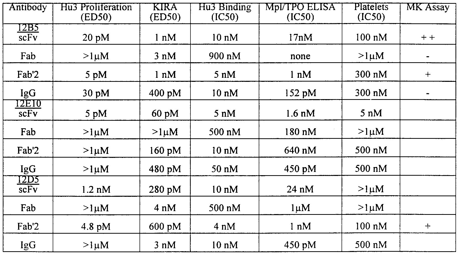
Claims
Priority Applications (6)
| Application Number | Priority Date | Filing Date | Title |
|---|---|---|---|
| DE69839270T DE69839270T2 (en) | 1997-08-25 | 1998-08-21 | AGONISTIC ANTIBODIES TO THE THROMBOPOIETIN RECEPTOR AND THEIR THERAPEUTIC USES |
| JP2000507802A JP2001513999A (en) | 1997-08-25 | 1998-08-21 | Agonist antibodies to thrombopoietin receptor and their therapeutic use |
| ES98939976T ES2304795T3 (en) | 1997-08-25 | 1998-08-21 | AGONIST ANTIBODIES FOR THE TROMBOPOYETINE RECEPTOR AND ITS THERAPEUTIC USES. |
| AU88312/98A AU755822B2 (en) | 1997-08-25 | 1998-08-21 | Agonist antibodies to the thrombopoietin receptor, and their therapeutic uses |
| EP98939976A EP1009831B1 (en) | 1997-08-25 | 1998-08-21 | Agonist antibodies to the thrombopoietin receptor, and their therapeutic uses |
| CA2301964A CA2301964C (en) | 1997-08-25 | 1998-08-21 | Agonist antibodies to the thrombopoietin receptor, and their therapeutic uses |
Applications Claiming Priority (2)
| Application Number | Priority Date | Filing Date | Title |
|---|---|---|---|
| US08/918,148 US6342220B1 (en) | 1997-08-25 | 1997-08-25 | Agonist antibodies |
| US08/918,148 | 1997-08-25 |
Publications (2)
| Publication Number | Publication Date |
|---|---|
| WO1999010494A2 true WO1999010494A2 (en) | 1999-03-04 |
| WO1999010494A3 WO1999010494A3 (en) | 1999-05-20 |
Family
ID=25439886
Family Applications (1)
| Application Number | Title | Priority Date | Filing Date |
|---|---|---|---|
| PCT/US1998/017364 WO1999010494A2 (en) | 1997-08-25 | 1998-08-21 | Agonist antibodies to the thrombopoietin receptor, and their therapeutic uses |
Country Status (9)
| Country | Link |
|---|---|
| US (1) | US6342220B1 (en) |
| EP (2) | EP1009831B1 (en) |
| JP (1) | JP2001513999A (en) |
| AT (1) | ATE389721T1 (en) |
| AU (1) | AU755822B2 (en) |
| CA (1) | CA2301964C (en) |
| DE (1) | DE69839270T2 (en) |
| ES (1) | ES2304795T3 (en) |
| WO (1) | WO1999010494A2 (en) |
Cited By (150)
| Publication number | Priority date | Publication date | Assignee | Title |
|---|---|---|---|---|
| WO2001021163A2 (en) * | 1999-09-21 | 2001-03-29 | Emory University | Methods and compositions for treating platelet-related disorders using mpl pathway inhibitory agents |
| WO2001072834A1 (en) * | 2000-03-30 | 2001-10-04 | Novartis Ag | Selectable marker genes |
| EP1149906A1 (en) * | 2000-04-25 | 2001-10-31 | Pliva, Farmaceutska, Industrija, Dionicko Drustvo | Thrombopoietin receptor modulating peptide |
| WO2001096601A2 (en) * | 2000-06-16 | 2001-12-20 | Isis Innovation Limited | Neurotransmission disorders |
| WO2002030954A1 (en) * | 2000-10-06 | 2002-04-18 | Kyowa Hakko Kogyo Co., Ltd. | Method of purifying antibody |
| WO2002046238A2 (en) * | 2000-12-05 | 2002-06-13 | Alexion Pharmaceuticals, Inc. | Rationally designed antibodies |
| EP1294769A1 (en) * | 2000-06-16 | 2003-03-26 | Human Genome Sciences, Inc. | Antibodies that immunospecifically bind to blys |
| WO2003091424A1 (en) | 2002-04-26 | 2003-11-06 | Chugai Seiyaku Kabushiki Kaisha | Method of screening agonistic antibody |
| WO2005033134A2 (en) * | 2003-09-30 | 2005-04-14 | Regeneron Pharmaceuticals, Inc. | Secreted protein therapeutics and uses thereof |
| EP1616880A1 (en) * | 2003-03-13 | 2006-01-18 | Chugai Seiyaku Kabushiki Kaisha | Ligand having agonistic activity to mutated receptor |
| US7064191B2 (en) | 2000-10-06 | 2006-06-20 | Kyowa Hakko Kogyo Co., Ltd. | Process for purifying antibody |
| WO2007087428A2 (en) * | 2006-01-25 | 2007-08-02 | Amgen Inc. | Thrombopoietic compounds |
| WO2007108559A1 (en) | 2006-03-23 | 2007-09-27 | Kirin Pharma Kabushiki Kaisha | Agonistic antibody directed against human thrombopoietin receptor |
| JPWO2005056604A1 (en) * | 2003-12-12 | 2007-12-06 | 中外製薬株式会社 | Anti-Mpl antibody |
| AU2003295623B2 (en) * | 2000-12-05 | 2008-06-05 | Alexion Pharmaceuticals, Inc. | Rationally designed antibodies |
| US7396917B2 (en) | 2000-12-05 | 2008-07-08 | Alexion Pharmaceuticals, Inc. | Rationally designed antibodies |
| US7531643B2 (en) | 1997-09-11 | 2009-05-12 | Chugai Seiyaku Kabushiki Kaisha | Monoclonal antibody inducing apoptosis |
| WO2009064944A2 (en) | 2007-11-16 | 2009-05-22 | Nuvelo, Inc. | Antibodies to lrp6 |
| EP2077279A1 (en) | 2000-06-28 | 2009-07-08 | Amgen Inc. | Thymic stromal lymphopoietin receptor molecules and uses thereof |
| WO2009086003A1 (en) | 2007-12-20 | 2009-07-09 | Xoma Technology Ltd. | Methods for the treatment of gout |
| EP2078729A1 (en) | 2005-02-04 | 2009-07-15 | Pfizer Products Inc. | PPY agonists and uses thereof |
| WO2009105269A1 (en) | 2008-02-20 | 2009-08-27 | Amgen Inc. | Antibodies directed to angiopoietin-1 and angiopoietin-2 and use thereof |
| US7662924B2 (en) | 2002-02-22 | 2010-02-16 | The Board Of Trustees Of The University Of Illinois | Beta chain-associated regulator of apoptosis |
| US7696325B2 (en) | 1999-03-10 | 2010-04-13 | Chugai Seiyaku Kabushiki Kaisha | Polypeptide inducing apoptosis |
| WO2010075238A1 (en) | 2008-12-23 | 2010-07-01 | Amgen Inc. | Human cgrp receptor binding proteins |
| EP2213685A1 (en) | 2002-09-06 | 2010-08-04 | Amgen Inc. | Therapeutic anti-IL-1R1 monoclonal antibody |
| EP2236519A1 (en) | 2007-09-18 | 2010-10-06 | Amgen, Inc | Human GM-CSF antigen binding proteins |
| US7879328B2 (en) | 2000-06-16 | 2011-02-01 | Human Genome Sciences, Inc. | Antibodies that immunospecifically bind to B lymphocyte stimulator |
| WO2011034660A1 (en) * | 2009-09-16 | 2011-03-24 | Immunomedics, Inc. | Class i anti-cea antibodies and uses thereof |
| WO2011038301A2 (en) | 2009-09-25 | 2011-03-31 | Xoma Technology Ltd. | Screening methods |
| WO2011038302A2 (en) | 2009-09-25 | 2011-03-31 | Xoma Technology Ltd. | Novel modulators |
| WO2011056600A1 (en) | 2009-10-26 | 2011-05-12 | Amgen Inc. | Human il-23 antigen binding proteins |
| WO2011071783A1 (en) | 2009-12-07 | 2011-06-16 | Amgen Inc. | Human antigen binding proteins that bind beta-klotho, fgf receptors and complexes thereof |
| EP2336177A1 (en) | 2004-08-04 | 2011-06-22 | Amgen, Inc | Antibodies to DKK-1 |
| EP2351838A1 (en) | 2000-10-20 | 2011-08-03 | Chugai Seiyaku Kabushiki Kaisha | Crosslinking agonistic antibodies |
| US8030275B2 (en) | 1999-09-07 | 2011-10-04 | Amgen Inc. | Methods for treating obesity using fibroblast growth factor-like polypeptides |
| US8034770B2 (en) | 2008-06-04 | 2011-10-11 | Amgen Inc. | FGF21 polypeptides comprising two or more mutations |
| US8034903B2 (en) | 2000-10-20 | 2011-10-11 | Chugai Seiyaku Kabushiki Kaisha | Degraded TPO agonist antibody |
| WO2011130417A2 (en) | 2010-04-15 | 2011-10-20 | Amgen Inc. | HUMAN FGF RECEPTOR AND β-KLOTHO BINDING PROTEINS |
| US8062906B2 (en) | 2000-08-18 | 2011-11-22 | Human Genome Sciences, Inc. | B-lymphocyte stimulator binding polypeptides and methods based thereon |
| US8062635B2 (en) | 2003-10-10 | 2011-11-22 | Chugai Seiyaku Kabushiki Kaisha | Bispecific antibody substituting for functional proteins |
| US8071092B1 (en) | 1996-10-25 | 2011-12-06 | Human Genome Sciences, Inc. | Methods of inhibiting B lymphocytes using antibodies to Neutrokine-alpha |
| WO2012010321A1 (en) | 2010-07-23 | 2012-01-26 | Erasmus University Medical Center Rotterdam | Foetal haemoglobin inhibitor |
| WO2012010855A1 (en) | 2010-07-23 | 2012-01-26 | Medical Research Council | Intracellular immunity |
| WO2012058393A2 (en) | 2010-10-27 | 2012-05-03 | Amgen Inc. | Dkk1 antibodies and methods of use |
| US8188040B2 (en) | 2009-05-05 | 2012-05-29 | Amgen Inc. | FGF21 mutants and uses thereof |
| US8211649B2 (en) | 2006-03-31 | 2012-07-03 | Human Genome Sciences, Inc. | Methods of diagnosing and prognosing hodgkin's lymphoma |
| WO2012097238A2 (en) | 2011-01-14 | 2012-07-19 | Five Prime Therapeutics, Inc. | Il-27 antagonists for treating inflammatory diseases |
| WO2012118903A2 (en) | 2011-03-01 | 2012-09-07 | Amgen Inc. | Bispecific binding agents |
| US8278421B2 (en) | 2006-03-20 | 2012-10-02 | Xoma Techolology Ltd. | Human antibodies specific for gastrin materials and methods |
| WO2012167143A1 (en) | 2011-06-03 | 2012-12-06 | Xoma Technology Ltd. | Antibodies specific for tgf-beta |
| WO2012170438A2 (en) | 2011-06-06 | 2012-12-13 | Amgen Inc. | HUMAN ANTIGEN BINDING PROTEINS THAT BIND TO A COMPLEX COMPRISING β-KLOTHO AND AN FGF RECEPTOR |
| US8334105B2 (en) | 2004-12-20 | 2012-12-18 | Northshore University Healthsystem | Methods for treating cardiac disease by modifying an N-terminal domain of troponin I |
| WO2012174534A2 (en) | 2011-06-17 | 2012-12-20 | Amgen Inc. | Method of treating or ameliorating metabolic disorders using clec-2 |
| US8409577B2 (en) | 2006-06-12 | 2013-04-02 | Emergent Product Development Seattle, Llc | Single chain multivalent binding proteins with effector function |
| EP2589610A1 (en) | 2007-08-21 | 2013-05-08 | Amgen, Inc | Human c-fms antigen binding proteins |
| WO2013086443A1 (en) | 2011-12-08 | 2013-06-13 | Amgen Inc. | Agonistic human lcat antigen binding proteins and their use in therapy |
| WO2013096516A1 (en) | 2011-12-19 | 2013-06-27 | Xoma Technology Ltd. | Methods for treating acne |
| WO2013117735A1 (en) | 2012-02-09 | 2013-08-15 | Medical Research Council | Conjugate comprising an agent and an antiviral ligand and its use in a method for delivering the agent intracellularly |
| WO2013163377A1 (en) | 2012-04-27 | 2013-10-31 | Novo Nordisk A/S | Human cd30 ligand antigen binding proteins |
| WO2013169264A1 (en) | 2012-05-11 | 2013-11-14 | Five Prime Therapeutics, Inc. | Methods of treating conditions with antibodies that bind colony stimulating factor 1 receptor (csf1r) |
| EP2671891A2 (en) | 2008-06-27 | 2013-12-11 | Amgen Inc. | Ang-2 inhibition to treat multiple sclerosis |
| US8623375B2 (en) | 2010-05-14 | 2014-01-07 | Baxter International Inc. | Chimeric OspA genes, proteins, and methods of use thereof |
| WO2014144632A2 (en) | 2013-03-15 | 2014-09-18 | Amgen Inc. | Human pac1 antibodies |
| WO2014144553A1 (en) | 2013-03-15 | 2014-09-18 | Amgen Inc. | Secreted frizzle-related protein 5 (sfrp5) binding proteins and methods of treatment |
| WO2014159764A1 (en) | 2013-03-14 | 2014-10-02 | Amgen Inc. | Chrdl-1 antigen binding proteins and methods of treatment |
| US8853366B2 (en) | 2001-01-17 | 2014-10-07 | Emergent Product Development Seattle, Llc | Binding domain-immunoglobulin fusion proteins |
| US9023367B2 (en) | 2012-07-27 | 2015-05-05 | Baxter International Inc. | Compositions comprising chimeric OSPA molecules and methods of use thereof |
| US9074186B2 (en) | 2012-08-15 | 2015-07-07 | Boston Medical Center Corporation | Production of red blood cells and platelets from stem cells |
| US9101609B2 (en) | 2008-04-11 | 2015-08-11 | Emergent Product Development Seattle, Llc | CD37 immunotherapeutic and combination with bifunctional chemotherapeutic thereof |
| US9145450B2 (en) | 1998-10-23 | 2015-09-29 | Amgen Inc. | Thrombopoietic compounds |
| US9168286B2 (en) | 2005-10-13 | 2015-10-27 | Human Genome Sciences, Inc. | Methods and compositions for use in treatment of patients with autoantibody positive disease |
| WO2015200089A1 (en) | 2014-06-23 | 2015-12-30 | Five Prime Therapeutics, Inc. | Methods of treating conditions with antibodies that bind colony stimulating factor 1 receptor (csf1r) |
| WO2016040767A2 (en) | 2014-09-12 | 2016-03-17 | Amgen Inc. | Chrdl-1 epitopes and antibodies |
| WO2016069727A1 (en) | 2014-10-29 | 2016-05-06 | Five Prime Therapeutics, Inc. | Combination therapy for cancer |
| WO2016106180A1 (en) | 2014-12-22 | 2016-06-30 | Five Prime Therapeutics, Inc. | Anti-csf1r antibodies for treating pvns |
| WO2016111947A2 (en) | 2015-01-05 | 2016-07-14 | Jounce Therapeutics, Inc. | Antibodies that inhibit tim-3:lilrb2 interactions and uses thereof |
| WO2016141111A1 (en) | 2015-03-03 | 2016-09-09 | Xoma (Us) Llc | Treatment of post-prandial hyperinsulinemia and hypoglycemia after bariatric surgery |
| WO2016154177A2 (en) | 2015-03-23 | 2016-09-29 | Jounce Therapeutics, Inc. | Antibodies to icos |
| WO2016161410A2 (en) | 2015-04-03 | 2016-10-06 | Xoma Technology Ltd. | Treatment of cancer using inhibitors of tgf-beta and pd-1 |
| US9464139B2 (en) | 2013-08-30 | 2016-10-11 | Amgen Inc. | GITR antigen binding proteins and methods of use thereof |
| WO2016168149A1 (en) | 2015-04-13 | 2016-10-20 | Five Prime Therapeutics, Inc. | Combination therapy for cancer |
| WO2016179194A1 (en) | 2015-05-04 | 2016-11-10 | Jounce Therapeutics, Inc. | Lilra3 and method of using the same |
| US9493530B2 (en) | 2009-05-05 | 2016-11-15 | Amgen Inc. | FGF21 mutants comprising a mutation at position 98, 171 and/or 180 |
| EP3124045A2 (en) | 2006-12-20 | 2017-02-01 | Xoma (Us) Llc | Treatment of il-1 beta related diseases |
| WO2017059371A1 (en) | 2015-10-01 | 2017-04-06 | Amgen Inc. | Treatment of bile acid disorders |
| WO2017070423A1 (en) | 2015-10-22 | 2017-04-27 | Jounce Therapeutics, Inc. | Gene signatures for determining icos expression |
| WO2017091577A1 (en) | 2015-11-23 | 2017-06-01 | Five Prime Therapeutics, Inc. | Fgfr2 inhibitors alone or in combination with immune stimulating agents in cancer treatment |
| US9670269B2 (en) | 2006-03-31 | 2017-06-06 | Chugai Seiyaku Kabushiki Kaisha | Methods of modifying antibodies for purification of bispecific antibodies |
| US9777066B2 (en) | 2005-06-10 | 2017-10-03 | Chugai Seiyaku Kabushiki Kaisha | Pharmaceutical compositions containing sc(Fv)2 |
| US9828429B2 (en) | 2007-09-26 | 2017-11-28 | Chugai Seiyaku Kabushiki Kaisha | Method of modifying isoelectric point of antibody via amino acid substitution in CDR |
| WO2018005950A1 (en) | 2016-07-01 | 2018-01-04 | Five Prime Therapeutics, Inc. | Combined anti tumor therapy with a gitr agonist and cpg |
| WO2018026748A1 (en) | 2016-08-01 | 2018-02-08 | Xoma (Us) Llc | Parathyroid hormone receptor 1 (pth1r) antibodies and uses thereof |
| WO2018064255A2 (en) | 2016-09-28 | 2018-04-05 | Xoma (Us) Llc | Antibodies that bind interleukin-2 and uses thereof |
| WO2018085358A1 (en) | 2016-11-02 | 2018-05-11 | Jounce Therapeutics, Inc. | Antibodies to pd-1 and uses thereof |
| US9975966B2 (en) | 2014-09-26 | 2018-05-22 | Chugai Seiyaku Kabushiki Kaisha | Cytotoxicity-inducing theraputic agent |
| WO2018091720A1 (en) | 2016-11-21 | 2018-05-24 | INSERM (Institut National de la Santé et de la Recherche Médicale) | Methods and pharmaceutical compositions for the prophylactic treatment of metastases |
| US10011858B2 (en) | 2005-03-31 | 2018-07-03 | Chugai Seiyaku Kabushiki Kaisha | Methods for producing polypeptides by regulating polypeptide association |
| WO2018128454A1 (en) | 2017-01-06 | 2018-07-12 | 에이비엘바이오 주식회사 | ANTI-α-SYN ANTIBODY AND USE THEREOF |
| EP3357510A1 (en) | 2010-05-04 | 2018-08-08 | Five Prime Therapeutics, Inc. | Antibodies that bind csf1r |
| US10080790B2 (en) | 2014-09-19 | 2018-09-25 | The Regents Of The University Of Michigan | Staphylococcus aureus materials and methods |
| WO2018183608A1 (en) | 2017-03-31 | 2018-10-04 | Five Prime Therapeutics, Inc. | Combination therapy for cancer using anti-gitr antibodies |
| WO2018213304A1 (en) | 2017-05-16 | 2018-11-22 | Five Prime Therapeutics, Inc. | Anti-fgfr2 antibodies in combination with chemotherapy agents in cancer treatment |
| US10143748B2 (en) | 2005-07-25 | 2018-12-04 | Aptevo Research And Development Llc | B-cell reduction using CD37-specific and CD20-specific binding molecules |
| WO2018222770A1 (en) | 2017-05-31 | 2018-12-06 | Amgen Inc. | Anti-jagged1 antigen binding proteins |
| US10156562B2 (en) | 2014-05-16 | 2018-12-18 | Amgen Inc. | Assay for detecting Th1 and Th2 cell populations |
| WO2018237097A1 (en) | 2017-06-20 | 2018-12-27 | Amgen Inc. | Method of treating or ameliorating metabolic disorders using binding proteins for gastric inhibitory peptide receptor (gipr) in combination with glp-1 agonists |
| WO2019020480A1 (en) | 2017-07-24 | 2019-01-31 | INSERM (Institut National de la Santé et de la Recherche Médicale) | Antibodies and peptides to treat hcmv related diseases |
| WO2019028382A1 (en) | 2017-08-04 | 2019-02-07 | Amgen Inc. | Method of conjugation of cys-mabs |
| WO2019035034A1 (en) | 2017-08-16 | 2019-02-21 | The Broad Institute, Inc. | Neuronal assay method involving calcineurin |
| WO2019055537A1 (en) | 2017-09-13 | 2019-03-21 | Five Prime Therapeutics, Inc. | Combination anti-csf1r and anti-pd-1 antibody combination therapy for pancreatic cancer |
| US10253101B2 (en) | 2015-08-06 | 2019-04-09 | Xoma (Us) Llc | Antibody fragments against the insulin receptor and uses thereof to treat hypoglycemia |
| US10294303B2 (en) | 2015-12-23 | 2019-05-21 | Amgen Inc. | Method of treating or ameliorating metabolic disorders using binding proteins for gastric inhibitory peptide receptor (GIPR) in combination with GLP-1 agonists |
| WO2019108806A1 (en) | 2017-11-29 | 2019-06-06 | Board Of Regents Of The University Of Texas System | Compositions and methods for cancer therapy |
| WO2019117684A1 (en) | 2017-12-14 | 2019-06-20 | 에이비엘바이오 주식회사 | Bispecific antibody to a-syn/igf1r and use thereof |
| WO2019126514A2 (en) | 2017-12-22 | 2019-06-27 | Jounce Therapeutics, Inc. | Antibodies for lilrb2 |
| US10450381B2 (en) | 2010-11-17 | 2019-10-22 | Chugai Seiyaku Kabushiki Kaisha | Methods of treatment that include the administration of bispecific antibodies |
| US10626176B2 (en) | 2014-10-31 | 2020-04-21 | Jounce Therapeutics, Inc. | Methods of treating conditions with antibodies that bind B7-H4 |
| WO2020089203A1 (en) | 2018-10-29 | 2020-05-07 | INSERM (Institut National de la Santé et de la Recherche Médicale) | Composition for the treatment of antibody deficiencies |
| WO2020094609A1 (en) | 2018-11-06 | 2020-05-14 | INSERM (Institut National de la Santé et de la Recherche Médicale) | Methods and pharmaceutical compositions for the treatment of acute myeloid leukemia by eradicating leukemic stem cells |
| WO2020120786A1 (en) | 2018-12-14 | 2020-06-18 | INSERM (Institut National de la Santé et de la Recherche Médicale) | Isolated mhc-derived human peptides and uses thereof for stimulating and activating the suppressive function of cd8+cd45rclow tregs |
| EP3679949A1 (en) | 2012-08-31 | 2020-07-15 | Five Prime Therapeutics, Inc. | Methods of treating conditions with antibodies that bind colony stimulating factor 1 receptor (csf1r) |
| WO2020169472A2 (en) | 2019-02-18 | 2020-08-27 | INSERM (Institut National de la Santé et de la Recherche Médicale) | Methods of inducing phenotypic changes in macrophages |
| US10759870B2 (en) | 2017-09-29 | 2020-09-01 | Chugai Seiyaku Kabushiki Kaisha | Multispecific antigen-binding molecules having blood coagulation factor VIII (FVIII) cofactor function-substituting activity and pharmaceutical formulations containing such a molecule as an active ingredient |
| WO2020176549A1 (en) * | 2019-02-26 | 2020-09-03 | Sorrento Therapeutics, Inc. | Antigen binding proteins that bind bcma |
| WO2020251316A1 (en) | 2019-06-14 | 2020-12-17 | 에이비엘바이오 주식회사 | BISPECIFIC ANTIBODY AGAINST α-SYN/IGF1R AND USE THEREOF |
| US10905772B2 (en) | 2017-01-17 | 2021-02-02 | Amgen Inc. | Method of treating or ameliorating metabolic disorders using GLP-1 receptor agonists conjugated to antagonists for gastric inhibitory peptide receptor (GIPR) |
| WO2021058729A1 (en) | 2019-09-27 | 2021-04-01 | INSERM (Institut National de la Santé et de la Recherche Médicale) | Anti-müllerian inhibiting substance type i receptor antibodies and uses thereof |
| WO2021058763A1 (en) | 2019-09-27 | 2021-04-01 | INSERM (Institut National de la Santé et de la Recherche Médicale) | Anti-müllerian inhibiting substance antibodies and uses thereof |
| WO2021076930A1 (en) | 2019-10-18 | 2021-04-22 | The Regents Of The University Of California | Plxdc activators and their use in the treatment of blood vessel disorders |
| WO2021099600A1 (en) | 2019-11-22 | 2021-05-27 | INSERM (Institut National de la Santé et de la Recherche Médicale) | Inhibitors of adrenomedullin for the treatment of acute myeloid leukemia by eradicating leukemic stem cells |
| US11046784B2 (en) | 2006-03-31 | 2021-06-29 | Chugai Seiyaku Kabushiki Kaisha | Methods for controlling blood pharmacokinetics of antibodies |
| WO2021130717A1 (en) | 2019-12-26 | 2021-07-01 | 에이비엘바이오 주식회사 | Method for purifying biologically active peptide by using protein a affinity chromatography |
| WO2021139682A1 (en) | 2020-01-07 | 2021-07-15 | Hifibio (Hk) Limited | Anti-galectin-9 antibody and uses thereof |
| WO2021141907A1 (en) | 2020-01-06 | 2021-07-15 | Hifibio (Hong Kong) Limited | Anti-tnfr2 antibody and uses thereof |
| WO2021163064A2 (en) | 2020-02-14 | 2021-08-19 | Jounce Therapeutics, Inc. | Antibodies and fusion proteins that bind to ccr8 and uses thereof |
| US11124576B2 (en) | 2013-09-27 | 2021-09-21 | Chungai Seiyaku Kabushiki Kaisha | Method for producing polypeptide heteromultimer |
| US11142587B2 (en) | 2015-04-01 | 2021-10-12 | Chugai Seiyaku Kabushiki Kaisha | Method for producing polypeptide hetero-oligomer |
| US11150254B2 (en) | 2014-09-26 | 2021-10-19 | Chugai Seiyaku Kabushiki Kaisha | Method for measuring reactivity of FVIII |
| WO2021228956A1 (en) | 2020-05-12 | 2021-11-18 | INSERM (Institut National de la Santé et de la Recherche Médicale) | New method to treat cutaneous t-cell lymphomas and tfh derived lymphomas |
| US11214623B2 (en) | 2014-09-26 | 2022-01-04 | Chugai Seiyaku Kabushiki Kaisha | Antibody capable of neutralizing substance having activity alternative to function of coagulation factor VIII (FVIII) |
| WO2022033419A2 (en) | 2020-08-10 | 2022-02-17 | Shanghai Xbh Biotechnology Co., Ltd. | Compositions and methods for treating autoimmune diseases and cancers by targeting igsf8 |
| WO2022081718A1 (en) | 2020-10-14 | 2022-04-21 | Five Prime Therapeutics, Inc. | Anti-c-c chemokine receptor 8 (ccr8) antibodies and methods of use thereof |
| US11352438B2 (en) | 2016-09-06 | 2022-06-07 | Chugai Seiyaku Kabushiki Kaisha | Methods of using a bispecific antibody that recognizes coagulation factor IX and/or activated coagulation factor IX and coagulation factor X and/or activated coagulation factor X |
| US11352426B2 (en) | 2015-09-21 | 2022-06-07 | Aptevo Research And Development Llc | CD3 binding polypeptides |
| US11401328B2 (en) | 2018-07-09 | 2022-08-02 | Five Prime Therapeutics, Inc. | Antibodies binding to ILT4 |
| US11649262B2 (en) | 2015-12-28 | 2023-05-16 | Chugai Seiyaku Kabushiki Kaisha | Method for promoting efficiency of purification of Fc region-containing polypeptide |
| WO2023139292A1 (en) | 2022-01-24 | 2023-07-27 | Cambridge Enterprise Limited | Tau therapy |
| WO2023235699A1 (en) | 2022-05-31 | 2023-12-07 | Jounce Therapeutics, Inc. | Antibodies to lilrb4 and uses thereof |
| US11891445B1 (en) | 2018-05-24 | 2024-02-06 | Abl Bio Inc. | Anti-B7-H3 antibody and use thereof |
Families Citing this family (38)
| Publication number | Priority date | Publication date | Assignee | Title |
|---|---|---|---|---|
| TWI242563B (en) * | 1998-04-30 | 2005-11-01 | Tanox Inc | Monoclonal agonist antibodies which specifically bind to or interact with human G-CSF receptor |
| USD464436S1 (en) * | 1999-11-19 | 2002-10-15 | Fox Lite, Inc. | Collapsible skylight tube having open ends and a light reflecting inner surface |
| CA2441953A1 (en) * | 2001-03-23 | 2002-10-03 | The Board Of Regents Of The University Of Texas System | Methods for inhibition of angiogenesis, tumor growth and metastasis by fully human anti-il8 and anti-muc18 in diverse types of tumors |
| WO2003015716A2 (en) * | 2001-08-13 | 2003-02-27 | Ige Therapeutics, Inc. | Immunoglobulin e vaccines and methods of use thereof |
| KR100467750B1 (en) * | 2001-11-29 | 2005-01-24 | 씨제이 주식회사 | Fusion protein having the enhanced in vivo activity of erythropoietin |
| DE60226499D1 (en) * | 2001-12-28 | 2008-06-19 | Amgen Fremont Inc | USE OF ANTIBODIES TO THE MUC18 ANTIGEN |
| JP2005516965A (en) * | 2001-12-28 | 2005-06-09 | アブジェニックス・インコーポレーテッド | Method using anti-MUC18 antibody |
| US20050069549A1 (en) | 2002-01-14 | 2005-03-31 | William Herman | Targeted ligands |
| GB0202206D0 (en) * | 2002-01-31 | 2002-03-20 | Bioinvent Int Ab | Method of making libraries of anti-ligands |
| EP1482973B1 (en) | 2002-03-15 | 2009-08-19 | Schering Corporation | Methods of modulating cd200 receptors |
| WO2004003144A2 (en) * | 2002-07-01 | 2004-01-08 | Human Genome Sciences, Inc. | Antibodies that specifically bind to reg iv |
| CA2492457A1 (en) * | 2002-08-02 | 2004-02-12 | Craig A. Rosen | Antibodies against c3a receptor |
| US20040028661A1 (en) * | 2002-08-07 | 2004-02-12 | Bartelmez Stephen H. | Expansion of cells using thrombopoietin and anti-transforming growth factor-beta |
| ATE414105T1 (en) * | 2002-10-11 | 2008-11-15 | Chugai Pharmaceutical Co Ltd | CELL DEATH-INDUCING AGENT |
| JP2004279086A (en) * | 2003-03-13 | 2004-10-07 | Konica Minolta Holdings Inc | Radiation image conversion panel and method for manufacturing it |
| JPWO2004087763A1 (en) * | 2003-03-31 | 2006-07-27 | 中外製薬株式会社 | Modified antibody against CD22 and use thereof |
| WO2004111233A1 (en) * | 2003-06-11 | 2004-12-23 | Chugai Seiyaku Kabushiki Kaisha | Process for producing antibody |
| US20070281327A1 (en) * | 2003-12-12 | 2007-12-06 | Kiyotaka Nakano | Methods of Screening for Modified Antibodies With Agonistic Activities |
| WO2005056603A1 (en) * | 2003-12-12 | 2005-06-23 | Chugai Seiyaku Kabushiki Kaisha | Cell death inducing agent |
| US20080274110A1 (en) * | 2004-04-09 | 2008-11-06 | Shuji Ozaki | Cell Death-Inducing Agents |
| TW200605906A (en) * | 2004-05-11 | 2006-02-16 | Chugai Pharmaceutical Co Ltd | Remedy for thrombopenia |
| US20060275288A1 (en) * | 2005-01-20 | 2006-12-07 | Grihalde Nelson D | GLP-1 receptor agonist and allosteric modulator monoclonal antibodies and uses thereof |
| JP5057967B2 (en) | 2005-03-31 | 2012-10-24 | 中外製薬株式会社 | sc (Fv) 2 structural isomer |
| EP1927367A4 (en) * | 2005-05-18 | 2009-08-12 | Univ Tokushima | Novel pharmaceutical using anti-hla antibody |
| KR101367544B1 (en) * | 2005-06-10 | 2014-02-26 | 추가이 세이야쿠 가부시키가이샤 | Stabilizer for protein preparation comprising meglumine and use thereof |
| WO2006132341A1 (en) * | 2005-06-10 | 2006-12-14 | Chugai Seiyaku Kabushiki Kaisha | sc(Fv)2 SITE-DIRECTED MUTANT |
| US20100040600A1 (en) * | 2006-06-14 | 2010-02-18 | Chugai Seiyaku Kabushiki Kaisha | Agents for Promoting the Growth of Hematopoietic Stem Cells |
| US20100150927A1 (en) * | 2006-07-13 | 2010-06-17 | Chugai Seiyaku Kabushiki Kaisha | Cell death inducer |
| DK3199180T3 (en) * | 2007-03-08 | 2022-03-21 | Humanigen Inc | EPHA3 ANTIBODIES FOR THE TREATMENT OF FIXED TUMORS |
| CL2008000719A1 (en) * | 2007-03-12 | 2008-09-05 | Univ Tokushima Chugai Seiyaku | THERAPEUTIC AGENT FOR CANCER RESISTANT TO CHEMOTHERAPEUTIC AGENTS THAT UNDERSTAND AN ANTIBODY THAT RECOGNIZES IT CLASS I AS ACTIVE INGREDIENT; PHARMACEUTICAL COMPOSITION THAT INCLUDES SUCH ANTIBODY; AND METHOD TO TREAT CANCER RESISTANT TO |
| CN102625811B (en) | 2008-10-10 | 2016-09-21 | 安姆根有限公司 | FGF21 mutant and application thereof |
| JP2014515476A (en) * | 2011-05-20 | 2014-06-30 | アボットジャパン株式会社 | Immunoassay methods and reagents for reducing non-specific binding |
| WO2013074636A1 (en) * | 2011-11-14 | 2013-05-23 | New York University | Muscle specific receptor kinase and modulation thereof |
| SI2870247T1 (en) * | 2012-07-05 | 2019-10-30 | Hoffmann La Roche | Expression and secretion system |
| WO2014035693A2 (en) * | 2012-08-31 | 2014-03-06 | The Scripps Research Institute | Methods and compositions related to modulators of eukaryotic cells |
| US20190112380A1 (en) | 2016-03-29 | 2019-04-18 | University Of Southern California | Chimeric antigen receptors targeting cancer |
| WO2018222901A1 (en) | 2017-05-31 | 2018-12-06 | Elstar Therapeutics, Inc. | Multispecific molecules that bind to myeloproliferative leukemia (mpl) protein and uses thereof |
| GB202003444D0 (en) * | 2020-03-10 | 2020-04-22 | argenx BV | Musk agonist antibody |
Citations (3)
| Publication number | Priority date | Publication date | Assignee | Title |
|---|---|---|---|---|
| WO1995014930A1 (en) * | 1993-11-23 | 1995-06-01 | Genentech, Inc. | Kinase receptor activation assay |
| WO1995027061A1 (en) * | 1994-04-04 | 1995-10-12 | Genentech, Inc. | Protein tyrosine kinase agonist antibodies |
| WO1995027062A1 (en) * | 1994-04-04 | 1995-10-12 | Genentech, Inc. | Agonist antibodies against the flk2/flt3 receptor and uses thereof |
Family Cites Families (70)
| Publication number | Priority date | Publication date | Assignee | Title |
|---|---|---|---|---|
| US3773919A (en) | 1969-10-23 | 1973-11-20 | Du Pont | Polylactide-drug mixtures |
| US4179337A (en) | 1973-07-20 | 1979-12-18 | Davis Frank F | Non-immunogenic polypeptides |
| USRE30985E (en) | 1978-01-01 | 1982-06-29 | Serum-free cell culture media | |
| US4263428A (en) | 1978-03-24 | 1981-04-21 | The Regents Of The University Of California | Bis-anthracycline nucleic acid function inhibitors and improved method for administering the same |
| JPS6023084B2 (en) | 1979-07-11 | 1985-06-05 | 味の素株式会社 | blood substitute |
| US4399216A (en) | 1980-02-25 | 1983-08-16 | The Trustees Of Columbia University | Processes for inserting DNA into eucaryotic cells and for producing proteinaceous materials |
| ZA811368B (en) | 1980-03-24 | 1982-04-28 | Genentech Inc | Bacterial polypedtide expression employing tryptophan promoter-operator |
| US4626507A (en) | 1980-09-25 | 1986-12-02 | The Salk Institute For Biological Studies | Hybridomas producing monoclonal antibodies specific for a human cell surface glycoprotein |
| DE3169595D1 (en) | 1980-11-10 | 1985-05-02 | Gersonde Klaus | Method of preparing lipid vesicles by ultrasonic treatment, the use of this method and apparatus for its application |
| US4419446A (en) | 1980-12-31 | 1983-12-06 | The United States Of America As Represented By The Department Of Health And Human Services | Recombinant DNA process utilizing a papilloma virus DNA as a vector |
| IE52535B1 (en) | 1981-02-16 | 1987-12-09 | Ici Plc | Continuous release pharmaceutical compositions |
| US4485045A (en) | 1981-07-06 | 1984-11-27 | Research Corporation | Synthetic phosphatidyl cholines useful in forming liposomes |
| NZ201705A (en) | 1981-08-31 | 1986-03-14 | Genentech Inc | Recombinant dna method for production of hepatitis b surface antigen in yeast |
| US4640835A (en) | 1981-10-30 | 1987-02-03 | Nippon Chemiphar Company, Ltd. | Plasminogen activator derivatives |
| JPS58118008A (en) | 1982-01-06 | 1983-07-13 | Nec Corp | Data processor |
| DE3374837D1 (en) | 1982-02-17 | 1988-01-21 | Ciba Geigy Ag | Lipids in the aqueous phase |
| DE3218121A1 (en) | 1982-05-14 | 1983-11-17 | Leskovar, Peter, Dr.-Ing., 8000 München | Pharmaceutical compositions for tumour treatment |
| US4943529A (en) | 1982-05-19 | 1990-07-24 | Gist-Brocades Nv | Kluyveromyces as a host strain |
| EP0102324A3 (en) | 1982-07-29 | 1984-11-07 | Ciba-Geigy Ag | Lipids and surfactants in an aqueous medium |
| US4558005A (en) | 1982-09-13 | 1985-12-10 | University Patents, Inc. | Monoclonal anti-erythropoietin |
| US4601978A (en) | 1982-11-24 | 1986-07-22 | The Regents Of The University Of California | Mammalian metallothionein promoter system |
| US4560655A (en) | 1982-12-16 | 1985-12-24 | Immunex Corporation | Serum-free cell culture medium and process for making same |
| US4657866A (en) | 1982-12-21 | 1987-04-14 | Sudhir Kumar | Serum-free, synthetic, completely chemically defined tissue culture media |
| US4713339A (en) | 1983-01-19 | 1987-12-15 | Genentech, Inc. | Polycistronic expression vector construction |
| AU2353384A (en) | 1983-01-19 | 1984-07-26 | Genentech Inc. | Amplification in eukaryotic host cells |
| US4816567A (en) | 1983-04-08 | 1989-03-28 | Genentech, Inc. | Recombinant immunoglobin preparations |
| US4544545A (en) | 1983-06-20 | 1985-10-01 | Trustees University Of Massachusetts | Liposomes containing modified cholesterol for organ targeting |
| US4710457A (en) | 1983-06-29 | 1987-12-01 | Sloan-Kettering Institute For Cancer Research | Monoclonal antibody for human hematopoietic glycoproteins and method |
| HUT35524A (en) | 1983-08-02 | 1985-07-29 | Hoechst Ag | Process for preparing pharmaceutical compositions containing regulatory /regulative/ peptides providing for the retarded release of the active substance |
| AU3145184A (en) | 1983-08-16 | 1985-02-21 | Zymogenetics Inc. | High expression of foreign genes in schizosaccharomyces pombe |
| ATE159858T1 (en) | 1983-09-26 | 1997-11-15 | Ehrenfeld Udo | AGENT AND PRODUCT FOR THE DIAGNOSIS AND THERAPY OF TUMORS AND FOR THE TREATMENT OF WEAKNESSES OF THE CELLULAR AND HUMORAL IMMUNE DEFENSE |
| US4767704A (en) | 1983-10-07 | 1988-08-30 | Columbia University In The City Of New York | Protein-free culture medium |
| US4615885A (en) | 1983-11-01 | 1986-10-07 | Terumo Kabushiki Kaisha | Pharmaceutical composition containing urokinase |
| US4496689A (en) | 1983-12-27 | 1985-01-29 | Miles Laboratories, Inc. | Covalently attached complex of alpha-1-proteinase inhibitor with a water soluble polymer |
| US4965199A (en) | 1984-04-20 | 1990-10-23 | Genentech, Inc. | Preparation of functional human factor VIII in mammalian cells using methotrexate based selection |
| US4879231A (en) | 1984-10-30 | 1989-11-07 | Phillips Petroleum Company | Transformation of yeasts of the genus pichia |
| US4683195A (en) | 1986-01-30 | 1987-07-28 | Cetus Corporation | Process for amplifying, detecting, and/or-cloning nucleic acid sequences |
| EP0206448B1 (en) | 1985-06-19 | 1990-11-14 | Ajinomoto Co., Inc. | Hemoglobin combined with a poly(alkylene oxide) |
| GB8516415D0 (en) | 1985-06-28 | 1985-07-31 | Celltech Ltd | Culture of animal cells |
| US4927762A (en) | 1986-04-01 | 1990-05-22 | Cell Enterprises, Inc. | Cell culture medium with antioxidant |
| GB8610600D0 (en) | 1986-04-30 | 1986-06-04 | Novo Industri As | Transformation of trichoderma |
| US4791192A (en) | 1986-06-26 | 1988-12-13 | Takeda Chemical Industries, Ltd. | Chemically modified protein with polyethyleneglycol |
| EP0266032A1 (en) | 1986-08-29 | 1988-05-04 | Beecham Group Plc | Modified fibrinolytic enzyme |
| IL87737A (en) | 1987-09-11 | 1993-08-18 | Genentech Inc | Method for culturing polypeptide factor dependent vertebrate recombinant cells |
| GB8724885D0 (en) | 1987-10-23 | 1987-11-25 | Binns M M | Fowlpox virus promotors |
| EP0321196A3 (en) | 1987-12-18 | 1990-07-18 | Mycogen Plant Science, Inc. | 780 t-dna gene transcription activator |
| DE3889546T2 (en) | 1987-12-21 | 1994-09-08 | Univ Toledo | TRANSFORMATION OF Germinating PLANT SEEDS WITH THE HELP OF AGROBACTERIUM. |
| US5258287A (en) | 1988-03-22 | 1993-11-02 | Genentech, Inc. | DNA encoding and methods of production of insulin-like growth factor binding protein BP53 |
| AU4005289A (en) | 1988-08-25 | 1990-03-01 | Smithkline Beecham Corporation | Recombinant saccharomyces |
| US5223409A (en) | 1988-09-02 | 1993-06-29 | Protein Engineering Corp. | Directed evolution of novel binding proteins |
| WO1990003430A1 (en) | 1988-09-23 | 1990-04-05 | Cetus Corporation | Cell culture medium for enhanced cell growth, culture longevity and product expression |
| FR2646437B1 (en) | 1989-04-28 | 1991-08-30 | Transgene Sa | NOVEL DNA SEQUENCES, THEIR APPLICATION AS A SEQUENCE ENCODING A SIGNAL PEPTIDE FOR THE SECRETION OF MATURE PROTEINS BY RECOMBINANT YEASTS, EXPRESSION CASSETTES, PROCESSED YEASTS AND PROCESS FOR PREPARING THE SAME |
| EP0402226A1 (en) | 1989-06-06 | 1990-12-12 | Institut National De La Recherche Agronomique | Transformation vectors for yeast yarrowia |
| DE3920358A1 (en) | 1989-06-22 | 1991-01-17 | Behringwerke Ag | BISPECIFIC AND OLIGO-SPECIFIC, MONO- AND OLIGOVALENT ANTI-BODY CONSTRUCTS, THEIR PRODUCTION AND USE |
| DK168302B1 (en) | 1989-06-29 | 1994-03-07 | Danisco | Method of introducing molecules, especially genetic material into plant cells |
| FR2649120B1 (en) | 1989-06-30 | 1994-01-28 | Cayla | NOVEL STRAIN AND ITS MUTANTS OF FILAMENTOUS MUSHROOMS, PROCESS FOR PRODUCING RECOMBINANT PROTEINS USING SAID STRAIN, AND STRAINS AND PROTEINS OBTAINED BY SAID METHOD |
| WO1991008291A2 (en) | 1989-11-22 | 1991-06-13 | Genentech, Inc. | Latency associated peptide and uses therefor |
| GB9015198D0 (en) | 1990-07-10 | 1990-08-29 | Brien Caroline J O | Binding substance |
| WO1992020791A1 (en) | 1990-07-10 | 1992-11-26 | Cambridge Antibody Technology Limited | Methods for producing members of specific binding pairs |
| WO1993006213A1 (en) | 1991-09-23 | 1993-04-01 | Medical Research Council | Production of chimeric antibodies - a combinatorial approach |
| JPH07501451A (en) | 1991-11-25 | 1995-02-16 | エンゾン・インコーポレイテッド | Multivalent antigen binding protein |
| PT1696031E (en) | 1991-12-02 | 2010-06-25 | Medical Res Council | Production of anti-self antibodies from antibody segment repertoires and displayed on phage |
| WO1993019172A1 (en) | 1992-03-24 | 1993-09-30 | Cambridge Antibody Technology Limited | Methods for producing members of specific binding pairs |
| GB9313509D0 (en) | 1993-06-30 | 1993-08-11 | Medical Res Council | Chemisynthetic libraries |
| WO1995015388A1 (en) | 1993-12-03 | 1995-06-08 | Medical Research Council | Recombinant binding proteins and peptides |
| SG47030A1 (en) | 1994-01-03 | 1998-03-20 | Genentech Inc | Thrombopoietin |
| US5571686A (en) | 1994-04-14 | 1996-11-05 | Massachusetts Institute Of Technology | Method of using megapoietin for prolonging the survival & viability of platlets |
| US5885574A (en) | 1994-07-26 | 1999-03-23 | Amgen Inc. | Antibodies which activate an erythropoietin receptor |
| JPH09289892A (en) | 1996-04-25 | 1997-11-11 | Yamanouchi Pharmaceut Co Ltd | C-mpl ligand agonist antibody |
| US5980893A (en) | 1997-07-17 | 1999-11-09 | Beth Israel Deaconess Medical Center, Inc. | Agonist murine monoclonal antibody as a stimulant for megakaryocytopoiesis |
-
1997
- 1997-08-25 US US08/918,148 patent/US6342220B1/en not_active Expired - Lifetime
-
1998
- 1998-08-21 WO PCT/US1998/017364 patent/WO1999010494A2/en active IP Right Grant
- 1998-08-21 CA CA2301964A patent/CA2301964C/en not_active Expired - Lifetime
- 1998-08-21 AU AU88312/98A patent/AU755822B2/en not_active Ceased
- 1998-08-21 JP JP2000507802A patent/JP2001513999A/en active Pending
- 1998-08-21 EP EP98939976A patent/EP1009831B1/en not_active Expired - Lifetime
- 1998-08-21 EP EP08002810A patent/EP1958965A3/en not_active Withdrawn
- 1998-08-21 AT AT98939976T patent/ATE389721T1/en active
- 1998-08-21 ES ES98939976T patent/ES2304795T3/en not_active Expired - Lifetime
- 1998-08-21 DE DE69839270T patent/DE69839270T2/en not_active Expired - Lifetime
Patent Citations (3)
| Publication number | Priority date | Publication date | Assignee | Title |
|---|---|---|---|---|
| WO1995014930A1 (en) * | 1993-11-23 | 1995-06-01 | Genentech, Inc. | Kinase receptor activation assay |
| WO1995027061A1 (en) * | 1994-04-04 | 1995-10-12 | Genentech, Inc. | Protein tyrosine kinase agonist antibodies |
| WO1995027062A1 (en) * | 1994-04-04 | 1995-10-12 | Genentech, Inc. | Agonist antibodies against the flk2/flt3 receptor and uses thereof |
Non-Patent Citations (4)
| Title |
|---|
| B. DENG ET AL.: "An agonist murine monoclonal antibody to the human c-mpl receptor stimulates in vitro megakaryocytopoiesis." EXPERIMENTAL HEMATOLOGY, vol. 24, no. 9, August 1996, page 260 XP002095741 Charlottesville, USA * |
| C. ADAMS ET AL.: "Development of potent agonist antibodies to c-mpl from a human scFv phage display library." BLOOD, vol. 90, no. 10 suppl. 1, part 1 of 2, 15 November 1997, page 55a XP002095743 New York, USA * |
| M-H. XIE ET AL.: "Direct demonstration of MuSK involvement in acetylcholine receptor clustering through identification of agonist scFv." NATURE BIOTECHNOLOGY, vol. 15, August 1997, pages 768-771, XP002095742 New York, USA * |
| PATENT ABSTRACTS OF JAPAN vol. 098, no. 003, 27 February 1998 & JP 09 289892 A (YAMANOUCHI PHARMACEUT CO LTD), 11 November 1997 * |
Cited By (272)
| Publication number | Priority date | Publication date | Assignee | Title |
|---|---|---|---|---|
| US8071092B1 (en) | 1996-10-25 | 2011-12-06 | Human Genome Sciences, Inc. | Methods of inhibiting B lymphocytes using antibodies to Neutrokine-alpha |
| US8303951B2 (en) | 1996-10-25 | 2012-11-06 | Human Genome Sciences, Inc. | Neutrokine-alpha antibodies and methods of use thereof |
| US8231873B2 (en) | 1996-10-25 | 2012-07-31 | Human Genome Sciences, Inc. | Methods of treatment using antibodies to Neutrokine-alpha |
| US8173122B2 (en) | 1996-10-25 | 2012-05-08 | Human Genome Sciences, Inc. | Methods of treatment using antibodies to neutrokine-alpha |
| US7531643B2 (en) | 1997-09-11 | 2009-05-12 | Chugai Seiyaku Kabushiki Kaisha | Monoclonal antibody inducing apoptosis |
| US9145450B2 (en) | 1998-10-23 | 2015-09-29 | Amgen Inc. | Thrombopoietic compounds |
| US9534032B2 (en) | 1998-10-23 | 2017-01-03 | Amgen Inc. | Thrombopoietic compounds |
| US7696325B2 (en) | 1999-03-10 | 2010-04-13 | Chugai Seiyaku Kabushiki Kaisha | Polypeptide inducing apoptosis |
| US8053408B2 (en) | 1999-09-07 | 2011-11-08 | Amgen Inc. | Methods for treating obesity using fibroblast growth factor-like polypeptides |
| US8030275B2 (en) | 1999-09-07 | 2011-10-04 | Amgen Inc. | Methods for treating obesity using fibroblast growth factor-like polypeptides |
| US6376242B1 (en) | 1999-09-21 | 2002-04-23 | Emory University | Methods and compositions for treating platelet-related disorders using MPL pathway inhibitory agents |
| WO2001021163A2 (en) * | 1999-09-21 | 2001-03-29 | Emory University | Methods and compositions for treating platelet-related disorders using mpl pathway inhibitory agents |
| US7022521B2 (en) | 1999-09-21 | 2006-04-04 | Emory University | Methods and compositions for treating platelet-related disorders using MPL pathway inhibitory agents |
| WO2001021163A3 (en) * | 1999-09-21 | 2002-03-07 | Univ Emory | Methods and compositions for treating platelet-related disorders using mpl pathway inhibitory agents |
| WO2001072834A1 (en) * | 2000-03-30 | 2001-10-04 | Novartis Ag | Selectable marker genes |
| WO2001080873A2 (en) * | 2000-04-25 | 2001-11-01 | Pliva, Farmaceutska Industrija, Dionicko Drustvo | Thrombopoietin receptor modulating peptide |
| WO2001080873A3 (en) * | 2000-04-25 | 2002-01-31 | Pliva Pharm & Chem Works | Thrombopoietin receptor modulating peptide |
| EP1149906A1 (en) * | 2000-04-25 | 2001-10-31 | Pliva, Farmaceutska, Industrija, Dionicko Drustvo | Thrombopoietin receptor modulating peptide |
| US7267820B2 (en) | 2000-06-16 | 2007-09-11 | Isis Innovation Limited | Neurotransmission disorders |
| EP1294769A4 (en) * | 2000-06-16 | 2004-07-28 | Human Genome Sciences Inc | Antibodies that immunospecifically bind to blys |
| US7605236B2 (en) | 2000-06-16 | 2009-10-20 | Human Genome Sciences, Inc. | Antibodies that immunospecifically bind to B lymphocyte stimulator protein |
| WO2001096601A2 (en) * | 2000-06-16 | 2001-12-20 | Isis Innovation Limited | Neurotransmission disorders |
| WO2001096601A3 (en) * | 2000-06-16 | 2002-03-28 | Isis Innovation | Neurotransmission disorders |
| US8101181B2 (en) | 2000-06-16 | 2012-01-24 | Human Genome Sciences, Inc. | Antibodies that immunospecifically bind to B lymphocyte stimulator protein |
| US7732147B2 (en) | 2000-06-16 | 2010-06-08 | Isis Innovation Limited | Neurotransmission disorders |
| US9187548B2 (en) | 2000-06-16 | 2015-11-17 | Human Genome Sciences, Inc. | Antibodies that immunospecifically bind to B lymphocyte stimulator protein |
| US7879328B2 (en) | 2000-06-16 | 2011-02-01 | Human Genome Sciences, Inc. | Antibodies that immunospecifically bind to B lymphocyte stimulator |
| EP1294769A1 (en) * | 2000-06-16 | 2003-03-26 | Human Genome Sciences, Inc. | Antibodies that immunospecifically bind to blys |
| EP2077279A1 (en) | 2000-06-28 | 2009-07-08 | Amgen Inc. | Thymic stromal lymphopoietin receptor molecules and uses thereof |
| US8062906B2 (en) | 2000-08-18 | 2011-11-22 | Human Genome Sciences, Inc. | B-lymphocyte stimulator binding polypeptides and methods based thereon |
| US7064191B2 (en) | 2000-10-06 | 2006-06-20 | Kyowa Hakko Kogyo Co., Ltd. | Process for purifying antibody |
| WO2002030954A1 (en) * | 2000-10-06 | 2002-04-18 | Kyowa Hakko Kogyo Co., Ltd. | Method of purifying antibody |
| JPWO2002030954A1 (en) * | 2000-10-06 | 2004-02-19 | 協和醗酵工業株式会社 | Methods for purifying antibodies |
| US8586039B2 (en) | 2000-10-20 | 2013-11-19 | Chugai Seiyaku Kabushiki Kaisha | Degraded TPO agonist antibody |
| US8034903B2 (en) | 2000-10-20 | 2011-10-11 | Chugai Seiyaku Kabushiki Kaisha | Degraded TPO agonist antibody |
| EP2351838A1 (en) | 2000-10-20 | 2011-08-03 | Chugai Seiyaku Kabushiki Kaisha | Crosslinking agonistic antibodies |
| US20120015436A1 (en) * | 2000-10-20 | 2012-01-19 | Chugai Seiyaku Kabushiki Kaisha | Degraded tpo agonist antibody |
| US7396917B2 (en) | 2000-12-05 | 2008-07-08 | Alexion Pharmaceuticals, Inc. | Rationally designed antibodies |
| EP1589034A3 (en) * | 2000-12-05 | 2005-11-02 | Alexion Pharmaceuticals, Inc. | Rationally designed antibodies |
| WO2002046238A2 (en) * | 2000-12-05 | 2002-06-13 | Alexion Pharmaceuticals, Inc. | Rationally designed antibodies |
| WO2002046238A3 (en) * | 2000-12-05 | 2003-07-10 | Alexion Pharma Inc | Rationally designed antibodies |
| US9409964B2 (en) | 2000-12-05 | 2016-08-09 | Alexion Pharmaceuticals, Inc. | Rationally designed antibodies |
| EP1589034A2 (en) * | 2000-12-05 | 2005-10-26 | Alexion Pharmaceuticals, Inc. | Rationally designed antibodies |
| EP1642910A1 (en) * | 2000-12-05 | 2006-04-05 | Alexion Pharmaceuticals, Inc. | Rationally designed antibodies |
| US7482435B2 (en) * | 2000-12-05 | 2009-01-27 | Alexion Pharmaceuticals, Inc. | Rationally designed antibodies |
| AU2002234001B2 (en) * | 2000-12-05 | 2008-05-01 | Alexion Pharmaceuticals, Inc. | Rationally designed antibodies |
| AU2003295623B2 (en) * | 2000-12-05 | 2008-06-05 | Alexion Pharmaceuticals, Inc. | Rationally designed antibodies |
| US8771932B2 (en) | 2000-12-05 | 2014-07-08 | Alexion Pharmaceuticals, Inc. | Rationally designed antibodies |
| US8674082B2 (en) | 2000-12-05 | 2014-03-18 | Alexion Pharmaceuticals Inc. | Rationally designed antibodies |
| US8853366B2 (en) | 2001-01-17 | 2014-10-07 | Emergent Product Development Seattle, Llc | Binding domain-immunoglobulin fusion proteins |
| US7662924B2 (en) | 2002-02-22 | 2010-02-16 | The Board Of Trustees Of The University Of Illinois | Beta chain-associated regulator of apoptosis |
| US7732149B2 (en) | 2002-04-26 | 2010-06-08 | Chugai Seiyaku Kabushiki Kaisha | Methods of screening agonistic antibodies |
| WO2003091424A1 (en) | 2002-04-26 | 2003-11-06 | Chugai Seiyaku Kabushiki Kaisha | Method of screening agonistic antibody |
| EP2213685A1 (en) | 2002-09-06 | 2010-08-04 | Amgen Inc. | Therapeutic anti-IL-1R1 monoclonal antibody |
| EP2277543A1 (en) | 2002-09-06 | 2011-01-26 | Amgen, Inc | Therapeutic anti-IL-1R1 monoclonal antibody |
| EP3020414A1 (en) | 2002-09-06 | 2016-05-18 | Amgen, Inc | Therapeutic anti-il-1r1 monoclonal antibody |
| EP1616880A1 (en) * | 2003-03-13 | 2006-01-18 | Chugai Seiyaku Kabushiki Kaisha | Ligand having agonistic activity to mutated receptor |
| EP1616880A4 (en) * | 2003-03-13 | 2006-07-26 | Chugai Pharmaceutical Co Ltd | Ligand having agonistic activity to mutated receptor |
| US7355018B2 (en) | 2003-09-30 | 2008-04-08 | Regeneron Pharmaceuticals, Inc. | Modified IGF1 polypeptides with increased stability and potency |
| WO2005033134A3 (en) * | 2003-09-30 | 2005-08-25 | Regeneron Pharma | Secreted protein therapeutics and uses thereof |
| WO2005033134A2 (en) * | 2003-09-30 | 2005-04-14 | Regeneron Pharmaceuticals, Inc. | Secreted protein therapeutics and uses thereof |
| US7632503B2 (en) | 2003-09-30 | 2009-12-15 | Regeneron Pharmaceuticals, Inc. | Method of treating a muscle-related condition with modified IGF1 polypeptides |
| US8062635B2 (en) | 2003-10-10 | 2011-11-22 | Chugai Seiyaku Kabushiki Kaisha | Bispecific antibody substituting for functional proteins |
| JPWO2005056604A1 (en) * | 2003-12-12 | 2007-12-06 | 中外製薬株式会社 | Anti-Mpl antibody |
| JP4708190B2 (en) * | 2003-12-12 | 2011-06-22 | 中外製薬株式会社 | Anti-Mpl antibody |
| EP2336177A1 (en) | 2004-08-04 | 2011-06-22 | Amgen, Inc | Antibodies to DKK-1 |
| US8334105B2 (en) | 2004-12-20 | 2012-12-18 | Northshore University Healthsystem | Methods for treating cardiac disease by modifying an N-terminal domain of troponin I |
| EP2078729A1 (en) | 2005-02-04 | 2009-07-15 | Pfizer Products Inc. | PPY agonists and uses thereof |
| US10011858B2 (en) | 2005-03-31 | 2018-07-03 | Chugai Seiyaku Kabushiki Kaisha | Methods for producing polypeptides by regulating polypeptide association |
| US11168344B2 (en) | 2005-03-31 | 2021-11-09 | Chugai Seiyaku Kabushiki Kaisha | Methods for producing polypeptides by regulating polypeptide association |
| US9777066B2 (en) | 2005-06-10 | 2017-10-03 | Chugai Seiyaku Kabushiki Kaisha | Pharmaceutical compositions containing sc(Fv)2 |
| US10143748B2 (en) | 2005-07-25 | 2018-12-04 | Aptevo Research And Development Llc | B-cell reduction using CD37-specific and CD20-specific binding molecules |
| US10307481B2 (en) | 2005-07-25 | 2019-06-04 | Aptevo Research And Development Llc | CD37 immunotherapeutics and uses thereof |
| US9168286B2 (en) | 2005-10-13 | 2015-10-27 | Human Genome Sciences, Inc. | Methods and compositions for use in treatment of patients with autoantibody positive disease |
| WO2007087428A2 (en) * | 2006-01-25 | 2007-08-02 | Amgen Inc. | Thrombopoietic compounds |
| WO2007087428A3 (en) * | 2006-01-25 | 2007-09-27 | Amgen Inc | Thrombopoietic compounds |
| US8278421B2 (en) | 2006-03-20 | 2012-10-02 | Xoma Techolology Ltd. | Human antibodies specific for gastrin materials and methods |
| US8999329B2 (en) | 2006-03-23 | 2015-04-07 | Kyowa Hakko Kirin Co., Ltd. | Agonist antibody to human thrombopoietin receptor |
| US8236314B2 (en) | 2006-03-23 | 2012-08-07 | Kyowa Hakko Kirin Co., Ltd. | Agonist antibody to human thrombopoietin receptor |
| US8048421B2 (en) | 2006-03-23 | 2011-11-01 | Kyowa Hakko Kirin Co., Ltd. | Agonist antibody to human thrombopoietin receptor |
| WO2007108559A1 (en) | 2006-03-23 | 2007-09-27 | Kirin Pharma Kabushiki Kaisha | Agonistic antibody directed against human thrombopoietin receptor |
| US10934344B2 (en) | 2006-03-31 | 2021-03-02 | Chugai Seiyaku Kabushiki Kaisha | Methods of modifying antibodies for purification of bispecific antibodies |
| US9670269B2 (en) | 2006-03-31 | 2017-06-06 | Chugai Seiyaku Kabushiki Kaisha | Methods of modifying antibodies for purification of bispecific antibodies |
| US8211649B2 (en) | 2006-03-31 | 2012-07-03 | Human Genome Sciences, Inc. | Methods of diagnosing and prognosing hodgkin's lymphoma |
| US11046784B2 (en) | 2006-03-31 | 2021-06-29 | Chugai Seiyaku Kabushiki Kaisha | Methods for controlling blood pharmacokinetics of antibodies |
| US8409577B2 (en) | 2006-06-12 | 2013-04-02 | Emergent Product Development Seattle, Llc | Single chain multivalent binding proteins with effector function |
| EP3124045A2 (en) | 2006-12-20 | 2017-02-01 | Xoma (Us) Llc | Treatment of il-1 beta related diseases |
| EP2592093A1 (en) | 2007-08-21 | 2013-05-15 | Amgen, Inc | Human c-fms antigen binding proteins |
| EP2589610A1 (en) | 2007-08-21 | 2013-05-08 | Amgen, Inc | Human c-fms antigen binding proteins |
| EP3330292A1 (en) | 2007-08-21 | 2018-06-06 | Amgen, Inc | Human c-fms antigen binding proteins |
| EP2236519A1 (en) | 2007-09-18 | 2010-10-06 | Amgen, Inc | Human GM-CSF antigen binding proteins |
| US9828429B2 (en) | 2007-09-26 | 2017-11-28 | Chugai Seiyaku Kabushiki Kaisha | Method of modifying isoelectric point of antibody via amino acid substitution in CDR |
| US11248053B2 (en) | 2007-09-26 | 2022-02-15 | Chugai Seiyaku Kabushiki Kaisha | Method of modifying isoelectric point of antibody via amino acid substitution in CDR |
| EP3211011A1 (en) | 2007-11-16 | 2017-08-30 | Nuvelo, Inc. | Antibodies to lrp6 |
| WO2009064944A2 (en) | 2007-11-16 | 2009-05-22 | Nuvelo, Inc. | Antibodies to lrp6 |
| EP2851373A1 (en) | 2007-12-20 | 2015-03-25 | Xoma (Us) Llc | Methods for the treatment of gout |
| WO2009086003A1 (en) | 2007-12-20 | 2009-07-09 | Xoma Technology Ltd. | Methods for the treatment of gout |
| US10336820B2 (en) | 2008-02-20 | 2019-07-02 | Amgen Inc. | Antibodies directed to angiopoietin-1 and angiopoietin-2 and uses thereof |
| WO2009105269A1 (en) | 2008-02-20 | 2009-08-27 | Amgen Inc. | Antibodies directed to angiopoietin-1 and angiopoietin-2 and use thereof |
| US9101609B2 (en) | 2008-04-11 | 2015-08-11 | Emergent Product Development Seattle, Llc | CD37 immunotherapeutic and combination with bifunctional chemotherapeutic thereof |
| US11840558B2 (en) | 2008-06-04 | 2023-12-12 | Amgen Inc. | Methods of treating non-alcoholic steatohepatitis using FGF21 mutants |
| US8034770B2 (en) | 2008-06-04 | 2011-10-11 | Amgen Inc. | FGF21 polypeptides comprising two or more mutations |
| US11072640B2 (en) | 2008-06-04 | 2021-07-27 | Amgen Inc. | Methods of treating non-alcoholic steatohepatitis using FGF21 mutants |
| US10011642B2 (en) | 2008-06-04 | 2018-07-03 | Amgen Inc. | Methods of treating of diabetes and obesity using FGF21 mutants |
| EP2671891A2 (en) | 2008-06-27 | 2013-12-11 | Amgen Inc. | Ang-2 inhibition to treat multiple sclerosis |
| EP3569620A1 (en) | 2008-12-23 | 2019-11-20 | Amgen Inc. | Human cgrp receptor binding antibodies |
| WO2010075238A1 (en) | 2008-12-23 | 2010-07-01 | Amgen Inc. | Human cgrp receptor binding proteins |
| US9493530B2 (en) | 2009-05-05 | 2016-11-15 | Amgen Inc. | FGF21 mutants comprising a mutation at position 98, 171 and/or 180 |
| US8835385B2 (en) | 2009-05-05 | 2014-09-16 | Amgen Inc. | FGF21 polypeptides comprising two or more mutations and uses thereof |
| US8795985B2 (en) | 2009-05-05 | 2014-08-05 | Amgen Inc. | FGF 21 polypeptides comprising two or more mutations and uses thereof |
| US8618053B2 (en) | 2009-05-05 | 2013-12-31 | Amgen Inc. | FGF21 mutants multimers and uses thereof |
| US8188040B2 (en) | 2009-05-05 | 2012-05-29 | Amgen Inc. | FGF21 mutants and uses thereof |
| US9139657B2 (en) | 2009-09-16 | 2015-09-22 | Immunomedics, Inc. | Class I anti-CEA antibodies and uses thereof |
| US8945554B2 (en) | 2009-09-16 | 2015-02-03 | Immunomedics, Inc. | Class I anti-CEA antibodies and uses thereof |
| US8771690B2 (en) | 2009-09-16 | 2014-07-08 | Immunomedics, Inc. | Class I anti-CEA antibodies and uses thereof |
| US8603479B1 (en) | 2009-09-16 | 2013-12-10 | Immunomedics, Inc. | Class I anti-CEA antibodies and uses thereof |
| US9371393B2 (en) | 2009-09-16 | 2016-06-21 | Immunomedics, Inc. | Class I anti-CEA antibodies and uses thereof |
| US8287865B2 (en) | 2009-09-16 | 2012-10-16 | Immunomedics, Inc. | Class I anti-CEA antibodies and uses thereof |
| WO2011034660A1 (en) * | 2009-09-16 | 2011-03-24 | Immunomedics, Inc. | Class i anti-cea antibodies and uses thereof |
| US8470994B2 (en) | 2009-09-16 | 2013-06-25 | Immunomedics, Inc. | Class I anti-CEA antibodies and uses thereof |
| US9885711B2 (en) | 2009-09-25 | 2018-02-06 | Xoma Technology Ltd. | Screening methods |
| WO2011038302A2 (en) | 2009-09-25 | 2011-03-31 | Xoma Technology Ltd. | Novel modulators |
| WO2011038301A2 (en) | 2009-09-25 | 2011-03-31 | Xoma Technology Ltd. | Screening methods |
| EP3187877A1 (en) | 2009-09-25 | 2017-07-05 | XOMA Technology Ltd. | Screening methods |
| EP2957296A1 (en) | 2009-09-25 | 2015-12-23 | Xoma (Us) Llc | Insulin receptor binding antibodies |
| US9487580B2 (en) | 2009-10-26 | 2016-11-08 | Amgen Inc. | Human IL-23 antigen binding proteins |
| WO2011056600A1 (en) | 2009-10-26 | 2011-05-12 | Amgen Inc. | Human il-23 antigen binding proteins |
| EP3656790A1 (en) | 2009-10-26 | 2020-05-27 | Amgen, Inc | Human il-23 antigen binding proteins |
| US9951129B2 (en) | 2009-10-26 | 2018-04-24 | Amgen Inc. | Human IL-23 antigen binding proteins |
| EP3181586A1 (en) | 2009-10-26 | 2017-06-21 | Amgen, Inc | Human il-23 antigen binding proteins |
| US10570205B2 (en) | 2009-12-07 | 2020-02-25 | Amgen, Inc. | Human antigen binding proteins that bind β-Klotho, FGF receptors and complexes thereof |
| EP3760642A1 (en) | 2009-12-07 | 2021-01-06 | Amgen Inc. | Human antigen binding proteins that bind beta-klotho, fgf receptors and complexes thereof |
| WO2011071783A1 (en) | 2009-12-07 | 2011-06-16 | Amgen Inc. | Human antigen binding proteins that bind beta-klotho, fgf receptors and complexes thereof |
| EP2711375A1 (en) | 2009-12-07 | 2014-03-26 | Amgen Inc. | Human Antigen Binding Proteins that bind Beta-Klotho, FGF Receptors and complexes thereof |
| EP3202787A1 (en) | 2009-12-07 | 2017-08-09 | Amgen, Inc | Human antigen binding proteins that bind beta-klotho, fgf receptors and complexes thereof |
| EP3670534A2 (en) | 2010-04-15 | 2020-06-24 | Amgen Inc. | Human fgf receptor and beta-klotho binding proteins |
| WO2011130417A2 (en) | 2010-04-15 | 2011-10-20 | Amgen Inc. | HUMAN FGF RECEPTOR AND β-KLOTHO BINDING PROTEINS |
| US9517264B2 (en) | 2010-04-15 | 2016-12-13 | Amgen Inc. | Human FGF receptor and β-Klotho binding proteins |
| EP3943154A1 (en) | 2010-05-04 | 2022-01-26 | Five Prime Therapeutics, Inc. | Antibodies that bind csf1r |
| EP3357510A1 (en) | 2010-05-04 | 2018-08-08 | Five Prime Therapeutics, Inc. | Antibodies that bind csf1r |
| US9895434B2 (en) | 2010-05-14 | 2018-02-20 | Research Foundation Of The State University Of New York | Chimeric OspA genes, proteins and methods of use thereof |
| US9303073B2 (en) | 2010-05-14 | 2016-04-05 | Baxalta Incorporated | Chimeric OspA proteins and methods of use thereof |
| US8623376B2 (en) | 2010-05-14 | 2014-01-07 | Baxter International Inc. | Chimeric OspA genes, proteins, and methods of use thereof |
| US8623375B2 (en) | 2010-05-14 | 2014-01-07 | Baxter International Inc. | Chimeric OspA genes, proteins, and methods of use thereof |
| US9334311B2 (en) | 2010-05-14 | 2016-05-10 | Baxalta Incorporated | Chimeric OspA genes, proteins and methods of use thereof |
| WO2012010855A1 (en) | 2010-07-23 | 2012-01-26 | Medical Research Council | Intracellular immunity |
| WO2012010321A1 (en) | 2010-07-23 | 2012-01-26 | Erasmus University Medical Center Rotterdam | Foetal haemoglobin inhibitor |
| EP3838922A1 (en) | 2010-10-27 | 2021-06-23 | Amgen Inc. | Dkk1 antibodies and methods of use |
| WO2012058393A2 (en) | 2010-10-27 | 2012-05-03 | Amgen Inc. | Dkk1 antibodies and methods of use |
| EP3219725A2 (en) | 2010-10-27 | 2017-09-20 | Amgen, Inc | Dkk1 antibodies and methods of use |
| US10800839B2 (en) | 2010-10-27 | 2020-10-13 | Amgen Inc. | DKK1 antibodies and methods of use |
| US10450381B2 (en) | 2010-11-17 | 2019-10-22 | Chugai Seiyaku Kabushiki Kaisha | Methods of treatment that include the administration of bispecific antibodies |
| WO2012097238A2 (en) | 2011-01-14 | 2012-07-19 | Five Prime Therapeutics, Inc. | Il-27 antagonists for treating inflammatory diseases |
| WO2012118903A2 (en) | 2011-03-01 | 2012-09-07 | Amgen Inc. | Bispecific binding agents |
| EP3954704A1 (en) | 2011-06-03 | 2022-02-16 | XOMA Technology Ltd. | Antibodies specific for tgf-beta |
| WO2012167143A1 (en) | 2011-06-03 | 2012-12-06 | Xoma Technology Ltd. | Antibodies specific for tgf-beta |
| US11248052B2 (en) | 2011-06-06 | 2022-02-15 | Amgen Inc. | Antigen binding proteins that bind to a complex comprising β-Klotho and an FGF receptor |
| EP3447074A2 (en) | 2011-06-06 | 2019-02-27 | Amgen, Inc | Human antigen binding proteins that bind to a complex comprising beta-klotho and an fgf receptor |
| WO2012170438A2 (en) | 2011-06-06 | 2012-12-13 | Amgen Inc. | HUMAN ANTIGEN BINDING PROTEINS THAT BIND TO A COMPLEX COMPRISING β-KLOTHO AND AN FGF RECEPTOR |
| US9574002B2 (en) | 2011-06-06 | 2017-02-21 | Amgen Inc. | Human antigen binding proteins that bind to a complex comprising β-Klotho and an FGF receptor |
| WO2012174534A2 (en) | 2011-06-17 | 2012-12-20 | Amgen Inc. | Method of treating or ameliorating metabolic disorders using clec-2 |
| WO2013086443A1 (en) | 2011-12-08 | 2013-06-13 | Amgen Inc. | Agonistic human lcat antigen binding proteins and their use in therapy |
| WO2013096516A1 (en) | 2011-12-19 | 2013-06-27 | Xoma Technology Ltd. | Methods for treating acne |
| EP3050900A1 (en) | 2011-12-19 | 2016-08-03 | Xoma (Us) Llc | Methods for treating acne |
| WO2013117735A1 (en) | 2012-02-09 | 2013-08-15 | Medical Research Council | Conjugate comprising an agent and an antiviral ligand and its use in a method for delivering the agent intracellularly |
| EP3929208A1 (en) | 2012-04-27 | 2021-12-29 | Amgen Inc. | Human cd30 ligand antigen binding proteins |
| EP3431492A1 (en) | 2012-04-27 | 2019-01-23 | Novo Nordisk A/S | Human cd30 ligand antigen binding proteins |
| WO2013163377A1 (en) | 2012-04-27 | 2013-10-31 | Novo Nordisk A/S | Human cd30 ligand antigen binding proteins |
| EP4230641A2 (en) | 2012-04-27 | 2023-08-23 | Amgen Inc. | Human cd30 ligand antigen binding proteins |
| EP3539984A1 (en) | 2012-05-11 | 2019-09-18 | Five Prime Therapeutics, Inc. | Methods of treating conditions with antibodies that bind colony stimulating factor 1 receptor (csf1r) |
| WO2013169264A1 (en) | 2012-05-11 | 2013-11-14 | Five Prime Therapeutics, Inc. | Methods of treating conditions with antibodies that bind colony stimulating factor 1 receptor (csf1r) |
| US9023367B2 (en) | 2012-07-27 | 2015-05-05 | Baxter International Inc. | Compositions comprising chimeric OSPA molecules and methods of use thereof |
| US10544393B2 (en) | 2012-08-15 | 2020-01-28 | Boston Medical Center Corporation | Production of red blood cells and platelets from stem cells |
| US11124769B2 (en) | 2012-08-15 | 2021-09-21 | Boston Medical Center Corporation | Production of red blood cells and platelets from stem cells |
| WO2014028749A3 (en) * | 2012-08-15 | 2015-07-16 | Boston Medical Center Corporation | Production of red blood cells and platelets from stem cells |
| US9896660B2 (en) | 2012-08-15 | 2018-02-20 | Boston Medical Center Corporation | Production of red blood cells and platelets from stem cells |
| US9074186B2 (en) | 2012-08-15 | 2015-07-07 | Boston Medical Center Corporation | Production of red blood cells and platelets from stem cells |
| US9919009B2 (en) | 2012-08-15 | 2018-03-20 | Boston Medical Center Corporation | Production of red blood cells and platelets from stem cells |
| EP3679949A1 (en) | 2012-08-31 | 2020-07-15 | Five Prime Therapeutics, Inc. | Methods of treating conditions with antibodies that bind colony stimulating factor 1 receptor (csf1r) |
| WO2014159764A1 (en) | 2013-03-14 | 2014-10-02 | Amgen Inc. | Chrdl-1 antigen binding proteins and methods of treatment |
| WO2014144553A1 (en) | 2013-03-15 | 2014-09-18 | Amgen Inc. | Secreted frizzle-related protein 5 (sfrp5) binding proteins and methods of treatment |
| WO2014144632A2 (en) | 2013-03-15 | 2014-09-18 | Amgen Inc. | Human pac1 antibodies |
| EP3683237A1 (en) | 2013-03-15 | 2020-07-22 | Amgen Inc. | Human pac1 antibodies |
| US9464139B2 (en) | 2013-08-30 | 2016-10-11 | Amgen Inc. | GITR antigen binding proteins and methods of use thereof |
| EP3427751A1 (en) | 2013-08-30 | 2019-01-16 | Amgen, Inc | Gitr antigen binding proteins |
| US11124576B2 (en) | 2013-09-27 | 2021-09-21 | Chungai Seiyaku Kabushiki Kaisha | Method for producing polypeptide heteromultimer |
| US10156562B2 (en) | 2014-05-16 | 2018-12-18 | Amgen Inc. | Assay for detecting Th1 and Th2 cell populations |
| US11156600B2 (en) | 2014-05-16 | 2021-10-26 | Amgen Inc. | Assay for detecting TH1 and TH2 cell populations |
| WO2015200089A1 (en) | 2014-06-23 | 2015-12-30 | Five Prime Therapeutics, Inc. | Methods of treating conditions with antibodies that bind colony stimulating factor 1 receptor (csf1r) |
| WO2016040767A2 (en) | 2014-09-12 | 2016-03-17 | Amgen Inc. | Chrdl-1 epitopes and antibodies |
| US10596245B2 (en) | 2014-09-19 | 2020-03-24 | The Regents Of The University Of Michigan | Staphylococcus aureus materials and methods |
| US10080790B2 (en) | 2014-09-19 | 2018-09-25 | The Regents Of The University Of Michigan | Staphylococcus aureus materials and methods |
| US11150254B2 (en) | 2014-09-26 | 2021-10-19 | Chugai Seiyaku Kabushiki Kaisha | Method for measuring reactivity of FVIII |
| US9975966B2 (en) | 2014-09-26 | 2018-05-22 | Chugai Seiyaku Kabushiki Kaisha | Cytotoxicity-inducing theraputic agent |
| US11001643B2 (en) | 2014-09-26 | 2021-05-11 | Chugai Seiyaku Kabushiki Kaisha | Cytotoxicity-inducing therapeutic agent |
| US11214623B2 (en) | 2014-09-26 | 2022-01-04 | Chugai Seiyaku Kabushiki Kaisha | Antibody capable of neutralizing substance having activity alternative to function of coagulation factor VIII (FVIII) |
| WO2016069727A1 (en) | 2014-10-29 | 2016-05-06 | Five Prime Therapeutics, Inc. | Combination therapy for cancer |
| EP3835323A1 (en) | 2014-10-29 | 2021-06-16 | Five Prime Therapeutics, Inc. | Combination therapy for cancer |
| US10626176B2 (en) | 2014-10-31 | 2020-04-21 | Jounce Therapeutics, Inc. | Methods of treating conditions with antibodies that bind B7-H4 |
| WO2016106180A1 (en) | 2014-12-22 | 2016-06-30 | Five Prime Therapeutics, Inc. | Anti-csf1r antibodies for treating pvns |
| WO2016111947A2 (en) | 2015-01-05 | 2016-07-14 | Jounce Therapeutics, Inc. | Antibodies that inhibit tim-3:lilrb2 interactions and uses thereof |
| WO2016141111A1 (en) | 2015-03-03 | 2016-09-09 | Xoma (Us) Llc | Treatment of post-prandial hyperinsulinemia and hypoglycemia after bariatric surgery |
| EP3735986A1 (en) | 2015-03-23 | 2020-11-11 | Jounce Therapeutics, Inc. | Antibodies to icos |
| WO2016154177A2 (en) | 2015-03-23 | 2016-09-29 | Jounce Therapeutics, Inc. | Antibodies to icos |
| US11142587B2 (en) | 2015-04-01 | 2021-10-12 | Chugai Seiyaku Kabushiki Kaisha | Method for producing polypeptide hetero-oligomer |
| US11685775B2 (en) | 2015-04-03 | 2023-06-27 | Xoma Technology Ltd. | Method of increasing the ratio of effector T cells to regulatory T cells in a tumor by administering to a subject a TGF-beta inhibitor and a PD-1 antibody |
| US10683347B2 (en) | 2015-04-03 | 2020-06-16 | Xoma Technology Ltd. | Treatment of cancer using anti-TGF-β and anti-PD-1 antibodies |
| US10167334B2 (en) | 2015-04-03 | 2019-01-01 | Xoma Technology Ltd. | Treatment of cancer using anti-TGF-BETA and PD-1 antibodies |
| WO2016161410A2 (en) | 2015-04-03 | 2016-10-06 | Xoma Technology Ltd. | Treatment of cancer using inhibitors of tgf-beta and pd-1 |
| EP3770171A1 (en) | 2015-04-03 | 2021-01-27 | XOMA Technology Ltd. | Treatment of cancer using inhibitors of tgf-beta and pd-1 |
| WO2016168149A1 (en) | 2015-04-13 | 2016-10-20 | Five Prime Therapeutics, Inc. | Combination therapy for cancer |
| EP3964527A2 (en) | 2015-04-13 | 2022-03-09 | Five Prime Therapeutics, Inc. | Combination therapy for cancer |
| WO2016179194A1 (en) | 2015-05-04 | 2016-11-10 | Jounce Therapeutics, Inc. | Lilra3 and method of using the same |
| US10253101B2 (en) | 2015-08-06 | 2019-04-09 | Xoma (Us) Llc | Antibody fragments against the insulin receptor and uses thereof to treat hypoglycemia |
| US11352426B2 (en) | 2015-09-21 | 2022-06-07 | Aptevo Research And Development Llc | CD3 binding polypeptides |
| WO2017059371A1 (en) | 2015-10-01 | 2017-04-06 | Amgen Inc. | Treatment of bile acid disorders |
| WO2017070423A1 (en) | 2015-10-22 | 2017-04-27 | Jounce Therapeutics, Inc. | Gene signatures for determining icos expression |
| WO2017091580A1 (en) | 2015-11-23 | 2017-06-01 | Five Prime Therapeutics, Inc. | Predicting response to cancer treatment with fgfr2 inhibitors |
| WO2017091577A1 (en) | 2015-11-23 | 2017-06-01 | Five Prime Therapeutics, Inc. | Fgfr2 inhibitors alone or in combination with immune stimulating agents in cancer treatment |
| US11046774B2 (en) | 2015-12-23 | 2021-06-29 | Amgen Inc. | Method of treating or ameliorating metabolic disorders using binding proteins for gastric inhibitory peptide receptor (GIPR) in combination with GLP-1 agonists |
| US10294303B2 (en) | 2015-12-23 | 2019-05-21 | Amgen Inc. | Method of treating or ameliorating metabolic disorders using binding proteins for gastric inhibitory peptide receptor (GIPR) in combination with GLP-1 agonists |
| US11649262B2 (en) | 2015-12-28 | 2023-05-16 | Chugai Seiyaku Kabushiki Kaisha | Method for promoting efficiency of purification of Fc region-containing polypeptide |
| WO2018005950A1 (en) | 2016-07-01 | 2018-01-04 | Five Prime Therapeutics, Inc. | Combined anti tumor therapy with a gitr agonist and cpg |
| US10519250B2 (en) | 2016-08-01 | 2019-12-31 | Xoma (Us) Llc | Parathyroid hormone receptor 1 (PTH1R) antibodies and uses thereof |
| US11787876B2 (en) | 2016-08-01 | 2023-10-17 | Xoma (Us) Llc | Parathyroid hormone receptor 1 (PTH1R) antibodies and uses thereof |
| WO2018026748A1 (en) | 2016-08-01 | 2018-02-08 | Xoma (Us) Llc | Parathyroid hormone receptor 1 (pth1r) antibodies and uses thereof |
| US11352438B2 (en) | 2016-09-06 | 2022-06-07 | Chugai Seiyaku Kabushiki Kaisha | Methods of using a bispecific antibody that recognizes coagulation factor IX and/or activated coagulation factor IX and coagulation factor X and/or activated coagulation factor X |
| WO2018064255A2 (en) | 2016-09-28 | 2018-04-05 | Xoma (Us) Llc | Antibodies that bind interleukin-2 and uses thereof |
| WO2018085358A1 (en) | 2016-11-02 | 2018-05-11 | Jounce Therapeutics, Inc. | Antibodies to pd-1 and uses thereof |
| EP3988569A1 (en) | 2016-11-02 | 2022-04-27 | Jounce Therapeutics, Inc. | Antibodies to pd-1 and uses thereof |
| WO2018091720A1 (en) | 2016-11-21 | 2018-05-24 | INSERM (Institut National de la Santé et de la Recherche Médicale) | Methods and pharmaceutical compositions for the prophylactic treatment of metastases |
| WO2018128454A1 (en) | 2017-01-06 | 2018-07-12 | 에이비엘바이오 주식회사 | ANTI-α-SYN ANTIBODY AND USE THEREOF |
| US10905772B2 (en) | 2017-01-17 | 2021-02-02 | Amgen Inc. | Method of treating or ameliorating metabolic disorders using GLP-1 receptor agonists conjugated to antagonists for gastric inhibitory peptide receptor (GIPR) |
| WO2018183608A1 (en) | 2017-03-31 | 2018-10-04 | Five Prime Therapeutics, Inc. | Combination therapy for cancer using anti-gitr antibodies |
| WO2018213304A1 (en) | 2017-05-16 | 2018-11-22 | Five Prime Therapeutics, Inc. | Anti-fgfr2 antibodies in combination with chemotherapy agents in cancer treatment |
| US11028180B2 (en) | 2017-05-31 | 2021-06-08 | Amgen Inc. | Anti-Jagged1 antigen binding proteins |
| US11897963B2 (en) | 2017-05-31 | 2024-02-13 | Amgen Inc. | Anti-Jagged1 antigen binding proteins |
| US11897965B2 (en) | 2017-05-31 | 2024-02-13 | Amgen Inc. | Anti-Jagged1 antigen binding proteins |
| WO2018222770A1 (en) | 2017-05-31 | 2018-12-06 | Amgen Inc. | Anti-jagged1 antigen binding proteins |
| WO2018237097A1 (en) | 2017-06-20 | 2018-12-27 | Amgen Inc. | Method of treating or ameliorating metabolic disorders using binding proteins for gastric inhibitory peptide receptor (gipr) in combination with glp-1 agonists |
| WO2019020480A1 (en) | 2017-07-24 | 2019-01-31 | INSERM (Institut National de la Santé et de la Recherche Médicale) | Antibodies and peptides to treat hcmv related diseases |
| WO2019028382A1 (en) | 2017-08-04 | 2019-02-07 | Amgen Inc. | Method of conjugation of cys-mabs |
| WO2019035034A1 (en) | 2017-08-16 | 2019-02-21 | The Broad Institute, Inc. | Neuronal assay method involving calcineurin |
| WO2019055537A1 (en) | 2017-09-13 | 2019-03-21 | Five Prime Therapeutics, Inc. | Combination anti-csf1r and anti-pd-1 antibody combination therapy for pancreatic cancer |
| US10759870B2 (en) | 2017-09-29 | 2020-09-01 | Chugai Seiyaku Kabushiki Kaisha | Multispecific antigen-binding molecules having blood coagulation factor VIII (FVIII) cofactor function-substituting activity and pharmaceutical formulations containing such a molecule as an active ingredient |
| WO2019108806A1 (en) | 2017-11-29 | 2019-06-06 | Board Of Regents Of The University Of Texas System | Compositions and methods for cancer therapy |
| WO2019117684A1 (en) | 2017-12-14 | 2019-06-20 | 에이비엘바이오 주식회사 | Bispecific antibody to a-syn/igf1r and use thereof |
| WO2019126514A2 (en) | 2017-12-22 | 2019-06-27 | Jounce Therapeutics, Inc. | Antibodies for lilrb2 |
| EP4219559A2 (en) | 2017-12-22 | 2023-08-02 | Jounce Therapeutics, Inc. | Antibodies for lilrb2 |
| US11891445B1 (en) | 2018-05-24 | 2024-02-06 | Abl Bio Inc. | Anti-B7-H3 antibody and use thereof |
| US11401328B2 (en) | 2018-07-09 | 2022-08-02 | Five Prime Therapeutics, Inc. | Antibodies binding to ILT4 |
| WO2020089203A1 (en) | 2018-10-29 | 2020-05-07 | INSERM (Institut National de la Santé et de la Recherche Médicale) | Composition for the treatment of antibody deficiencies |
| WO2020094609A1 (en) | 2018-11-06 | 2020-05-14 | INSERM (Institut National de la Santé et de la Recherche Médicale) | Methods and pharmaceutical compositions for the treatment of acute myeloid leukemia by eradicating leukemic stem cells |
| WO2020120786A1 (en) | 2018-12-14 | 2020-06-18 | INSERM (Institut National de la Santé et de la Recherche Médicale) | Isolated mhc-derived human peptides and uses thereof for stimulating and activating the suppressive function of cd8+cd45rclow tregs |
| WO2020169472A2 (en) | 2019-02-18 | 2020-08-27 | INSERM (Institut National de la Santé et de la Recherche Médicale) | Methods of inducing phenotypic changes in macrophages |
| WO2020176549A1 (en) * | 2019-02-26 | 2020-09-03 | Sorrento Therapeutics, Inc. | Antigen binding proteins that bind bcma |
| EP4268842A2 (en) | 2019-06-14 | 2023-11-01 | ABL Bio, Inc. | BISPECIFIC ANTIBODY AGAINST a-SYN/IGF1R AND USE THEREOF |
| WO2020251316A1 (en) | 2019-06-14 | 2020-12-17 | 에이비엘바이오 주식회사 | BISPECIFIC ANTIBODY AGAINST α-SYN/IGF1R AND USE THEREOF |
| WO2021058729A1 (en) | 2019-09-27 | 2021-04-01 | INSERM (Institut National de la Santé et de la Recherche Médicale) | Anti-müllerian inhibiting substance type i receptor antibodies and uses thereof |
| WO2021058763A1 (en) | 2019-09-27 | 2021-04-01 | INSERM (Institut National de la Santé et de la Recherche Médicale) | Anti-müllerian inhibiting substance antibodies and uses thereof |
| WO2021076930A1 (en) | 2019-10-18 | 2021-04-22 | The Regents Of The University Of California | Plxdc activators and their use in the treatment of blood vessel disorders |
| WO2021099600A1 (en) | 2019-11-22 | 2021-05-27 | INSERM (Institut National de la Santé et de la Recherche Médicale) | Inhibitors of adrenomedullin for the treatment of acute myeloid leukemia by eradicating leukemic stem cells |
| WO2021130717A1 (en) | 2019-12-26 | 2021-07-01 | 에이비엘바이오 주식회사 | Method for purifying biologically active peptide by using protein a affinity chromatography |
| WO2021141907A1 (en) | 2020-01-06 | 2021-07-15 | Hifibio (Hong Kong) Limited | Anti-tnfr2 antibody and uses thereof |
| WO2021139682A1 (en) | 2020-01-07 | 2021-07-15 | Hifibio (Hk) Limited | Anti-galectin-9 antibody and uses thereof |
| WO2021163064A2 (en) | 2020-02-14 | 2021-08-19 | Jounce Therapeutics, Inc. | Antibodies and fusion proteins that bind to ccr8 and uses thereof |
| US11692038B2 (en) | 2020-02-14 | 2023-07-04 | Gilead Sciences, Inc. | Antibodies that bind chemokine (C-C motif) receptor 8 (CCR8) |
| WO2021228956A1 (en) | 2020-05-12 | 2021-11-18 | INSERM (Institut National de la Santé et de la Recherche Médicale) | New method to treat cutaneous t-cell lymphomas and tfh derived lymphomas |
| WO2022033419A2 (en) | 2020-08-10 | 2022-02-17 | Shanghai Xbh Biotechnology Co., Ltd. | Compositions and methods for treating autoimmune diseases and cancers by targeting igsf8 |
| WO2022081718A1 (en) | 2020-10-14 | 2022-04-21 | Five Prime Therapeutics, Inc. | Anti-c-c chemokine receptor 8 (ccr8) antibodies and methods of use thereof |
| WO2023139292A1 (en) | 2022-01-24 | 2023-07-27 | Cambridge Enterprise Limited | Tau therapy |
| WO2023235699A1 (en) | 2022-05-31 | 2023-12-07 | Jounce Therapeutics, Inc. | Antibodies to lilrb4 and uses thereof |
Also Published As
| Publication number | Publication date |
|---|---|
| ATE389721T1 (en) | 2008-04-15 |
| EP1009831A2 (en) | 2000-06-21 |
| DE69839270T2 (en) | 2009-04-30 |
| EP1958965A3 (en) | 2010-12-08 |
| CA2301964A1 (en) | 1999-03-04 |
| EP1958965A2 (en) | 2008-08-20 |
| AU8831298A (en) | 1999-03-16 |
| CA2301964C (en) | 2012-02-07 |
| DE69839270D1 (en) | 2008-04-30 |
| US6342220B1 (en) | 2002-01-29 |
| JP2001513999A (en) | 2001-09-11 |
| EP1009831B1 (en) | 2008-03-19 |
| ES2304795T3 (en) | 2008-10-16 |
| AU755822B2 (en) | 2002-12-19 |
| WO1999010494A3 (en) | 1999-05-20 |
Similar Documents
| Publication | Publication Date | Title |
|---|---|---|
| AU755822B2 (en) | Agonist antibodies to the thrombopoietin receptor, and their therapeutic uses | |
| US20060177441A1 (en) | Agonist antibodies | |
| CA2178482C (en) | Thrombopoietin | |
| EP0885299B1 (en) | Ob receptor and ligands | |
| EP1370589B1 (en) | Rationally designed antibodies | |
| WO1995001997A1 (en) | RECOMBINANT AND HUMANIZED IL-1β ANTIBODIES FOR TREATMENT OF IL-1 MEDIATED INFLAMMATORY DISORDERS IN MAN | |
| SK280610B6 (en) | Humanised monoclonal antibodies against human interleukin-5 or a fragment thereof, hybridoma, polypeptide, isolated dna, recombinant vector, host cells, process for the preparation of polypeptide, and pharmaceutical compositions | |
| WO2002078612A2 (en) | Thrombopoietin (tpo) synthebody for stimulation of platelet production | |
| CA2292415A1 (en) | B7-binding molecules for treating immune diseases | |
| AU2003201322B2 (en) | Agonist antibodies to the thrombopoietin receptor, and their therapeutic uses | |
| RU2245365C2 (en) | Thrombopoietin | |
| WO2004108078A2 (en) | Rationally designed antibodies | |
| BE1009021A5 (en) | Thrombopoietin | |
| US20040136980A1 (en) | Thrombopoietin(tpo) synthebody for stimulation of platelet production | |
| US8278099B1 (en) | Monoclonal antibody to human thrombopoietin | |
| US8147844B1 (en) | Mpl ligand (thrombopoietin), nucleic acids encoding such, and methods of treatment using mpl ligand | |
| KR100607613B1 (en) | Thrombopoietin | |
| US8241900B1 (en) | mpl ligand | |
| US6660256B1 (en) | Porcine mpl ligand | |
| KR20040065249A (en) | Thrombopoeitin | |
| IL161813A (en) | CHIMERIC POLYPEPTIDES COMPRISING A HETEROLOGOUS POLYPEPTIDE AND A FRAGMENT OF A mpl LIGAND |
Legal Events
| Date | Code | Title | Description |
|---|---|---|---|
| AK | Designated states |
Kind code of ref document: A2 Designated state(s): AL AM AT AU AZ BA BB BG BR BY CA CH CN CU CZ DE DK EE ES FI GB GE GH GM HR HU ID IL IS JP KE KG KP KR KZ LC LK LR LS LT LU LV MD MG MK MN MW MX NO NZ PL PT RO RU SD SE SG SI SK SL TJ TM TR TT UA UG UZ VN YU ZW |
|
| AL | Designated countries for regional patents |
Kind code of ref document: A2 Designated state(s): GH GM KE LS MW SD SZ UG ZW AM AZ BY KG KZ MD RU TJ TM AT BE CH CY DE DK ES FI FR GB GR IE IT LU MC NL PT SE BF BJ CF CG CI CM GA GN GW ML MR NE SN TD TG |
|
| AK | Designated states |
Kind code of ref document: A3 Designated state(s): AL AM AT AU AZ BA BB BG BR BY CA CH CN CU CZ DE DK EE ES FI GB GE GH GM HR HU ID IL IS JP KE KG KP KR KZ LC LK LR LS LT LU LV MD MG MK MN MW MX NO NZ PL PT RO RU SD SE SG SI SK SL TJ TM TR TT UA UG UZ VN YU ZW |
|
| AL | Designated countries for regional patents |
Kind code of ref document: A3 Designated state(s): GH GM KE LS MW SD SZ UG ZW AM AZ BY KG KZ MD RU TJ TM AT BE CH CY DE DK ES FI FR GB GR IE IT LU MC NL PT SE BF BJ CF CG CI CM GA GN GW ML MR NE SN TD TG |
|
| 121 | Ep: the epo has been informed by wipo that ep was designated in this application | ||
| DFPE | Request for preliminary examination filed prior to expiration of 19th month from priority date (pct application filed before 20040101) | ||
| ENP | Entry into the national phase |
Ref document number: 2301964 Country of ref document: CA Ref country code: JP Ref document number: 2000 507802 Kind code of ref document: A Format of ref document f/p: F Ref country code: CA Ref document number: 2301964 Kind code of ref document: A Format of ref document f/p: F |
|
| NENP | Non-entry into the national phase |
Ref country code: KR |
|
| WWE | Wipo information: entry into national phase |
Ref document number: 88312/98 Country of ref document: AU |
|
| WWE | Wipo information: entry into national phase |
Ref document number: 1998939976 Country of ref document: EP |
|
| WWP | Wipo information: published in national office |
Ref document number: 1998939976 Country of ref document: EP |
|
| REG | Reference to national code |
Ref country code: DE Ref legal event code: 8642 |
|
| WWG | Wipo information: grant in national office |
Ref document number: 88312/98 Country of ref document: AU |