WO1990013261A1 - A method and an apparatus for taking out a sample of an amniotic fluid - Google Patents

A method and an apparatus for taking out a sample of an amniotic fluid Download PDF

Info

Publication number
WO1990013261A1
WO1990013261A1 PCT/DK1990/000122 DK9000122W WO9013261A1 WO 1990013261 A1 WO1990013261 A1 WO 1990013261A1 DK 9000122 W DK9000122 W DK 9000122W WO 9013261 A1 WO9013261 A1 WO 9013261A1
Authority
WO
WIPO (PCT)
Prior art keywords
amniotic
amniotic fluid
fluid
cells
cavity
Prior art date
Application number
PCT/DK1990/000122
Other languages
French (fr)
Inventor
Karin Sundberg
Original Assignee
Karin Sundberg
Priority date (The priority date is an assumption and is not a legal conclusion. Google has not performed a legal analysis and makes no representation as to the accuracy of the date listed.)
Filing date
Publication date
Application filed by Karin Sundberg filed Critical Karin Sundberg
Priority to DE69018629T priority Critical patent/DE69018629T2/en
Priority to EP90907180A priority patent/EP0471721B1/en
Publication of WO1990013261A1 publication Critical patent/WO1990013261A1/en

Links

Classifications

    • AHUMAN NECESSITIES
    • A61MEDICAL OR VETERINARY SCIENCE; HYGIENE
    • A61BDIAGNOSIS; SURGERY; IDENTIFICATION
    • A61B10/00Other methods or instruments for diagnosis, e.g. instruments for taking a cell sample, for biopsy, for vaccination diagnosis; Sex determination; Ovulation-period determination; Throat striking implements
    • A61B10/02Instruments for taking cell samples or for biopsy
    • A61B10/0291Instruments for taking cell samples or for biopsy for uterus
    • AHUMAN NECESSITIES
    • A61MEDICAL OR VETERINARY SCIENCE; HYGIENE
    • A61BDIAGNOSIS; SURGERY; IDENTIFICATION
    • A61B10/00Other methods or instruments for diagnosis, e.g. instruments for taking a cell sample, for biopsy, for vaccination diagnosis; Sex determination; Ovulation-period determination; Throat striking implements
    • A61B10/0045Devices for taking samples of body liquids
    • A61B10/0048Devices for taking samples of body liquids for taking amniotic fluid samples

Definitions

  • the present invention relates to a method of taking out a sample of a cell-containing amniotic fluid from an amniotic cavity, said method comprising penetrating the wall of the amniotic cavity by means of a hollow needle or cannula and extracting a volume of cell-containing amniotic fluid therethrough.
  • sampling of amniotic fluid is offered to pregnant women having an increased risk of bearing children with chromosome disorders. If the sample collected indicates that the child to be born is diseased an induced abortion is offered.
  • chorionic villi biopsy is an expensive examination. It is estimated that a chorionic villi biopsy sample requires the double number of technician hours to be processed when compared to an amniotic fluid sample.
  • amniocentesis technique Preliminary results from USA indicate that amniocentesis carried out at an early stage, even as early as the 9th week of pregnancy, implies a low risk of spontaneous abortion, and that the risk of complications at later stages of the pregnancy does not seem to be increased (Hanson F.W. et al., 1987; Elejalde B.R. and Elejalde M.M., 1988; Goodmilow L. et al., 1988).
  • amniocentesis is independent of the concentration of cells.
  • the cells in the amniotic fluid are considered to be waste products without any significance for the development of the fetus.
  • the present invention provides a method of the above type and the method according to the invention is characterized in separating outside the amniotic cavity amniotic fluid with a reduced content of cells from the extracted volume of amniotic fluid and recirculating at least part of the separated fluid to the amniotic cavity substantially immediately after the separation, whereby a sample with an increased cell concentration is retained.
  • a sample containing the necessary amount of cells may be collected by permanently removing only a relatively small volume of amniotic fluid from the amniotic cavity. Because of the relatively small volume of amniotic fluid necessary the sample collection can be carried out at an earlier stage of pregnancy without an increase of the abortion risk and even with a reduced risk of abortion.
  • the method according to the invention renders it possible to collect a sample containing a relatively large amount of cell material. This means that the necessary time for carrying out the laboratory examination may be reduced, because the cell material may be cultivated in a time period which is shorter than hitherto.
  • amniotic fluid removed from the amniotic cavity may be separated continuously and the separated amniotic fluid with its reduced cell content may then simultaneously and continuously be returned to the amniotic cavity through a return passage. This means that in principle a rather considerable part of the cell material present within the amniotic cavity may be removed therefrom while only a rather small amount of amniotic fluid is present outside the amniotic cavity at any time.
  • volume of amniotic fluid may be withdrawn from the amniotic cavity in two or more part volumes and after the withdrawal of each of these volumes the separation and the subsequent
  • recirculation of amniotic fluid to the amniotic cavity may be take place before the next part volume is withdrawn or taken out.
  • the separation of amniotic fluid with a reduced content of cells may be performed in any suitable manner, for example by centrifuging. However, the separation is preferably performed by filtration by passing at least part of the volume of amniotic fluid withdrawn from the amniotic cavity through a cell filter which may be arranged at a suitable position within the flow passage of the amniotic fluid outside the amniotic cavity.
  • part of the cells present In the amniotic fluid will be filtered from the fluid and be retained by the filter.
  • the amniotic fluid having flown through the filter whereby its content of cells has been reduced may then continuously or intermittently be returned to the amniotic cavity.
  • the cells which are filtered from the amniotic fluid and which are deposited on the filter may be removed together with the cell filter and subsequent cultivation of the cells may take place on the filter. Such a procedure involves a substantial saving of time and rationalization in cultivating the cell material.
  • the volume of amniotic fluid may be withdrawn from the amniotic cavity in any suitable manner.
  • the amniotic fluid may be continuously withdrawn from the amniotic cavity and recirculated thereto through an external flow passage through which the fluid is circulated by means of a peristaltic pump or another kind of pump.
  • the volume of amniotic fluid is withdrawn from the amniotic cavity by means of a syringe, and amniotic fluid is then drawn or sucked into a chamber defined within the syringe and thereafter displaced from the syringe chamber and returned to the amniotic cavity, the amniotic fluid being passed through a cell filter when the fluid is drawn into or displaced from the syringe chamber.
  • the syringe should be adapted so as to cause the amniotic fluid to flow through the cell filter mainly In one direction.
  • the invention also relates to an apparatus or sampling device for use in carrying out the method as described above and comprising a hollow needle or cannula for penetrating the wall of the amniotic cavity, a sample receiving chamber communicating with the hollow needle or cannula, and means for drawing a volume of amniotic fluid into the sample receiving chamber through the hollow needle, and the
  • apparatus is characterized in that the apparatus further comprises separating means for separating amniotic fluid with a reduced content of cells from said volume of amniotic fluid drawn into the sample receiving chamber, and fluid returning means for returning at least part of the separated amniotic fluid to the amniotic cavity.
  • the apparatus or sampling device may be in the form of a syringe and the return means may comprise a one-way valve for causing the amniotic fluid to flow through the cell filter in one direction only during the suction and pressure strokes of the piston of the syringe.
  • the separating means may comprise a cell filter which may form part of the one-way valve.
  • the one-way valve may be adapted to open during the suction stroke of the syringe so that amniotic fluid containing cell material may be sucked into the syringe cylinder through the hollow needle of the syringe.
  • the one-way valve closes so as to cause the cell- containing amniotic fluid to be forced through the cell filter arranged within the one-way valve, and the filtered amniotic fluid with a reduced cell content may then be returned to the amniotic cavity through the hollow needle .
  • cell material deposited on the cell filter and a small residual volume of fluid are left in the syringe.
  • the residual amount of amniotic fluid may be used for alpha-fetoprotein determination. In order to avoid excessive sudden changes in pressure within the amniotic cavity small amounts of amniotic fluid may be sucked into the syringe at a time.
  • the syringe piston may be moved through a number of short consecutive suction and pressure strokes. In this manner a substantial amount of cells may be removed from the amniotic fluid with a rather small physical influence of the pregnancy.
  • the filter material within the syringe has been filled with cells the resistance against the piston movements will increase and the sampling procedure may then be terminated.
  • amniocentesis at a. late stage of pregnancy it may possibly be preferred to use a single relatively long suction stroke and a subsequent corresponding pre ⁇ ssure stroke.
  • the cells filtered from the amniotic fltiSrd may be rinsed off the cell filter with culturing medium diresctly into a culturing flask whereby eentrifugation of the sample is ⁇ imecessary.
  • the cells may be cultivated directly on t he filter as mentioned above.
  • Tdhe system implies more independence as regards a specific stage of pegnancy at the time of sample collection, since the collection of sample may be continued until the resistance from the filter is incrreased so as to indicate that a sufficient number of cells has beem collected.
  • Tihe risk of inducing abortions due to the sample collection may be decreased, since only a very small proportion of the amniotic fluid is psarmanently removed from the amniotic cavity, whereby the conditions of pressure remain substantially unchanged in the amniotic cavxlty.
  • Ehe risk of amniotic bandlng/amniotic prolapse leading to congenital malformations or abortions may be decreased by carrying out ismnlocentesis at the above early stage since the cell membranes are rsot adherent until the 10th-12th week of pregnancy.
  • the risk of developing lung complications in the newborns may also be reduced, since several studies have indicated a connection between the amount of amniotic fluid at the time of sampling collection and the risk of developing lung problems (Jo-Anne Finegan, 1984).
  • Fig. 1 is a diagrammatic side view and partial sectional view illustrating a first embodiment of a syringe or sampling device according to the invention
  • Fig. 2 is a side view and partial sectional view of a second
  • Fig. 3 is a side view and partial sectional view showing a third embodiment of a sampling device according to the invention
  • Fig. 4 and 5 are diagrammatic side view showing a fourth embodiment of a sampling device according to the invention and Illustrating the device during suction and pressure strokes, respectively, and
  • Fig. 6 is a diagrammatic side view Illustrating a fifth embodiment of a syringe according to the present invention.
  • the syringe or sampling device shown in Fig. 1 comprises a cylinder
  • the cylinder 10 having a piston 11 with a piston rod 12 displaceably mounted therein.
  • the piston 11 defines a cylinder chamber 13 having a volume which may be changed by displacing the piston 11.
  • the cylinder 10 comprises at one end a cone or needle seat 14 for mounting a hollow needle or cannula 15.
  • An annular shoulder 16 is defined within the cylinder 10 at the end of which the cone 14 is formed, and a cell filter 17 is mounted on the shoulder 16.
  • the cell filter 17, which is fluid penetrable, but able to retain amnion cells is formed like a valve flap which may be moved between an open position indicated by dotted lines 18 in Fig. 1 and a closed position in which the peripheral portion of the filter is in engagement with the shoulder 16.
  • valve flap or cell filter 17 closes automatically so that the cell-containing amniotic fluid is pressed through the filter whereby a substantial part of the cells in the fluid is filtered therefrom and remains on the inner side of the filter, and the amniotic fluid which is returned to the amniotic cavity has a substantially reduced content of cells.
  • the procedure may be repeated one or several times if desired so that a small amount of amniotic fluid is once more sucked into the cylinder and subsequently returned to the amniotic cavity.
  • the sampling procedure is terminated and the hollow needle is withdrawn from the wall of the amniotic cavity.
  • a possible small residual amount of amniotic fluid present within the cylinder chamber 13 when the piston 11 is in its advanced position may be used for alpha-fetoprotein determination, if desired.
  • the filter 17 with the amniotic cells deposited thereon may now be removed from the syringe and the cells may be rinsed, if desired.
  • Fig. 2 shows a modified embodiment of the syringe according to the invention.
  • the cell filter 17 is fastened to the shoulder 16, and the inner bore of the cone 14 communicates with the cylinder chamber 13 not only through the cell filter 17, but also through a by-pass passage 19.
  • the by-pass passage is controlled by a one-way valve or non-return valve 20 allowing fluid to flow through the by-pass passage 19 in a direction from the cone 14 and into the cylinder chamber 13, but not in the opposite direction.
  • One or more abutment fins 21 may be formed on the inner surface of the cylinder, and the outer ends of these fins may determine the innermost position of the piston 11 and may prevent the piston from coming into contact with the one-way valve 20.
  • the syringe shown in Fig. 2 may be used in substantially the same manner as described above in connection with Fig. 1.
  • the one-way valve 20 is open as shown in Fig. 2, and the main part of fluid sucked into the syringe will flow into the cylinder chamber 13 through the by-pass passage 19 where the flow resistance is smallest.
  • the one-way valve 20 is closed so that the fluid is forced to flow out from the cylinder chamber 13 through the cell filter 17 whereby the outflowing fluid is filtered.
  • the cell filter 17 with the filtered amniotic cells is removed, and these cells may then be cultivated as described above.
  • the embodiment of the syringe shown in Fig. 3 corresponds to that shown in Fig. 1 apart from the fact that the cell filter 17 in Fig. 1 Is formed like a flap which also functions as a one-way valve, while the cell filter 17 in Fig. 3 is loosely arranged within the cone 14 which is a separate part.
  • the separately formed cone 14 may be releasably fastened to the adjacent end of the cylinder 10 by a frictional fit 22, a thread connection, a bayonet socket or another releasable connection.
  • the filter 17, which may, for example, be filter material which is surrounded by a grid-shaped or perforated stiff housing, has a diameter which is somewhat smaller than the inner diameter of the cylinder 10 and the filter 17 is prevented from penetrating into the cylinder 10 by an abutment means 23 which may, for example, be a cruciformed member as shown in Fig. 3.
  • the cell filter 17 is moved into abutting engagement with the abutment means 23 as shown in Fig. 3, but the fluid sucked into the syringe may freely flow into the cylinder along the periphery of the filter 17 as indicated by arrows in Fig. 3.
  • the filter 17 is moved away from the abutment means 23 so that the circumferential part thereof is pressed into abutting engagement with the inner wall of the cone 14 as indicated by dotted lines 24 in Fig. 3. That means that the cell- containing amniotic fluid displaced from the cylinder chamber 13 during the pressure stroke is forced to pass the cell filter 17 whereby a substantial part of the cells is filtered from the amniotic fluid.
  • the sampling procedure may be terminated and the cone 14 may be removed from the cylinder 10 by releasing the connection 22.
  • the filter 17 with the cell material filtered from the amniotic fluid may now easily be removed.
  • Figs. 4 and 5 illustrate a sampling syringe or sampling device comprising a standard amniocentesis syringe and a standard needle 15.
  • the device further comprises a filtering unit 25 having a housing 26 with an outwardly extending socket 27 for receiving the cone 14 of the syringe cylinder 10 and an oppositely directed cone 28 for mounting the needle 15 thereon.
  • the filtering unit 25 may be of the disposable type delivered in a sterilized package.
  • a plate or disclike filtering member 17 is arranged within the housing 26 so as to be freely movable between a first position in which the filtering member is in abutting engagement with an inner annular shoulder formed in the housing 26 (Fig.
  • the filtering member may comprise a filtering medium which is sandwiched between a pair of opposite, perforated disc members.
  • the sample device or syringe illustrated in Figs. 4 and 5 may be operated as follows: When the piston 11 is in its innermost position as shown in Fig. 5 and the pointed end of the hollow needle 15 has been inserted through the wall of the amniotic cavity as described above, the piston 11 may be moved outwardly as indicated by an arrow in Fig. 4, whereby amniotic cell-containing fluid is sucked into the housing 26 of the filtering unit 25. The fluid flow through the housing 26 moves the filtering member 17 to its open position as shown in Fig. 4 so that the fluid may freely flow through the housing 26 and through the cone 14 into the cylinder chamber 13 as indicated by arrows 30 in Fig. 4.
  • the annular shoulder 16 may be provided with an annular sealing member, such as a sealing ring made of silicon, so as to provide a tight engagement between the peripheral part of the filtering disc 17 and the shoulder 16. Consequently, fluid expelled from the syringe chamber 13 is forced to flow through the filtering member 17 whereby amniotic cells are filtered from the fluid and deposited on the filtering member 17 while the filtered fluid having a reduced cell content is returned to the amniotic cavity through the needle 15.
  • the suction-expulsion procedure described may now be repeated several times and the amount of cell material retained by the filter member is increased each time amniotic fluid is circulated through the sampling device. A substantially increased flow
  • the filtering unit 25 may be removed from the cone 14 of the syringe. Thereafter, a culturing medium may be sucked into the syringe.
  • the housing 26 is composed by two separate parts which are releasably interconnected for example by means of a screw connection 31, and the housing parts may now be disconnected by releasing the connection 31. Now, the filtering member 17 is turned around, for example by means of pincers, whereafter the housing members are reconnected by means of the screw connection 31 and the socket 27 is remounted on the cone 14.
  • the culturing medium may be expelled from the syringe chamber 13 through the housing 26 and through the filtering member 17 which is in its closed position as shown in Fig. 5.
  • the filtering member 17 is flushed by the culturing medium and the amniotic cells retained by the filtering member is removed therefrom and entrained by the culturing medium.
  • the culturing medium containing the filtered cell material may be discharged directly into a culturing flask which may be placed in an incubator. In this manner the tedious eentrifugation procedure used in the conventional technique may be avoided.
  • the filtering unit 25 may be removed from the syringe and the needle 15 may be removed from the housing 26.
  • the socket 27 and the cone 28 may be plugged or otherwise closed by means of suitable closing means.
  • the small amount of amniotic fluid present in the filtering unit 25 is sufficient to keep the cells alive during transportation or mailing.
  • Fig, 6 shows a modified embodiment of the sampling device shown in Figs. 4 and 5.
  • the filtering member 17 is mounted substantially stationarily within the housing 26 of the filtering unit 25, and the filtering member comprises a central ring 32 defining a central opening 33 and forming a valve seat for a valve body 34, which may, for example, be a ball-like member made from plastic material.
  • the valve body 34 cooperates with the annular valve seat 32 so as to form a one-way valve allowing fluid to flow through the central opening 33 when amniotic fluid is sucked into the syringe 10 through the housing 26.
  • the housing 26 may consist of two separate parts interconnected by a releasable connection, such as a screw connection 31 and the cell material retained by the filtering member 17 may then be flushed from the filtering member in a manner similar to that described above.
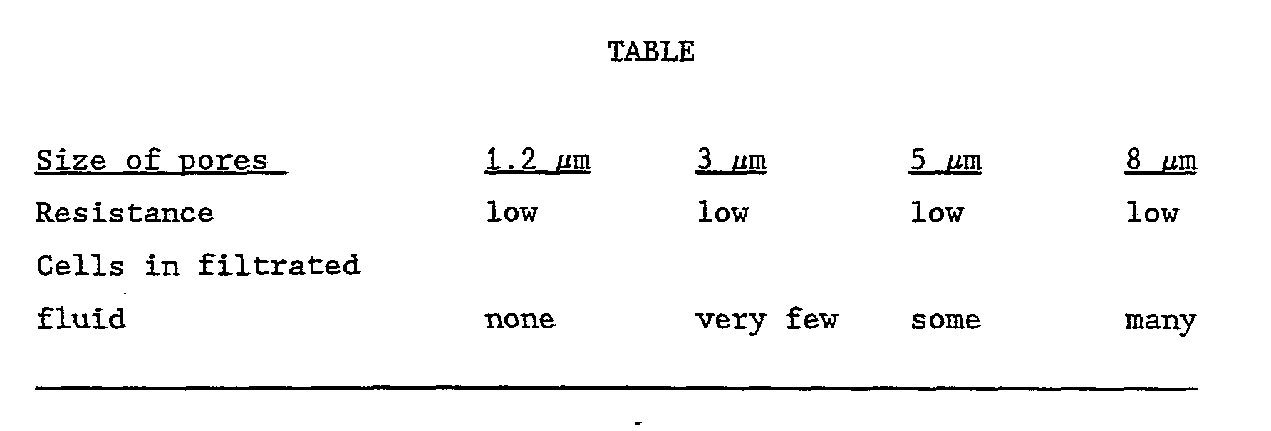
  • a series of amniotic samples which had been cultured for a long time was treated with trypsin EDTA in order to loosen all the cells from the bottom of the culturing flask whereby a suspension containing free floating amnion cells was prepared. This suspension was diluted so as to obtain a concentration of cells comparable to that of an untreated amniotic fluid sample.
  • a pore size of 1.2 ⁇ m is a good size because all of the cells are retained by the filtration and the flow resistance through the filter is low. It was found that with such size of pores 50 mm of the cell suspension could be filtered completely without any appreciable flow resistance. The cells retained by the filter could be flushed from the filter without any problems.
  • cellulose is probably not the most suited material for use in connection with the present invention because this material is to some extend protein binding.
  • the filtration procedure itself implies a very small loss of cells, which does not exceed the loss seen with the presently used procedure of eentrifugation. This was examined by using polycarbonate filters which are partially transparent. After filtration the filters were examined for adherent cells by microscopy, and only very few cells were found. The filtered samples were all compared with control samples from the same pool of amniotic fluid which had only been centrifuged. The same number and the same size of clones were obtained, and the cell types were the same for the two procedures.
  • any sampling device or sampling system by means of which amniotic fluid may be withdrawn from the amniotic cavity filtered outside the amniotic cavity and returned to the amniotic cavity with a more or less reduced content of cells could be used.
  • the syrlngs described above need not be provided with a displaceable piston, but the walls of the syringe chamber may be deformable so that the syringe chamber may be compressed.

Abstract

A sample of amniotic fluid containing cells may be taken out from the amniotic cavity by penetrating the wall of the amniotic cavity by means of a hollow needle (15) through which a volume of the amniotic fluid may be withdrawn. Outside the amniotic cavity the cells or part thereof are separated from the amniotic fluid which is immediately thereafter returned to the amniotic cavity. This means that it is possible to withdraw a relatively large amount of cells without considerably reducing the total volume of the amniotic fluid present in the amniotic cavity. Therefore, it is possible to take out a sample at an early stage of the pregnancy period. The sample may, for example, be withdrawn by means of a sampling device comprising a one-way valve (16, 17) including a cell filter (17). The sampling device may further comprise a syringe with cylinder (10) and a piston (11) movably arranged therein. By moving the piston (11) through a suitable number of consecutive suction and pressure strokes a suitable amount of cell material is deposited on the cell filter (17). The filter (17) may be withdrawn from the sampling device and the cells may be cultured on the cell filter or the cells may be flushed from the filter by means of a culturing medium.

Description

A METHOD AND AN APPARATUS FOR TAKING OUT A SAMPLE OF AN AMNIOTIC FLUID
The present invention relates to a method of taking out a sample of a cell-containing amniotic fluid from an amniotic cavity, said method comprising penetrating the wall of the amniotic cavity by means of a hollow needle or cannula and extracting a volume of cell-containing amniotic fluid therethrough.
In some countries sampling of amniotic fluid is offered to pregnant women having an increased risk of bearing children with chromosome disorders. If the sample collected indicates that the child to be born is diseased an induced abortion is offered.
With the presently applied sampling technique 15-20 ml amniotic fluid is collected in the 16th week of pregnancy, and normally a further 3- 4 weeks elapse before a result of the laboratory examination is available. Consequently, the pregnant woman in most cases will be in the 19th-20th week of pregnancy before an abortion can be induced. Of course this represents a heavy strain for the woman at this advanced stage of pregnancy and also from a medical point of view it is inappropriate. With the present technique it is not possible to carry out the sampling of amniotic fluid earlier, since this involves a
considerable risk that the collected sample fails to give the desired result due to a too low content of cells. When routine amniocentesis is carried out approximately 10% of the total volume of the amniotic fluid is collected. It has been shown that if more than 17 ml is collected in the 16th week of pregnancy, the risk of respiratory distress in the neonatals is increased (Jo-Anne K. Finegan, 1984). Thus, the motivation for collecting samples at this late stage of pregnancy has been to achieve a successful culturing of cells and partly to avoid the risk of spontaneous abortion which risk is known to be relatively low at this stage (A. Tabor, Thesis, 1988).
During recent years, chorionic villi biopsy has been developed in order to obtain early chromosomal examinations of the fetus. By this technique, it is normally possible to obtain a result in the late first trimester. However, this method of sampling involves the problem that the placenta tissue is not always chromosomally identical with the fetus which is a prerequisite for a correct diagnosis (Verjaal M. et al., 1987; Eiben B. et al., 1989).
Consequently, it is necessary to carry out amniocentesis in a significant number of pregnant women in the 16th pregnancy week in order to determine whether the diagnosis is correct or not, since amniocentesis is still the most reliable method for chromosomal examinations of fetuses. Furthermore, chorionic villi biopsy is an expensive examination. It is estimated that a chorionic villi biopsy sample requires the double number of technician hours to be processed when compared to an amniotic fluid sample.
It is therefore of considerable relevance to improve the
amniocentesis technique. Preliminary results from USA indicate that amniocentesis carried out at an early stage, even as early as the 9th week of pregnancy, implies a low risk of spontaneous abortion, and that the risk of complications at later stages of the pregnancy does not seem to be increased (Hanson F.W. et al., 1987; Elejalde B.R. and Elejalde M.M., 1988; Goodmilow L. et al., 1988).
With the presently used technique, only the cells in a sample of amniotic fluid are used for chromosomal fetus examination. After eentrifugation of the sample, the supernatant is discarded, so that only the cells remain. Subsequently, these cells are cultivated for 2-3 weeks in order to obtain a sufficient number of dividing cells for the chromosomal examination. Only 2 ml of amniotic fluid is used for alpha-fetoprotein determination, and this part of the
amniocentesis is independent of the concentration of cells. The cells in the amniotic fluid are considered to be waste products without any significance for the development of the fetus.
The present invention provides a method of the above type and the method according to the invention is characterized in separating outside the amniotic cavity amniotic fluid with a reduced content of cells from the extracted volume of amniotic fluid and recirculating at least part of the separated fluid to the amniotic cavity substantially immediately after the separation, whereby a sample with an increased cell concentration is retained.
Because the separated amniotic fluid with reduced content of cells is substantially immediately returned to the amniotic cavity, a sample containing the necessary amount of cells may be collected by permanently removing only a relatively small volume of amniotic fluid from the amniotic cavity. Because of the relatively small volume of amniotic fluid necessary the sample collection can be carried out at an earlier stage of pregnancy without an increase of the abortion risk and even with a reduced risk of abortion.
Since the cells in the amniotic fluid are considered to be waste products which may be removed from the amniotic cavity without any risk increase the method according to the invention renders it possible to collect a sample containing a relatively large amount of cell material. This means that the necessary time for carrying out the laboratory examination may be reduced, because the cell material may be cultivated in a time period which is shorter than hitherto.
It is primarily the relative amount of amniotic fluid being removed from the amniotic cavity which is decisive for how early in the pregnancy period a sample can be collected. In the method according to the invention the amniotic fluid removed from the amniotic cavity may be separated continuously and the separated amniotic fluid with its reduced cell content may then simultaneously and continuously be returned to the amniotic cavity through a return passage. This means that in principle a rather considerable part of the cell material present within the amniotic cavity may be removed therefrom while only a rather small amount of amniotic fluid is present outside the amniotic cavity at any time.
Alternatively the volume of amniotic fluid may be withdrawn from the amniotic cavity in two or more part volumes and after the withdrawal of each of these volumes the separation and the subsequent
recirculation of amniotic fluid to the amniotic cavity may be take place before the next part volume is withdrawn or taken out.
Consequently only one part volume is removed from the amniotic cavity at a time and the amount of fluid present in such part volume may then be adapted to the total amount of amniotic fluid present in the amniotic cavity at the time when the sample is to be taken out, whereby the risk may be reduced to an acceptable level. The separation of amniotic fluid with a reduced content of cells may be performed in any suitable manner, for example by centrifuging. However, the separation is preferably performed by filtration by passing at least part of the volume of amniotic fluid withdrawn from the amniotic cavity through a cell filter which may be arranged at a suitable position within the flow passage of the amniotic fluid outside the amniotic cavity. Thus, part of the cells present In the amniotic fluid will be filtered from the fluid and be retained by the filter. The amniotic fluid having flown through the filter whereby its content of cells has been reduced may then continuously or intermittently be returned to the amniotic cavity. The cells which are filtered from the amniotic fluid and which are deposited on the filter may be removed together with the cell filter and subsequent cultivation of the cells may take place on the filter. Such a procedure involves a substantial saving of time and rationalization in cultivating the cell material.
The volume of amniotic fluid may be withdrawn from the amniotic cavity in any suitable manner. As an example, the amniotic fluid may be continuously withdrawn from the amniotic cavity and recirculated thereto through an external flow passage through which the fluid is circulated by means of a peristaltic pump or another kind of pump. In the preferred embodiment, however, the volume of amniotic fluid is withdrawn from the amniotic cavity by means of a syringe, and amniotic fluid is then drawn or sucked into a chamber defined within the syringe and thereafter displaced from the syringe chamber and returned to the amniotic cavity, the amniotic fluid being passed through a cell filter when the fluid is drawn into or displaced from the syringe chamber. This means that the syringe should be adapted so as to cause the amniotic fluid to flow through the cell filter mainly In one direction. The invention also relates to an apparatus or sampling device for use in carrying out the method as described above and comprising a hollow needle or cannula for penetrating the wall of the amniotic cavity, a sample receiving chamber communicating with the hollow needle or cannula, and means for drawing a volume of amniotic fluid into the sample receiving chamber through the hollow needle, and the
apparatus according to the invention is characterized in that the apparatus further comprises separating means for separating amniotic fluid with a reduced content of cells from said volume of amniotic fluid drawn into the sample receiving chamber, and fluid returning means for returning at least part of the separated amniotic fluid to the amniotic cavity.
The apparatus or sampling device may be in the form of a syringe and the return means may comprise a one-way valve for causing the amniotic fluid to flow through the cell filter in one direction only during the suction and pressure strokes of the piston of the syringe. Furthermore, the separating means may comprise a cell filter which may form part of the one-way valve. The one-way valve may be adapted to open during the suction stroke of the syringe so that amniotic fluid containing cell material may be sucked into the syringe cylinder through the hollow needle of the syringe. When the syringe piston is subsequently moved in the opposite direction through a pressure stroke the one-way valve closes so as to cause the cell- containing amniotic fluid to be forced through the cell filter arranged within the one-way valve, and the filtered amniotic fluid with a reduced cell content may then be returned to the amniotic cavity through the hollow needle . After the pressure stroke of the syringe piston cell material deposited on the cell filter and a small residual volume of fluid are left in the syringe. The residual amount of amniotic fluid may be used for alpha-fetoprotein determination. In order to avoid excessive sudden changes in pressure within the amniotic cavity small amounts of amniotic fluid may be sucked into the syringe at a time. Thus, the syringe piston may be moved through a number of short consecutive suction and pressure strokes. In this manner a substantial amount of cells may be removed from the amniotic fluid with a rather small physical influence of the pregnancy. When the filter material within the syringe has been filled with cells the resistance against the piston movements will increase and the sampling procedure may then be terminated. In case of amniocentesis at a. late stage of pregnancy it may possibly be preferred to use a single relatively long suction stroke and a subsequent corresponding preεssure stroke. After sampling the cells filtered from the amniotic fltiSrd may be rinsed off the cell filter with culturing medium diresctly into a culturing flask whereby eentrifugation of the sample is ϊαimecessary. Alternatively, the cells may be cultivated directly on t he filter as mentioned above. Summarizing, the following advantages may be obtained by the method and apparatus according to the invention: a) Εhe collection of the samples can be carried out at an earlier stagrfi of pregnancy due to the fact that an increased number of cells resmlts in better cell growth. b) The time required to obtain results is reduced considerably. c) Tdhe system implies more independence as regards a specific stage of pegnancy at the time of sample collection, since the collection of sample may be continued until the resistance from the filter is incrreased so as to indicate that a sufficient number of cells has beem collected. d) Tihe risk of inducing abortions due to the sample collection may be decreased, since only a very small proportion of the amniotic fluid is psarmanently removed from the amniotic cavity, whereby the conditions of pressure remain substantially unchanged in the amniotic cavxlty. e) Ehe risk of amniotic bandlng/amniotic prolapse leading to congenital malformations or abortions may be decreased by carrying out ismnlocentesis at the above early stage since the cell membranes are rsot adherent until the 10th-12th week of pregnancy. f) The risk of developing lung complications in the newborns may also be reduced, since several studies have indicated a connection between the amount of amniotic fluid at the time of sampling collection and the risk of developing lung problems (Jo-Anne Finegan, 1984). g) An increased proportion of living cells in the sample as compared to the presently used technique where only approximately 10% of the cells in the 16th week of pregnancy are alive. It is assumed that it is possible to obtain this larger proportion of living cells since the recirculation of amniotic fluid results in a larger fluidal flow around the needle point whereby an increased number of vital cells will be detached from the fetus and the membranes . h) A reduction of non-successful samplings due to lack of cell growth since each individual sample will contain a large number of cells. (D.E. Rooney et al., 1989). i) The costs involved in amniocentesis as compared to chorionic villi biopsy are lower. j) An increased number of cell clones from which chromosomal analysis can be carried out will result in a more reliable chromosomal diagnosis as compared to the present situation where an answer in some cases is to be given on the basis of only 5 clones. k) An improved diagnosis of chromosomal mosaicism due to the presence of more cells in the starting material.
The invention will now be further described with reference to the drawings, wherein
Fig. 1 is a diagrammatic side view and partial sectional view illustrating a first embodiment of a syringe or sampling device according to the invention,
Fig. 2 is a side view and partial sectional view of a second
embodiment of the sampling device according to the invention,
Fig. 3 is a side view and partial sectional view showing a third embodiment of a sampling device according to the invention, Fig. 4 and 5 are diagrammatic side view showing a fourth embodiment of a sampling device according to the invention and Illustrating the device during suction and pressure strokes, respectively, and
Fig. 6 is a diagrammatic side view Illustrating a fifth embodiment of a syringe according to the present invention.
The syringe or sampling device shown in Fig. 1 comprises a cylinder
10 having a piston 11 with a piston rod 12 displaceably mounted therein. Within the cylinder 10 the piston 11 defines a cylinder chamber 13 having a volume which may be changed by displacing the piston 11. The cylinder 10 comprises at one end a cone or needle seat 14 for mounting a hollow needle or cannula 15. An annular shoulder 16 is defined within the cylinder 10 at the end of which the cone 14 is formed, and a cell filter 17 is mounted on the shoulder 16. The cell filter 17, which is fluid penetrable, but able to retain amnion cells is formed like a valve flap which may be moved between an open position indicated by dotted lines 18 in Fig. 1 and a closed position in which the peripheral portion of the filter is in engagement with the shoulder 16.
When a sample of amniotic fluid is to be taken out from the amniotic cavity by means of the syringe shown in Fig. 1 the piston 11 is in its advanced position in which it is in abutting engagement with the cell filter 17. The pointed end of the hollow needle 15 is now passed through the wall of the amniotic cavity while the insertion of the needle is surveyed by ultrasound scanning. When the wall of the amniotic cavity has been penetrated by the hollow needle the piston
11 is moved outwardly during a suction stroke whereby cell containing amniotic fluid flows through the hollow needle 15 into the cylinder chamber 13 while the valve flap or cell filter 17 is in its open position indicated by dotted lines 18. When a sufficiently small amount of cell-containing amniotic fluid has been sucked into the cylinder 10 the piston 11 is pressed towards its advanced position whereby the fluid sucked into the cylinder chamber is again
displaced therefrom through the hollow needle 15 and returned to the amniotic cavity. During the pressure stroke the valve flap or cell filter 17 closes automatically so that the cell-containing amniotic fluid is pressed through the filter whereby a substantial part of the cells in the fluid is filtered therefrom and remains on the inner side of the filter, and the amniotic fluid which is returned to the amniotic cavity has a substantially reduced content of cells. The procedure may be repeated one or several times if desired so that a small amount of amniotic fluid is once more sucked into the cylinder and subsequently returned to the amniotic cavity. When a suitable amount of cell material has been collected on the inner side of the cell filter 17, which functions as a one way valve, the sampling procedure is terminated and the hollow needle is withdrawn from the wall of the amniotic cavity. A possible small residual amount of amniotic fluid present within the cylinder chamber 13 when the piston 11 is in its advanced position may be used for alpha-fetoprotein determination, if desired. The filter 17 with the amniotic cells deposited thereon may now be removed from the syringe and the cells may be rinsed, if desired.
If the piston is moved only a very short length during the sampling so that the suction stroke as well as the pressure stroke are rather short it is possible to withdraw only a very small amount of fluid from the amniotic cavity at a time. This means that a cell containing amniotic fluid sample may be collected at a rather early stage of the pregnancy period when the total amount of fluid in the amniotic cavity is relatively small, without the risk of any substantial adverse influence.
Fig. 2 shows a modified embodiment of the syringe according to the invention. In the embodiment shown in Fig. 2 the cell filter 17 is fastened to the shoulder 16, and the inner bore of the cone 14 communicates with the cylinder chamber 13 not only through the cell filter 17, but also through a by-pass passage 19. The by-pass passage is controlled by a one-way valve or non-return valve 20 allowing fluid to flow through the by-pass passage 19 in a direction from the cone 14 and into the cylinder chamber 13, but not in the opposite direction. One or more abutment fins 21 may be formed on the inner surface of the cylinder, and the outer ends of these fins may determine the innermost position of the piston 11 and may prevent the piston from coming into contact with the one-way valve 20. The syringe shown in Fig. 2 may be used in substantially the same manner as described above in connection with Fig. 1. During the suction stroke of the piston 11 the one-way valve 20 is open as shown in Fig. 2, and the main part of fluid sucked into the syringe will flow into the cylinder chamber 13 through the by-pass passage 19 where the flow resistance is smallest. During the pressure stroke of the piston the one-way valve 20 is closed so that the fluid is forced to flow out from the cylinder chamber 13 through the cell filter 17 whereby the outflowing fluid is filtered. After a suitable number of successive suction and pressure strokes the cell filter 17 with the filtered amniotic cells is removed, and these cells may then be cultivated as described above.
The embodiment of the syringe shown in Fig. 3 corresponds to that shown in Fig. 1 apart from the fact that the cell filter 17 in Fig. 1 Is formed like a flap which also functions as a one-way valve, while the cell filter 17 in Fig. 3 is loosely arranged within the cone 14 which is a separate part. The separately formed cone 14 may be releasably fastened to the adjacent end of the cylinder 10 by a frictional fit 22, a thread connection, a bayonet socket or another releasable connection. The filter 17, which may, for example, be filter material which is surrounded by a grid-shaped or perforated stiff housing, has a diameter which is somewhat smaller than the inner diameter of the cylinder 10 and the filter 17 is prevented from penetrating into the cylinder 10 by an abutment means 23 which may, for example, be a cruciformed member as shown in Fig. 3.
During the suction stroke the cell filter 17 is moved into abutting engagement with the abutment means 23 as shown in Fig. 3, but the fluid sucked into the syringe may freely flow into the cylinder along the periphery of the filter 17 as indicated by arrows in Fig. 3. During the pressure stroke the filter 17 is moved away from the abutment means 23 so that the circumferential part thereof is pressed into abutting engagement with the inner wall of the cone 14 as indicated by dotted lines 24 in Fig. 3. That means that the cell- containing amniotic fluid displaced from the cylinder chamber 13 during the pressure stroke is forced to pass the cell filter 17 whereby a substantial part of the cells is filtered from the amniotic fluid. When a sufficient amount of cells has been collected on the filter 17 after two or more pressure and suction strokes, the sampling procedure may be terminated and the cone 14 may be removed from the cylinder 10 by releasing the connection 22. The filter 17 with the cell material filtered from the amniotic fluid may now easily be removed.
Figs. 4 and 5 illustrate a sampling syringe or sampling device comprising a standard amniocentesis syringe and a standard needle 15. The device further comprises a filtering unit 25 having a housing 26 with an outwardly extending socket 27 for receiving the cone 14 of the syringe cylinder 10 and an oppositely directed cone 28 for mounting the needle 15 thereon. The filtering unit 25 may be of the disposable type delivered in a sterilized package. A plate or disclike filtering member 17 is arranged within the housing 26 so as to be freely movable between a first position in which the filtering member is in abutting engagement with an inner annular shoulder formed in the housing 26 (Fig. 5) and an open position in which the filtering member is in abutting engagement with spacer members 29 projecting into the housing from an inner wall part adjacent to the socket 27 (Fig. 4). The filtering member may comprise a filtering medium which is sandwiched between a pair of opposite, perforated disc members.
The sample device or syringe illustrated in Figs. 4 and 5 may be operated as follows: When the piston 11 is in its innermost position as shown in Fig. 5 and the pointed end of the hollow needle 15 has been inserted through the wall of the amniotic cavity as described above, the piston 11 may be moved outwardly as indicated by an arrow in Fig. 4, whereby amniotic cell-containing fluid is sucked into the housing 26 of the filtering unit 25. The fluid flow through the housing 26 moves the filtering member 17 to its open position as shown in Fig. 4 so that the fluid may freely flow through the housing 26 and through the cone 14 into the cylinder chamber 13 as indicated by arrows 30 in Fig. 4. When a desired amount of amniotic fluid has been sucked into the cylinder chamber 13, the movement of the piston 11 is reversed as indicated by an arrow in Fig. 5 so as to expel the fluid from the syringe chamber 13. When fluid is discharged from the syringe chamber 13 through the pressure stroke the fluid flow through the housing 26 from the cone 14 to the needle
15 causes the filtering disc 17 to move to its closed position as shown in Fig. 5. The annular shoulder 16 may be provided with an annular sealing member, such as a sealing ring made of silicon, so as to provide a tight engagement between the peripheral part of the filtering disc 17 and the shoulder 16. Consequently, fluid expelled from the syringe chamber 13 is forced to flow through the filtering member 17 whereby amniotic cells are filtered from the fluid and deposited on the filtering member 17 while the filtered fluid having a reduced cell content is returned to the amniotic cavity through the needle 15. The suction-expulsion procedure described may now be repeated several times and the amount of cell material retained by the filter member is increased each time amniotic fluid is circulated through the sampling device. A substantially increased flow
resistance during the pressure stroke indicates that a sufficient amount of cell material has been retained by the filter member 17, and the sampling may then be terminated. When the needle 15 has been retracted from the amniotic cavity, the filtering unit 25 may be removed from the cone 14 of the syringe. Thereafter, a culturing medium may be sucked into the syringe. The housing 26 is composed by two separate parts which are releasably interconnected for example by means of a screw connection 31, and the housing parts may now be disconnected by releasing the connection 31. Now, the filtering member 17 is turned around, for example by means of pincers, whereafter the housing members are reconnected by means of the screw connection 31 and the socket 27 is remounted on the cone 14. Now, the culturing medium may be expelled from the syringe chamber 13 through the housing 26 and through the filtering member 17 which is in its closed position as shown in Fig. 5. Thus, the filtering member 17 is flushed by the culturing medium and the amniotic cells retained by the filtering member is removed therefrom and entrained by the culturing medium. The culturing medium containing the filtered cell material may be discharged directly into a culturing flask which may be placed in an incubator. In this manner the tedious eentrifugation procedure used in the conventional technique may be avoided. In cases where the collected sample of cell material is not to be cultivated immediately after the sampling procedure but is to be sent to a laboratory, the filtering unit 25 may be removed from the syringe and the needle 15 may be removed from the housing 26.
Thereafter, the socket 27 and the cone 28 may be plugged or otherwise closed by means of suitable closing means. The small amount of amniotic fluid present in the filtering unit 25 is sufficient to keep the cells alive during transportation or mailing.
Fig, 6 shows a modified embodiment of the sampling device shown in Figs. 4 and 5. In the embodiment shown in Fig. 6 the filtering member 17 is mounted substantially stationarily within the housing 26 of the filtering unit 25, and the filtering member comprises a central ring 32 defining a central opening 33 and forming a valve seat for a valve body 34, which may, for example, be a ball-like member made from plastic material. The valve body 34 cooperates with the annular valve seat 32 so as to form a one-way valve allowing fluid to flow through the central opening 33 when amniotic fluid is sucked into the syringe 10 through the housing 26. On the other hand, when fluid is expelled from the syringe the one-way valve closes the opening 33 whereby the amniotic fluid being expelled is forced to flow through the filter member 17. Otherwise, substantially the same sampling procedure as described above may be used in connection with the sampling device shown in Fig. 6. The housing 26 may consist of two separate parts interconnected by a releasable connection, such as a screw connection 31 and the cell material retained by the filtering member 17 may then be flushed from the filtering member in a manner similar to that described above.
Type of filter and size of pores therein
In order to find the best cell filtering material a test has been made with cellulose filters having different size of pores, namely 1.2 μm, 3 μm, 5 μm and 8 μm. Fluid containing cultured amnion cells in concentrations corresponding to the concentrations of untreated amniotic fluid samples was filtered. The smallest cells in an amniotic fluid sample are the living cells and therefore the optimum pore size must be determined on the basis of the smallest cells. Cultured amnion cells are all alive and therefore they are well suited for such determination of the best size of pores.
A series of amniotic samples which had been cultured for a long time was treated with trypsin EDTA in order to loosen all the cells from the bottom of the culturing flask whereby a suspension containing free floating amnion cells was prepared. This suspension was diluted so as to obtain a concentration of cells comparable to that of an untreated amniotic fluid sample.
By means of a 20 ml syringe of the type normally used for
conventional amniocentesis approximately 20 ml of the cell suspension was forced through a cellulose paper filter arranged in a filter holder. The filtered fluid was examined under microscope. During the filtration, the force necessary for expelling the suspension from the syringe was also noticed. The filter diameter was 47 mm. The results of the test are stated in the following table
Figure imgf000016_0001
From the above test is was concluded that a filter having a pore size of less than 3 μm are best suited. A pore size of 1.2 μm, for example, is a good size because all of the cells are retained by the filtration and the flow resistance through the filter is low. It was found that with such size of pores 50 mm of the cell suspension could be filtered completely without any appreciable flow resistance. The cells retained by the filter could be flushed from the filter without any problems. However, cellulose is probably not the most suited material for use in connection with the present invention because this material is to some extend protein binding. Therefore, three different filter materials from Millipore have been examined for toxicity by means of the so-called mouse test in which the ability of mouse embryos to develop from the 2-cell stage to the blastocyst stage is examined. Small pieces of filter material were placed with the mouse embryos and the degree of blastocyst development was evaluated and compared with a control without any additions. The following materials have been tested: Cellulose esters, polycarbonate, polyvinyldifluoride, and the plastic materials of the applied 3-way plugs. All these materials were found non-toxic and it was concluded that the studied materials are applicable. Furthermore, repeated amniotic fluid cultivations have been carried out from pooled amniotic fluid remnants, in which small pieces of filter material have been added to the culturing flasks. The cell growth of these flasks were compared with control cultivations from the same pool. No significant differences in growth were found for the different materials.
By comparative cultivations of small and larger aliquots of amniotic fluid from the same pool, it was shown that the larger volumes of amniotic fluid containing a larger number of cells also results in correspondingly more clones, which is a prerequisite for the new amniocentesis system. In six experimental series from different pools of amniotic fluid unequivocal results from culturing have been found.
After filtration of pooled amniotic fluid remnants the cells were removed from the filters by rinsing with the culturing medium and it was shown that it is possible to grow cells successfully after this filtration procedure. This experiment was carried out using
different filter materials and pore sizes. Different filter housings have been applied and varying volumes of amniotic fluid were
filtered. The filtration procedure itself implies a very small loss of cells, which does not exceed the loss seen with the presently used procedure of eentrifugation. This was examined by using polycarbonate filters which are partially transparent. After filtration the filters were examined for adherent cells by microscopy, and only very few cells were found. The filtered samples were all compared with control samples from the same pool of amniotic fluid which had only been centrifuged. The same number and the same size of clones were obtained, and the cell types were the same for the two procedures.
It should be understood that various changes and modifications of the sampling devices and methods described above could be made within the scope of the present invention. Thus, in principle any sampling device or sampling system by means of which amniotic fluid may be withdrawn from the amniotic cavity filtered outside the amniotic cavity and returned to the amniotic cavity with a more or less reduced content of cells could be used. As an example, the syrlngs described above need not be provided with a displaceable piston, but the walls of the syringe chamber may be deformable so that the syringe chamber may be compressed.

Claims

1. A method of taking out a sample of a cell-containing amniotic fluid from an amniotic cavity, said method comprising penetrating the wall of the amniotic cavity by means of a hollow needle or canula (15) and extracting a volume of cell-containing amniotic fluid therethrough,
c h a r a c t e r i z e d in separating outside the amniotic cavity amniotic fluid with reduced content of cells from the extracted volume of amniotic fluid and returning at least part of the separated fluid to the amniotic cavity immediately after the separation, whereby a sample with an increased cell concentration is retained.
2. A method according to claim 1,
c h a r a c t e r i z e d in performing the separation continuously and in continuously and simultaneously returning separated amniotic fluid to the amniotic cavity through a return passage.
3. A method according to claim 1,
c h a r a c t e r i z e d in taking out the said volume of amniotic fluid as two or more part volumes and separating amniotic fluid from each of the these part volumes after taking out the same and
subsequently returning separated amniotic fluid prior to taking out the next part volume.
4. A method according to claim 1 or 2,
c h a r a c t e r i z e d in separating amniotic fluid by passing at least part of said volume of amniotic fluid taken out through a cell filter (17).
5. A method according to claim 4,
c h a r a c t e r i z e d in that said volume of amniotic fluid is taken out by means of a syringe, amniotic fluid from the amniotic cavity being drawn into a chamber (13) of the syringe and
subsequently displaced from said chamber and returned to the amniotic cavity, the amniotic fluid being passed through a cell filter (17) when drawn in or displaced from the syringe chamber.
6. An apparatus for use in carrying out a method according to claim 1 and comprising a hollow needle or cannula (15) for penetrating the wall of the amniotic cavity, a sample receiving chamber (13) communicating with the hollow needle or cannula, and means (11) for drawing a volume of amniotic fluid into the sample receiving chamber through the hollow needle,
ch a r a c t e r i z e d in that the apparatus further comprises separating means (17, 19, 20) for separating amniotic fluid with a reduced content of cells from said volume of amniotic fluid drawn Into the sample receiving chamber, and fluid returning means (11) for returning at least part of the separated amniotic fluid to the amniotic cavity.
7. An apparatus according to claim 6 ,
ch a r a c t e r i z e d in that the separating means comprise a cell filter (17).
8. An apparatus according to claim 7 , which is in the form of a syringe,
ch a r a c t e r i z e d in that the return means comprise a oneway valve (17, 20) for securing that the amniotic fluid is to flow in one direction only through the cell filter (17) during suction and pressure strokes of the syringe piston (11).
9. An apparatus according to claim 8,
ch ar a c t e r i z e d in that the cell filter forms part of the one-way valve (17).
10. An apparatus according to claim 8,
ch a r a c t e r i z e d in that the drawing means or returning means comprise a by-pass passage (19), which is defined within the syringe cylinder and provided with a one-way valve (20), and which is adapted to let the amniotic fluid by-pass the cell filter (17) during the pressure and suction strokes of the piston (11).
PCT/DK1990/000122 1989-05-10 1990-05-10 A method and an apparatus for taking out a sample of an amniotic fluid WO1990013261A1 (en)

Priority Applications (2)

Application Number Priority Date Filing Date Title
DE69018629T DE69018629T2 (en) 1989-05-10 1990-05-10 DEVICE FOR REMOVING AMNIOTIC LIQUID.
EP90907180A EP0471721B1 (en) 1989-05-10 1990-05-10 Apparatus for taking a sample of amniotic fluid

Applications Claiming Priority (2)

Application Number Priority Date Filing Date Title
DK2293/89 1989-05-10
DK229389A DK229389D0 (en) 1989-05-10 1989-05-10 PROCEDURE AND APPARATUS FOR SAMPLING A CELL SUSTAINABLE WATER FROM THE AMNION HOLE

Publications (1)

Publication Number Publication Date
WO1990013261A1 true WO1990013261A1 (en) 1990-11-15

Family

ID=8111392

Family Applications (1)

Application Number Title Priority Date Filing Date
PCT/DK1990/000122 WO1990013261A1 (en) 1989-05-10 1990-05-10 A method and an apparatus for taking out a sample of an amniotic fluid

Country Status (8)

Country Link
EP (1) EP0471721B1 (en)
JP (1) JPH05501965A (en)
AU (1) AU5568490A (en)
CA (1) CA2064193A1 (en)
DE (1) DE69018629T2 (en)
DK (2) DK229389D0 (en)
HK (1) HK35696A (en)
WO (1) WO1990013261A1 (en)

Cited By (7)

* Cited by examiner, † Cited by third party
Publication number Priority date Publication date Assignee Title
GB2344526A (en) * 1999-01-12 2000-06-14 Dumaresq Lucas Alison Jayne Syringe with filter, and filter therefor
US6623959B2 (en) 2001-06-13 2003-09-23 Ethicon, Inc. Devices and methods for cell harvesting
US7399278B1 (en) 2003-05-05 2008-07-15 Los Angeles Biomedical Research Institute At Harbor-Ucla Medical Center Method and system for measuring amniotic fluid volume and/or assessing fetal weight
WO2009075001A1 (en) * 2007-12-13 2009-06-18 Biocell Center S.P.A. Method of collection and preservation of fluids and/or materials, in particular of organic fluids and/or materials containing stem cells, and device employable in such method
CN103492009A (en) * 2011-04-08 2014-01-01 佳迈特股份有限公司 Filter needle
US10112015B2 (en) * 2013-12-18 2018-10-30 Ge Healthcare Uk Limited Continuous use syringe filter
EP3649961A1 (en) * 2013-03-15 2020-05-13 Amniotics AB Apparatuses for amniotic fluid collection

Families Citing this family (6)

* Cited by examiner, † Cited by third party
Publication number Priority date Publication date Assignee Title
DE102009022350B4 (en) * 2009-05-15 2011-11-03 Fraunhofer-Gesellschaft zur Förderung der angewandten Forschung e.V. Pipette head, pipetting device
CA2898467A1 (en) 2013-01-18 2014-07-24 Biomeme Incorporated Analytic device
MX2016005480A (en) 2013-11-01 2016-08-03 Biomeme Inc Sample extraction and preparation device.
KR101594447B1 (en) * 2013-12-05 2016-02-17 주식회사 엠에스피 Filter valve and syringe comprising the valve
JP2015112409A (en) * 2013-12-13 2015-06-22 サン—ア フロンテック カンパニー,リミテッド Syringe
CA3192080A1 (en) 2020-09-18 2022-03-24 Marc DEJOHN Portable devices and methods for analyzing samples

Citations (6)

* Cited by examiner, † Cited by third party
Publication number Priority date Publication date Assignee Title
US3757780A (en) * 1971-02-25 1973-09-11 Ishikawa Susakusho Kk Needle assembly with longitudinally movable filter
US3938513A (en) * 1974-01-03 1976-02-17 Hargest Thomas S Syringe filter and valve combination
US4066079A (en) * 1976-11-03 1978-01-03 Chiarolla Victor D Filter needle
US4308875A (en) * 1981-03-11 1982-01-05 Universal Medical Instrument Corporation Amniocentesis needle
US4366822A (en) * 1979-12-03 1983-01-04 Applied Medical Devices, Inc. Method and apparatus for bone marrow cell separation and analysis
US4820276A (en) * 1988-02-08 1989-04-11 Enrique Moreno Filter assembly for use with a hypodermic syringe

Patent Citations (6)

* Cited by examiner, † Cited by third party
Publication number Priority date Publication date Assignee Title
US3757780A (en) * 1971-02-25 1973-09-11 Ishikawa Susakusho Kk Needle assembly with longitudinally movable filter
US3938513A (en) * 1974-01-03 1976-02-17 Hargest Thomas S Syringe filter and valve combination
US4066079A (en) * 1976-11-03 1978-01-03 Chiarolla Victor D Filter needle
US4366822A (en) * 1979-12-03 1983-01-04 Applied Medical Devices, Inc. Method and apparatus for bone marrow cell separation and analysis
US4308875A (en) * 1981-03-11 1982-01-05 Universal Medical Instrument Corporation Amniocentesis needle
US4820276A (en) * 1988-02-08 1989-04-11 Enrique Moreno Filter assembly for use with a hypodermic syringe

Cited By (14)

* Cited by examiner, † Cited by third party
Publication number Priority date Publication date Assignee Title
GB2344526A (en) * 1999-01-12 2000-06-14 Dumaresq Lucas Alison Jayne Syringe with filter, and filter therefor
GB2344526B (en) * 1999-01-12 2001-05-23 Dumaresq Lucas Alison Jayne Syringe with filter,and filter therefor
US6796965B2 (en) 1999-01-12 2004-09-28 Alison Jayne Dumaresq-Lucas Syringe with filter, and filter therefor
US6623959B2 (en) 2001-06-13 2003-09-23 Ethicon, Inc. Devices and methods for cell harvesting
US7399278B1 (en) 2003-05-05 2008-07-15 Los Angeles Biomedical Research Institute At Harbor-Ucla Medical Center Method and system for measuring amniotic fluid volume and/or assessing fetal weight
WO2009075001A1 (en) * 2007-12-13 2009-06-18 Biocell Center S.P.A. Method of collection and preservation of fluids and/or materials, in particular of organic fluids and/or materials containing stem cells, and device employable in such method
CN103492009A (en) * 2011-04-08 2014-01-01 佳迈特股份有限公司 Filter needle
EP2695629A2 (en) * 2011-04-08 2014-02-12 Zamart Co.,ltd Filter needle
EP2695629A4 (en) * 2011-04-08 2014-12-31 Zamart Co Ltd Filter needle
CN106237444A (en) * 2011-04-08 2016-12-21 佳迈特股份有限公司 Filter pin
CN106362253A (en) * 2011-04-08 2017-02-01 佳迈特股份有限公司 Filter needle
CN106362253B (en) * 2011-04-08 2020-02-11 佳迈特股份有限公司 Filter needle
EP3649961A1 (en) * 2013-03-15 2020-05-13 Amniotics AB Apparatuses for amniotic fluid collection
US10112015B2 (en) * 2013-12-18 2018-10-30 Ge Healthcare Uk Limited Continuous use syringe filter

Also Published As

Publication number Publication date
HK35696A (en) 1996-03-08
EP0471721A1 (en) 1992-02-26
DK229389D0 (en) 1989-05-10
DK0471721T3 (en) 1995-06-26
EP0471721B1 (en) 1995-04-12
DE69018629D1 (en) 1995-05-18
JPH05501965A (en) 1993-04-15
CA2064193A1 (en) 1990-11-11
AU5568490A (en) 1990-11-29
DE69018629T2 (en) 1996-01-25

Similar Documents

Publication Publication Date Title
US5494044A (en) Method for taking a sample of amniotic fluid
JP5425200B2 (en) Bioparticle capture device and method of use thereof
US10519420B2 (en) Method for obtaining sterile human amniotic fluid and uses thereof
EP0471721B1 (en) Apparatus for taking a sample of amniotic fluid
US20200077987A1 (en) Method for obtaining sterile human amniotic fluid and uses thereof
US4194513A (en) Antenatal cell extracting device and method
US5139031A (en) Method and device for cytology and microbiological testing
JP6452695B2 (en) Bone marrow fat portion isolation device and method
EP0693182A1 (en) Method of separating particles from a filter
US20070173737A1 (en) Cell collection device
US5000192A (en) Prenatal specimen collection method
US20060122534A1 (en) Container for suspension and filtration of stool
CN209485831U (en) The extracellular vesica enriching apparatus of the urine of hospital
EP2167642A1 (en) Separation method of biological objects relative to their viscoelastic properties
CN209485830U (en) The device of hospital and the home-use extracellular vesica of enrichment urine that can make
Byrne et al. Amnifiltration in the first trimester: feasibility, technical aspects and cytological outcome
CN107669300A (en) It is easy to specimens from pri collection device
CN204521718U (en) A kind of can the delicate metering negative-pressure suction bottle of collector soma
JP2004236527A (en) Nucleated cell-separating device
Baringer A simplified procedure for spinal fluid cytology
WO1992000039A1 (en) Method and instrument for cytological examination of body fluids and fine-needle aspirates
CN110713968A (en) Method for rapidly obtaining large amount of purified plasmodium sporozoites
US4004322A (en) Aseptic serum collection method
CN219374719U (en) Multi-purpose collector of amniotic fluid puncture
CN218009900U (en) Gynaecology and obstetrics uses negative pressure suction filter equipment

Legal Events

Date Code Title Description
AK Designated states

Kind code of ref document: A1

Designated state(s): AT AU BB BG BR CA CH DE DK ES FI GB HU JP KP KR LK LU MC MG MW NL NO RO SD SE SU US

AL Designated countries for regional patents

Kind code of ref document: A1

Designated state(s): AT BE BF BJ CF CG CH CM DE DK ES FR GA GB IT LU ML MR NL SE SN TD TG

WWE Wipo information: entry into national phase

Ref document number: 1990907180

Country of ref document: EP

WWE Wipo information: entry into national phase

Ref document number: 2064193

Country of ref document: CA

REG Reference to national code

Ref country code: DE

Ref legal event code: 8642

WWP Wipo information: published in national office

Ref document number: 1990907180

Country of ref document: EP

WWG Wipo information: grant in national office

Ref document number: 1990907180

Country of ref document: EP