WO1989009788A2 - Biosynthetic osteogenic proteins and osteogenic devices containing them - Google Patents

Biosynthetic osteogenic proteins and osteogenic devices containing them Download PDF

Info

Publication number
WO1989009788A2
WO1989009788A2 PCT/US1989/001469 US8901469W WO8909788A2 WO 1989009788 A2 WO1989009788 A2 WO 1989009788A2 US 8901469 W US8901469 W US 8901469W WO 8909788 A2 WO8909788 A2 WO 8909788A2
Authority
WO
WIPO (PCT)
Prior art keywords
protein
amino acid
sequence
bone
acid sequences
Prior art date
Application number
PCT/US1989/001469
Other languages
French (fr)
Other versions
WO1989009788A3 (en
Inventor
Hermann Oppermann
Thangavel Kuberasampath
David C. Rueger
Engin Ozkaynak
Original Assignee
Creative Biomolecules, Inc.
Priority date (The priority date is an assumption and is not a legal conclusion. Google has not performed a legal analysis and makes no representation as to the accuracy of the date listed.)
Filing date
Publication date
Family has litigation
First worldwide family litigation filed litigation Critical https://patents.darts-ip.com/?family=27391116&utm_source=google_patent&utm_medium=platform_link&utm_campaign=public_patent_search&patent=WO1989009788(A2) "Global patent litigation dataset” by Darts-ip is licensed under a Creative Commons Attribution 4.0 International License.
Priority claimed from US07/179,406 external-priority patent/US4968590A/en
Priority claimed from US07/315,342 external-priority patent/US5011691A/en
Application filed by Creative Biomolecules, Inc. filed Critical Creative Biomolecules, Inc.
Priority to DE68927153T priority Critical patent/DE68927153T2/en
Priority to EP89904959A priority patent/EP0372031B1/en
Publication of WO1989009788A2 publication Critical patent/WO1989009788A2/en
Publication of WO1989009788A3 publication Critical patent/WO1989009788A3/en

Links

Classifications

    • AHUMAN NECESSITIES
    • A61MEDICAL OR VETERINARY SCIENCE; HYGIENE
    • A61KPREPARATIONS FOR MEDICAL, DENTAL OR TOILETRY PURPOSES
    • A61K9/00Medicinal preparations characterised by special physical form
    • A61K9/0012Galenical forms characterised by the site of application
    • A61K9/0019Injectable compositions; Intramuscular, intravenous, arterial, subcutaneous administration; Compositions to be administered through the skin in an invasive manner
    • A61K9/0024Solid, semi-solid or solidifying implants, which are implanted or injected in body tissue
    • AHUMAN NECESSITIES
    • A61MEDICAL OR VETERINARY SCIENCE; HYGIENE
    • A61LMETHODS OR APPARATUS FOR STERILISING MATERIALS OR OBJECTS IN GENERAL; DISINFECTION, STERILISATION OR DEODORISATION OF AIR; CHEMICAL ASPECTS OF BANDAGES, DRESSINGS, ABSORBENT PADS OR SURGICAL ARTICLES; MATERIALS FOR BANDAGES, DRESSINGS, ABSORBENT PADS OR SURGICAL ARTICLES
    • A61L27/00Materials for grafts or prostheses or for coating grafts or prostheses
    • A61L27/14Macromolecular materials
    • A61L27/22Polypeptides or derivatives thereof, e.g. degradation products
    • A61L27/227Other specific proteins or polypeptides not covered by A61L27/222, A61L27/225 or A61L27/24
    • AHUMAN NECESSITIES
    • A61MEDICAL OR VETERINARY SCIENCE; HYGIENE
    • A61LMETHODS OR APPARATUS FOR STERILISING MATERIALS OR OBJECTS IN GENERAL; DISINFECTION, STERILISATION OR DEODORISATION OF AIR; CHEMICAL ASPECTS OF BANDAGES, DRESSINGS, ABSORBENT PADS OR SURGICAL ARTICLES; MATERIALS FOR BANDAGES, DRESSINGS, ABSORBENT PADS OR SURGICAL ARTICLES
    • A61L27/00Materials for grafts or prostheses or for coating grafts or prostheses
    • A61L27/14Macromolecular materials
    • A61L27/22Polypeptides or derivatives thereof, e.g. degradation products
    • A61L27/24Collagen
    • AHUMAN NECESSITIES
    • A61MEDICAL OR VETERINARY SCIENCE; HYGIENE
    • A61LMETHODS OR APPARATUS FOR STERILISING MATERIALS OR OBJECTS IN GENERAL; DISINFECTION, STERILISATION OR DEODORISATION OF AIR; CHEMICAL ASPECTS OF BANDAGES, DRESSINGS, ABSORBENT PADS OR SURGICAL ARTICLES; MATERIALS FOR BANDAGES, DRESSINGS, ABSORBENT PADS OR SURGICAL ARTICLES
    • A61L27/00Materials for grafts or prostheses or for coating grafts or prostheses
    • A61L27/28Materials for coating prostheses
    • A61L27/34Macromolecular materials
    • AHUMAN NECESSITIES
    • A61MEDICAL OR VETERINARY SCIENCE; HYGIENE
    • A61LMETHODS OR APPARATUS FOR STERILISING MATERIALS OR OBJECTS IN GENERAL; DISINFECTION, STERILISATION OR DEODORISATION OF AIR; CHEMICAL ASPECTS OF BANDAGES, DRESSINGS, ABSORBENT PADS OR SURGICAL ARTICLES; MATERIALS FOR BANDAGES, DRESSINGS, ABSORBENT PADS OR SURGICAL ARTICLES
    • A61L27/00Materials for grafts or prostheses or for coating grafts or prostheses
    • A61L27/36Materials for grafts or prostheses or for coating grafts or prostheses containing ingredients of undetermined constitution or reaction products thereof, e.g. transplant tissue, natural bone, extracellular matrix
    • A61L27/3604Materials for grafts or prostheses or for coating grafts or prostheses containing ingredients of undetermined constitution or reaction products thereof, e.g. transplant tissue, natural bone, extracellular matrix characterised by the human or animal origin of the biological material, e.g. hair, fascia, fish scales, silk, shellac, pericardium, pleura, renal tissue, amniotic membrane, parenchymal tissue, fetal tissue, muscle tissue, fat tissue, enamel
    • A61L27/3608Bone, e.g. demineralised bone matrix [DBM], bone powder
    • AHUMAN NECESSITIES
    • A61MEDICAL OR VETERINARY SCIENCE; HYGIENE
    • A61LMETHODS OR APPARATUS FOR STERILISING MATERIALS OR OBJECTS IN GENERAL; DISINFECTION, STERILISATION OR DEODORISATION OF AIR; CHEMICAL ASPECTS OF BANDAGES, DRESSINGS, ABSORBENT PADS OR SURGICAL ARTICLES; MATERIALS FOR BANDAGES, DRESSINGS, ABSORBENT PADS OR SURGICAL ARTICLES
    • A61L27/00Materials for grafts or prostheses or for coating grafts or prostheses
    • A61L27/36Materials for grafts or prostheses or for coating grafts or prostheses containing ingredients of undetermined constitution or reaction products thereof, e.g. transplant tissue, natural bone, extracellular matrix
    • A61L27/3641Materials for grafts or prostheses or for coating grafts or prostheses containing ingredients of undetermined constitution or reaction products thereof, e.g. transplant tissue, natural bone, extracellular matrix characterised by the site of application in the body
    • A61L27/3645Connective tissue
    • A61L27/365Bones
    • AHUMAN NECESSITIES
    • A61MEDICAL OR VETERINARY SCIENCE; HYGIENE
    • A61LMETHODS OR APPARATUS FOR STERILISING MATERIALS OR OBJECTS IN GENERAL; DISINFECTION, STERILISATION OR DEODORISATION OF AIR; CHEMICAL ASPECTS OF BANDAGES, DRESSINGS, ABSORBENT PADS OR SURGICAL ARTICLES; MATERIALS FOR BANDAGES, DRESSINGS, ABSORBENT PADS OR SURGICAL ARTICLES
    • A61L27/00Materials for grafts or prostheses or for coating grafts or prostheses
    • A61L27/36Materials for grafts or prostheses or for coating grafts or prostheses containing ingredients of undetermined constitution or reaction products thereof, e.g. transplant tissue, natural bone, extracellular matrix
    • A61L27/3641Materials for grafts or prostheses or for coating grafts or prostheses containing ingredients of undetermined constitution or reaction products thereof, e.g. transplant tissue, natural bone, extracellular matrix characterised by the site of application in the body
    • A61L27/3645Connective tissue
    • A61L27/3654Cartilage, e.g. meniscus
    • AHUMAN NECESSITIES
    • A61MEDICAL OR VETERINARY SCIENCE; HYGIENE
    • A61LMETHODS OR APPARATUS FOR STERILISING MATERIALS OR OBJECTS IN GENERAL; DISINFECTION, STERILISATION OR DEODORISATION OF AIR; CHEMICAL ASPECTS OF BANDAGES, DRESSINGS, ABSORBENT PADS OR SURGICAL ARTICLES; MATERIALS FOR BANDAGES, DRESSINGS, ABSORBENT PADS OR SURGICAL ARTICLES
    • A61L27/00Materials for grafts or prostheses or for coating grafts or prostheses
    • A61L27/50Materials characterised by their function or physical properties, e.g. injectable or lubricating compositions, shape-memory materials, surface modified materials
    • A61L27/56Porous materials, e.g. foams or sponges
    • AHUMAN NECESSITIES
    • A61MEDICAL OR VETERINARY SCIENCE; HYGIENE
    • A61PSPECIFIC THERAPEUTIC ACTIVITY OF CHEMICAL COMPOUNDS OR MEDICINAL PREPARATIONS
    • A61P1/00Drugs for disorders of the alimentary tract or the digestive system
    • A61P1/02Stomatological preparations, e.g. drugs for caries, aphtae, periodontitis
    • AHUMAN NECESSITIES
    • A61MEDICAL OR VETERINARY SCIENCE; HYGIENE
    • A61PSPECIFIC THERAPEUTIC ACTIVITY OF CHEMICAL COMPOUNDS OR MEDICINAL PREPARATIONS
    • A61P43/00Drugs for specific purposes, not provided for in groups A61P1/00-A61P41/00
    • CCHEMISTRY; METALLURGY
    • C07ORGANIC CHEMISTRY
    • C07KPEPTIDES
    • C07K14/00Peptides having more than 20 amino acids; Gastrins; Somatostatins; Melanotropins; Derivatives thereof
    • C07K14/435Peptides having more than 20 amino acids; Gastrins; Somatostatins; Melanotropins; Derivatives thereof from animals; from humans
    • C07K14/475Growth factors; Growth regulators
    • C07K14/51Bone morphogenetic factor; Osteogenins; Osteogenic factor; Bone-inducing factor
    • AHUMAN NECESSITIES
    • A61MEDICAL OR VETERINARY SCIENCE; HYGIENE
    • A61CDENTISTRY; APPARATUS OR METHODS FOR ORAL OR DENTAL HYGIENE
    • A61C8/00Means to be fixed to the jaw-bone for consolidating natural teeth or for fixing dental prostheses thereon; Dental implants; Implanting tools
    • A61C8/0003Not used, see subgroups
    • A61C8/0004Consolidating natural teeth
    • A61C8/0006Periodontal tissue or bone regeneration
    • AHUMAN NECESSITIES
    • A61MEDICAL OR VETERINARY SCIENCE; HYGIENE
    • A61FFILTERS IMPLANTABLE INTO BLOOD VESSELS; PROSTHESES; DEVICES PROVIDING PATENCY TO, OR PREVENTING COLLAPSING OF, TUBULAR STRUCTURES OF THE BODY, e.g. STENTS; ORTHOPAEDIC, NURSING OR CONTRACEPTIVE DEVICES; FOMENTATION; TREATMENT OR PROTECTION OF EYES OR EARS; BANDAGES, DRESSINGS OR ABSORBENT PADS; FIRST-AID KITS
    • A61F2310/00Prostheses classified in A61F2/28 or A61F2/30 - A61F2/44 being constructed from or coated with a particular material
    • A61F2310/00005The prosthesis being constructed from a particular material
    • A61F2310/00365Proteins; Polypeptides; Degradation products thereof
    • AHUMAN NECESSITIES
    • A61MEDICAL OR VETERINARY SCIENCE; HYGIENE
    • A61KPREPARATIONS FOR MEDICAL, DENTAL OR TOILETRY PURPOSES
    • A61K38/00Medicinal preparations containing peptides
    • AHUMAN NECESSITIES
    • A61MEDICAL OR VETERINARY SCIENCE; HYGIENE
    • A61LMETHODS OR APPARATUS FOR STERILISING MATERIALS OR OBJECTS IN GENERAL; DISINFECTION, STERILISATION OR DEODORISATION OF AIR; CHEMICAL ASPECTS OF BANDAGES, DRESSINGS, ABSORBENT PADS OR SURGICAL ARTICLES; MATERIALS FOR BANDAGES, DRESSINGS, ABSORBENT PADS OR SURGICAL ARTICLES
    • A61L2430/00Materials or treatment for tissue regeneration
    • A61L2430/02Materials or treatment for tissue regeneration for reconstruction of bones; weight-bearing implants

Definitions

  • This invention relates to osteogenic devices, to synthetic genes encoding proteins which can induce osteogenesis in mammals and methods for their production using recombinant DNA techniques, to synthetic forms of osteogenic protein, and to bone and cartilage repair procedures using osteogenic device comprising the synthetic proteins.
  • Mammalian bone tissue is known to contain one or more proteinaceous materials, presumably active during growth and natural bone healing, which can induce a developmental cascade of cellular events resulting in endochondral bone formation.
  • This active factor (or factors) has variously been referred to in the literature as bone morphogenetic or morphogenic protein, bone inductive protein, osteogenic protein, osteogenin, or osteoinductive protein.
  • the developmental cascade of bone differentiation consists of recruitment of mesenchymal cells, proliferation of progenitor cells, calcification of cartilage, vascular invasion, bone formation, remodeling, and finally marrow differentiation (Reddi (1981) Collagen Rel. Res. 1:209-226). Though the precise mechanisms underlying these phenotypic transformations are unclear, it has been shown that the natural endochondral bone differentiation activity of bone matrix can be dissociatively extracted and reconstituted with inactive residual collagenous matrix to restore full bone induction activity (Sampath and Reddi, (1981) Proc. Natl. Acad. Sci. USA 78:7599-7603). This provides an experimental method for assaying protein extracts for their ability to induce endochondral bone in vivo.
  • This putative bone inductive protein has been shown to have a molecular mass of less than 50 kilodaltons (kD).
  • kD kilodaltons
  • Several species of mammals produce closely related protein as demonstrated by cross species implant experiments (Sampath and Reddi (1983) Proc. Natl. Acad. Sci. USA 80:6591-6595).
  • Urist et al. (Proc. Natl. Acad. Sci. USA (1984) 81:371-375) disclose a bovine bone morphogenetic protein extract having the properties of an acidic polypeptide and a molecular weight of approximately 18 kD. The authors reported that the protein was present in a fraction separated by hydroxyapatite chromatography, and that it induced bone formation in mouse hindquarter muscle and bone regeneration in trephine defects in rat and dog skulls. Their method of obtaining the extract from bone results in ill-defined and impure preparations.
  • Wozney et al. discloses the isolation of full-length cDNA's encoding the human equivalents of three polypeptides originally purified from bovine bone. The authors report that each of the three recombinantly expressed human proteins are independently or in combination capable of inducing cartilage formation. No evidence of bone formation is reported.
  • It is an object of this invention to provide osteogenic devices comprising matrices containing dispersed osteogenic protein capable of bone induction in allogenic and xenogenic implants. Another object is to provide synthetic osteogenic proteins capable of inducing endochondral bone formation in mammals, including humans. Yet another object is to provide genes encoding non-native osteogenic proteins and methods for their production using recombinant DNA techniques. Another object is to provide novel biosynthetic forms of osteogenic proteins and a structural design for novel, functional osteogenic proteins. Another object is to provide methods for inducing cartilage formation.
  • This invention involves osteogenic devices which, when implanted in a mammalian body, can induce at the locus of the implant the full developmental cascade of endochondral bone formation and bone marrow differentiation. Suitably modified as disclosed herein, the devices also may be used to induce cartilage formation.
  • the devices comprise a carrier material, referred to herein as a matrix, having the characteristics disclosed below, containing dispersed osteogenic protein in the form of a biosynthetic construct.
  • a series of consensus DNA sequences were designed with the goal of producing an active osteogenic protein.
  • the sequences were based on partial amino acid sequence data obtained from the naturally sourced product and on observed homologies with unrelated genes reported in the literature, or the sequences they encode, having a presumed or demonstrated developmental function.
  • biosynthetic consensus sequences have been expressed as fusion proteins in procaryotes, purified, cleaved, refolded, combined with a matrix, implanted in an established animal model, and shown to have endochondral bone-inducing activity.
  • the currently preferred active proteins comprise sequences designated COP5, COP7, COP16, and OP1. The amino acid sequences of these proteins are set forth below.
  • the dashes (-) are used as fillers only to line up comparable sequences in related proteins, and have no other function.
  • amino acids 45-50 of COP7 for example, are NHAVV.
  • the numbering of amino acids is selected solely for purposes of facilitating comparisons between sequences.
  • the DF residues numbered at 9 and 10 of COP5 and COP7 may comprise residues, e.g., 35 and 36, of an osteogenic protein embodying the invention.
  • Various leader or trailer sequences may be attached to the operative active region provided the osteogenic or chondrogenic activity of the protein is not destroyed.
  • the invention comprises a protein comprising an amino acid sequence sufficiently duplicative of the sequence of COP5, COP7, COP16, or OP1 such that it is capable of inducing endochondral bone formation when properly folded and implanted in a mammal in association with a matrix.
  • Some of these sequences induce cartilage, but not bone.
  • the bone forming materials may be used to produce cartilage if implanted in an avascular locus, or if an inhibitor to full bone development is implanted together with the active protein.
  • the invention comprises a protein less than about 200 amino acids long (for each chain) including a sequence sufficiently duplicative of the sequence of COP5, COP7, COP16, or OP1 such that it is capable at least of cartilage formation when properly folded and implanted in a mammal in association with a matrix.
  • the phrase "sufficiently duplicative", as used herein, is used to describe proteins having a degree of homology with the specific sequences disclosed herein and other, different amino acids but which nevertheless exhibit osteogenic or chondrogenic activity.
  • these proteins comprise species of the generic amino acid sequences:
  • each of the amino acids arranged vertically at each position in the sequence may be used alternatively in various combinations.
  • these generic sequences have 6 and preferably 7 cysteine residues where inter- or intramolecular disulfide bonds can form, and contain other critical amino acids which influence the tertiary structure of the proteins.
  • Particular useful sequences include:
  • Vgl is a known Xenopus sequence heretofore not associated with bone formation.
  • DPP is an amino acid sequence encoded by a Drosophila gene responsible for development of the dorsoventral pattern.
  • OP1 is a region of a natural sequence encoded by exons of a genomic DNA sequence retrieved by applicants.
  • the CBMPs are amino acid sequences comprising subparts of mammalian proteins encoded by genomic DNAs and cDNAs retrieved by applicants.
  • the COPs are totally biosynthetic protein sequences expressed by novel consensus gene constructs, designed using the criteria set forth herein, and not yet found in nature.
  • COP5, COP7, COP16, and OP1 constructs constitute the first instances of the design of a bioactive protein without preexisting knowledge of the active region of a native form nucleotide or amino acid sequence.
  • the invention thus provides synthetic osteogenic protein produced using recombinant DNA techniques.
  • the protein may include forms having varying glycosylation patterns, varying N-termini, a family of related proteins having regions of amino acid sequence homology, and active truncated or mutated forms of native protein, no matter how derived.
  • skilled genetic engineers can design and synthesize genes which encode appropriate amino acid sequences, and then can express them in various types of host cells, including, both procaryotes and eucaryotes, to produce large quantities of active synthetic proteins comprising truncated analogs, muteins, fusion proteins, and other constructs mimicking the biological activity of the native forms and capable of inducing bone formation in mammals including humans.
  • the synthetic proteins are useful in clinical applications in conjunction with a suitable delivery or support system (matrix).
  • the matrix is made up of particles or porous materials.
  • the pores must be of a dimension to permit progenitor cell migration and subsequent differentiation and proliferation.
  • the particle size should be within the range of 70. - 850 ⁇ m, preferably 70 - 420 ⁇ m. It may be fabricated by close packing particulate material into a shape spanning the bone defect, or by otherwise structuring as desired a material that is biocompatible (non-inflammatory) and, biodegradable in vivo to serve as a "temporary scaffold” and substratum for recruitment of migratory progenitor cells, and as a base for their subsequent anchoring and proliferation.
  • preferred carriers include particulate, demineralized, guanidine extracted, species-specific (allogenic) bone, and particulate, deglycosglated (or HF treated), protein extracted, demineralized, xenogenic bone.
  • xenogenic bone powder matrices also may be treated with proteases such as trypsin.
  • Other useful matrix materials comprise collagen, homopolymers and copolymers of glycolic acid and lactic acid, hydroxyapatite, tricalcium phosphate and other calcium phosphates.
  • the osteogenic proteins and implantable osteogenic devices enabled and disclosed herein will permit the physician to obtain optimal predictable bone formation to correct, for example, acquired and congenital craniofacial and other skeletal or dental anomalies (Glowacki et al. (1981) Lancet 1:959-963).
  • the devices may be used to induce local endochondral bone formation in non-union fractures, and in other clinical applications including periodontal applications where bone formation is required.
  • Another potential clinical application is in cartilage repair, for example, in the treatment of osteoarthritis.
  • FIGURE 1 is a comparison of the amino acid sequence of various osteogenic proteins to those of the TGF-beta family.
  • COP1, COP3, COP4, COP5, and COP7 are a family of analogs of synthetic osteogenic proteins developed from the consensus gene that was joined to a leader protein via a hinge region having the sequence D-P-N-G that permitted chemical cleavage at the D-P site (by acid) or N-G (by hydroxylamine) resulting in the release of the analog protein;
  • VGI is a Xenopus protein
  • DPP is a Drosophila protein;
  • OP1 is a native osteogenic protein;
  • CBMP2a and 2b, and CBMP3 are subparts of proteins disclosed in PCT application 087/01537;
  • MIS Mullerian inhibitory substance; and "consensus choices" represent various substitutions of amino acids that may be made at various positions in osteogenic proteins;
  • FIGURE 2A is an E. coli expression vector containing a gene of an osteogenic protein fused to a leader protein;
  • FIGURE 2B is the DNA sequence comprising a modified trp-LE leader, two Fb domains of protein A, an ASP-PRO cleavage site, and the COP5 sequence;
  • FIGURES 3A and 3B are photomicrographs of implants showing the histology (day 12) of COP5 active recombinant protein.
  • A is a control (rat matrix alone, 25 mg).
  • B is rat matrix plus COP5, showing +++ cartilage formation and ++ bone formation (see key infra). Similar results are achieved with COP7; and
  • FIGURE 4 is a schematic representation of the DNA sequence and corresponding amino acid sequence of a consensus gene for osteogenic protein (COPO).
  • COPO osteogenic protein
  • BOP has been characterized significantly; its ability to induce cartilage and ultimately endochondral bone growth in cat, rabbit, and rat have been studied; it has been shown to be able to induce the full developmental cascade of bone formation previously ascribed to unknown protein or proteins in heterogeneous bone extracts; and it may be used to induce formation of endochondral bone in orthopedic defects including non-union fractures.
  • DNAs encoding totally novel, non-native protein constructs which individually and combined are capable of producing true endochondral bone.
  • the DNAs may be expressed using well established recombinant DNA technologies in procaryotic or . eucaryotic host cells, and the expressed proteins may be oxidized and refolded in vitro if necessary for biological activity.
  • a synthetic consensus gene shown in FIGURE 4 was designed to encode a consensus protein based on amino acid predictions from homology with the TGF-beta gene family.
  • the designed concensus sequence was then constructed using known techniques involving assembly of oligonucleotides manufactured in a DNA synthesizer.
  • Tryptic peptides derived from Bovine Osteogenic Protein isolated by applicants and sequenced by Edman degradation provided amino acid sequences that showed strong homology with the Drosophila DPP protein sequence (as inferred from the gene), the Xenopus VG1 protein, and somewhat less homology to inhibin and TGF-beta, as demonstrated below in TABLE 1.
  • an appropriate amino acid sequence of an osteogenic protein (from which the nucleic acid sequence can be determined), the following points were considered: (1) the amino acid sequence determined by Edman degradation of natural source osteogenic protein tryptic fragments is ranked highest as long as it has a strong signal and shows homology or conservative changes when aligned with the other members of the gene family; (2) where the sequence matches for all four proteins, it is used in the synthetic gene sequence; (3) matching amino acids in DPP and Vgl are used; (4) If Vgl or DPP diverged but either one were matched by inhibin or by TGF-beta, this matched amino acid is chosen; (5) where all sequences diverged, the DPP sequence is initially chosen, with a later plan of creating the Vgl sequence by mutagenesis kept as a possibility.
  • the consensus sequence is designed to preserve the disulfide crosslinking and the apparent structural homology among the related proteins.
  • the recombinant osteogenic/chondrogenic proteins are "novel" in the sense that, as far as applicants are aware, they do not exist in nature or, if they do exist, have never before been associated with bone or cartilage formation.
  • the approach to design of these proteins is to employ amino acid sequences, found in the native OP isolates, in polypeptide structures are patterned after certain proteins reported in the literature, or the amino acid sequences inferred from DNAs reported in the literature.
  • each X independently represents an amino acid.
  • Expression experiments of two of. these constructs demonstrate activity.
  • Expression experiments with constructs patterned after this template amino acid sequence with a shorter sequence having only six cysteines also show activity: wherein each X independently represents an amino acid.
  • Within these generic structures are a multiplicity of specific sequences which have osteogenic or chondrogenic activity. Preferred structures are those having the amino acid sequence:
  • Novel active proteins also are defined by amino acid sequences comprising an active domain beginning at residue number 6 of this sequence, i.e, omitting the N terminal CXXXX, or omitting any of the preferred specific combinations such as CKRHP, CRRKQ, CKRHE, etc, resulting in a construct having only six cysteine residues.
  • PCT 87/01537 was published, and it was observed that the proteins there identified as BMPII a and b and BMPIII each included" a region embodying this generic structure. These proteins were not demonstrated to be osteogenic in the published application.
  • the preferred osteogenic proteins are expressed from recombinant DNA and comprise amino acid sequences including any of the following sequences:
  • these sequences have considerable homology with the alpha and beta inhibins, three forms of TGF beta, and MIS.
  • the synthetic genes designed to express the proteins as described above preferably are produced by assembly of chemically synthesized oligonucleotides.
  • 15-100mer oligonucleotides may be synthesized on a Biosearch DNA Model 8600 Synthesizer, and purified by polyacrylamide gel electrophoresis (PAGE) in Tris-Borate-EDTA buffer (TBE). The DNA is then electroeluted from the gel.
  • Overlapping oligomers may be phosphorylated by T4 polynucleotide kinase and ligated into larger blocks which may also be purifed by PAGE. Natural gene sequences and cDNAs also may be used for expression.
  • the genes can be expressed in appropriate prokaryotic hosts such as various strains of E. coli and also in bacillus, yeasts, and various animal cells such as CHO, myeloma, etc. Generally, expression may be achieved using many cell types and expression systems well known to those skilled in the art. If the gene is to be expressed in E. coli, it must first be cloned into an expression vector.
  • FIGURE 2A An expression vector (FIGURE 2A) based on pBR322 and containing a synthetic trp promoter operator and the modified trp LE leader can be opened at the EcoRI and PSTI restriction sites, and a FB-FB COP1, COP3, COP5, and COP7 gene fragments (FIGURE 2B) can be inserted between these sites, where FB is fragment B of Staphylococcal Protein A.
  • the expressed fusion protein results from attachment of the COP gene to a fragment encoding FB.
  • the COP protein is joined to the leader protein via a hinge region having the sequence Asp-Pro-Asn-Gly.
  • This hinge permits chemical cleavage of the fusion protein with dilute acid at the asp-pro site or cleavage at Asn-Gly with hydroxylamine, resulting in release of the COP protein.
  • the proteins are expressed as fusion products, using the modified trp-LE as a leader.
  • Antisera to COP7 and COP5 were produced in New Zealand white rabbits. Western blots demonstrate that the antisera react with COP7 and COP5 preparations. Antisera to COP7 has been tested for reactivity to naturally sourced bovine osteogenic protein samples. Western blots show a clear reaction with the 30 kD protein and, when reduced, with the 16 kD subunit. The immunoreactive species appears as a closely-spaced doublet in the 16 kD subunit region, similar to the 16 kD doublet seen in Con A blots.
  • the carrier described in the bioassay section, infra may be replaced by either a biodegradable-synthetic or synthetic-inorganic matrix (e.g., HAP, collagen, tricalcium phosphate, or polylactic acid, polyglycolic acid and various copolymers thereof).
  • xenogeneic bone may be used if pretreated as described below.
  • surface charge, particle size, the presence of mineral, and the methodology for combining matrix and osteogenic protein all play a role in achieving successful bone induction. Perturbation of the charge by chemical modification abolishes the inductive response.
  • Particle size influences the quantitative response of new bone; particles between 75 and 420 ⁇ m elicit the maximum response. Contamination of the matrix with bone mineral will inhibit bone formation.
  • the procedures used to formulate osteogenic protein onto the matrix are extremely sensitive to the physical and chemical state of both the osteogenic protein and the matrix.
  • the sequential cellular reactions at the interface of the bone matrix/OP implants are complex.
  • the multistep cascade includes: binding of fibrin and fibronectin to implanted matrix, chemotaxis of cells, proliferation of fibroblasts, differentiation into chondroblasts, cartilage formation, vascular invasion, bone formation, remodeling, and bone marrow differentiation.
  • a successful carrier for osteogenic protein must perform several important functions. It must bind osteogenic protein and act as a slow release delivery system, accommodate each step of the cellular response during bone development, and protect the osteogenic protein from nonspecific proteolysis.
  • selected materials must be biocompatible in vivo and biodegradable; the carrier must act as a temporary scaffold until replaced completely by new bone. Biocompatibility requires that the matrix not induce significant inflammation when implanted and not be rejected by the host animal. Biodegradability requires that the matrix be slowly absorbed by the body of the host during development of new bone or cartilage.
  • Polylactic acid (PLA), polyglycolic acid (PGA), and various combinations have different dissolution rates in vivo. In bones, the dissolution rates can vary according to whether the implant is placed in cortical or trabecular bone.
  • Matrix geometry, particle size, the presence of surface charge, and porosity or the presence of interstices among the particles of a size sufficient to permit cell infiltration, are all important to successful matrix performance. It is preferred to shape the matrix to the desired form of the new bone and to have dimensions which span non-union defects. Rat studies show that the new bone is formed essentially having the dimensions of the device implanted.
  • the matrix may comprise a shape-retaining solid made of loosely adhered particulate material, e.g., with collagen. It may also comprise a molded, porous solid, or simply an aggregation of close-packed particles held in place by surrounding tissue. Masticated muscle or other tissue may also be used. Large allogeneic bone implants can act as a carrier for the matrix if their marrow cavities are cleaned and packed with particles and the dispersed osteogenic protein.
  • Demineralized bone matrix is prepared from the dehydrated diaphyseal shafts of rat femur and tibia as described herein to produce a bone particle size which pass through a 420 ⁇ m sieve.
  • the bone particles are subjected to dissociative extraction with 4 M guanidine-HCl.
  • Such treatment results in a complete loss of the inherent ability of the bone matrix to induce endochondral bone differentiation.
  • the remaining insoluble material is used to fabricate the matrix.
  • the material is mostly collagenous in nature, and upon implantation, does not induce cartilage and bone. All new preparations are tested for mineral content and false positives before use.
  • the total loss of biological activity of bone matrix is restored when an active ⁇ steoinductive protein fraction or a pure protein is reconstituted with the biologically inactive insoluble collagenous matrix.
  • the osteoinductive protein can be obtained from any vertebrate, e.g., bovine, porcine, monkey, or human, or produced using recombinant DNA techniques.
  • osteogenic protein When osteogenic protein is reconstituted with collagenous bone matrix from other species and implanted in rat, no bone is formed. This suggests that while the osteogenic protein is xenogenic (not species specific), while the matrix is species specific and cannot be implanted cross species perhaps due to intrinsic immunogenic or inhibitory components. Thus, heretofore, for bone-based matrices, in order for the osteogenic protein to exhibit its full bone inducing activity, a species specific collagenous bone matrix was required.
  • Type I collagen The major component of all bone matrices is Type I collagen. Iri addition to collagen, extracted bone includes non-collagenous proteins which may account for 5% of its mass. Many non-collagenous components of bone matrix are glycoproteins. Although the biological significance of the glycoproteins in bone formation is not known, they may present themselves as potent antigens by virtue of their carbohydrate content and may constitute immunogenic and/or inhibitory components that are present in xenogenic matrix.
  • a collagenous bone matrix may be used as a carrier to effect bone inducing activity in xenogenic implants, if one first removes the immunogenic and inhibitory components from the matrix.
  • the matrix is deglycosglated chemically using, for example, hydrogen fluoride to achieve this purpose.
  • Bovine bone residue prepared as described above is sieved, and particles of the 74-420 ⁇ M are collected.
  • the sample is dried in vacuo over P 2 O 5 , transferred to the reaction vessel and anhydrous hydrogen fluoride (HF) (10-20 ml/g of matrix) is then distilled onto the sample at -70°C.
  • HF hydrous hydrogen fluoride
  • the vessel is allowed to warm to 0°C. and the reaction mixture is stirred at this temperature for 120 min. After evaporation of the HF in vacuo. the residue is dried thoroughly in vacuo over KOH pellets to remove any remaining traces of acid.
  • Extent of deglycosylation can be determined from carbohydrate analysis of matrix samples takenbefore and after treatment with HF, after washing the samples appropriately to remove non-covalently bound carbohydrates.
  • the deglycosylated bone matrix is next treated as set forth below:
  • Fabrication of osteogenic devices using any of the matrices set forth above with any of the osteogenic proteins described above may be performed as follows.
  • matrix is added to osteogenic protein in guanidine-HCl. Samples are vortexed and incubated at a low temperature. Samples are then further vortexed. Cold absolute ethanol is added to the mixture which is then stirred and incubated. After centrifugation (microfuge high speed) the supernatant is discarded. The reconstituted matrix is washed with cold concentrated ethanol in water and then lyophilized.
  • the protein is mixed with the matrix, vortexed many times, and then lyophilized.
  • the lyophilized material may be used "as is" for implants
  • osteogenic devices have been incorporated in matrices to produce osteogenic devices, and assayed in rat for endochondral bone. Studies in rats show the osteogenic effect to be dependent on the dose of osteogenic protein dispersed in the osteogenic device. No activity is observed if the matrix is implanted alone. The following sets forth guidelines for how the osteogenic devices disclosed herein can be assayed for evaluating protein constructs and matrices for biological activity.
  • This assay consists of implanting the test samples in subcutaneous sites in allogenic recipient rats under ether anesthesia. Male Long-Evans rats, aged 28-32 days, were used. A vertical incision (1 cm) is made under sterile conditions in the skin over the thoraic region, and a pocket is prepared by blunt dissection. Approximately 25 mg of the test sample is implanted deep into the pocket and the incision is closed with a metallic skin clip. The day of implantation is designated as day one of the experiment. Implants were removed on day 12. The heterotropic site allows for the study of bone induction without the possible ambiguities resulting from the use of orthotopic sites.
  • the implant model in rats exhibits a controlled progression through the stages of matrix induced endochondral bone development including: (1) transient infiltration by polymorphonuclear leukocytes on day one; (2) mesenchymal cell migration and proliferation on days two and three; (3) chondrocyte appearance on days five and six; (4) cartilage matrix formation on day seven; (5) cartiliage calcification on day eight; (6) vascul-ar invasion, appearance of osteoblasts, and formation of new bone on days nine and ten; (7) appearance of osteoblastic and bone remodeling and dissolution of the implanted matrix on days twelve to eighteen; and (8) hematopoietic bone marrow differentiation in the ossicle on day twenty-one. The results show that the shape of the new bone conforms to the shape of the implanted matrix.
  • Implants are fixed in Bouins Solution, embedded in parafilm, cut into 6-8 mm sections. Staining with toluidine blue or hemotoxylin/eosin demonstrates clearly the ultimate development of endochondrial bone. Twelve day implants are usually sufficient to determine whether the implants show bone inducing activity.
  • Alkaline phosphatase activity may be used as a marker for osteogenesis.
  • the enzyme activity may be determined spectrophotometrically after homogenization of the implant. The activity peaks at 9-10 days in vivo and thereafter slowly declines. Implants showing no bone development by histology should have little or no alkaline phosphatase activity under these assay conditions.
  • the assay is useful for quantitation and obtaining an estimate of bone formation very quickly after the implants are removed from the rat. Alternatively the amount of bone formation can be determined by measuring the calcium content of the implant.
  • the osteogenic activity due to osteogenic protein is represented by "bone forming units". One bone forming unit represents the amount of protein that is needed for half maximal bone forming activity as compared to rat demineralized bone matrix as control and determined by calcium content of the implant on day 12.
  • rat carrier Devices that contained only rat carrier show complete absence of new bone formation.
  • the implant consists of carrier rat matrix and surrounding mesenchymal cells. Again, the devices that contained rat carrier and not correctly folded (or biologically inactive) recombinant protein also showed complete absence of bone formation. These implants are scored as cartilage formation (-) and bone formation (-). The endochondral bone formation activity is scored as zero percent (0%) (FIGURE 3A).
  • Implants included biologically active recombinant protein, however, showed evidence of endochondral bone formation. Histologically they showed new cartilage and bone formation.
  • the cartilage formation is scored as (+) by the presence of metachromatically stained chondrocytes in the center of the implant, as (++) by the presence of numerous chondrocytes in many areas of the implant and as (+++) by the presence of abundant chondrocytes forming cartilage matrix and the appearance of hypertrophied chondrocytes accompanying cartilage calcification (FIGURE 3B).
  • the bone formation is scored as (+) by the presence of osteoblast surrounding vascular endothelium forming new matrix, as (++) by the formation of bone due to osteoblasts (as indicated by arrows) and further bone remodeling by the appearance of osteoclasts in opposition to the newly formed bone matrix. Vascular invasion is evident in these implants (FIGURE 3B). Formation is scored as (+++) by the presence of extensive remodeled bone which results in the formation of ossicles.
  • the overall bone inducing activity due to recombinant protein is represented as percent response of endochondral bone formation (see TABLE 2 below).

Abstract

Disclosed are 1) osteogenic devices comprising a matrix containing osteogenic protein and methods of inducing endochondral bone growth and cartilage in mammals using the devices; 2) amino acid sequence data, amino acid composition, solubility properties, structural features, homologies and various other data characterizing osteogenic proteins, 3) methods of producing osteogenic proteins using recombinant DNA technology, and 4) osteogenically and chondrogenically active synthetic protein constructs.

Description

BIOSYNTHETIC OSTEOGENIC PROTEINS AND OSTEOGENIC DEVICES CONTAINING THEM
This invention relates to osteogenic devices, to synthetic genes encoding proteins which can induce osteogenesis in mammals and methods for their production using recombinant DNA techniques, to synthetic forms of osteogenic protein, and to bone and cartilage repair procedures using osteogenic device comprising the synthetic proteins.
Mammalian bone tissue is known to contain one or more proteinaceous materials, presumably active during growth and natural bone healing, which can induce a developmental cascade of cellular events resulting in endochondral bone formation. This active factor (or factors) has variously been referred to in the literature as bone morphogenetic or morphogenic protein, bone inductive protein, osteogenic protein, osteogenin, or osteoinductive protein.
The developmental cascade of bone differentiation consists of recruitment of mesenchymal cells, proliferation of progenitor cells, calcification of cartilage, vascular invasion, bone formation, remodeling, and finally marrow differentiation (Reddi (1981) Collagen Rel. Res. 1:209-226). Though the precise mechanisms underlying these phenotypic transformations are unclear, it has been shown that the natural endochondral bone differentiation activity of bone matrix can be dissociatively extracted and reconstituted with inactive residual collagenous matrix to restore full bone induction activity (Sampath and Reddi, (1981) Proc. Natl. Acad. Sci. USA 78:7599-7603). This provides an experimental method for assaying protein extracts for their ability to induce endochondral bone in vivo.
This putative bone inductive protein has been shown to have a molecular mass of less than 50 kilodaltons (kD). Several species of mammals produce closely related protein as demonstrated by cross species implant experiments (Sampath and Reddi (1983) Proc. Natl. Acad. Sci. USA 80:6591-6595).
The potential utility of these proteins has been widely recognized. It is contemplated that the availability of the pure protein would revolutionize orthopedic medicine, certain types of plastic surgery, and various periodontal and craniofacial reconstructive procedures.
The observed properties of these protein fractions have induced an intense research effort in various laboratories directed to isolating and identifying the pure factor or factors responsible for osteogenic activity. The current state of the art of purification of osteogenic protein from mammalian bone is disclosed by Sampath et al. (Proc. Natl. Acad. Sci. USA (1987) M). Urist et al. (Proc. Soc. Exp. Biol. Med. (1984) 173:194-199) disclose a human osteogenic protein fraction which was extracted from demineralized cortical bone by means of a calcium chloride-urea inorganic-organic solvent mixture, and retrieved by differential precipitation in guanidine-hydrochloride and preparative gel electrophoresis. The authors report that the protein fraction has an amino acid composition of an acidic polypeptide and a molecular weight in a range of 17-18 kD.
Urist et al. (Proc. Natl. Acad. Sci. USA (1984) 81:371-375) disclose a bovine bone morphogenetic protein extract having the properties of an acidic polypeptide and a molecular weight of approximately 18 kD. The authors reported that the protein was present in a fraction separated by hydroxyapatite chromatography, and that it induced bone formation in mouse hindquarter muscle and bone regeneration in trephine defects in rat and dog skulls. Their method of obtaining the extract from bone results in ill-defined and impure preparations.
European Patent Application Serial No. 148,155, published October 7, 1985, purports to disclose osteogenic proteins derived from bovine, porcine, and human origin. One of the proteins, designated by the inventors as a P3 protein having a molecular weight of 22-24 kD, is said to have been purified to an essentially homogeneous state. This material is reported to induce bone formation when implanted into animals. International Application No. PCT/087/01537, published January 14, 1988, discloses an impure fraction from bovine bone which has bone induction qualities. The named applicants also disclose putative bone inductive factors produced by recombinant DNA techniques. Four DNA sequences were retrieved from human or bovine genomic or cDNA libraries and apparently .expressed in recombinant host cells. While the applicants stated that the expressed proteins may be bone morphogenic proteins, bone induction was not demonstrated. See also Urist et al., EP 0,212,474. entitled Bone Morphogenic Agents,
Wang et al. (Proc. Nat. Acad. Sci. USA (1988) 85 : 9484-9488) discloses the purification of a bovine bone morphogenetic protein from guanidine extracts of demineralized bone having cartilage and bone formation activity as a basic protein corresponding to a molecular weight of 30 kD determined from gel elution. Purification of the protein yielded proteins of 30, 18 and 16 kD which, upon separation, were inactive. In view of this result, the authors acknowledged that the exact identity of the active material had not been determined.
Wozney et al. (Science (1988) 242: 1528-1534) discloses the isolation of full-length cDNA's encoding the human equivalents of three polypeptides originally purified from bovine bone. The authors report that each of the three recombinantly expressed human proteins are independently or in combination capable of inducing cartilage formation. No evidence of bone formation is reported.
It is an object of this invention to provide osteogenic devices comprising matrices containing dispersed osteogenic protein capable of bone induction in allogenic and xenogenic implants. Another object is to provide synthetic osteogenic proteins capable of inducing endochondral bone formation in mammals, including humans. Yet another object is to provide genes encoding non-native osteogenic proteins and methods for their production using recombinant DNA techniques. Another object is to provide novel biosynthetic forms of osteogenic proteins and a structural design for novel, functional osteogenic proteins. Another object is to provide methods for inducing cartilage formation.
These and other objects and features of the invention will be apparent from the description, drawings, and claims which follow.
Summary of the Invention
This invention involves osteogenic devices which, when implanted in a mammalian body, can induce at the locus of the implant the full developmental cascade of endochondral bone formation and bone marrow differentiation. Suitably modified as disclosed herein, the devices also may be used to induce cartilage formation. The devices comprise a carrier material, referred to herein as a matrix, having the characteristics disclosed below, containing dispersed osteogenic protein in the form of a biosynthetic construct.
Key to these developments was the successful preparation of substantially pure osteogenic protein by purification from bone, the elucidation of amino acid sequence and structure data of the native osteogenic protein, and insights involving study of the DNA and amino acid sequences of the natural source product. A protocol was developed which results in retrieval of active, substantially pure osteogenic protein from mammalian bone. Investigation of the properties and structure of the native form osteogenic protein then permitted the inventors to develop a rational design for non-native forms, i.e., forms never before known in nature, capable of inducing bone formation. As far as applicants are aware, the constructs disclosed herein constitute the first instance of the design of a functional, active protein without preexisting knowledge of the active region of a native form nucleotide or amino acid sequence. A series of consensus DNA sequences were designed with the goal of producing an active osteogenic protein. The sequences were based on partial amino acid sequence data obtained from the naturally sourced product and on observed homologies with unrelated genes reported in the literature, or the sequences they encode, having a presumed or demonstrated developmental function. Several of the biosynthetic consensus sequences have been expressed as fusion proteins in procaryotes, purified, cleaved, refolded, combined with a matrix, implanted in an established animal model, and shown to have endochondral bone-inducing activity. The currently preferred active proteins comprise sequences designated COP5, COP7, COP16, and OP1. The amino acid sequences of these proteins are set forth below.
Figure imgf000009_0001
Figure imgf000010_0001
In. these sequences and all other amino acid sequences disclosed herein, the dashes (-) are used as fillers only to line up comparable sequences in related proteins, and have no other function. Thus, amino acids 45-50 of COP7, for example, are NHAVV. Also, the numbering of amino acids is selected solely for purposes of facilitating comparisons between sequences. Thus, for example, the DF residues numbered at 9 and 10 of COP5 and COP7 may comprise residues, e.g., 35 and 36, of an osteogenic protein embodying the invention. Various leader or trailer sequences may be attached to the operative active region provided the osteogenic or chondrogenic activity of the protein is not destroyed.
Thus, in one aspect, the invention comprises a protein comprising an amino acid sequence sufficiently duplicative of the sequence of COP5, COP7, COP16, or OP1 such that it is capable of inducing endochondral bone formation when properly folded and implanted in a mammal in association with a matrix. Some of these sequences induce cartilage, but not bone. Also, the bone forming materials may be used to produce cartilage if implanted in an avascular locus, or if an inhibitor to full bone development is implanted together with the active protein. Thus, in another aspect, the invention comprises a protein less than about 200 amino acids long (for each chain) including a sequence sufficiently duplicative of the sequence of COP5, COP7, COP16, or OP1 such that it is capable at least of cartilage formation when properly folded and implanted in a mammal in association with a matrix. The phrase "sufficiently duplicative", as used herein, is used to describe proteins having a degree of homology with the specific sequences disclosed herein and other, different amino acids but which nevertheless exhibit osteogenic or chondrogenic activity.
In one preferred aspect, these proteins comprise species of the generic amino acid sequences:
Figure imgf000011_0001
where the letters indicate the amino acid residues of standard single letter code, and the Xs each represent any one of the 22 naturally occurring amino acid residues. Preferred amino acid sequences within the foregoing generic sequences are:
Figure imgf000012_0001
wherein each of the amino acids arranged vertically at each position in the sequence may be used alternatively in various combinations. Note that these generic sequences have 6 and preferably 7 cysteine residues where inter- or intramolecular disulfide bonds can form, and contain other critical amino acids which influence the tertiary structure of the proteins. These generic structural features are found in previously published sequences, none of which have been described as capable of osteogenic activity, and most of which never have been linked with such activity.
Particular useful sequences include:
Figure imgf000013_0001
Figure imgf000014_0001
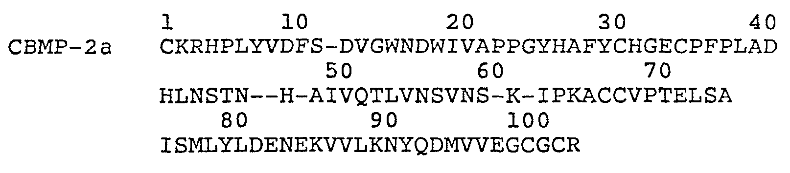
Vgl is a known Xenopus sequence heretofore not associated with bone formation. DPP is an amino acid sequence encoded by a Drosophila gene responsible for development of the dorsoventral pattern. OP1 is a region of a natural sequence encoded by exons of a genomic DNA sequence retrieved by applicants. The CBMPs are amino acid sequences comprising subparts of mammalian proteins encoded by genomic DNAs and cDNAs retrieved by applicants. The COPs are totally biosynthetic protein sequences expressed by novel consensus gene constructs, designed using the criteria set forth herein, and not yet found in nature.
These proteins are believed to be dimers. They appear not to be active when reduced. Various combinations of species of the proteins may exist as heterodimers or homodimers. As far as applicants are aware, the COP5, COP7, COP16, and OP1 constructs constitute the first instances of the design of a bioactive protein without preexisting knowledge of the active region of a native form nucleotide or amino acid sequence.
The invention thus provides synthetic osteogenic protein produced using recombinant DNA techniques. The protein may include forms having varying glycosylation patterns, varying N-termini, a family of related proteins having regions of amino acid sequence homology, and active truncated or mutated forms of native protein, no matter how derived. In view of this disclosure, skilled genetic engineers can design and synthesize genes which encode appropriate amino acid sequences, and then can express them in various types of host cells, including, both procaryotes and eucaryotes, to produce large quantities of active synthetic proteins comprising truncated analogs, muteins, fusion proteins, and other constructs mimicking the biological activity of the native forms and capable of inducing bone formation in mammals including humans.
The synthetic proteins are useful in clinical applications in conjunction with a suitable delivery or support system (matrix). The matrix is made up of particles or porous materials. The pores must be of a dimension to permit progenitor cell migration and subsequent differentiation and proliferation. The particle size should be within the range of 70. - 850 μm, preferably 70 - 420 μm. It may be fabricated by close packing particulate material into a shape spanning the bone defect, or by otherwise structuring as desired a material that is biocompatible (non-inflammatory) and, biodegradable in vivo to serve as a "temporary scaffold" and substratum for recruitment of migratory progenitor cells, and as a base for their subsequent anchoring and proliferation. Currently preferred carriers include particulate, demineralized, guanidine extracted, species-specific (allogenic) bone, and particulate, deglycosglated (or HF treated), protein extracted, demineralized, xenogenic bone. Optionally, such xenogenic bone powder matrices also may be treated with proteases such as trypsin. Other useful matrix materials comprise collagen, homopolymers and copolymers of glycolic acid and lactic acid, hydroxyapatite, tricalcium phosphate and other calcium phosphates.
The osteogenic proteins and implantable osteogenic devices enabled and disclosed herein will permit the physician to obtain optimal predictable bone formation to correct, for example, acquired and congenital craniofacial and other skeletal or dental anomalies (Glowacki et al. (1981) Lancet 1:959-963). The devices may be used to induce local endochondral bone formation in non-union fractures, and in other clinical applications including periodontal applications where bone formation is required. Another potential clinical application is in cartilage repair, for example, in the treatment of osteoarthritis.
Brief Description of the Drawing
The foregoing and other objects of this invention, the various features thereof, as well as the invention itself, may be more fully understood from the following description, when read together with the accompanying drawings, in which:
FIGURE 1 is a comparison of the amino acid sequence of various osteogenic proteins to those of the TGF-beta family. COP1, COP3, COP4, COP5, and COP7 are a family of analogs of synthetic osteogenic proteins developed from the consensus gene that was joined to a leader protein via a hinge region having the sequence D-P-N-G that permitted chemical cleavage at the D-P site (by acid) or N-G (by hydroxylamine) resulting in the release of the analog protein; VGI is a Xenopus protein, DPP is a Drosophila protein; OP1 is a native osteogenic protein; CBMP2a and 2b, and CBMP3 are subparts of proteins disclosed in PCT application 087/01537; MIS is Mullerian inhibitory substance; and "consensus choices" represent various substitutions of amino acids that may be made at various positions in osteogenic proteins;
FIGURE 2A is an E. coli expression vector containing a gene of an osteogenic protein fused to a leader protein;
FIGURE 2B is the DNA sequence comprising a modified trp-LE leader, two Fb domains of protein A, an ASP-PRO cleavage site, and the COP5 sequence; FIGURES 3A and 3B are photomicrographs of implants showing the histology (day 12) of COP5 active recombinant protein. A is a control (rat matrix alone, 25 mg). B is rat matrix plus COP5, showing +++ cartilage formation and ++ bone formation (see key infra). Similar results are achieved with COP7; and
FIGURE 4 is a schematic representation of the DNA sequence and corresponding amino acid sequence of a consensus gene for osteogenic protein (COPO).
Description
Purification protocols have been developed which enable isolation of the osteogenic protein present in crude protein extracts from mammalian bone. The isolation procedure enables the production of significant quantities of substantially pure osteogenic protein from any mammalian species, provided sufficient amounts of fresh bone from the species is available. The empirical development of the procedure, coupled with the availability of fresh calf bone, enabled isolation of substantially pure bovine osteogenic protein (BOP). BOP has been characterized significantly; its ability to induce cartilage and ultimately endochondral bone growth in cat, rabbit, and rat have been studied; it has been shown to be able to induce the full developmental cascade of bone formation previously ascribed to unknown protein or proteins in heterogeneous bone extracts; and it may be used to induce formation of endochondral bone in orthopedic defects including non-union fractures. In its native form it is a glycosylated, dimeric protein. However, it is active in deglycosylated form. It has been partially sequenced.
Elucidation of the amino acid sequence of BOP enabled the construction of a consensus nucleic acid sequence designed as disclosed herein based on the sequence data, inferred codons for the sequences, and observation of partial homology with known genes. These consensus sequences have been refined by comparison with the sequences present in certain regulatory genes from drosophila, xenopus, and human followed by point mutation, expression, and assay for activity. This approach has been successful in producing several active totally synthetic constructs not found in nature (as far as applicants are aware) which have true osteogenic activity.
These discoveries enable the construction of DNAs encoding totally novel, non-native protein constructs which individually and combined are capable of producing true endochondral bone. The DNAs may be expressed using well established recombinant DNA technologies in procaryotic or . eucaryotic host cells, and the expressed proteins may be oxidized and refolded in vitro if necessary for biological activity.
The design and production of such biosynthetic proteins, the nature of the matrix, and other material aspects concerning the nature, utility, how to make, and how to use the subject matter claimed herein will be further understood from the following, which constitutes the best method currently known for practicing the various aspects of the invention.
CONSENSUS SEQUENCE DESIGN
A synthetic consensus gene shown in FIGURE 4 was designed to encode a consensus protein based on amino acid predictions from homology with the TGF-beta gene family. The designed concensus sequence was then constructed using known techniques involving assembly of oligonucleotides manufactured in a DNA synthesizer.
Tryptic peptides derived from Bovine Osteogenic Protein isolated by applicants and sequenced by Edman degradation provided amino acid sequences that showed strong homology with the Drosophila DPP protein sequence (as inferred from the gene), the Xenopus VG1 protein, and somewhat less homology to inhibin and TGF-beta, as demonstrated below in TABLE 1.
Figure imgf000023_0001
Figure imgf000024_0001
In determining an appropriate amino acid sequence of an osteogenic protein (from which the nucleic acid sequence can be determined), the following points were considered: (1) the amino acid sequence determined by Edman degradation of natural source osteogenic protein tryptic fragments is ranked highest as long as it has a strong signal and shows homology or conservative changes when aligned with the other members of the gene family; (2) where the sequence matches for all four proteins, it is used in the synthetic gene sequence; (3) matching amino acids in DPP and Vgl are used; (4) If Vgl or DPP diverged but either one were matched by inhibin or by TGF-beta, this matched amino acid is chosen; (5) where all sequences diverged, the DPP sequence is initially chosen, with a later plan of creating the Vgl sequence by mutagenesis kept as a possibility. In addition, the consensus sequence is designed to preserve the disulfide crosslinking and the apparent structural homology among the related proteins.
RECOMBINANT OSTEOGENIC PROTEIN CONSTRUCTS
This approach resulted in the production of novel recombinant proteins capable of inducing formation of cartilage and endochondral bone comprising a protein structure analogous to or duplicative of the functional domain of the naturally sourced material. The amino acid sequences encoded by the consensus DNA sequences were derived from a family of natural proteins implicated in tissue development. These gene products/proteins are known to exist in active form as dimers and are, in general, processed from a precursor protein to produce an active C-terminal domain of the precursor.
The recombinant osteogenic/chondrogenic proteins are "novel" in the sense that, as far as applicants are aware, they do not exist in nature or, if they do exist, have never before been associated with bone or cartilage formation. The approach to design of these proteins is to employ amino acid sequences, found in the native OP isolates, in polypeptide structures are patterned after certain proteins reported in the literature, or the amino acid sequences inferred from DNAs reported in the literature. Thus, using the design criteria set forth above, and refining the amino acid sequence as more protein sequence information was learned, a series of synthetic proteins were designed with the hope and intent that they might have osteogenic or chondrogenic activity when tested in the bioassay system disclosed below.
It was noted, for example, that DPP from drosophila, VGl from Xenopus, the TGF beta family of proteins, and to a lesser extent, alpha and beta inhibins, had significant homologies with certain of the sequences derived from the naturally sourced OP product. (FIGURE 1.) Study of these proteins led to the realization that a portion of the sequence of each had a structural similarity observable by analysis of the positional relationship of cysteines and other amino acids which have an important influence on three dimensional protein conformation. It was noted that a region of these sequences had a series of seven cysteines, placed very nearly in the same relative positions, and certain other amino acids in sequence as set forth below:
Figure imgf000026_0001
wherein each X independently represents an amino acid. Expression experiments of two of. these constructs demonstrate activity. Expression experiments with constructs patterned after this template amino acid sequence with a shorter sequence having only six cysteines also show activity:
Figure imgf000027_0001
wherein each X independently represents an amino acid. Within these generic structures are a multiplicity of specific sequences which have osteogenic or chondrogenic activity. Preferred structures are those having the amino acid sequence:
Figure imgf000027_0002
wherein, in each position where more than one amino acid is shown, any one of the amino acids shown may be used. Novel active proteins also are defined by amino acid sequences comprising an active domain beginning at residue number 6 of this sequence, i.e, omitting the N terminal CXXXX, or omitting any of the preferred specific combinations such as CKRHP, CRRKQ, CKRHE, etc, resulting in a construct having only six cysteine residues. After this work, PCT 87/01537 was published, and it was observed that the proteins there identified as BMPII a and b and BMPIII each included" a region embodying this generic structure. These proteins were not demonstrated to be osteogenic in the published application. However, applicants discovered that a subpart of the amino acid sequence of these proteins, properly folded, and implanted as set forth herein, is active. These are disclosed herein as CBMPIIa, CBMPIIb, and CBMPIII. Also, applicants retrieved a previously unreported gene by probing a human genomic DNA library with COPO. This protein was designated OP1. It comprises a region exhibiting the same generic structure.
Thus, the preferred osteogenic proteins are expressed from recombinant DNA and comprise amino acid sequences including any of the following sequences:
Figure imgf000028_0001
Figure imgf000029_0001
Figure imgf000030_0001
As shown in FIGURE 1, these sequences have considerable homology with the alpha and beta inhibins, three forms of TGF beta, and MIS.
Gene Preparation
The synthetic genes designed to express the proteins as described above preferably are produced by assembly of chemically synthesized oligonucleotides. 15-100mer oligonucleotides may be synthesized on a Biosearch DNA Model 8600 Synthesizer, and purified by polyacrylamide gel electrophoresis (PAGE) in Tris-Borate-EDTA buffer (TBE). The DNA is then electroeluted from the gel. Overlapping oligomers may be phosphorylated by T4 polynucleotide kinase and ligated into larger blocks which may also be purifed by PAGE. Natural gene sequences and cDNAs also may be used for expression.
Expression
The genes can be expressed in appropriate prokaryotic hosts such as various strains of E. coli and also in bacillus, yeasts, and various animal cells such as CHO, myeloma, etc. Generally, expression may be achieved using many cell types and expression systems well known to those skilled in the art. If the gene is to be expressed in E. coli, it must first be cloned into an expression vector. An expression vector (FIGURE 2A) based on pBR322 and containing a synthetic trp promoter operator and the modified trp LE leader can be opened at the EcoRI and PSTI restriction sites, and a FB-FB COP1, COP3, COP5, and COP7 gene fragments (FIGURE 2B) can be inserted between these sites, where FB is fragment B of Staphylococcal Protein A. The expressed fusion protein results from attachment of the COP gene to a fragment encoding FB. The COP protein is joined to the leader protein via a hinge region having the sequence Asp-Pro-Asn-Gly. This hinge permits chemical cleavage of the fusion protein with dilute acid at the asp-pro site or cleavage at Asn-Gly with hydroxylamine, resulting in release of the COP protein. For COP16 and OP1, the proteins are expressed as fusion products, using the modified trp-LE as a leader.
Production of Active Proteins
The following procedure was followed for production of active recombinant proteins. E. coli cells containing the fusion proteins were lysed. The fusion proteins were purified by differential solubilization. In the case of the COP1, 3, 4, 5, and 7 fusion proteins, cleavage was with dilute acid, and the resulting cleavage products were passed through a Sephacryl-200HR column. The Sephacryl column separated most of the uncleaved fusion products from the COP1, 3, 4, 5, and 7 analogs. In the case of the COP16 or OP1 fusion protein, cleavage was with a more concentrated acid, and an SP-Trisacryl column was used as an additional purification step. The COP or OP fractions were then subjected to HPLC on a semi-prep C-18 column.
Initial conditions for refolding of COP analogs or OP1 were at pH 8.0 using Tris, Gu-HCl, dithiothreitol. Final conditions for refolding of COP analogs were at pH 8.0 using Tris, oxidized glutathione, and lower amounts of Gu-HCl and dithiothreitol. Alternatively, the COP or OP1 proteins are suspended in 50 mM HCl, 6 M guanidine-HCl, pH 8.0, for 18 hours at 4°C. Refolding may not be required if the proteins are expressed in animal cells.
Production of Antisera
Antisera to COP7 and COP5 were produced in New Zealand white rabbits. Western blots demonstrate that the antisera react with COP7 and COP5 preparations. Antisera to COP7 has been tested for reactivity to naturally sourced bovine osteogenic protein samples. Western blots show a clear reaction with the 30 kD protein and, when reduced, with the 16 kD subunit. The immunoreactive species appears as a closely-spaced doublet in the 16 kD subunit region, similar to the 16 kD doublet seen in Con A blots.
MATRIX PREPARATION
General Consideration of Matrix Properties
The carrier described in the bioassay section, infra, may be replaced by either a biodegradable-synthetic or synthetic-inorganic matrix (e.g., HAP, collagen, tricalcium phosphate, or polylactic acid, polyglycolic acid and various copolymers thereof). Also xenogeneic bone may be used if pretreated as described below. Studies have shown that surface charge, particle size, the presence of mineral, and the methodology for combining matrix and osteogenic protein all play a role in achieving successful bone induction. Perturbation of the charge by chemical modification abolishes the inductive response. Particle size influences the quantitative response of new bone; particles between 75 and 420 μm elicit the maximum response. Contamination of the matrix with bone mineral will inhibit bone formation. Most importantly, the procedures used to formulate osteogenic protein onto the matrix are extremely sensitive to the physical and chemical state of both the osteogenic protein and the matrix.
The sequential cellular reactions at the interface of the bone matrix/OP implants are complex. The multistep cascade includes: binding of fibrin and fibronectin to implanted matrix, chemotaxis of cells, proliferation of fibroblasts, differentiation into chondroblasts, cartilage formation, vascular invasion, bone formation, remodeling, and bone marrow differentiation.
A successful carrier for osteogenic protein must perform several important functions. It must bind osteogenic protein and act as a slow release delivery system, accommodate each step of the cellular response during bone development, and protect the osteogenic protein from nonspecific proteolysis. In addition, selected materials must be biocompatible in vivo and biodegradable; the carrier must act as a temporary scaffold until replaced completely by new bone. Biocompatibility requires that the matrix not induce significant inflammation when implanted and not be rejected by the host animal. Biodegradability requires that the matrix be slowly absorbed by the body of the host during development of new bone or cartilage. Polylactic acid (PLA), polyglycolic acid (PGA), and various combinations have different dissolution rates in vivo. In bones, the dissolution rates can vary according to whether the implant is placed in cortical or trabecular bone.
Matrix geometry, particle size, the presence of surface charge, and porosity or the presence of interstices among the particles of a size sufficient to permit cell infiltration, are all important to successful matrix performance. It is preferred to shape the matrix to the desired form of the new bone and to have dimensions which span non-union defects. Rat studies show that the new bone is formed essentially having the dimensions of the device implanted.
The matrix may comprise a shape-retaining solid made of loosely adhered particulate material, e.g., with collagen. It may also comprise a molded, porous solid, or simply an aggregation of close-packed particles held in place by surrounding tissue. Masticated muscle or other tissue may also be used. Large allogeneic bone implants can act as a carrier for the matrix if their marrow cavities are cleaned and packed with particles and the dispersed osteogenic protein.
Preparation of Biologically Active Allogenic Matrix
Demineralized bone matrix is prepared from the dehydrated diaphyseal shafts of rat femur and tibia as described herein to produce a bone particle size which pass through a 420 μm sieve. The bone particles are subjected to dissociative extraction with 4 M guanidine-HCl. Such treatment results in a complete loss of the inherent ability of the bone matrix to induce endochondral bone differentiation. The remaining insoluble material is used to fabricate the matrix. The material is mostly collagenous in nature, and upon implantation, does not induce cartilage and bone. All new preparations are tested for mineral content and false positives before use. The total loss of biological activity of bone matrix is restored when an active όsteoinductive protein fraction or a pure protein is reconstituted with the biologically inactive insoluble collagenous matrix. The osteoinductive protein can be obtained from any vertebrate, e.g., bovine, porcine, monkey, or human, or produced using recombinant DNA techniques.
Preparation of Deolycosylated Bone Matrix for Use in Xenogenic Implant
When osteogenic protein is reconstituted with collagenous bone matrix from other species and implanted in rat, no bone is formed. This suggests that while the osteogenic protein is xenogenic (not species specific), while the matrix is species specific and cannot be implanted cross species perhaps due to intrinsic immunogenic or inhibitory components. Thus, heretofore, for bone-based matrices, in order for the osteogenic protein to exhibit its full bone inducing activity, a species specific collagenous bone matrix was required.
The major component of all bone matrices is Type I collagen. Iri addition to collagen, extracted bone includes non-collagenous proteins which may account for 5% of its mass. Many non-collagenous components of bone matrix are glycoproteins. Although the biological significance of the glycoproteins in bone formation is not known, they may present themselves as potent antigens by virtue of their carbohydrate content and may constitute immunogenic and/or inhibitory components that are present in xenogenic matrix.
It has now been discovered that a collagenous bone matrix may be used as a carrier to effect bone inducing activity in xenogenic implants, if one first removes the immunogenic and inhibitory components from the matrix. The matrix is deglycosglated chemically using, for example, hydrogen fluoride to achieve this purpose.
Bovine bone residue prepared as described above is sieved, and particles of the 74-420 μM are collected. The sample is dried in vacuo over P2O5, transferred to the reaction vessel and anhydrous hydrogen fluoride (HF) (10-20 ml/g of matrix) is then distilled onto the sample at -70°C. The vessel is allowed to warm to 0°C. and the reaction mixture is stirred at this temperature for 120 min. After evaporation of the HF in vacuo. the residue is dried thoroughly in vacuo over KOH pellets to remove any remaining traces of acid.
Extent of deglycosylation can be determined from carbohydrate analysis of matrix samples takenbefore and after treatment with HF, after washing the samples appropriately to remove non-covalently bound carbohydrates.
The deglycosylated bone matrix is next treated as set forth below:
1) suspend in TBS (Tris-buffered Saline)
1g/200 ml and stir at 4°C for 2 hrs; or in 6 M urea, 50 mM Tris-HCl, 500 mM NaCl, pH 7.0 (UTBS) and stir at RT for 30 min; 2) centrifuge and wash with TBS or UTBS as in step 1; and
3) centrifuge; discard supernatant; water wash residue; and then lyophilize.
FABRICATION OF DEVICE
Fabrication of osteogenic devices using any of the matrices set forth above with any of the osteogenic proteins described above may be performed as follows.
A. Ethanol precipitation
In this procedure, matrix is added to osteogenic protein in guanidine-HCl. Samples are vortexed and incubated at a low temperature. Samples are then further vortexed. Cold absolute ethanol is added to the mixture which is then stirred and incubated. After centrifugation (microfuge high speed) the supernatant is discarded. The reconstituted matrix is washed with cold concentrated ethanol in water and then lyophilized.
B. Acetonitrile Trifluoroacetic Acid Lvophilization
In this procedure, osteogenic protein in an acetonitrile trifluroacetic acid (ACN/TFA) solution is added to the carrier. Samples are vigorously vortexed many times and then lyophilized. C. Urea Lyophilization
For those proteins that are prepared in urea buffer, the protein is mixed with the matrix, vortexed many times, and then lyophilized. The lyophilized material may be used "as is" for implants
IN VIVO RAT BIOASSAY
Several of the synthetic proteins have been incorporated in matrices to produce osteogenic devices, and assayed in rat for endochondral bone. Studies in rats show the osteogenic effect to be dependent on the dose of osteogenic protein dispersed in the osteogenic device. No activity is observed if the matrix is implanted alone. The following sets forth guidelines for how the osteogenic devices disclosed herein can be assayed for evaluating protein constructs and matrices for biological activity.
A. Subcutaneous Implantation
The bioassay for bone induction as described by Sampath and Reddi (Proc. Natl. Acad. Sci. USA (1983) 80: 6591-6595), herein incorporated by reference, is used to assess endochondral bone differentiation activity. This assay consists of implanting the test samples in subcutaneous sites in allogenic recipient rats under ether anesthesia. Male Long-Evans rats, aged 28-32 days, were used. A vertical incision (1 cm) is made under sterile conditions in the skin over the thoraic region, and a pocket is prepared by blunt dissection. Approximately 25 mg of the test sample is implanted deep into the pocket and the incision is closed with a metallic skin clip. The day of implantation is designated as day one of the experiment. Implants were removed on day 12. The heterotropic site allows for the study of bone induction without the possible ambiguities resulting from the use of orthotopic sites.
B. Cellular Events
The implant model in rats exhibits a controlled progression through the stages of matrix induced endochondral bone development including: (1) transient infiltration by polymorphonuclear leukocytes on day one; (2) mesenchymal cell migration and proliferation on days two and three; (3) chondrocyte appearance on days five and six; (4) cartilage matrix formation on day seven; (5) cartiliage calcification on day eight; (6) vascul-ar invasion, appearance of osteoblasts, and formation of new bone on days nine and ten; (7) appearance of osteoblastic and bone remodeling and dissolution of the implanted matrix on days twelve to eighteen; and (8) hematopoietic bone marrow differentiation in the ossicle on day twenty-one. The results show that the shape of the new bone conforms to the shape of the implanted matrix. C. Histological Evaluation
Histological sectioning and staining is preferred to determine the extent of osteogenesis in the implants. Implants are fixed in Bouins Solution, embedded in parafilm, cut into 6-8 mm sections. Staining with toluidine blue or hemotoxylin/eosin demonstrates clearly the ultimate development of endochondrial bone. Twelve day implants are usually sufficient to determine whether the implants show bone inducing activity.
D. Biological Markers
Alkaline phosphatase activity may be used as a marker for osteogenesis. The enzyme activity may be determined spectrophotometrically after homogenization of the implant. The activity peaks at 9-10 days in vivo and thereafter slowly declines. Implants showing no bone development by histology should have little or no alkaline phosphatase activity under these assay conditions. The assay is useful for quantitation and obtaining an estimate of bone formation very quickly after the implants are removed from the rat. Alternatively the amount of bone formation can be determined by measuring the calcium content of the implant. The osteogenic activity due to osteogenic protein is represented by "bone forming units". One bone forming unit represents the amount of protein that is needed for half maximal bone forming activity as compared to rat demineralized bone matrix as control and determined by calcium content of the implant on day 12.
Devices that contained only rat carrier show complete absence of new bone formation. The implant consists of carrier rat matrix and surrounding mesenchymal cells. Again, the devices that contained rat carrier and not correctly folded (or biologically inactive) recombinant protein also showed complete absence of bone formation. These implants are scored as cartilage formation (-) and bone formation (-). The endochondral bone formation activity is scored as zero percent (0%) (FIGURE 3A).
Implants included biologically active recombinant protein, however, showed evidence of endochondral bone formation. Histologically they showed new cartilage and bone formation.
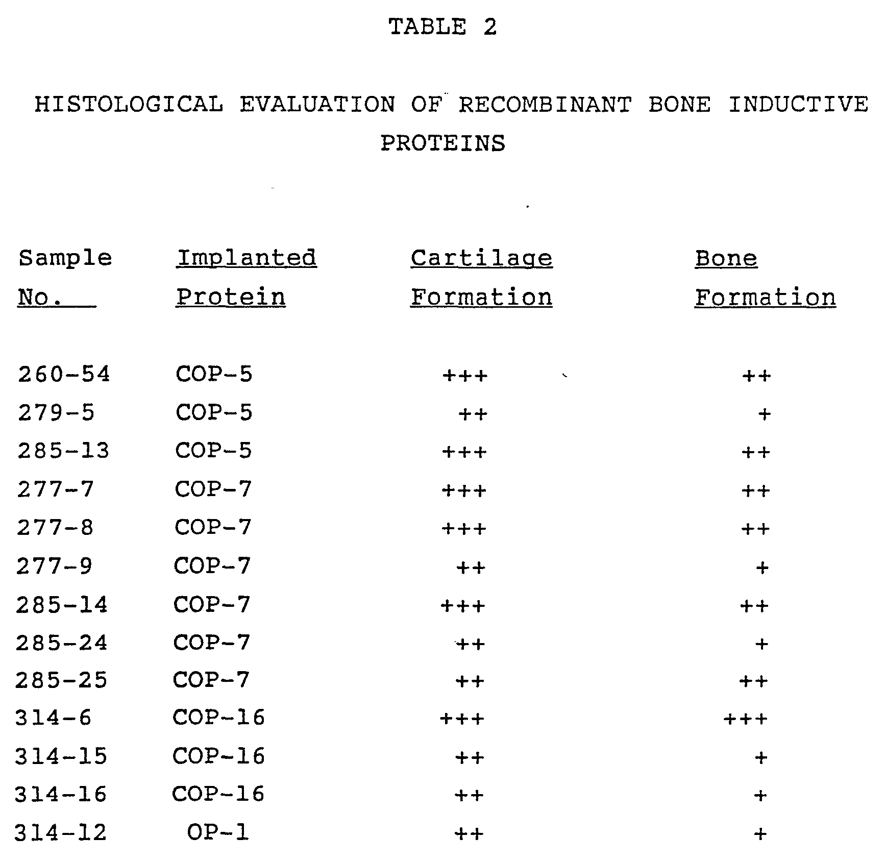
The cartilage formation is scored as (+) by the presence of metachromatically stained chondrocytes in the center of the implant, as (++) by the presence of numerous chondrocytes in many areas of the implant and as (+++) by the presence of abundant chondrocytes forming cartilage matrix and the appearance of hypertrophied chondrocytes accompanying cartilage calcification (FIGURE 3B). The bone formation is scored as (+) by the presence of osteoblast surrounding vascular endothelium forming new matrix, as (++) by the formation of bone due to osteoblasts (as indicated by arrows) and further bone remodeling by the appearance of osteoclasts in opposition to the newly formed bone matrix. Vascular invasion is evident in these implants (FIGURE 3B). Formation is scored as (+++) by the presence of extensive remodeled bone which results in the formation of ossicles.
The overall bone inducing activity due to recombinant protein is represented as percent response of endochondral bone formation (see TABLE 2 below).
Figure imgf000045_0001
The invention may be embodied in other specific forms without departing from the spirit or essential characteristics thereof. The present embodiments are therefore to be considered in all respects as illustrative and not restrictive, the scope of the invention being indicated by the appended claims rather than by the foregoing description, and all changes which come within the meaning and range of equivalency of the claims are therefore intended to be embraced therein.
Figure imgf000071_0001
Figure imgf000072_0001
Figure imgf000079_0001
Figure imgf000080_0001

Claims

Claims
1. An osteogenic device for implantation in a mammal, said device comprising:
a biocompatible, in vivo biodegradable matrix defining pores of a dimension sufficient to permit influx, proliferation and differentiation of migratory progenitor cells from the body of said mammal; and
a protein, produced by expression of recombinant DNA in a host cell, comprising one or more polypeptide chains, each of which comprises an amino acid sequence sufficiently duplicative of the sequence of COP5, COP7, COP16, or OP1 such that said protein is capable of inducing endochondral bone formation in association with said matrix when implanted in a mammal.
2. A device for implantation in a mammal, said device comprising:
a biocompatible, in vivo biodegradable matrix defining pores of a dimension sufficient to permit influx, proliferation and differentiation of migratory progenitor cells from the body of said mammal; and a protein, produced by expression of recombinant DNA in a host cell, comprising one or more polypeptide chains, each of which has less than about 200 amino acids, and each of which comprises a sequence sufficiently duplicative of the sequence of COP5, COP7, COP16, or OP1 such that said protein is capable of inducing cartilage formation in association with said matrix when implanted in a mammal.
3. The device of claim 1 or 2 wherein the sequence comprises:
wherein each X independently represents an amino acid.
4. The device of claim 1 or 2 wherein the sequence comprises:
Figure imgf000048_0002
wherein each X independently represents an amino acid.
5. The device of claim 1 or 2 wherein the sequence comprises:
Figure imgf000049_0001
wherein, in each position where more than one amino acid is shown, any one of the amino acids shown may be in that position.
6. The device of claim 1 or 2 wherein the sequence comprises:
Figure imgf000049_0002
wherein, in each position where more than one amino acid is shown, any one of the amino acids shown may be in that position.
7. The device of claim 1 or 2 wherein the sequence comprises:
Figure imgf000050_0001
8. The device of claim 1 or 2 wherein the sequence comprises:
Figure imgf000050_0002
9 . The device of claim 1 or 2 wherein the sequence comprises :
Figure imgf000050_0003
10. The device of claim 1 or 2 wherein the sequence comprises:
Figure imgf000051_0001
11. The device of claim 1 or 2 wherein the sequence comprises:
Figure imgf000051_0002
12. The device of claim 1 or 2 wherein the sequence comprises:
Figure imgf000051_0003
13. The device of claim 1 or 2 wherein the sequence comprises:
Figure imgf000052_0001
14. The device of claim 1 or 2 wherein the sequence comprises:
Figure imgf000052_0002
15. The device of claim 1 or 2 wherein the sequence comprises:
Figure imgf000052_0003
16. The device of claim 1 or 2 wherein the sequence comprises:
Figure imgf000052_0004
17. The device of claim 1 or 2 wherein the sequence comprises:
Figure imgf000053_0001
18. The device of claim 1 or 2 wherein the sequence comprises:
Figure imgf000053_0002
19. The device of claim 1 or 2 wherein the sequence comprises:
Figure imgf000053_0003
20. The device of claim 1 or 2 wherein the osteogenics protein comprises a pair of separate polypeptide chains.
21. Osteogenic protein, produced by expression of recombinant DNA in a host cell, comprising one or more polypeptide chains each of which comprises a sequence sufficiently duplicative of the sequence of COP5, COP7, COP16, or OP1 such that said protein is capable of inducing endochondral bone formation in association with a matrix when implanted in a mammal.
22. A protein, produced by expression of recombinant DNA in a host cell, comprising one or more polypeptide chains less than about 200 amino acids long, and comprising a sequence sufficiently duplicative of the sequence of COP5, COP7, COP16, or OP1 such that said protein is capable of inducing cartilage formation in association with a matrix when implanted in a mammal.
23. The protein of claim 21 or 22 further characterized by being unglycosylated.
24. The protein of claim 21 or 22 comprising a pair of separate polypeptide chains.
25. The protein of claim 21 or 22 comprising the amino acid sequences:
Figure imgf000054_0001
wherein each X independently represents an amino acid,
26. The protein of claim 21 or 22 comprising the amino acid sequences:
Figure imgf000055_0001
wherein each X independently represents an amino acid,
27. The protein of claim 21 or 22 comprising the amino acid sequences:
Figure imgf000055_0002
wherein, in each position where more than one amino acid is shown, any one of the amino acids shown may be in that position.
28. The protein of claim 21 or 22 comprising the amino acid sequences:
Figure imgf000056_0001
wherein, in each position where more than one amino acid is shown, any one of the amino acids shown may be in that position.
29. The protein of claim 21 or 22 comprising the amino acid sequences:
Figure imgf000056_0002
30. The protein of claim 21 or 22 comprising the amino acid sequences:
Figure imgf000056_0003
31. The protein of claim 21 or 22 comprising the amino acid sequence:
Figure imgf000057_0001
32. The protein of claim 21 or 22 comprising the amino acid sequences:
Figure imgf000057_0002
33. The protein of claim 21 or 22 comprising the amino acid sequences:
Figure imgf000057_0003
34. The protein of claim 21 or 22 comprising the amino acid sequences:
Figure imgf000058_0001
35. The protein of claim 21 or 22 comprising the amino acid sequences:
Figure imgf000058_0002
36. The protein of claim 21 or 22 comprising the amino acid sequences:
Figure imgf000058_0003
37. The protein of claim 21 or 22 comprising the amino acid sequences:
Figure imgf000058_0004
38. The protein of claim 21 or 22 comprising the amino acid sequences:
Figure imgf000059_0001
39. The protein of claim 21 or 22 comprising the amino acid sequences:
Figure imgf000059_0002
40. The protein of claim 21 or 22 comprising the amino acid sequences:
Figure imgf000059_0003
41. The protein of claim 21 or 22 comprising the amino acid sequences:
Figure imgf000059_0004
42. The protein of claim 21 or 22,comprising the product of expression of a DNA in a procaryotic cell.
43. A cell line engineered to express the protein of claim 21 or 22.
44. The protein of claim 21 having a half maximum bone forming activity of about 20 - 25 ng per 25 mg of implant.
45. Antibodies reactive with an epitope of the protein of claim 21 or 22.
46. The device of claim 1 or 2 wherein said matrix comprises demineralized, deglycosylated, protein extracted, particulate, xenogenic bone.
47. The device of claim 1 or 2 wherein said matrix comprises demineralized, protein-extracted, particulate, xenogenic bone treated with HF.
PCT/US1989/001469 1988-04-08 1989-04-07 Biosynthetic osteogenic proteins and osteogenic devices containing them WO1989009788A2 (en)

Priority Applications (2)

Application Number Priority Date Filing Date Title
DE68927153T DE68927153T2 (en) 1988-04-08 1989-04-07 BIOSYNTHETIC OSTEOGENE PROTEINS AND OSTEOGENE DEVICES CONTAINING THEM
EP89904959A EP0372031B1 (en) 1988-04-08 1989-04-07 Biosynthetic osteogenic proteins and osteogenic devices containing them

Applications Claiming Priority (6)

Application Number Priority Date Filing Date Title
US07/179,406 US4968590A (en) 1988-04-08 1988-04-08 Osteogenic proteins and polypeptides
US23263088A 1988-08-15 1988-08-15
US232,630 1989-02-23
US315,342 1989-02-23
US07/315,342 US5011691A (en) 1988-08-15 1989-02-23 Osteogenic devices
US179,406 1989-02-23

Publications (2)

Publication Number Publication Date
WO1989009788A2 true WO1989009788A2 (en) 1989-10-19
WO1989009788A3 WO1989009788A3 (en) 1990-02-08

Family

ID=27391116

Family Applications (2)

Application Number Title Priority Date Filing Date
PCT/US1989/001453 WO1989009787A2 (en) 1988-04-08 1989-04-07 Osteogenic devices
PCT/US1989/001469 WO1989009788A2 (en) 1988-04-08 1989-04-07 Biosynthetic osteogenic proteins and osteogenic devices containing them

Family Applications Before (1)

Application Number Title Priority Date Filing Date
PCT/US1989/001453 WO1989009787A2 (en) 1988-04-08 1989-04-07 Osteogenic devices

Country Status (8)

Country Link
US (4) US5814604A (en)
EP (6) EP0714665B1 (en)
JP (5) JP2548414B2 (en)
AT (4) ATE134647T1 (en)
AU (2) AU628050B2 (en)
CA (2) CA1338663C (en)
DE (6) DE01201546T1 (en)
WO (2) WO1989009787A2 (en)

Cited By (61)

* Cited by examiner, † Cited by third party
Publication number Priority date Publication date Assignee Title
EP0416578A2 (en) * 1989-09-06 1991-03-13 Takeda Chemical Industries, Ltd. Protein, DNA and use thereof
EP0448704A1 (en) * 1989-10-17 1991-10-02 Stryker Corporation Osteogenic devices
US5106626A (en) * 1988-10-11 1992-04-21 International Genetic Engineering, Inc. Osteogenic factors
US5106748A (en) * 1986-07-01 1992-04-21 Genetics Institute, Inc. Dna sequences encoding 5 proteins
WO1992007073A1 (en) * 1990-10-18 1992-04-30 Creative Biomolecules, Inc. Osteogenic peptides
US5116738A (en) * 1986-07-01 1992-05-26 Genetics Institute, Inc. DNA sequences encoding
US5118667A (en) * 1991-05-03 1992-06-02 Celtrix Pharmaceuticals, Inc. Bone growth factors and inhibitors of bone resorption for promoting bone formation
WO1992015323A1 (en) * 1991-03-11 1992-09-17 Creative Biomolecules, Inc. Protein-induced morphogenesis
EP0512844A1 (en) 1991-05-10 1992-11-11 Celtrix Pharmaceuticals, Inc. Targeted delivery of bone growth factors
US5187076A (en) * 1986-07-01 1993-02-16 Genetics Institute, Inc. DNA sequences encoding BMP-6 proteins
WO1993004692A1 (en) * 1991-08-30 1993-03-18 Creative Biomolecules, Inc. Morphogen-induced modulation of inflammatory response
WO1993005751A2 (en) * 1991-08-30 1993-04-01 Creative Biomolecules, Inc. Osteogenic proteins in the treatment of bone deseases
US5206023A (en) * 1991-01-31 1993-04-27 Robert F. Shaw Method and compositions for the treatment and repair of defects or lesions in cartilage
US5270300A (en) * 1991-09-06 1993-12-14 Robert Francis Shaw Methods and compositions for the treatment and repair of defects or lesions in cartilage or bone
US5284756A (en) * 1988-10-11 1994-02-08 Lynn Grinna Heterodimeric osteogenic factor
WO1994010203A2 (en) * 1992-11-03 1994-05-11 Creative Biomolecules, Inc. Op-3-induced morphogenesis
WO1994020539A1 (en) * 1993-03-04 1994-09-15 Creative Biomolecules, Inc. Method and compositions for recombinant osteogenic protein production
US5366875A (en) * 1986-07-01 1994-11-22 Genetics Institute, Inc. Methods for producing BMP-7 proteins
US5393739A (en) * 1990-11-30 1995-02-28 Celtrix Pharmaceuticals, Inc. Use of bone morphogenetic protein in synergistic combination with TGF-β for bone repair
US5459047A (en) * 1986-07-01 1995-10-17 Genetics Institute, Inc. BMP-6 proteins
WO1995033502A1 (en) * 1994-06-03 1995-12-14 Creative Biomolecules, Inc. Manufacture of autogenous replacement body parts
US5543394A (en) * 1986-07-01 1996-08-06 Genetics Institute, Inc. Bone morphogenetic protein 5(BMP-5) compositions
EP0591392B1 (en) * 1991-06-21 1996-09-11 Genetics Institute, Inc. Pharmaceutical formulations of osteogenic proteins
US5650276A (en) * 1991-03-11 1997-07-22 Creative Biomolecules, Inc. Morphogenic protein screening method
WO1997026277A2 (en) * 1996-01-22 1997-07-24 Creative Biomolecules, Inc. Morphogen analogs and methods for producing them
US5652118A (en) * 1991-03-11 1997-07-29 Creative Biomolecules, Inc. Nucleic acid encoding a novel morphogenic protein, OP-3
US5656593A (en) * 1991-03-11 1997-08-12 Creative Biomolecules, Inc. Morphogen induced periodontal tissue regeneration
US5674844A (en) * 1991-03-11 1997-10-07 Creative Biomolecules, Inc. Treatment to prevent loss of and/or increase bone mass in metabolic bone diseases
US5688678A (en) * 1990-05-16 1997-11-18 Genetics Institute, Inc. DNA encoding and methods for producing BMP-8 proteins
US5693615A (en) * 1991-06-05 1997-12-02 The Procter & Gamble Company Therapeutic compositions for osteoinduction
US5707810A (en) * 1991-03-11 1998-01-13 Creative Biomolecules, Inc. Method of diagnosing renal tissue damage or disease
US5739107A (en) * 1991-03-11 1998-04-14 Creative Biomolecules, Inc. Morphogen treatment of gastrointestinal ulcers
US5849686A (en) * 1991-03-11 1998-12-15 Creative Biomolecules, Inc. Morphogen-induced liver regeneration
US5854071A (en) * 1991-03-11 1998-12-29 Creative Biomolecules, Inc. OP-3- induced morphongenesis
US5866364A (en) * 1991-11-04 1999-02-02 Genetics Institute, Inc. Recombinant bone morphogenetic protein heterodimers
US5939388A (en) * 1986-07-01 1999-08-17 Rosen; Vicki A. Methods of administering BMP-5 compositions
US5972884A (en) * 1991-03-11 1999-10-26 Creative Biomolecules, Inc. Morphogen treatment of gastrointestinal ulcers
US6022853A (en) * 1991-08-30 2000-02-08 Creative Biomolecules, Inc. Morphogen-enriched dietary composition
US6034061A (en) * 1991-06-25 2000-03-07 Genetics Institute, Inc. BMP-9 compositions
US6034062A (en) * 1997-03-13 2000-03-07 Genetics Institute, Inc. Bone morphogenetic protein (BMP)-9 compositions and their uses
US6040431A (en) * 1995-06-07 2000-03-21 Stryker Corporation Single chain analogs of the TGF-β superfamily (morphons)
US6071708A (en) * 1991-03-11 2000-06-06 Stryker Biotech Morphogenic protein compositions of matter
US6077823A (en) * 1991-03-11 2000-06-20 Creative Biomolecules, Inc. Method for reducing tissue damage associated with ischemia-reperfusion or hypoxia injury
US6090776A (en) * 1991-03-11 2000-07-18 Creative Bio Molecules, Inc. Morphogen treatment of organ implants
US6150328A (en) * 1986-07-01 2000-11-21 Genetics Institute, Inc. BMP products
US6177406B1 (en) 1986-07-01 2001-01-23 Genetics Institute, Inc. BMP-3 products
US6194376B1 (en) 1991-03-11 2001-02-27 Creative Biomolecules, Inc. Method for modulating inflammatory response comprising administering morphogen
US6211146B1 (en) * 1991-03-11 2001-04-03 Curis, Inc. 60A protein-induced morphogenesis
US6461630B1 (en) 1995-06-07 2002-10-08 Stryker Corporation Terminally sterilized osteogenic devices and preparation thereof
US6492508B1 (en) 1996-06-03 2002-12-10 United States Surgical Corp. A Division Of Tyco Healthcare Group Nucleic acids encoding extracellular matrix proteins
US6495513B1 (en) 1991-03-11 2002-12-17 Curis, Inc. Morphogen-enhanced survival and repair of neural cells
US6531445B1 (en) 1991-03-11 2003-03-11 Curis, Inc. Protein-induced morphogenesis in liver tissue
US6617307B1 (en) * 1998-11-12 2003-09-09 Kyocera Corporation Peptide and osteogenetic accelerator
US6800603B2 (en) 1991-03-11 2004-10-05 Curis, Inc. Morphogen-induced neural cell adhesion
US7056882B2 (en) 1991-03-11 2006-06-06 Curis, Inc. Treatment to prevent loss of and/or increase bone mass in metabolic bone diseases
US7160725B2 (en) 2001-11-13 2007-01-09 Curis, Inc. Hedgehog signaling promotes the formation of three dimensional cartilage matrices
US7378392B1 (en) 1990-05-16 2008-05-27 Genetics Institute, Llc Bone and cartilage inductive proteins
JP2009227679A (en) * 1992-07-31 2009-10-08 Stryker Corp Histogenetic-factor-induced nerve regeneration and repair
EP2529764A1 (en) 2011-05-31 2012-12-05 Curasan AG Biodegradable composite material
EP2550006A1 (en) * 2010-03-24 2013-01-30 Lifenet Health Bmp-4 peptides&methods of use
US11078248B2 (en) 2010-03-19 2021-08-03 Lifenet Health BMP peptides and methods of use

Families Citing this family (60)

* Cited by examiner, † Cited by third party
Publication number Priority date Publication date Assignee Title
US5902741A (en) * 1986-04-18 1999-05-11 Advanced Tissue Sciences, Inc. Three-dimensional cartilage cultures
US6586388B2 (en) 1988-04-08 2003-07-01 Stryker Corporation Method of using recombinant osteogenic protein to repair bone or cartilage defects
US5266683A (en) * 1988-04-08 1993-11-30 Stryker Corporation Osteogenic proteins
US20070166353A1 (en) * 1988-04-08 2007-07-19 Stryker Corporation Osteogenic proteins
AU645244B2 (en) * 1988-04-08 1994-01-13 Genetics Institute, Llc Bone and cartilage inductive compositions
DE01201546T1 (en) * 1988-04-08 2004-07-08 Stryker Corp., Kalamazoo Osteogenic facilities
US6919308B2 (en) * 1988-04-08 2005-07-19 Stryker Corporation Osteogenic devices
US5108436A (en) * 1988-09-29 1992-04-28 Collagen Corporation Implant fixation
ES2113857T3 (en) * 1989-03-28 1998-05-16 Genetics Inst OSTEOINDUCTIVE COMPOSITIONS.
US5645591A (en) * 1990-05-29 1997-07-08 Stryker Corporation Synthetic bone matrix
WO1992007004A1 (en) * 1990-10-18 1992-04-30 Creative Biomolecules, Inc. Osteogenic protein
US20080233170A1 (en) * 1992-02-21 2008-09-25 Stryker Corporation Osteogenic Proteins
ZA9711580B (en) * 1996-12-25 1999-09-23 Hoechst Marion Roussel Ltd Process for the production of purified dimeric bone morphogenetic factors.
US20010016646A1 (en) 1998-03-20 2001-08-23 David C. Rueger Osteogenic devices and methods of use thereof for repair of endochondral bone, osteochondral and chondral defects
US7041641B2 (en) 1997-03-20 2006-05-09 Stryker Corporation Osteogenic devices and methods of use thereof for repair of endochondral bone and osteochondral defects
EP0985149A1 (en) * 1997-05-30 2000-03-15 Creative Biomolecules, Inc. Methods for evaluating tissue morphogenesis and activity
US5972368A (en) * 1997-06-11 1999-10-26 Sdgi Holdings, Inc. Bone graft composites and spacers
US7147839B2 (en) 1998-05-29 2006-12-12 Curis, Inc. Methods for evaluating tissue morphogenesis and activity
US6846906B1 (en) * 1998-10-07 2005-01-25 Stryker Corporation Modified proteins of the TGF-β superfamily, including morphogenic proteins
US6231528B1 (en) 1999-01-15 2001-05-15 Jonathan J. Kaufman Ultrasonic and growth factor bone-therapy: apparatus and method
DE19906096A1 (en) 1999-02-13 2000-08-17 Walter Sebald Protein with a heparin-binding epitope
DE10026713A1 (en) * 2000-05-30 2001-12-06 Walter Sebald Mutein a chain of a protein from the superfamily of the growth factor TGF-beta
US20020114795A1 (en) 2000-12-22 2002-08-22 Thorne Kevin J. Composition and process for bone growth and repair
JP4566515B2 (en) * 2001-04-25 2010-10-20 アイトゲネシッシェ テヒニッシェ ホーホシューレ チューリッヒ Drug delivery matrix to promote wound healing
WO2003043673A1 (en) 2001-11-19 2003-05-30 Scil Technology Gmbh Device having osteoinductive and osteoconductive properties
US20050008629A1 (en) * 2002-05-08 2005-01-13 Interpore Orthopaedics, A Delaware Corporation Encapsulated AGF cells
US7166133B2 (en) 2002-06-13 2007-01-23 Kensey Nash Corporation Devices and methods for treating defects in the tissue of a living being
EP1539261B1 (en) 2002-09-10 2006-04-12 Scil Technology GmbH Metal implant coated under reduced oxygen concentration with osteoinductive protein
US20050020506A1 (en) * 2003-07-25 2005-01-27 Drapeau Susan J. Crosslinked compositions comprising collagen and demineralized bone matrix, methods of making and methods of use
US7534264B2 (en) * 2004-01-28 2009-05-19 Ultradent Products, Inc. Delivery system for bone growth promoting material
CA2558395C (en) 2004-03-10 2011-11-29 Scil Technology Gmbh Coated implants, their manufacturing and use thereof
US7621963B2 (en) * 2005-04-13 2009-11-24 Ebi, Llc Composite bone graft material
US20060233849A1 (en) * 2005-04-13 2006-10-19 Simon Bruce J Composite bone graft material
US8506983B2 (en) * 2006-05-01 2013-08-13 Warsaw Orthopedic, Inc. Bone filler material
US20100209470A1 (en) * 2006-05-01 2010-08-19 Warsaw Orthopedic, Inc. An Indiana Corporation Demineralized bone matrix devices
US7838022B2 (en) * 2006-05-01 2010-11-23 Warsaw Orthopedic, Inc Malleable implants containing demineralized bone matrix
US7771741B2 (en) * 2006-05-01 2010-08-10 Warsaw Orthopedic, Inc Demineralized bone matrix devices
US7718616B2 (en) 2006-12-21 2010-05-18 Zimmer Orthobiologics, Inc. Bone growth particles and osteoinductive composition thereof
US9056150B2 (en) 2007-12-04 2015-06-16 Warsaw Orthopedic, Inc. Compositions for treating bone defects
WO2009083544A2 (en) 2007-12-28 2009-07-09 Kuros Biosurgery Ag Pdgf fusion proteins incorporated into fibrin foams
US8840913B2 (en) 2008-03-27 2014-09-23 Warsaw Orthopedic, Inc. Malleable multi-component implants and materials therefor
WO2010074039A1 (en) 2008-12-26 2010-07-01 旭硝子株式会社 Ethylene/tetrafluoroethylene copolymer granulation method
US8895291B2 (en) 2010-10-08 2014-11-25 Terumo Bct, Inc. Methods and systems of growing and harvesting cells in a hollow fiber bioreactor system with control conditions
CN103313733A (en) 2010-11-15 2013-09-18 捷迈整形外科生物材料有限公司 Bone void fillers
US10207027B2 (en) 2012-06-11 2019-02-19 Globus Medical, Inc. Bioactive bone graft substitutes
US9486483B2 (en) 2013-10-18 2016-11-08 Globus Medical, Inc. Bone grafts including osteogenic stem cells, and methods relating to the same
US9539286B2 (en) 2013-10-18 2017-01-10 Globus Medical, Inc. Bone grafts including osteogenic stem cells, and methods relating to the same
CN105992816B (en) 2013-11-16 2018-04-17 泰尔茂比司特公司 Cell amplification in bioreactor
US9579421B2 (en) 2014-02-07 2017-02-28 Globus Medical Inc. Bone grafts and methods of making and using bone grafts
US9463264B2 (en) 2014-02-11 2016-10-11 Globus Medical, Inc. Bone grafts and methods of making and using bone grafts
JP6783143B2 (en) 2014-03-25 2020-11-11 テルモ ビーシーティー、インコーポレーテッド Passive replenishment of medium
WO2016049421A1 (en) 2014-09-26 2016-03-31 Terumo Bct, Inc. Scheduled feed
US11426489B2 (en) 2015-06-10 2022-08-30 Globus Medical, Inc. Biomaterial compositions, implants, and methods of making the same
US10016529B2 (en) 2015-06-10 2018-07-10 Globus Medical, Inc. Biomaterial compositions, implants, and methods of making the same
WO2017004592A1 (en) 2015-07-02 2017-01-05 Terumo Bct, Inc. Cell growth with mechanical stimuli
US11685883B2 (en) 2016-06-07 2023-06-27 Terumo Bct, Inc. Methods and systems for coating a cell growth surface
US11104874B2 (en) 2016-06-07 2021-08-31 Terumo Bct, Inc. Coating a bioreactor
US11624046B2 (en) 2017-03-31 2023-04-11 Terumo Bct, Inc. Cell expansion
EP3656842A1 (en) 2017-03-31 2020-05-27 Terumo BCT, Inc. Cell expansion
US11896736B2 (en) 2020-07-13 2024-02-13 Globus Medical, Inc Biomaterial implants and methods of making the same

Citations (5)

* Cited by examiner, † Cited by third party
Publication number Priority date Publication date Assignee Title
EP0128041A2 (en) * 1983-06-06 1984-12-12 David Jeston Baylink Polypeptides exhibiting skeletal growth factor activity
US4563350A (en) * 1984-10-24 1986-01-07 Collagen Corporation Inductive collagen based bone repair preparations
EP0169016A2 (en) * 1984-07-16 1986-01-22 Celtrix Pharmaceuticals, Inc. Polypeptide cartilage-inducing factors found in bone
EP0212474A2 (en) * 1985-08-07 1987-03-04 The Regents Of The University Of California Bone morphogenetic peptides
WO1988000205A1 (en) * 1986-07-01 1988-01-14 Genetics Institute, Inc. Novel osteoinductive compositions

Family Cites Families (51)

* Cited by examiner, † Cited by third party
Publication number Priority date Publication date Assignee Title
US621146A (en) * 1899-03-14 Presser-foot for sewing-machines
CH595293A5 (en) * 1975-02-20 1978-02-15 Battelle Memorial Institute
US4294753A (en) * 1980-08-04 1981-10-13 The Regents Of The University Of California Bone morphogenetic protein process
US4455256A (en) * 1981-05-05 1984-06-19 The Regents Of The University Of California Bone morphogenetic protein
US4394370A (en) * 1981-09-21 1983-07-19 Jefferies Steven R Bone graft material for osseous defects and method of making same
US4434094A (en) * 1983-04-12 1984-02-28 Collagen Corporation Partially purified osteogenic factor and process for preparing same from demineralized bone
NZ210699A (en) * 1984-01-04 1989-06-28 Int Genetic Eng Isolation of an osteogenic protein of the p3 immunologically related family
US4804744A (en) * 1984-01-04 1989-02-14 International Genetic Engineering, Inc. Osteogenic factors
US4563489A (en) * 1984-02-10 1986-01-07 University Of California Biodegradable organic polymer delivery system for bone morphogenetic protein
GB8413319D0 (en) * 1984-05-24 1984-06-27 Oliver Roy Frederick Biological material
US4620327A (en) * 1984-07-05 1986-11-04 Caplan Arnold I Process of adapting soluble bone protein for use in stimulating osteoinduction
US4843063A (en) * 1984-07-16 1989-06-27 Collagen Corporation Polypeptide cartilage-inducing factors found in bone
US4817190A (en) 1985-08-27 1989-03-28 Motorola, Inc. Method for dynamically regrouping subscribers on a communications system
ZA874681B (en) * 1986-07-01 1988-04-27 Genetics Inst Novel osteoinductive factors
US5187076A (en) * 1986-07-01 1993-02-16 Genetics Institute, Inc. DNA sequences encoding BMP-6 proteins
US5106748A (en) * 1986-07-01 1992-04-21 Genetics Institute, Inc. Dna sequences encoding 5 proteins
US5013649A (en) * 1986-07-01 1991-05-07 Genetics Institute, Inc. DNA sequences encoding osteoinductive products
US5108922A (en) * 1986-07-01 1992-04-28 Genetics Institute, Inc. DNA sequences encoding BMP-1 products
US4877864A (en) * 1987-03-26 1989-10-31 Genetics Institute, Inc. Osteoinductive factors
DE3775363D1 (en) * 1986-10-22 1992-01-30 Biotechnolog Forschung Gmbh GROWTH-STIMULATING MATERIAL, MANUFACTURING PROCESS AND THERAPEUTIC COMPOSITION.
WO1989009605A1 (en) * 1988-04-06 1989-10-19 Collagen Corporation Bone-inducing protein
US5108753A (en) * 1988-04-08 1992-04-28 Creative Biomolecules Osteogenic devices
AU645244B2 (en) * 1988-04-08 1994-01-13 Genetics Institute, Llc Bone and cartilage inductive compositions
US5266683A (en) * 1988-04-08 1993-11-30 Stryker Corporation Osteogenic proteins
US5324819A (en) * 1988-04-08 1994-06-28 Stryker Corporation Osteogenic proteins
US5354557A (en) * 1988-04-08 1994-10-11 Stryker Corporation Osteogenic devices
US5162114A (en) * 1989-02-23 1992-11-10 Stryker Corporation Bone collagen matrix for xenogenic implants
US4975526A (en) * 1989-02-23 1990-12-04 Creative Biomolecules, Inc. Bone collagen matrix for zenogenic implants
US5258494A (en) * 1988-04-08 1993-11-02 Stryker Corporation Osteogenic proteins
DE01201546T1 (en) * 1988-04-08 2004-07-08 Stryker Corp., Kalamazoo Osteogenic facilities
US5250302A (en) * 1988-04-08 1993-10-05 Stryker Corporation Osteogenic devices
US6586388B2 (en) * 1988-04-08 2003-07-01 Stryker Corporation Method of using recombinant osteogenic protein to repair bone or cartilage defects
US6919308B2 (en) * 1988-04-08 2005-07-19 Stryker Corporation Osteogenic devices
US5344654A (en) * 1988-04-08 1994-09-06 Stryker Corporation Prosthetic devices having enhanced osteogenic properties
US5011691A (en) * 1988-08-15 1991-04-30 Stryker Corporation Osteogenic devices
US5106626A (en) * 1988-10-11 1992-04-21 International Genetic Engineering, Inc. Osteogenic factors
US5284756A (en) * 1988-10-11 1994-02-08 Lynn Grinna Heterodimeric osteogenic factor
ES2113857T3 (en) * 1989-03-28 1998-05-16 Genetics Inst OSTEOINDUCTIVE COMPOSITIONS.
JPH05500503A (en) * 1989-08-21 1993-02-04 セルトリックス ファーマシューティカルズ、インコーポレイテッド bone specific protein
US5645591A (en) * 1990-05-29 1997-07-08 Stryker Corporation Synthetic bone matrix
US5652337A (en) * 1991-03-11 1997-07-29 Creative Biomolecules, Inc. OP-3-induced morphogenesis
US5656593A (en) * 1991-03-11 1997-08-12 Creative Biomolecules, Inc. Morphogen induced periodontal tissue regeneration
US5674844A (en) * 1991-03-11 1997-10-07 Creative Biomolecules, Inc. Treatment to prevent loss of and/or increase bone mass in metabolic bone diseases
US6022853A (en) * 1991-08-30 2000-02-08 Creative Biomolecules, Inc. Morphogen-enriched dietary composition
US5906827A (en) * 1994-06-03 1999-05-25 Creative Biomolecules, Inc. Matrix for the manufacture of autogenous replacement body parts
US5674292A (en) * 1995-06-07 1997-10-07 Stryker Corporation Terminally sterilized osteogenic devices and preparation thereof
CA2280931C (en) * 1997-02-07 2009-05-05 Stryker Corporation Matrix-free osteogenic devices, implants and methods of use thereof
US20010016646A1 (en) * 1998-03-20 2001-08-23 David C. Rueger Osteogenic devices and methods of use thereof for repair of endochondral bone, osteochondral and chondral defects
US20030032586A1 (en) * 1997-05-15 2003-02-13 David C. Rueger Compositions for morphogen-induced osteogenesis
EP0937776A3 (en) * 1998-02-18 2002-06-12 Kabushiki Kaisha Toyota Chuo Kenkyusho Immobilized enzymes
US6677432B1 (en) * 1998-10-07 2004-01-13 Stryker Corporation Mutations of the C-terminal portion of TGF-β superfamily proteins

Patent Citations (5)

* Cited by examiner, † Cited by third party
Publication number Priority date Publication date Assignee Title
EP0128041A2 (en) * 1983-06-06 1984-12-12 David Jeston Baylink Polypeptides exhibiting skeletal growth factor activity
EP0169016A2 (en) * 1984-07-16 1986-01-22 Celtrix Pharmaceuticals, Inc. Polypeptide cartilage-inducing factors found in bone
US4563350A (en) * 1984-10-24 1986-01-07 Collagen Corporation Inductive collagen based bone repair preparations
EP0212474A2 (en) * 1985-08-07 1987-03-04 The Regents Of The University Of California Bone morphogenetic peptides
WO1988000205A1 (en) * 1986-07-01 1988-01-14 Genetics Institute, Inc. Novel osteoinductive compositions

Non-Patent Citations (1)

* Cited by examiner, † Cited by third party
Title
See also references of EP0372031A1 *

Cited By (107)

* Cited by examiner, † Cited by third party
Publication number Priority date Publication date Assignee Title
US5187076A (en) * 1986-07-01 1993-02-16 Genetics Institute, Inc. DNA sequences encoding BMP-6 proteins
US5849880A (en) * 1986-07-01 1998-12-15 Genetics Institute, Inc. Bone morphogenetic protein (BMP)--6
US6432919B1 (en) 1986-07-01 2002-08-13 Genetics Institute, Inc. Bone morphogenetic protein-3 and compositions
US5106748A (en) * 1986-07-01 1992-04-21 Genetics Institute, Inc. Dna sequences encoding 5 proteins
US5366875A (en) * 1986-07-01 1994-11-22 Genetics Institute, Inc. Methods for producing BMP-7 proteins
US5166058A (en) * 1986-07-01 1992-11-24 Genetics Institute, Inc. DNA sequences encoding the osteoinductive proteins
US5116738A (en) * 1986-07-01 1992-05-26 Genetics Institute, Inc. DNA sequences encoding
US5635373A (en) * 1986-07-01 1997-06-03 Genetics Institute, Inc. Bone morphogenic protein-5(BMP-5) and DNA encoding same
US5618924A (en) * 1986-07-01 1997-04-08 Genetics Institute, Inc. BMP-2 products
US6613744B2 (en) 1986-07-01 2003-09-02 Genetics Institute, Llc BMP-6 proteins
US5939388A (en) * 1986-07-01 1999-08-17 Rosen; Vicki A. Methods of administering BMP-5 compositions
US5543394A (en) * 1986-07-01 1996-08-06 Genetics Institute, Inc. Bone morphogenetic protein 5(BMP-5) compositions
US6245889B1 (en) 1986-07-01 2001-06-12 Genetics Institute, Inc. BMP-4 products
US6150328A (en) * 1986-07-01 2000-11-21 Genetics Institute, Inc. BMP products
US6177406B1 (en) 1986-07-01 2001-01-23 Genetics Institute, Inc. BMP-3 products
US5459047A (en) * 1986-07-01 1995-10-17 Genetics Institute, Inc. BMP-6 proteins
US6207813B1 (en) 1986-07-01 2001-03-27 Genetics Institute, Inc. BMP-6 proteins
US5411941A (en) * 1988-10-11 1995-05-02 Xoma Corporation Heterodimeric osteogenic factor
US5284756A (en) * 1988-10-11 1994-02-08 Lynn Grinna Heterodimeric osteogenic factor
US5508263A (en) * 1988-10-11 1996-04-16 Xoma Corporation Heterodimeric osteogenic factor
US5106626A (en) * 1988-10-11 1992-04-21 International Genetic Engineering, Inc. Osteogenic factors
EP0416578A2 (en) * 1989-09-06 1991-03-13 Takeda Chemical Industries, Ltd. Protein, DNA and use thereof
US5453419A (en) * 1989-09-06 1995-09-26 Chichibu Cement Co., Ltd. Xenopus laevis bone morphogenetic proteins and use thereof
EP0416578A3 (en) * 1989-09-06 1992-04-22 Takeda Chemical Industries, Ltd. Protein, dna and use thereof
EP0448704A4 (en) * 1989-10-17 1992-07-08 Creative Biomolecules, Inc. Osteogenic devices
EP0448704A1 (en) * 1989-10-17 1991-10-02 Stryker Corporation Osteogenic devices
US5688678A (en) * 1990-05-16 1997-11-18 Genetics Institute, Inc. DNA encoding and methods for producing BMP-8 proteins
US7378392B1 (en) 1990-05-16 2008-05-27 Genetics Institute, Llc Bone and cartilage inductive proteins
WO1992007073A1 (en) * 1990-10-18 1992-04-30 Creative Biomolecules, Inc. Osteogenic peptides
US5393739A (en) * 1990-11-30 1995-02-28 Celtrix Pharmaceuticals, Inc. Use of bone morphogenetic protein in synergistic combination with TGF-β for bone repair
US5368858A (en) * 1991-01-31 1994-11-29 Robert F. Shaw Methods and compositions for the treatment and repair of defects or lesions in cartilage
US5206023A (en) * 1991-01-31 1993-04-27 Robert F. Shaw Method and compositions for the treatment and repair of defects or lesions in cartilage
US6399569B1 (en) 1991-03-11 2002-06-04 Curis, Inc. Morphogen treatments for limiting proliferation of epithelial cells
US6090776A (en) * 1991-03-11 2000-07-18 Creative Bio Molecules, Inc. Morphogen treatment of organ implants
US6395883B1 (en) * 1991-03-11 2002-05-28 Curis, Inc. Soluble morphogenic protein complex compositions of matter
US6211146B1 (en) * 1991-03-11 2001-04-03 Curis, Inc. 60A protein-induced morphogenesis
US5650276A (en) * 1991-03-11 1997-07-22 Creative Biomolecules, Inc. Morphogenic protein screening method
US7268208B2 (en) 1991-03-11 2007-09-11 Stryker Corporation Morphogen solubilizing peptides
US5652118A (en) * 1991-03-11 1997-07-29 Creative Biomolecules, Inc. Nucleic acid encoding a novel morphogenic protein, OP-3
US5656593A (en) * 1991-03-11 1997-08-12 Creative Biomolecules, Inc. Morphogen induced periodontal tissue regeneration
US7196056B2 (en) 1991-03-11 2007-03-27 Curis, Inc. Protein-induced morphogenesis of kidney tissue
US5674844A (en) * 1991-03-11 1997-10-07 Creative Biomolecules, Inc. Treatment to prevent loss of and/or increase bone mass in metabolic bone diseases
AU660019B2 (en) * 1991-03-11 1995-06-08 Stryker Corporation Protein-induced morphogenesis
US6194376B1 (en) 1991-03-11 2001-02-27 Creative Biomolecules, Inc. Method for modulating inflammatory response comprising administering morphogen
US5707810A (en) * 1991-03-11 1998-01-13 Creative Biomolecules, Inc. Method of diagnosing renal tissue damage or disease
US5733878A (en) * 1991-03-11 1998-03-31 Creative Biomolecules, Inc. Morphogen-induced periodontal tissue regeneration
US5739107A (en) * 1991-03-11 1998-04-14 Creative Biomolecules, Inc. Morphogen treatment of gastrointestinal ulcers
US5741641A (en) * 1991-03-11 1998-04-21 Creative Biomolecules, Inc. Morphogenic protein screening method
US7060680B2 (en) 1991-03-11 2006-06-13 Curis, Inc. Morphogen treatments for limiting proliferation of epithelial cells
US5849686A (en) * 1991-03-11 1998-12-15 Creative Biomolecules, Inc. Morphogen-induced liver regeneration
US5854071A (en) * 1991-03-11 1998-12-29 Creative Biomolecules, Inc. OP-3- induced morphongenesis
US6153583A (en) * 1991-03-11 2000-11-28 Stryker Corporation OP-3 induced morphogenesis
US7056882B2 (en) 1991-03-11 2006-06-06 Curis, Inc. Treatment to prevent loss of and/or increase bone mass in metabolic bone diseases
US6800603B2 (en) 1991-03-11 2004-10-05 Curis, Inc. Morphogen-induced neural cell adhesion
US5972884A (en) * 1991-03-11 1999-10-26 Creative Biomolecules, Inc. Morphogen treatment of gastrointestinal ulcers
US5994131A (en) * 1991-03-11 1999-11-30 Creative Biomolecules, Inc. Morphogenic protein screening method
US6495513B1 (en) 1991-03-11 2002-12-17 Curis, Inc. Morphogen-enhanced survival and repair of neural cells
WO1992015323A1 (en) * 1991-03-11 1992-09-17 Creative Biomolecules, Inc. Protein-induced morphogenesis
US6288031B1 (en) 1991-03-11 2001-09-11 Curis, Inc. Method for reducing extravasation of effector cells
US6565843B1 (en) * 1991-03-11 2003-05-20 Curis, Inc. Protein-induced tissue morphogenesis
US6531445B1 (en) 1991-03-11 2003-03-11 Curis, Inc. Protein-induced morphogenesis in liver tissue
US6071708A (en) * 1991-03-11 2000-06-06 Stryker Biotech Morphogenic protein compositions of matter
US6077823A (en) * 1991-03-11 2000-06-20 Creative Biomolecules, Inc. Method for reducing tissue damage associated with ischemia-reperfusion or hypoxia injury
US5118667A (en) * 1991-05-03 1992-06-02 Celtrix Pharmaceuticals, Inc. Bone growth factors and inhibitors of bone resorption for promoting bone formation
EP0512844A1 (en) 1991-05-10 1992-11-11 Celtrix Pharmaceuticals, Inc. Targeted delivery of bone growth factors
US5693615A (en) * 1991-06-05 1997-12-02 The Procter & Gamble Company Therapeutic compositions for osteoinduction
US5597897A (en) * 1991-06-21 1997-01-28 Genetics Institute, Inc. Pharmaceutical formulations of osteogenic proteins
EP0591392B1 (en) * 1991-06-21 1996-09-11 Genetics Institute, Inc. Pharmaceutical formulations of osteogenic proteins
US6034061A (en) * 1991-06-25 2000-03-07 Genetics Institute, Inc. BMP-9 compositions
US6287816B1 (en) 1991-06-25 2001-09-11 Genetics Institute, Inc. BMP-9 compositions
US6022853A (en) * 1991-08-30 2000-02-08 Creative Biomolecules, Inc. Morphogen-enriched dietary composition
WO1993004692A1 (en) * 1991-08-30 1993-03-18 Creative Biomolecules, Inc. Morphogen-induced modulation of inflammatory response
WO1993005751A3 (en) * 1991-08-30 1993-05-13 Creative Biomolecules Inc Osteogenic proteins in the treatment of bone deseases
WO1993005751A2 (en) * 1991-08-30 1993-04-01 Creative Biomolecules, Inc. Osteogenic proteins in the treatment of bone deseases
US5270300A (en) * 1991-09-06 1993-12-14 Robert Francis Shaw Methods and compositions for the treatment and repair of defects or lesions in cartilage or bone
US5866364A (en) * 1991-11-04 1999-02-02 Genetics Institute, Inc. Recombinant bone morphogenetic protein heterodimers
US6593109B1 (en) 1991-11-04 2003-07-15 Genetics Institute, Inc. Recombinant bone morphogenetic protein heterodimers, compositions and methods of use
US6190880B1 (en) 1991-11-04 2001-02-20 Genetics Institute Recombinant bone morphogenetic protein heterodimers, compositions and methods of use
JP2009227679A (en) * 1992-07-31 2009-10-08 Stryker Corp Histogenetic-factor-induced nerve regeneration and repair
WO1994010203A2 (en) * 1992-11-03 1994-05-11 Creative Biomolecules, Inc. Op-3-induced morphogenesis
WO1994010203A3 (en) * 1992-11-03 1994-08-18 Creative Biomolecules Inc Op-3-induced morphogenesis
WO1994020539A1 (en) * 1993-03-04 1994-09-15 Creative Biomolecules, Inc. Method and compositions for recombinant osteogenic protein production
WO1995033502A1 (en) * 1994-06-03 1995-12-14 Creative Biomolecules, Inc. Manufacture of autogenous replacement body parts
US6110482A (en) * 1994-06-03 2000-08-29 Styker Corporation Manufacture of autogenous replacement body parts
US5906827A (en) * 1994-06-03 1999-05-25 Creative Biomolecules, Inc. Matrix for the manufacture of autogenous replacement body parts
US6027743A (en) * 1994-06-03 2000-02-22 Stryker Corporation Manufacture of autogenous replacement body parts
US6504079B2 (en) 1995-06-07 2003-01-07 Stryker Corporation Terminally sterilized osteogenic devices and preparation thereof
US6479643B1 (en) 1995-06-07 2002-11-12 Stryker Corporation Single chain analogs of the TGF-β superfamily (morphons)
US6461630B1 (en) 1995-06-07 2002-10-08 Stryker Corporation Terminally sterilized osteogenic devices and preparation thereof
US6040431A (en) * 1995-06-07 2000-03-21 Stryker Corporation Single chain analogs of the TGF-β superfamily (morphons)
US6273598B1 (en) 1996-01-22 2001-08-14 Creative Biomolecules, Inc. Computer system and methods for producing morphogen analogs of human OP-1
WO1997026277A3 (en) * 1996-01-22 1997-09-25 Creative Biomolecules Inc Morphogen analogs and methods for producing them
WO1997026277A2 (en) * 1996-01-22 1997-07-24 Creative Biomolecules, Inc. Morphogen analogs and methods for producing them
US6492508B1 (en) 1996-06-03 2002-12-10 United States Surgical Corp. A Division Of Tyco Healthcare Group Nucleic acids encoding extracellular matrix proteins
US6958223B2 (en) 1996-06-03 2005-10-25 United States Surgical Corporation Methods for producing extracellular matrix proteins
US6034062A (en) * 1997-03-13 2000-03-07 Genetics Institute, Inc. Bone morphogenetic protein (BMP)-9 compositions and their uses
US7049293B2 (en) 1998-11-12 2006-05-23 Kyocera Corporation Peptide and osteogenetic accelerator
US6617307B1 (en) * 1998-11-12 2003-09-09 Kyocera Corporation Peptide and osteogenetic accelerator
US7160725B2 (en) 2001-11-13 2007-01-09 Curis, Inc. Hedgehog signaling promotes the formation of three dimensional cartilage matrices
US11078248B2 (en) 2010-03-19 2021-08-03 Lifenet Health BMP peptides and methods of use
EP2550006A4 (en) * 2010-03-24 2013-11-27 Lifenet Health Bmp-4 peptides&methods of use
EP2550006A1 (en) * 2010-03-24 2013-01-30 Lifenet Health Bmp-4 peptides&methods of use
JP2013521831A (en) * 2010-03-24 2013-06-13 ライフネット ヘルス BMP-4 peptide and method of use
US9951112B2 (en) 2010-03-24 2018-04-24 Lifenet Health BMP-4 peptides and methods of use
WO2012163532A2 (en) 2011-05-31 2012-12-06 Curasan Ag Biodegradable composite material
US9907884B2 (en) 2011-05-31 2018-03-06 Curasan Ag Biodegradable composite material
EP2529764A1 (en) 2011-05-31 2012-12-05 Curasan AG Biodegradable composite material

Also Published As

Publication number Publication date
CA1338663C (en) 1996-10-22
JPH08336390A (en) 1996-12-24
JPH08322570A (en) 1996-12-10
EP0723013A2 (en) 1996-07-24
EP0362367B1 (en) 1996-02-28
JPH03500655A (en) 1991-02-14
DE68929453T2 (en) 2003-11-20
ATE231523T1 (en) 2003-02-15
EP0714665A2 (en) 1996-06-05
EP0372031B1 (en) 1996-09-11
JP2869381B2 (en) 1999-03-10
EP0723013B1 (en) 2003-06-18
US20030124169A1 (en) 2003-07-03
JPH03502579A (en) 1991-06-13
DE68925773D1 (en) 1996-04-04
EP0362367A1 (en) 1990-04-11
AU3444989A (en) 1989-11-03
ATE142644T1 (en) 1996-09-15
EP0714665A3 (en) 1997-12-03
WO1989009788A3 (en) 1990-02-08
EP1225225A2 (en) 2002-07-24
WO1989009787A2 (en) 1989-10-19
AU628050B2 (en) 1992-09-10
ATE243254T1 (en) 2003-07-15
JP2933867B2 (en) 1999-08-16
DE68929472D1 (en) 2003-07-24
EP1221484A3 (en) 2002-09-18
JP2548414B2 (en) 1996-10-30
DE68927153T2 (en) 1997-03-06
AU618357B2 (en) 1991-12-19
EP0723013A3 (en) 1998-01-14
DE68929472T2 (en) 2004-05-13
US5814604A (en) 1998-09-29
JP2522568B2 (en) 1996-08-07
DE68929453D1 (en) 2003-02-27
DE68927153D1 (en) 1996-10-17
CA1341610C (en) 2011-02-01
DE01201555T1 (en) 2004-07-08
DE68925773T2 (en) 1996-10-17
EP1225225A3 (en) 2003-01-08
US20050054825A1 (en) 2005-03-10
US6297213B1 (en) 2001-10-02
JPH08187084A (en) 1996-07-23
EP0372031A1 (en) 1990-06-13
EP0714665B1 (en) 2003-01-22
DE01201546T1 (en) 2004-07-08
AU3530589A (en) 1989-11-03
ATE134647T1 (en) 1996-03-15
WO1989009787A3 (en) 1990-02-08
EP1221484A2 (en) 2002-07-10

Similar Documents

Publication Publication Date Title
AU628050B2 (en) Biosynthetic osteogenic proteins and osteogenic devices containing them
US6261835B1 (en) Nucleotide sequences encoding osteogenic proteins
US5011691A (en) Osteogenic devices
US5324819A (en) Osteogenic proteins
CA2094027C (en) Osteogenic peptides
EP0448704B1 (en) Osteogenic devices
US5496552A (en) Osteogenic devices
US5670336A (en) Method for recombinant production of osteogenic protein
US20060093640A1 (en) Osteoinductive biomaterials
WO1992007004A1 (en) Osteogenic protein
US7429484B2 (en) Nucleic acid molecules encoding osteogenic proteins
AU648997C (en) Osteogenic devices
ZA200500427B (en) Osteoinductive biomaterials

Legal Events

Date Code Title Description
AK Designated states

Kind code of ref document: A2

Designated state(s): AU BB BG BR DK FI HU JP KP KR LK MC MG MW NO RO SD SU US US US

AL Designated countries for regional patents

Kind code of ref document: A2

Designated state(s): AT BE BF BJ CF CG CH CM DE FR GA GB IT LU ML MR NL SE SN TD TG

WWE Wipo information: entry into national phase

Ref document number: 1989904959

Country of ref document: EP

AK Designated states

Kind code of ref document: A3

Designated state(s): AU BB BG BR DK FI HU JP KP KR LK MC MG MW NO RO SD SU US US US

AL Designated countries for regional patents

Kind code of ref document: A3

Designated state(s): AT BE BF BJ CF CG CH CM DE FR GA GB IT LU ML MR NL SE SN TD TG

WWP Wipo information: published in national office

Ref document number: 1989904959

Country of ref document: EP

WWG Wipo information: grant in national office

Ref document number: 1989904959

Country of ref document: EP