US9457179B2 - Systems for monitoring and applying electrical currents in an organ perfusion system - Google Patents
Systems for monitoring and applying electrical currents in an organ perfusion system Download PDFInfo
- Publication number
- US9457179B2 US9457179B2 US11/822,495 US82249507A US9457179B2 US 9457179 B2 US9457179 B2 US 9457179B2 US 82249507 A US82249507 A US 82249507A US 9457179 B2 US9457179 B2 US 9457179B2
- Authority
- US
- United States
- Prior art keywords
- electrode
- heart
- explanted
- organ
- signals
- Prior art date
- Legal status (The legal status is an assumption and is not a legal conclusion. Google has not performed a legal analysis and makes no representation as to the accuracy of the status listed.)
- Active, expires
Links
- 230000010412 perfusion Effects 0.000 title claims abstract description 81
- 210000000056 organ Anatomy 0.000 title claims description 125
- 238000012544 monitoring process Methods 0.000 title description 13
- 210000002216 heart Anatomy 0.000 claims abstract description 214
- 239000012530 fluid Substances 0.000 claims abstract description 77
- 210000005240 left ventricle Anatomy 0.000 claims abstract description 22
- 238000010009 beating Methods 0.000 claims abstract description 10
- 229920001296 polysiloxane Polymers 0.000 claims description 33
- 230000001746 atrial effect Effects 0.000 claims description 24
- 238000000034 method Methods 0.000 claims description 19
- 229910001220 stainless steel Inorganic materials 0.000 claims description 6
- 239000010935 stainless steel Substances 0.000 claims description 6
- 239000004593 Epoxy Substances 0.000 claims description 4
- 239000004417 polycarbonate Substances 0.000 claims description 4
- 229920000515 polycarbonate Polymers 0.000 claims description 4
- 239000004020 conductor Substances 0.000 claims description 2
- 210000002837 heart atrium Anatomy 0.000 claims description 2
- 230000008878 coupling Effects 0.000 claims 1
- 238000010168 coupling process Methods 0.000 claims 1
- 238000005859 coupling reaction Methods 0.000 claims 1
- 210000004369 blood Anatomy 0.000 abstract description 13
- 239000008280 blood Substances 0.000 abstract description 13
- 230000000694 effects Effects 0.000 abstract description 13
- 210000005245 right atrium Anatomy 0.000 abstract description 5
- 235000015097 nutrients Nutrition 0.000 abstract description 4
- 230000004962 physiological condition Effects 0.000 abstract 1
- 239000012528 membrane Substances 0.000 description 18
- 239000010963 304 stainless steel Substances 0.000 description 9
- 229910000589 SAE 304 stainless steel Inorganic materials 0.000 description 9
- 239000007789 gas Substances 0.000 description 9
- 210000000709 aorta Anatomy 0.000 description 7
- 230000008901 benefit Effects 0.000 description 7
- 230000006378 damage Effects 0.000 description 7
- 239000000463 material Substances 0.000 description 7
- 238000005086 pumping Methods 0.000 description 7
- RYGMFSIKBFXOCR-UHFFFAOYSA-N Copper Chemical compound [Cu] RYGMFSIKBFXOCR-UHFFFAOYSA-N 0.000 description 5
- 239000000560 biocompatible material Substances 0.000 description 5
- 238000009413 insulation Methods 0.000 description 5
- 210000003492 pulmonary vein Anatomy 0.000 description 5
- 208000027418 Wounds and injury Diseases 0.000 description 4
- 239000006260 foam Substances 0.000 description 4
- 208000014674 injury Diseases 0.000 description 4
- 230000007794 irritation Effects 0.000 description 4
- 238000012423 maintenance Methods 0.000 description 4
- 230000007246 mechanism Effects 0.000 description 4
- 238000000465 moulding Methods 0.000 description 4
- 230000008569 process Effects 0.000 description 4
- 210000001147 pulmonary artery Anatomy 0.000 description 4
- 230000001954 sterilising effect Effects 0.000 description 4
- 238000004659 sterilization and disinfection Methods 0.000 description 4
- 210000001519 tissue Anatomy 0.000 description 4
- 238000002054 transplantation Methods 0.000 description 4
- PCHJSUWPFVWCPO-UHFFFAOYSA-N gold Chemical compound [Au] PCHJSUWPFVWCPO-UHFFFAOYSA-N 0.000 description 3
- 239000010931 gold Substances 0.000 description 3
- 229910052737 gold Inorganic materials 0.000 description 3
- 208000028867 ischemia Diseases 0.000 description 3
- 238000012986 modification Methods 0.000 description 3
- 230000004048 modification Effects 0.000 description 3
- 210000004165 myocardium Anatomy 0.000 description 3
- 230000033764 rhythmic process Effects 0.000 description 3
- 230000035939 shock Effects 0.000 description 3
- 238000012360 testing method Methods 0.000 description 3
- 230000002792 vascular Effects 0.000 description 3
- 230000009471 action Effects 0.000 description 2
- 238000013459 approach Methods 0.000 description 2
- QVGXLLKOCUKJST-UHFFFAOYSA-N atomic oxygen Chemical compound [O] QVGXLLKOCUKJST-UHFFFAOYSA-N 0.000 description 2
- 230000007797 corrosion Effects 0.000 description 2
- 238000005260 corrosion Methods 0.000 description 2
- 230000007547 defect Effects 0.000 description 2
- 238000001514 detection method Methods 0.000 description 2
- 239000003814 drug Substances 0.000 description 2
- 238000001914 filtration Methods 0.000 description 2
- 210000005003 heart tissue Anatomy 0.000 description 2
- 230000002631 hypothermal effect Effects 0.000 description 2
- 230000036512 infertility Effects 0.000 description 2
- 210000005246 left atrium Anatomy 0.000 description 2
- 230000013011 mating Effects 0.000 description 2
- 238000005259 measurement Methods 0.000 description 2
- 239000002184 metal Substances 0.000 description 2
- 229910052751 metal Inorganic materials 0.000 description 2
- 230000003680 myocardial damage Effects 0.000 description 2
- 239000000082 organ preservation Substances 0.000 description 2
- 239000001301 oxygen Substances 0.000 description 2
- 229910052760 oxygen Inorganic materials 0.000 description 2
- 238000006213 oxygenation reaction Methods 0.000 description 2
- 239000004033 plastic Substances 0.000 description 2
- 229920003023 plastic Polymers 0.000 description 2
- 229920000642 polymer Polymers 0.000 description 2
- 238000004321 preservation Methods 0.000 description 2
- 239000003761 preservation solution Substances 0.000 description 2
- 230000008439 repair process Effects 0.000 description 2
- 230000004044 response Effects 0.000 description 2
- HTTJABKRGRZYRN-UHFFFAOYSA-N Heparin Chemical compound OC1C(NC(=O)C)C(O)OC(COS(O)(=O)=O)C1OC1C(OS(O)(=O)=O)C(O)C(OC2C(C(OS(O)(=O)=O)C(OC3C(C(O)C(O)C(O3)C(O)=O)OS(O)(=O)=O)C(CO)O2)NS(O)(=O)=O)C(C(O)=O)O1 HTTJABKRGRZYRN-UHFFFAOYSA-N 0.000 description 1
- 208000029549 Muscle injury Diseases 0.000 description 1
- 229920002323 Silicone foam Polymers 0.000 description 1
- 229920004482 WACKER® Polymers 0.000 description 1
- 239000000853 adhesive Substances 0.000 description 1
- 230000001070 adhesive effect Effects 0.000 description 1
- 230000004103 aerobic respiration Effects 0.000 description 1
- 230000004075 alteration Effects 0.000 description 1
- 150000001413 amino acids Chemical class 0.000 description 1
- 238000004458 analytical method Methods 0.000 description 1
- 239000003146 anticoagulant agent Substances 0.000 description 1
- 229940127219 anticoagulant drug Drugs 0.000 description 1
- 239000000427 antigen Substances 0.000 description 1
- 102000036639 antigens Human genes 0.000 description 1
- 108091007433 antigens Proteins 0.000 description 1
- 238000005452 bending Methods 0.000 description 1
- 238000001815 biotherapy Methods 0.000 description 1
- 230000017531 blood circulation Effects 0.000 description 1
- 210000005242 cardiac chamber Anatomy 0.000 description 1
- 230000001101 cardioplegic effect Effects 0.000 description 1
- 239000008148 cardioplegic solution Substances 0.000 description 1
- 238000002512 chemotherapy Methods 0.000 description 1
- GTKRFUAGOKINCA-UHFFFAOYSA-M chlorosilver;silver Chemical compound [Ag].[Ag]Cl GTKRFUAGOKINCA-UHFFFAOYSA-M 0.000 description 1
- 238000013329 compounding Methods 0.000 description 1
- 210000003748 coronary sinus Anatomy 0.000 description 1
- 210000004351 coronary vessel Anatomy 0.000 description 1
- 238000011161 development Methods 0.000 description 1
- 230000018109 developmental process Effects 0.000 description 1
- 238000010586 diagram Methods 0.000 description 1
- 230000004064 dysfunction Effects 0.000 description 1
- 238000010292 electrical insulation Methods 0.000 description 1
- 230000003511 endothelial effect Effects 0.000 description 1
- 238000011156 evaluation Methods 0.000 description 1
- 229920005570 flexible polymer Polymers 0.000 description 1
- 230000005484 gravity Effects 0.000 description 1
- 238000010438 heat treatment Methods 0.000 description 1
- 238000005534 hematocrit Methods 0.000 description 1
- 229960002897 heparin Drugs 0.000 description 1
- 229920000669 heparin Polymers 0.000 description 1
- 238000007373 indentation Methods 0.000 description 1
- 230000028709 inflammatory response Effects 0.000 description 1
- 238000001802 infusion Methods 0.000 description 1
- 230000000302 ischemic effect Effects 0.000 description 1
- 238000002955 isolation Methods 0.000 description 1
- 210000000265 leukocyte Anatomy 0.000 description 1
- 239000007788 liquid Substances 0.000 description 1
- 208000037891 myocardial injury Diseases 0.000 description 1
- 235000016709 nutrition Nutrition 0.000 description 1
- 230000004768 organ dysfunction Effects 0.000 description 1
- 230000001706 oxygenating effect Effects 0.000 description 1
- 229920005597 polymer membrane Polymers 0.000 description 1
- 229920002635 polyurethane Polymers 0.000 description 1
- 239000004814 polyurethane Substances 0.000 description 1
- 230000002035 prolonged effect Effects 0.000 description 1
- 230000001681 protective effect Effects 0.000 description 1
- 238000001959 radiotherapy Methods 0.000 description 1
- 230000010410 reperfusion Effects 0.000 description 1
- 239000000523 sample Substances 0.000 description 1
- 229920006268 silicone film Polymers 0.000 description 1
- 239000013514 silicone foam Substances 0.000 description 1
- 210000002460 smooth muscle Anatomy 0.000 description 1
- 239000000243 solution Substances 0.000 description 1
- 239000000126 substance Substances 0.000 description 1
- 239000000758 substrate Substances 0.000 description 1
- 238000002604 ultrasonography Methods 0.000 description 1
- 210000005166 vasculature Anatomy 0.000 description 1
- 230000001457 vasomotor Effects 0.000 description 1
- 230000000007 visual effect Effects 0.000 description 1
- 238000003466 welding Methods 0.000 description 1
- 238000009736 wetting Methods 0.000 description 1
Images


Classifications
-
- A—HUMAN NECESSITIES
- A01—AGRICULTURE; FORESTRY; ANIMAL HUSBANDRY; HUNTING; TRAPPING; FISHING
- A01N—PRESERVATION OF BODIES OF HUMANS OR ANIMALS OR PLANTS OR PARTS THEREOF; BIOCIDES, e.g. AS DISINFECTANTS, AS PESTICIDES OR AS HERBICIDES; PEST REPELLANTS OR ATTRACTANTS; PLANT GROWTH REGULATORS
- A01N1/00—Preservation of bodies of humans or animals, or parts thereof
- A01N1/02—Preservation of living parts
- A01N1/0236—Mechanical aspects
- A01N1/0242—Apparatuses, i.e. devices used in the process of preservation of living parts, such as pumps, refrigeration devices or any other devices featuring moving parts and/or temperature controlling components
- A01N1/0247—Apparatuses, i.e. devices used in the process of preservation of living parts, such as pumps, refrigeration devices or any other devices featuring moving parts and/or temperature controlling components for perfusion, i.e. for circulating fluid through organs, blood vessels or other living parts
-
- A—HUMAN NECESSITIES
- A01—AGRICULTURE; FORESTRY; ANIMAL HUSBANDRY; HUNTING; TRAPPING; FISHING
- A01N—PRESERVATION OF BODIES OF HUMANS OR ANIMALS OR PLANTS OR PARTS THEREOF; BIOCIDES, e.g. AS DISINFECTANTS, AS PESTICIDES OR AS HERBICIDES; PEST REPELLANTS OR ATTRACTANTS; PLANT GROWTH REGULATORS
- A01N1/00—Preservation of bodies of humans or animals, or parts thereof
- A01N1/02—Preservation of living parts
- A01N1/0278—Physical preservation processes
- A01N1/0294—Electromagnetic, i.e. using electromagnetic radiation or electromagnetic fields
-
- A—HUMAN NECESSITIES
- A61—MEDICAL OR VETERINARY SCIENCE; HYGIENE
- A61N—ELECTROTHERAPY; MAGNETOTHERAPY; RADIATION THERAPY; ULTRASOUND THERAPY
- A61N1/00—Electrotherapy; Circuits therefor
- A61N1/02—Details
- A61N1/04—Electrodes
- A61N1/0404—Electrodes for external use
- A61N1/0408—Use-related aspects
- A61N1/046—Specially adapted for shock therapy, e.g. defibrillation
-
- A—HUMAN NECESSITIES
- A61—MEDICAL OR VETERINARY SCIENCE; HYGIENE
- A61N—ELECTROTHERAPY; MAGNETOTHERAPY; RADIATION THERAPY; ULTRASOUND THERAPY
- A61N1/00—Electrotherapy; Circuits therefor
- A61N1/02—Details
- A61N1/04—Electrodes
- A61N1/0404—Electrodes for external use
- A61N1/0472—Structure-related aspects
- A61N1/0488—Details about the lead
-
- A—HUMAN NECESSITIES
- A61—MEDICAL OR VETERINARY SCIENCE; HYGIENE
- A61N—ELECTROTHERAPY; MAGNETOTHERAPY; RADIATION THERAPY; ULTRASOUND THERAPY
- A61N1/00—Electrotherapy; Circuits therefor
- A61N1/02—Details
- A61N1/04—Electrodes
- A61N1/0404—Electrodes for external use
- A61N1/0472—Structure-related aspects
- A61N1/0492—Patch electrodes
Definitions
- Organ preservation techniques typically involve hypothermic storage of the organ in a chemical perfusate solution on ice.
- a heart In the case of a heart, it is typically arrested, and cooled with a cardioplegic solution until it reaches a hypothermic, non-functioning state and then is stored in or perfused with a cold preservation solution.
- These techniques utilize a variety of cardioplegic and cold preservation solutions, none of which sufficiently protect the heart from myocardial damage resulting from ischemia.
- Such injuries are particularly undesirable when an organ, such as a heart, is intended to be transplanted from a donor into a recipient.
- reperfusion of a heart may exacerbate the myocardial injury and may cause coronary vascular endothelial and smooth muscle injury, which may lead to coronary vasomotor dysfunction.
- ex-vivo maintenance of an organ in a living, functioning, near-physiologic state would permit more careful monitoring and evaluation of the harvested organ. This would in turn allow earlier detection and potential repair of defects in the harvested organ, further improving transplant at outcomes. The ability to perform simple repairs on the organ would also allow many organs with minor defects to be saved, whereas current transplantation techniques require them to be discarded.
- HLA Human Leukocyte Antigen
- Prolonged and reliable ex-vivo organ care would also provide benefits outside the context of organ transplantation.
- a patient's body as a whole, can typically tolerate much lower levels of chemo-, bio- and radiation therapy than many particular organs.
- An ex-vivo organ care system would permit an organ to be removed from the body and treated in isolation, reducing the risk of damage to other parts of the body.
- Electrodes are used in some heart perfusion systems to measure the electrical activity of the explanted heart and to deliver defibrillation energy. There are a number of issues associated with these electrodes, such as their size, which makes them difficult to position and may cause them to come in contact with each other resulting in erroneous signals, particularly on smaller hearts. In addition, these electrodes require wetting with blood to establish electrical contact with the heart, have a tendency to move around due to vibration during transport and beating of the heart resulting in a loss of signal fidelity, have biocompatibility issues, and are incompatible with the sterilization method (ETO) used to sterilize components of the perfusion systems.
- ETO sterilization method
- Electrode systems have been developed for use in perfusion systems to measure the electrical activity of an explanted heart and to provide defibrillation energy as necessary.
- the perfusion systems maintain the heart in a beating state at, or near, normal physiologic conditions; circulating oxygenated, nutrient enriched perfusion fluid to the heart at or near physiologic temperature, pressure and flow rate.
- These systems include a pair of electrodes that are placed epicardially on the right atrium and left ventricle of the explanted heart, as well as an electrode placed in the aortic blood path.
- An advantage of this configuration allows an electrode to be held against the right atrium of the explanted heart under the heart's own weight, which reduces the likelihood that the electrode will shift during transport of the heart due to vibrations or the beating of the heart itself.
- placing the electrode epicardially allows the electrode to be manipulated to ensure better electrical connection as well as adjustments for differently shaped and sized hearts.
- an electrode in the aortic bloodpath supplies a more stable position for the sensing and detection of electrocardiogram (ECG) signals from the heart.
- ECG electrocardiogram
- This configuration provides an electrical connection for sensing and detecting ECG signals from the electrode in the aortic bloodpath, through the blood and heart muscle to the electrode, placed epicardially, on the right atrium.
- This electrode configuration has been shown to provide more stable ECG signals than two electrodes placed epicardially on the heart.
- the right atrial electrode in combination with a left ventricle electrode, is used to deliver defibrillation energy and/or pacing signals to the explanted heart after being placed in a perfusion system to ensure the heart is beating normally before the organ chamber is sealed.
- the left ventricle electrode may be moved aside, such that fewer elements are in contact with the heart that may cause irritation to the tissue.
- the left ventricle electrode may be left in place after a normal heart beat is achieved so defibrillation energy and/or pacing signals may be delivered to the heart after the organ chamber is sealed without the need for further manipulating the electrode through the membrane.
- a perfusion system for maintaining an organ ex-vivo may include a housing comprising an outer lid and an intermediate lid.
- the intermediate lid covers an opening to the housing for substantially enclosing the organ within the housing, and includes a frame and a flexible membrane suspended within the frame.
- the flexible membrane includes sufficient excess membrane material to contact an organ contained within the chamber, which enables a medical operator to touch/examine the organ indirectly through the membrane or manipulate one or more electrodes contained within the organ chamber while still maintaining sterility of the system and the organ.
- the outer lid opens and closes over the intermediate lid independently from the intermediate lid.
- the outer lid is rigid enough to protect the organ from physical contact, indirect or direct, and provide structural integrity to the organ chamber assembly.
- the organ chamber assembly includes a pad or a sac assembly sized and shaped for interfitting within a bottom of the housing.
- the pad assembly includes a pad formed from a material resilient enough to cushion the organ from mechanical vibrations and shocks during transport.
- the pad assembly is formed from silicone, which is biocompatible, impervious to liquids, capable of surviving sterilization processes (ETO, etc.) and provides a non-slip surface for electrodes.
- the pad of the invention includes a mechanism for receiving at least one electrode. The mechanism allows for adjustable placement of the at least one electrode on or in the pad to accommodate differently sized and shaped hearts.
- the pad may include a through-aperture through which an electrical lead of the at least one electrode may pass.
- the sac assembly may be two or more layers of silicone film sealed together and filled with air or fluid.
- all blood and tissue contacting materials have been selected for their high degree of biocompatibility.
- FIG. 1 illustrates a schematic diagram of a portable organ care system
- FIG. 2 illustrates an embodiment of an organ chamber assembly of the type employed in the organ care system of FIGS. 1 and 3 ;
- FIG. 3 illustrates an embodiment of an interconnection of the electrodes and signals flows of a system for monitoring organ electrical activity
- FIG. 4 a illustrates an embodiment of an aortic electrode and interconnections for a an interface to a system for monitoring organ electrical activity
- FIG. 4 b illustrates an exploded view of an embodiment of an aortic electrode
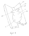
- FIG. 5 a illustrates an embodiment of an electrode for epicardial placement
- FIG. 5 b illustrates a first side of an electrode for epicardial placement
- FIG. 5 c illustrates a perspective view of an electrode for epicardial placement
- FIG. 6 a - c illustrate an embodiment of a cable assembly for use with the electrodes of a system for delivering electrical energy to an organ
- FIG. 7 illustrates an embodiment of a specially designed wire for use with the electrodes of a system for monitoring organ electrical activity
- FIG. 8 illustrates an exploded view of an organ chamber assembly for use in a system for monitoring organ electrical activity
- FIG. 9 illustrates the placement of an explanted heart on a pad containing electrodes for epicardial placement.
- Three electrodes are provided such that various connections may be made to a system for monitoring organ electrical activity in a perfusion system and providing, when appropriate, electrical energy to the organ.
- Two electrodes are placed proximate an explanted heart, preferably within a sterile environment.
- a third electrode is placed in the flow of the aortic perfusion fluid. This configuration allows for the monitoring of ECG signals of the explanted heart as well as for the delivery of defibrillation energy and/or pacing signals to the heart.
- Electrodes for epicardial placement are constructed of 304 stainless steel and are partially covered with silicone, which provides electrical insulation, is impervious to fluids, is biocompatible and provides a non-slip surface to aid in maintaining placement of the electrodes.
- the metal surface of the stainless steel electrodes is passivated to improve electrical performance, provide corrosion resistance and enhance biocompatibility.
- Electrodes for epicardial placement are resistance welded to 304 stainless steel wire contained within silicone insulation.
- the silicone wire insulation and silicone electrode covering are joined to provide protection for the weld as well as flexibility in the wire.
- the electrode placed in the flow of the aortic perfusion fluid is a thermal well constructed of 304 stainless steel and polycarbonate, into which has been potted a gold plated pin using electrically conductive epoxy.
- at least a portion of the electrode placed in the flow of the aortic perfusion fluid is covered with silicone to improved biocompatibility.
- Placement of one electrode in the flow of the aortic perfusion fluid allows for more stable ECG readings as the electrode is less susceptible to vibrations during transport as well as movement from a beating heart.
- one electrode for epicardial placement may be removed or moved aside, which may reduce any potential irritation of the heart tissue, provide fewer opportunities for the electrodes to touch, as well as provide more maneuverability of the remaining electrode for obtaining better placement on the heart.
- the electrodes for epicardial placement are maintained in position, at least partially, by the weight of the explanted heart.
- a completed electrical circuit for measuring ECG signals from the explanted heart exists from the electrode in the flow of the aortic perfusion fluid to an electrode for epicardial placement on the heart through the perfusion fluid and heart muscle.
- Defibrillation energy and/or pacing signals may be provided to the explanted heart by the electrodes for
- a perfusion system 10 which includes an organ chamber assembly 104 for containing the heart 102 (not shown) during ex-vivo maintenance, a reservoir 160 for holding, defoaming and filtering the perfusion fluid 108 , portal 774 for loading perfusion fluid 108 into the reservoir 160 and a portal 762 for applying therapeutics to the fluid 108 contained in the reservoir 160 , a perfusion fluid pump 106 for pumping/circulating perfusion fluid 108 to and from the harvested heart 102 ; a heater assembly 110 for maintaining the temperature of the perfusion fluid 108 at or near physiologic temperatures; a flow mode selector valve 112 for switching between normal and retrograde aortic flow modes (also referred to as “normal flow mode” and “retrograde flow mode,” respectively); an oxygenator 114 for oxygenating the perfusion fluid 108 subsequent to it being deoxygenated by the heart 102 from aerobic respiration; a nutritional subsystem 115 containing an infusion pump 182 for replenishing energy substrates
- the illustrative perfusion system 10 also includes a plurality of sensors, including without limitation: temperature sensors 120 , 122 and 124 ; pressure sensors 126 , 128 , 130 and 132 ; perfusion flow rate sensors 134 , 136 and 138 ; a perfusion fluid oxygenation and hematocrit sensor 140 ; and sensor/defib electrodes 12 , 50 and 52 , and defibrillation source 143 .
- sensors including without limitation: temperature sensors 120 , 122 and 124 ; pressure sensors 126 , 128 , 130 and 132 ; perfusion flow rate sensors 134 , 136 and 138 ; a perfusion fluid oxygenation and hematocrit sensor 140 ; and sensor/defib electrodes 12 , 50 and 52 , and defibrillation source 143 .
- the system 10 further includes: various components employed for maintaining suitable flow conditions to and from the heart 102 ; an operator interface 146 for assisting an operator in monitoring operation of the system 10 , and the condition of the heart 102 , and for enabling the operator to select various operating parameters; a power subsystem 148 for providing fault tolerant power to the system 10 ; and a controller 150 for controlling operation of the organ care system 10 .
- the perfusion fluid 108 flows from the pulmonary artery interface 166 into the oxygenator 114 .
- the oxygenator 114 receives gas from an external or onboard source 172 through a gas regulator 174 and a gas flow chamber 176 , which can be a pulse-width modulated solenoid valve that controls gas flow, or any other gas control device that allows for precise control of gas flow rate.
- a gas pressure gauge 178 provides a visual indication of amount remaining in the gas supply 172 .
- the transducer 132 provides similar information to the controller 150 .
- the controller 150 can regulate automatically the gas flow into the oxygenator 114 in dependence, for example, on the perfusion fluid oxygen content measured at the sensor 140 .
- the oxygenator 114 returns the perfusion fluid 108 to the reservoir 160 .
- the pulmonary vein interface 170 returns oxygenated blood to the left atrium of the heart 102 . Blood leaves the left ventricle and enters the aorta interface 162 .
- the aortic interface delivers oxygenated blood to the coronary arteries via the aorta.
- the pumping of the warm, oxygen and nutrient enriched perfusion fluid 108 through the heart 102 allows the heart 102 to function ex vivo in a near normal physiologic state.
- the warm perfusion fluid 108 warms the heart 102 as it perfuses through it, which may cause the heart 102 to resume beating in its natural fashion.
- the system 10 also includes a plurality of compliance chambers 184 , 186 and 188 .
- the compliance chambers 184 , 186 and 188 are essentially small inline fluid accumulators with flexible, resilient walls designed to simulate the human body's vascular compliance by aiding the system in more accurately mimicking blood flow in the human body, for example, by providing flow back-pressure and/or by filtering/reducing fluid pressure spikes due, for example, to flow rate changes and/or the pumping of the pump 106 .
- the compliance chamber 184 is located between an output 112 a of the mode valve 112 and the reservoir 160 and operates in combination with an adjustable clamp 190 during normal flow mode to provide back pressure to the aorta to cause perfusion fluid to flow into the coronary sinus to feed the heart 102 .
- the compliance chamber 186 is located between an output 112 b of the mode valve 112 and the pulmonary vein cannulation interface of the organ chamber assembly 104 .
- the primary function of the compliance chamber 186 is to provide back-pressure to the left atrium and to smooth pressure/flow spikes caused from the pumping action of the perfusion fluid pump 106 , which delivers blood to the heart without causing substantial fluid pressure spikes.
- the compliance chamber 188 is located between an output of a one way valve 310 and an inlet 110 a of the heater 110 .
- the primary function of the compliance chamber 188 is also to smooth pressure/flow spikes caused by the pumping action of the perfusion fluid pump 106 .
- FIG. 2 depicts an embodiment of an organ chamber assembly 104 of the type employed in the organ care system of FIG. 1 .
- an explanted heart 102 is perfused and transported to a donor site under sterile conditions while being monitored by a plurality of electrodes.
- the heart rests and is supported by a foam pad or sac 222 , preferably made of a biocompatible material resilient enough to cushion the heart 102 from vibrations and shocks during transport.
- the foam pad or sac 222 is comprised of silicone, although other biocompatible materials are envisioned.
- the heart is placed in a posterior arrangement, with the right atria in the top right and the left ventricle in the left-bottom. As shown, a right atrium electrode 52 and left ventricle electrode 50 are placed epicardially on the explanted heart 102 and are held in place by the weight of the heart 102 against the foam pad or sac 222 .
- At least one side of at least one of the right atrial electrode 52 and left ventricle electrode 50 are over-molded with silicone, and friction created by the contact between the silicone over-molding of the at least one electrode and the silicone pad or sac 222 further aids in maintaining the epicardial placement of the electrode.
- the structure of the electrode is described in more detail below.
- At least one of the right atrial electrode 52 and the left ventricle electrode 50 may be electrodes 142 and 144 , described in U.S. application Ser. No. 11/246,902.
- An aortic electrode 12 is placed in the aortic bloodpath for use in detecting ECG signals from the heart 102 during transport as blood travels to or from the aorta 158 .
- the organ chamber assembly 104 includes apertures for the pulmonary artery interface 166 , which carries perfusion fluid 108 from the pulmonary artery 164 , and the pulmonary vein interface 170 , which carries perfusion fluid to the pulmonary vein 168 .
- FIG. 3 depicts an interconnection of the electrodes and signal flows for monitoring organ electrical activity in a perfusion system 10 .
- the system 10 allows monitoring of heart 102 [the heart is there, just need to add 102 to the figure] electrical activity in a perfusion system as well as the delivery of defibrillation energy or pacing signals.
- the system 10 includes three electrodes: a right atrial electrode 52 , a left ventricle electrode 50 and an aortic electrode 12 .
- the aortic electrode 12 is placed in the aortic blood path outside the organ chamber 102 , which provides a stable position from which ECG signals from the heart may be measured and is less susceptible to the electrode shifting due to movements from the beating heart or the vibrations in the system during transport.
- the right atrial electrode 52 and left ventricle electrode 50 are placed epicardially on the heart within the organ chamber. Reference to “epicardially” includes, but is not limited to, on or near the heart. A silicone covering on at least a portion of the right atrial electrode 52 and the left ventricle electrode 50 aids in providing a non-slip surface to maintain the position of the electrodes.
- the perfusion-fluid contacting components may be coated or bonded with heparin or other anticoagulant or biocompatible material to reduce the inflammatory response that may otherwise arise when the perfusion fluid contacts the surfaces of the components.
- ECG signals are detected by both the aortic electrode 12 and the right atrial electrode 52 .
- An electric circuit is completed between the aortic electrode 12 , through the blood and heart muscle, to the right atrial electrode 52 .
- This placement allows more variability in the placement of the right atrial electrode 52 within the organ chamber 104 to accommodate differently shaped and sized hearts while maintaining a completed circuit.
- the right atrial electrode 52 is at least partially held in place by the weight of the heart 102 , which further aids in maintaining a completed circuit for detecting ECG signals.
- Electrode connection is made by placing the heart 102 on the one or more electrodes.
- One advantage of the invention is that it does not require the electrodes to be permanently or temporarily sutured or otherwise mechanically connected to the heart 102 .
- the present invention can be equally useful in such circumstances.
- one or more electrodes are provided for placement in the bloodpath and one or more electrodes are provided for epicardial placement on an explanted heart.
- ECG signals may be received by varying circuits comprising two electrodes placed in the bloodstream, two electrodes placed epicardially on the explanted heart, one electrode in the bloodstream and one electrode placed epicardially on the explanted heart, or any combination of the above.
- two electrodes are required to measure ECG signals, and as such, numerable combinations of electrode placements will provide ECG measurements.
- defibrillation energy and/or pacing signals may be necessary to restore a normal heart beat during transport to a donor site.
- the right atrial electrode 52 in conjunction with a left ventricle electrode 50 , may be used to provide defibrillation energy and/or pacing signals to the explanted heart 102 .
- the left ventricle electrode 50 may be removed from the heart 102 by manipulating the electrode through the flexible membrane. Removing the electrode reduces the likelihood of irritation to the heart tissue during transport.
- an operator may allow the left ventricle electrode 50 to remain epicardially placed should further defibrillation energy and/or pacing signals be required and without further need of manipulating the heart 102 and or electrodes 50 and 52 .
- a front end board connector 16 is provided as an interface between at least one electrode and one or more subsystems of the system 10 .
- At least one binding post 20 is provided to allow electrical connections to at least one electrode within the heart chamber 104 while maintaining the sterile integrity of the chamber.
- the aortic electrode 12 is connected to the front end board connector 16 by a first wire 30 .
- the right atrial electrode 52 is connected to a binding post 20 b by a second wire 32 , which is connected to the front end board connector by a third wire 34 .
- This connection configuration allows a completed circuit for the measurement of ECG signals from the explanted heart 102 .
- One of ordinary skill in the art will recognize that various other connections utilizing either fewer electrodes, wires or both could be used to achieve the same electrical circuit.
- a defibrillator connector 18 is provided as an interface between at least one electrode and a defibrillation source for providing defibrillation energy and/or pacing signals to the heart 102 .
- the right atrial electrode 52 is connected to a binding post 20 b by a second wire 32 , which is connected to the defibrillator connector 18 by a fourth wire 36 .
- the left ventricle electrode 50 is connected to a binding post 20 a by a fifth wire 38 , which is connected to the defibrillator connector 18 by a sixth wire 40 .
- This connection configuration allows a completed circuit for the delivery of defibrillation energy and/or pacing signals to the explanted heart 102 .
- One of ordinary skill in the art will recognize that various other connections utilizing either fewer electrodes, wires or both could be used to achieve the same electrical circuit.
- At least one of the first wire 30 , third wire 34 , fourth wire 36 , and sixth wire 40 is a custom-made wire preferably comprised of tinned soft copped with a PVC jacket. At least one of the third wire 34 , fourth wire 36 , fifth wire 38 , and sixth wire 40 is modified for purposes of defibrillation.
- FIGS. 4 a and 4 b depicts an embodiment of an aortic electrode and various interconnections that may be used to connect to the system.
- an aortic electrode 12 is comprised of a thermal well 80 comprised of 304 stainless steel and polycarbonate, into which a gold plated pin 82 has been potted using electrically conductive epoxy.
- the epoxy must cure for two hours at 65° C. to fully cure.
- the aortic electrode may be comprised of other electrically conductive and biocompatible materials.
- the aortic electrode is connected to a first wire 30 .
- the first wire 30 and a third wire 34 are twisted together for approximately six inches and are covered in a heat shrink jacket 84 .
- FIGS. 5 a - c illustrate one embodiment of an electrode 60 for epicardial placement.
- the epicardial electrodes are comprised of 304 stainless steel and over-molded with silicone. At least one aperture 68 in the stainless steel is provided to aid in securing the silicone to the stainless steel.
- the metal surface of the stainless steel is passivated to increase electrical performance, provide corrosion resistance and improve biocompatibility.
- Reference to “over-molded” includes, but is not limited to, covering or partially covering the electrode by means of molding, or other process that results in an electrode at least partially surrounded with silicone.
- Each epicardial electrode is resistance welded to 304 stainless steel wire 90 at a weld point 72 , which is surrounded with silicone and which is terminated in a gold plated pin.
- the over-molding of the wire 90 and the electrode 60 is overlapped at an interface 74 to reduce stress on the wire at the welding point but maintain wire flexibility.
- the electrode 60 is approximately a one inch by one inch square (2.5 cm by 2.5 cm), with a rounded edge 70 to reduce irritation to the tissue. It is large enough to easily contact at least part of the critical heart area and small enough to not have two electrodes touch, particularly on a small heart. These dimensions allow the electrodes to be placed precisely as well as maintain sufficient current density, i.e. keep it below damage threshold, although other electrode sizes and shapes are contemplated. In alternative embodiments, it is envisioned that each of the epicardial electrodes and wire may be comprised of other electrically conductive materials and biocompatible materials.
- the electrode 60 is provided with a first side 62 and a second side 64 .
- a portion 66 of the first side of the electrode 60 is exposed such that an electrical connection may be made epicardially with the heart 102 by placing the heart 102 on the first side 62 .
- the second side 64 of the electrode 60 is over-molded with silicone such that it is electrically insulated.
- the silicone is General Electric LIM 6050 silicone with 50 Shore A hardness, or other similar silicones from Wacker, Bayer or Dow Corning. 304 stainless steel and silicone are chosen for their biocompatibility as well as resistance to fluids.
- the materials chosen are also sufficiently resistant to the sterilization process (ETO) and to vacuum.
- ETO sterilization process
- other materials e.g., non-pourous foams
- biocompatibility issues e.g. silver-silver chloride
- the silicone over-molding of the electrode 60 provides a non-slip surface when the electrode is placed against the pad or sac 222 , which may also be constructed of silicone or have a surface that allows a reduced likelihood of slipping, which preferably aids in maintaining the positioning of the electrode after it has been epicardially placed on the heart 102 .
- the wires are comprised of tinned soft copper wire 90 with PVC insulation or heat shrink tubing 92 , illustrated in FIGS. 6 a and 6 b . Heat shrink tubing shown in exaggerated scale.
- Ring connectors 94 are provide to allow multiple connectors to the cabling.
- a connector 96 is provided for interconnection with the system 10 .
- the connector 96 is the defibrillator connector 18 .
- the cabling is modified for delivering defibrillation energy and/or pacing signals.
- FIG. 7 a schematic view of an embodiment of at least one of the second wire 32 and the fifth wire 38 is shown.
- at least one of the second wire 32 and fifth wire 38 is 304 stainless steel wire 90 over-molded with silicone insulation 92 .
- at least one of the second wire 32 and the fifth wire 38 is twenty gauge, multi-stranded soft-type 304 stainless steel and is over-molded with a 0.2 mm thick layer of silicone insulation.
- FIG. 8 depicts an exploded view of the illustrative organ chamber assembly 104 of FIGS. 1, 2 and 3 .
- the organ chamber assembly 104 includes a housing 194 , an outer lid 196 and an intermediate lid 198 .
- the housing includes a bottom 194 g and one or more walls 194 a - 194 d for containing the heart 102 .
- the intermediate lid 198 covers an opening 200 to the housing 194 for substantially enclosing the heart 102 (not shown) within the housing 194 .
- the intermediate lid 198 includes a frame 198 a and a flexible membrane 198 b suspended within the frame 198 a .
- the flexible membrane 198 b preferably, is transparent but may be opaque, translucent, or substantially transparent.
- the flexible membrane includes sufficient excess membrane material to contact the heart 102 when contained within the housing 194 .
- This feature enables a medical operator to touch/examine the heart 102 indirectly through the membrane 198 b , or apply an ultrasound probe to the heart 102 through the membrane 198 b , while maintaining sterility of the housing 194 .
- the membrane 198 b may be made, for example, from any suitable flexible polymer plastic, for example polyurethane. Apertures 199 a and 199 b in the membrane 198 b are provided through which electrodes 50 and 52 may be fed.
- the outer lid 196 opens and closes over the intermediate lid 198 independently from the intermediate lid 198 .
- the outer lid 196 is rigid enough to protect the heart 102 from physical contact, direct or indirect.
- the outer lid 196 and the chamber 194 may also be made from any suitable polymer plastic, for example polycarbonate.
- the housing 194 includes two hinge sections 202 a and 202 b
- the intermediate lid frame 198 a includes two corresponding mating hinge sections 204 a and 204 b , respectively.
- the hinge sections 202 a and 202 b on the housing 194 interfit with the hinge sections 204 a and 204 b on the intermediate lid frame 198 a to enable the intermediate lid 198 to open and close relative to the opening of the housing 194 .
- the organ chamber assembly 104 also includes two latches 206 a and 206 b for securing the intermediate lid 198 closed over the opening 200 .
- the latches 206 a and 206 b rotatably snap fit onto latch hinge section 208 a and 208 b , respectively, of the housing 194 .
- the intermediate lid frame 198 a also includes a hinge section 210 .
- the hinge section 210 rotatably snap fits with a mating hinge section 212 on the outer lid 196 to enable the outer lid 196 to open without opening the intermediate lid 198 .
- the outer lid 196 also includes two cutouts 214 a and 214 b for enabling the latches 206 a and 206 b to clamp down on the edge 216 of the intermediate lid frame 198 a.
- the organ chamber assembly 104 also includes a latch 218 , which rotatably snap fits onto a hinge part (not shown) on the wall 194 c of the housing 194 .
- the latch 218 engages a tab 221 on the edge 225 of the outer lid 196 to secure the outer lid 196 closed over the intermediate lid 198 .
- the intermediate lid also includes two gaskets 198 c and 198 d .
- the gasket 198 d interfits between a periphery of the intermediate lid frame 198 a and a periphery of the outer lid 196 to form a fluid seal between the intermediate lid 198 and the outer lid 196 when the outer lid 196 is closed.
- the gasket 198 c interfits between an outer rim 194 f of the housing 194 and the intermediate lid frame 198 a to form a fluid seal between the intermediate lid 198 and the periphery 194 f of the housing 194 when the intermediate lid 198 is closed, thereby providing a sterile environment for the heart once the organ care system is removed from the sterile operating room.
- the organ chamber assembly 104 includes a pad 222 or a sac assembly sized and shaped for interfitting over an inner bottom surface 194 g of the housing 194 .
- the pad 222 is formed from a material resilient enough to cushion the heart 102 from mechanical vibrations and shocks during transport, for example a silicone foam.
- the mechanism includes two through-apertures 224 a and 224 b for passing electrical leads from the under side of the pad 222 to corresponding electrodes on the heart-contacting surface of the pad. Passing the electrical leads through the pad 222 to the electrodes enables the electrodes to be adjustably positioned within the pad 222 to accommodate variously sized hearts.
- the mechanism may include, without limitation, one or more differently oriented slots, indentations, protrusions, through apertures, partially through apertures, hooks, eyelets, adhesive patches, or the like.
- the pad 222 may be configured with one or more sleeve-like structures that allow an electrode to be inserted within the pad 222 , thus providing a membrane-like surface of the pad 222 positioned between the electrode and the heart 102 .
- the pad 222 is configured as a pad assembly, with the assembly including one or more electrodes, such as the electrodes 50 and 52 , adjustably located in or on the pad 222 .
- the pad/electrode configuration of the invention facilitates contact between the electrodes and the heart 102 placed on the pad 222 , without temporarily or permanently suturing or otherwise mechanically connecting the electrodes to the heart 102 .
- the weight of the heart 102 (illustrated in FIG. 9 ) itself can also help stabilize the electrodes during transport.
- the organ chamber assembly 104 includes electrical interface connections 235 a - 235 b , which mount into the apertures 234 a - 234 b , respectively, in the wall 194 b of the housing 194 .
- a cover 226 is provided for protecting the electrical interface connections 235 a - 235 b .
- the electrical interface connections 235 a - 235 b are at least one of the binding posts 20 a - b of FIG. 3 .
- the interface connections 235 a and 235 b and aorta electrode 12 couple electrical signals, such as ECG signals, from the electrodes out of the housing 194 , for example, to a controller and/or an operator interface.
- the electrodes couple to the controller and/or the operator interface via the front end board connector 16 (not shown).
- the interface connections 235 a and 235 b may also couple to a defibrillation source, which may be either provided by external instrumentation or through circuitry within the system 10 , and which can send a defibrillation and/or pacing signal through electrodes to the heart 102 .
- the interface connections 235 a and 235 b are coupled to a defibrillation source via the defibrillation connector 18 .
- the organ chamber assembly 104 includes a resealable membrane interface 230 , which mounts in an interface aperture 232 .
- the interface 230 includes a frame 230 a and a resealable polymer membrane 230 b mounted in the frame 230 a .
- the membrane 230 b may be made of silicone or any other suitable polymer.
- the interface 230 is used to provide pacing leads, when necessary, to the heart 102 , without having to open the chamber lids 196 and 198 .
- the membrane 230 b seals around the pacing leads to maintain a closed environment around the heart 102 .
- the membrane 230 b also reseals in response to removing the pacing leads.
- the organ chamber assembly also includes a drain 201 for draining perfusion fluid 108 out of the housing 194 back into the reservoir 160 . Further, at least one mounting receptacle 203 is provided for mounting the organ chamber assembly 104 onto further components of the system 10 . As well, a plurality of apertures 228 a - c located on the organ chamber assembly 104 are provided for cannulation to vascular tissue of the heart 102 .
- FIG. 9 depicts the placement of an explanted heart on a pad containing electrodes for epicardial placement. At least one of the right atrial electrode 52 and the left ventricle electrode 50 are at least partially held in place by the weight of the explanted heart 102 against the pad 222 . As shown, the pulmonary artery 164 , aorta 158 , and pulmonary vein 168 are presented for cannulation.
- the heart 102 is harvested from a donor and cannulated into the organ chamber assembly 104 .
- the perfusion fluid 108 is prepared for use within system 10 by being loaded into the reservoir 160 via portal 774 and, optionally, being treated with therapeutics via portal 762 .
- the pump 106 pumps the loaded perfusion fluid 108 from a reservoir 160 to the heater assembly 110 .
- the heater assembly 110 heats the perfusion fluid 108 to or near a normal physiological temperature.
- embodiments of the disclosed subject matter are directed to a method of perserving a heart ex vivo, the method including the steps of placing a heart on one or more electrodes in a protective chamber of portable organ care system, pumping a perfusion fluid to the heart, the perfusion fluid being at a temperature of between about 25 ° C. and about 37° C., and at a volume of between about 200 ml/min and about 5 L/min, and monitoring electrical signals from the electrodes while pumping the perfusion fluid to the heart to preserve the heart ex vivo.
- the heater assembly 110 heats the perfusion fluid to between about 32° C. and about 37° C.
- the heater assembly 110 has an internal flow channel with a cross-sectional flow area that is approximately equal to the inside cross-sectional area of fluid conduits that carry the perfusion fluid 108 into and/or away from the heater assembly 110 , so as to minimize disturbance of fluid flow. From the heater assembly 110 , the perfusion fluid 108 flows to the flow mode selector valve 112 .
- One or more electrical signals related to the activity of the heart 102 are received by one or more electrodes 50 and 52 placed epicardially on the explanted heart 102 .
- the one or more electrical signals are transmitted along at least one wire 32 and 38 inside the organ chamber to one or more binding posts 20 a - b located at an interface between the inside of the organ chamber 104 and the outside of the organ chamber.
- This binding post configuration allows one or more signals to enter and exit the organ chamber 104 while maintaining the sterile environment within the organ chamber during transport of the explanted organ.
- the binding posts 20 a and 20 b may send or receive one or more signals to one or more units, systems, controllers or the like for the maintenance of the heart 102 .
- one or more signals from electrodes 50 and 52 placed epicardially on an explanted heart 102 are transmitted to the binding posts 20 a - b at the interface of the organ chamber 104 and are received by a front end board connector 16 , which may be connected to one or more units, systems or controllers for measuring signals from the explanted heart 102 and providing responses to the one or more signals.
- the one or more signals received by the front end board connector 16 are used to determine at least one of, but not limited to, the rate of a pump for providing perfusion fluid to the explanted heart 102 , the temperature to which the heating elements inside the heater should be set, determining whether pacing signals to maintain regular heart rhythm are required, the timing of pacing signals to be delivered to the heart 102 , etc.
- the binding posts 20 a,b may send or receive at least one signal to a defibrillator connector 18 .
- the defibrillator connector 18 sends signals to the binding posts 20 a - b , which are received by electrodes placed epicardially on an explanted heart 102 .
- the electrodes are a right atrial electrode 52 and a left ventricle electrode 50 .
- the signals sent by the defibrillator connector 18 are pacing signals for maintaining a proper heart 102 rhythm of the explanted heart 102 .
- signals received by the front end board connector 16 are transduced and analyzed; the analysis determining at least one output signal from the defibrillator connector 18 to be transmitted to an explanted heart 102 by the binding posts 20 a - b and electrodes placed on the explanted heart 102 , respectively.
- the enumerated listing of items does not imply that any or all of the items are mutually exclusive.
- the enumerated listing of items does not imply that any or all of the items are collectively exhaustive of anything, unless expressly specified otherwise.
- the enumerated listing of items does not imply that the items are ordered in any manner according to the order in which they are enumerated.
Abstract
Description
Claims (33)
Priority Applications (3)
| Application Number | Priority Date | Filing Date | Title |
|---|---|---|---|
| US11/822,495 US9457179B2 (en) | 2007-03-20 | 2007-07-06 | Systems for monitoring and applying electrical currents in an organ perfusion system |
| US15/207,303 US10327443B2 (en) | 2007-03-20 | 2016-07-11 | Systems for monitoring and applying electrical currents in an organ perfusion system |
| US16/418,250 US11917991B2 (en) | 2007-03-20 | 2019-05-21 | Systems for monitoring and applying electrical currents in an organ perfusion system |
Applications Claiming Priority (2)
| Application Number | Priority Date | Filing Date | Title |
|---|---|---|---|
| US91930607P | 2007-03-20 | 2007-03-20 | |
| US11/822,495 US9457179B2 (en) | 2007-03-20 | 2007-07-06 | Systems for monitoring and applying electrical currents in an organ perfusion system |
Related Child Applications (1)
| Application Number | Title | Priority Date | Filing Date |
|---|---|---|---|
| US15/207,303 Continuation US10327443B2 (en) | 2007-03-20 | 2016-07-11 | Systems for monitoring and applying electrical currents in an organ perfusion system |
Publications (2)
| Publication Number | Publication Date |
|---|---|
| US20080234768A1 US20080234768A1 (en) | 2008-09-25 |
| US9457179B2 true US9457179B2 (en) | 2016-10-04 |
Family
ID=39775533
Family Applications (3)
| Application Number | Title | Priority Date | Filing Date |
|---|---|---|---|
| US11/822,495 Active 2031-07-08 US9457179B2 (en) | 2007-03-20 | 2007-07-06 | Systems for monitoring and applying electrical currents in an organ perfusion system |
| US15/207,303 Active US10327443B2 (en) | 2007-03-20 | 2016-07-11 | Systems for monitoring and applying electrical currents in an organ perfusion system |
| US16/418,250 Active US11917991B2 (en) | 2007-03-20 | 2019-05-21 | Systems for monitoring and applying electrical currents in an organ perfusion system |
Family Applications After (2)
| Application Number | Title | Priority Date | Filing Date |
|---|---|---|---|
| US15/207,303 Active US10327443B2 (en) | 2007-03-20 | 2016-07-11 | Systems for monitoring and applying electrical currents in an organ perfusion system |
| US16/418,250 Active US11917991B2 (en) | 2007-03-20 | 2019-05-21 | Systems for monitoring and applying electrical currents in an organ perfusion system |
Country Status (1)
| Country | Link |
|---|---|
| US (3) | US9457179B2 (en) |
Cited By (11)
| Publication number | Priority date | Publication date | Assignee | Title |
|---|---|---|---|---|
| US9756851B2 (en) | 1997-09-23 | 2017-09-12 | The Department Of Veteran Affairs | Compositions, methods and devices for maintaining an organ |
| US9814230B2 (en) | 2008-01-31 | 2017-11-14 | Transmedics, Inc. | Systems and methods for ex vivo lung care |
| US9894894B2 (en) | 2004-10-07 | 2018-02-20 | Transmedics, Inc. | Systems and methods for ex-vivo organ care and for using lactate as an indication of donor organ status |
| US10039276B2 (en) | 2005-06-28 | 2018-08-07 | Transmedics, Inc. | Systems, methods, compositions and solutions for perfusing an organ |
| US10076112B2 (en) | 2014-06-02 | 2018-09-18 | Transmedic, Inc. | Ex vivo organ care system |
| US10194655B2 (en) | 2015-09-09 | 2019-02-05 | Transmedics, Inc. | Aortic cannula for ex vivo organ care system |
| US10314303B2 (en) * | 2004-10-07 | 2019-06-11 | Transmedics, Inc. | Systems and methods for ex-vivo organ care |
| US10327443B2 (en) | 2007-03-20 | 2019-06-25 | Transmedics, Inc. | Systems for monitoring and applying electrical currents in an organ perfusion system |
| US11571585B2 (en) | 2020-06-19 | 2023-02-07 | The Methodist Hospital | Method and apparatus for oncomagnetic treatment |
| US11785938B2 (en) | 2016-07-22 | 2023-10-17 | Eth Zurich | Perfusion loop assembly for an ex-vivo liver perfusion and a liver chamber assembly |
| US11856944B2 (en) | 2011-04-14 | 2024-01-02 | Transmedics, Inc. | Organ care solution for ex-vivo machine perfusion of donor lungs |
Families Citing this family (21)
| Publication number | Priority date | Publication date | Assignee | Title |
|---|---|---|---|---|
| US9301519B2 (en) | 2004-10-07 | 2016-04-05 | Transmedics, Inc. | Systems and methods for ex-vivo organ care |
| EP3677118A1 (en) | 2006-04-19 | 2020-07-08 | Transmedics, Inc. | Systems for ex vivo organ care |
| US20100196743A1 (en) * | 2009-02-02 | 2010-08-05 | Hyundai Motor Company | Apparatus and method for purging residual water and hydrogen during shutdown of fuel cell |
| WO2011002926A2 (en) | 2009-07-01 | 2011-01-06 | The General Hospital Corporation | Isolated adult cells, artificial organs,rehabilitated organs, research rools, organ encasements, organ perfusion systems, and methods for preparing and utilizing the same |
| WO2011140241A2 (en) | 2010-05-04 | 2011-11-10 | The General Hospital Corporation | Methods and compositions for preserving tissues and organs |
| US9426979B2 (en) | 2011-03-15 | 2016-08-30 | Paragonix Technologies, Inc. | Apparatus for oxygenation and perfusion of tissue for organ preservation |
| US9253976B2 (en) | 2011-03-15 | 2016-02-09 | Paragonix Technologies, Inc. | Methods and devices for preserving tissues |
| US8828710B2 (en) | 2011-03-15 | 2014-09-09 | Paragonix Technologies, Inc. | System for hypothermic transport of samples |
| US9867368B2 (en) | 2011-03-15 | 2018-01-16 | Paragonix Technologies, Inc. | System for hypothermic transport of samples |
| US11178866B2 (en) | 2011-03-15 | 2021-11-23 | Paragonix Technologies, Inc. | System for hypothermic transport of samples |
| WO2012125782A2 (en) | 2011-03-15 | 2012-09-20 | Paragonix Technologies, Inc. | Apparatus for oxygenation and perfusion of tissue for organ preservation |
| WO2013128013A1 (en) * | 2012-03-01 | 2013-09-06 | Medical Device Works Nv | Perfusion-occlusion device |
| US8785116B2 (en) | 2012-08-10 | 2014-07-22 | Paragonix Technologies, Inc. | Methods for evaluating the suitability of an organ for transplant |
| US9560846B2 (en) | 2012-08-10 | 2017-02-07 | Paragonix Technologies, Inc. | System for hypothermic transport of biological samples |
| US10111418B2 (en) * | 2013-06-07 | 2018-10-30 | Perfusion Solutions Pty Ltd | Organ perfusion system and device |
| US10918102B2 (en) | 2014-03-13 | 2021-02-16 | The General Hospital Corporation | Devices and methods to improve and assess viability of human livers |
| USD765874S1 (en) | 2014-10-10 | 2016-09-06 | Paragonix Technologies, Inc. | Transporter for a tissue transport system |
| US10091986B2 (en) | 2016-05-09 | 2018-10-09 | Xor-Labs Toronto Inc. | Organ perfusion device and method |
| US11166452B2 (en) | 2017-06-07 | 2021-11-09 | Paragonix Technologies, Inc. | Apparatus for tissue transport and preservation |
| WO2021036742A1 (en) * | 2019-08-26 | 2021-03-04 | 无锡市人民医院 | Ex vivo organ perfusion apparatus |
| US11632951B2 (en) | 2020-01-31 | 2023-04-25 | Paragonix Technologies, Inc. | Apparatus for tissue transport and preservation |
Citations (193)
| Publication number | Priority date | Publication date | Assignee | Title |
|---|---|---|---|---|
| US3253595A (en) * | 1963-08-07 | 1966-05-31 | Cordis Corp | Cardiac pacer electrode system |
| US3388803A (en) | 1965-04-16 | 1968-06-18 | Applied Biolog Sciences Lab In | Wearable dialysis apparatus |
| US3406531A (en) | 1964-08-25 | 1968-10-22 | Emil S. Swenson | Apparatus for maintaining organs in a completely viable state |
| US3468136A (en) | 1964-08-25 | 1969-09-23 | Emil S Swenson | Method for maintaining organs in a completely viable state |
| US3537956A (en) | 1967-06-26 | 1970-11-03 | Joseph R Falcone | Petrie dish |
| US3545221A (en) | 1967-05-22 | 1970-12-08 | Swenko Research & Dev Inc | Apparatus for maintaining organs in vitro in a completely viable state |
| US3545605A (en) | 1969-01-24 | 1970-12-08 | Ruth Robins | Paint roller package |
| US3587567A (en) * | 1968-12-20 | 1971-06-28 | Peter Paul Schiff | Mechanical ventricular assistance assembly |
| US3607646A (en) | 1968-04-04 | 1971-09-21 | Air Liquide | Method for the preservation of animal or human organs in living condition |
| US3632473A (en) | 1969-04-21 | 1972-01-04 | Univ California | Method and apparatus for preserving human organs extracorporeally |
| US3639084A (en) | 1970-04-06 | 1972-02-01 | Baxter Laboratories Inc | Mechanism for control pulsatile fluid flow |
| US3654085A (en) | 1968-11-26 | 1972-04-04 | Aga Ab | Storage device for organ transplants |
| US3738914A (en) | 1969-10-06 | 1973-06-12 | Baxter Laboratories Inc | Apparatus for preservation of organs |
| US3772153A (en) | 1969-04-02 | 1973-11-13 | Air Liquide | Apparatus for the preservation of animal or human organs in living condition |
| US3777507A (en) | 1971-11-24 | 1973-12-11 | Waters Instr Inc | Renal preservation system |
| US3843455A (en) | 1972-09-13 | 1974-10-22 | M Bier | Apparatus and technique for preservation of isolated organs through perfusion |
| US3851646A (en) | 1973-04-13 | 1974-12-03 | Sarns Inc | Connector for open heart surgery |
| US3881990A (en) | 1971-11-24 | 1975-05-06 | Waters Instr Inc | Method of transporting and storing organs while retaining the organs in a viable condition |
| US3995444A (en) | 1974-11-08 | 1976-12-07 | American Hospital Supply Corporation | Organ perfusion system |
| US4186565A (en) | 1978-05-19 | 1980-02-05 | Henry Ford Hospital | Perfusion system for organ preservation |
| US4231354A (en) | 1978-07-14 | 1980-11-04 | Howmedica, Incorporated | Pulsatile blood pumping apparatus and method |
| US4415556A (en) | 1980-12-23 | 1983-11-15 | Dr. Franz Kohler Chemie Gmbh | Protective solution for heart and kidney and process for its preparation |
| US4598697A (en) | 1983-12-29 | 1986-07-08 | Senko Medical Instrument Mfg. Co., Ltd. | Blood pump apparatus |
| US4605644A (en) | 1985-02-07 | 1986-08-12 | Regents Of The University Of Minnesota | Method for stimulating recovery from ischemia employing ribose and adenine |
| US4666425A (en) | 1985-12-17 | 1987-05-19 | The Dis Corporation | Device for perfusing an animal head |
| US4719201A (en) | 1985-02-07 | 1988-01-12 | Regents Of The University Of Minnesota | Method for stimulating recovery from ischemia |
| US4723939A (en) | 1986-07-31 | 1988-02-09 | The Research Foundation Of State Univ. Of New York | Apparatus and method for multiple organ procurement |
| US4745759A (en) | 1986-12-23 | 1988-05-24 | Bauer Dan O | Kidney preservation machine |
| US4759371A (en) | 1986-05-02 | 1988-07-26 | Siemens Aktiengesellschaft | Implantable, calibrateable measuring instrument for a body substance and a calibrating method |
| WO1988005261A1 (en) * | 1987-01-16 | 1988-07-28 | Tops Systems, Inc. | Total organ perfusion system |
| US4847470A (en) | 1987-12-14 | 1989-07-11 | Bakke Allan P | Electric blood warmer utilizing metallic ribbon flow cartridge and low thermal mass heating units |
| EP0347923A1 (en) | 1988-06-23 | 1989-12-27 | Nicholas J. Perrotta | Portable pulsatile organ perfusion device and method |
| US4920044A (en) | 1988-11-08 | 1990-04-24 | The Cleveland Clinic Foundation | Intracellular flush solution for preserving organs |
| EP0376763A2 (en) | 1988-10-26 | 1990-07-04 | McKelvey, Karen | A device for transportation of human organs used for transplantation |
| JPH02306901A (en) | 1989-05-19 | 1990-12-20 | Olympus Optical Co Ltd | Organ-storing device |
| US5051352A (en) | 1987-10-07 | 1991-09-24 | The Regents Of The University Of California | Apparatus and method of preserving the viability of animal organs |
| US5066578A (en) | 1989-12-21 | 1991-11-19 | The Regents Of The University Of California | Long-term preservation of organs for transplantation |
| JPH0499701A (en) | 1988-10-26 | 1992-03-31 | Mckelvey Karen | Transporting device for human internal organs for use in transplant |
| US5141847A (en) | 1989-08-18 | 1992-08-25 | Kureha Chemical Industry Co., Ltd. | Method and apparatus for producing pulsation |
| US5145771A (en) | 1990-04-12 | 1992-09-08 | The University Of North Carolina At Chapel Hill | Rinse solution for organs and tissues |
| US5157930A (en) | 1991-04-22 | 1992-10-27 | Mcghee Samuel C | Organ preservation apparatus |
| US5200398A (en) | 1991-09-12 | 1993-04-06 | Mount Sinai Hospital Corporation | Composition for the preservation of organs comprising glucuronic acid or a physiologically tolerated salt or ester thereof |
| US5217860A (en) | 1991-07-08 | 1993-06-08 | The American National Red Cross | Method for preserving organs for transplantation by vitrification |
| DE4201259A1 (en) | 1992-01-18 | 1993-07-22 | Sachs Elektronik Kg Hugo | Isolated heart perfusion appts. - has closed vessel with fluid and adjustable gas vol. to give timed aorta pressure |
| US5285657A (en) | 1990-09-28 | 1994-02-15 | Electrolux S.A.R.L. | Controlled-environment medical container |
| US5306711A (en) | 1992-06-24 | 1994-04-26 | Georgetown University | Organ preservative solution |
| US5326706A (en) | 1989-07-17 | 1994-07-05 | Research Foundation Of State University Of New York | Homeostatic organ preservation system |
| US5338662A (en) | 1992-09-21 | 1994-08-16 | Bio-Preserve Medical Corporation | Organ perfusion device |
| US5356771A (en) | 1993-03-11 | 1994-10-18 | Board Of Regents, The University Of Texas System | Combined perfusion and oxygenation organ preservation apparatus |
| US5356593A (en) | 1991-02-15 | 1994-10-18 | Cobe Laboratories, Inc. | Apparatus for measurement of blood saturation and hematocrit |
| US5362622A (en) | 1993-03-11 | 1994-11-08 | Board Of Regents, The University Of Texas System | Combined perfusion and oxygenation apparatus |
| US5370989A (en) | 1992-04-03 | 1994-12-06 | The Trustees Of Columbia University In The City Of New York | Solution for prolonged organ preservation |
| US5381510A (en) | 1991-03-15 | 1995-01-10 | In-Touch Products Co. | In-line fluid heating apparatus with gradation of heat energy from inlet to outlet |
| US5395314A (en) | 1990-10-10 | 1995-03-07 | Life Resuscitation Technologies, Inc. | Brain resuscitation and organ preservation device and method for performing the same |
| US5405742A (en) | 1993-07-16 | 1995-04-11 | Cyromedical Sciences, Inc. | Solutions for tissue preservation and bloodless surgery and methods using same |
| US5407793A (en) | 1991-10-18 | 1995-04-18 | University Of Pittsburgh Of The Commonwealth System Of Higher Education | An aqueous heart preservation and cardioplegia solution |
| US5407669A (en) | 1990-03-05 | 1995-04-18 | Lindstrom; Richard L. | Method and apparatus of a defined serumfree medical solution |
| WO1995031897A1 (en) | 1994-05-20 | 1995-11-30 | Vec Tec, Inc. | Method and apparatus monitoring viability of transplantable organs |
| US5473791A (en) | 1994-09-22 | 1995-12-12 | Holcomb; Tim C. | Paint roller and tray apparatus |
| US5498427A (en) | 1990-11-20 | 1996-03-12 | Pasteur Merieux Serums Et Vaccines | Solutions for the perfusion, preservation and reperfusion of organs |
| US5505709A (en) | 1994-09-15 | 1996-04-09 | Minimed, Inc., A Delaware Corporation | Mated infusion pump and syringe |
| WO1996018293A1 (en) | 1994-12-12 | 1996-06-20 | Charlotte-Mecklenburg Hospital Authority | Organ transplant solutions and method for transplanting an organ |
| US5552267A (en) | 1992-04-03 | 1996-09-03 | The Trustees Of Columbia University In The City Of New York | Solution for prolonged organ preservation |
| US5554497A (en) | 1994-12-12 | 1996-09-10 | Charlotte-Mecklenburg Hospital Authority | Cardioplegic solution for arresting an organ |
| US5554123A (en) | 1994-10-31 | 1996-09-10 | Glenn Herskowitz | Portable infusion pump |
| WO1996029865A1 (en) | 1995-03-27 | 1996-10-03 | Organ, Inc. | Organ evaluation and resuscitation device and method |
| US5571801A (en) | 1993-06-04 | 1996-11-05 | Biotime, Inc. | Plasma expander and blood substitute |
| US5584804A (en) | 1990-10-10 | 1996-12-17 | Life Resuscitation Technologies, Inc. | Brain resuscitation and organ preservation device and method for performing the same |
| US5586438A (en) * | 1995-03-27 | 1996-12-24 | Organ, Inc. | Portable device for preserving organs by static storage or perfusion |
| US5588816A (en) | 1993-05-26 | 1996-12-31 | Quest Medical, Inc. | Disposable cassette for cardioplegia delivery system |
| US5599173A (en) | 1994-02-10 | 1997-02-04 | Baxter International, Inc. | Blood pump system |
| US5599659A (en) | 1993-03-11 | 1997-02-04 | Breonics, Inc. | Preservation solution for ex vivo, warm preservation of tissues, explants,organs and vascular endothelial cells comprising retinal-derived fibroblast growth factor, cyclodextrin and chondroitin sulfate |
| US5643712A (en) | 1994-05-20 | 1997-07-01 | Brasile; Lauren | Method for treating and rendering grafts nonthrombogenic and substantially nonimmunogenic using an extracellular matrix coating |
| US5656420A (en) | 1995-02-24 | 1997-08-12 | University Of Kentucky Research Foundation | Method for employing the delta opioid dadle to extend tissue survival time during ischemia |
| US5679565A (en) | 1995-04-10 | 1997-10-21 | The Regents Of The University Of California | Method of preserving pancreatic islets |
| WO1997046091A1 (en) | 1996-06-06 | 1997-12-11 | Medtronic, Inc. | Heart preservation and transportation apparatus and method |
| US5702881A (en) | 1993-03-16 | 1997-12-30 | Alliance Pharmaceutical Corp. | Method and solution for organ preservation comprising retinal-derived growth factor, cyclodextrin, mucopolysaccharide and fluorocarbon |
| US5770149A (en) | 1995-10-31 | 1998-06-23 | Baxter International | Extracorporeal blood oxygenation system having integrated blood pump, heat exchanger and membrane oxygenator |
| US5776063A (en) | 1996-09-30 | 1998-07-07 | Molecular Biosystems, Inc. | Analysis of ultrasound images in the presence of contrast agent |
| US5786136A (en) | 1993-06-07 | 1998-07-28 | Mayer; Berndt | Method and device for conserving organs, body extremities and pieces of body tissue |
| US5787544A (en) | 1996-12-20 | 1998-08-04 | Ppg Industries, Inc. | Combined paint package and roller tray |
| US5807737A (en) | 1996-07-26 | 1998-09-15 | Schill Enterprises, Inc. | Heart and lung support assembly |
| US5823799A (en) | 1996-06-25 | 1998-10-20 | Thomas & Betts Corporation | Single-sided, straddle mount printed circuit board connector |
| US5843024A (en) | 1996-05-17 | 1998-12-01 | Breonics, Inc. | Solution and process for resuscitation and preparation of ischemically damaged tissue |
| US5856081A (en) | 1991-07-08 | 1999-01-05 | The American National Red Cross | Computer controlled cryoprotectant perfusion apparatus |
| US5882328A (en) | 1995-01-13 | 1999-03-16 | Qlt Phototherapeutics, Inc. | Method to prevent transplant rejection |
| WO1999015011A1 (en) | 1997-09-23 | 1999-04-01 | Hassanein Waleed H | Compositions, methods and devices for maintaining an organ |
| US5965433A (en) | 1996-05-29 | 1999-10-12 | Trans D.A.T.A. Service, Inc. | Portable perfusion/oxygenation module having mechanically linked dual pumps and mechanically actuated flow control for pulsatile cycling of oxygenated perfusate |
| CN1232723A (en) | 1998-04-21 | 1999-10-27 | 瓜拉喷注公开有限公司 | Hand-operated pump with trigger, for dispensing liquids |
| US5998240A (en) | 1996-07-22 | 1999-12-07 | Northrop Grumman Corporation | Method of extracting heat from a semiconductor body and forming microchannels therein |
| US6034109A (en) | 1994-12-22 | 2000-03-07 | The Regents Of The University Of California | Method for protection of heart by limiting metabolic and ionic abnormalities developed during ischemia following ischemia or resulting from ischemia |
| US6042550A (en) | 1998-09-09 | 2000-03-28 | Ntc Technology, Inc. | Methods of non-invasively estimating intrapulmonary shunt fraction and measuring cardiac output |
| US6046046A (en) | 1997-09-23 | 2000-04-04 | Hassanein; Waleed H. | Compositions, methods and devices for maintaining an organ |
| US6050987A (en) | 1998-09-21 | 2000-04-18 | The United States Of America As Represented By The Administrator Of The National Aeronautics And Space Administration | Tubular coupling |
| US6110139A (en) | 1997-10-21 | 2000-08-29 | Loubser; Paul Gerhard | Retrograde perfusion monitoring and control system |
| WO2000060936A1 (en) | 1999-04-12 | 2000-10-19 | Cambridge University Technical Services Limited | Methods and means for extracorporeal organ perfusion |
| US6144444A (en) | 1998-11-06 | 2000-11-07 | Medtronic Avecor Cardiovascular, Inc. | Apparatus and method to determine blood parameters |
| US6168877B1 (en) | 1998-12-18 | 2001-01-02 | Aer Energy Resources, Inc. | Air-managing system for metal-air battery using resealable septum |
| JP2001061956A (en) | 1999-06-24 | 2001-03-13 | Aisin Seiki Co Ltd | Autoperfusion apparatus for coronary artery bypass surgery under heartbeat |
| US20010003652A1 (en) | 1995-03-28 | 2001-06-14 | Thomas Charles Freeman | Relating to sample processing |
| US20020012988A1 (en) | 1999-04-14 | 2002-01-31 | Lauren Brasile | Organ chamber for exsanguinous metabolic support system |
| US6365338B1 (en) | 1999-04-27 | 2002-04-02 | David A. Bull | Organ preservative solution containing trehalose, anti-oxidant, cations and an energy source |
| US6375613B1 (en) | 1994-05-20 | 2002-04-23 | Breonics, Inc. | Prognostic testing of organs intended for transplantation |
| US6375611B1 (en) | 2000-01-07 | 2002-04-23 | Origin Medsystems, Inc. | Organ stabilizer |
| US6389308B1 (en) | 2000-05-30 | 2002-05-14 | Vladimir Shusterman | System and device for multi-scale analysis and representation of electrocardiographic data |
| US20020102720A1 (en) | 1995-12-15 | 2002-08-01 | Stig Steen | Preservation solution |
| US20020132220A1 (en) | 2000-12-27 | 2002-09-19 | Berens Kurt L. | Use of selectin antagonists in organ preservation solutions |
| US20020151950A1 (en) * | 1998-11-04 | 2002-10-17 | Acorn Cardiovascular, Inc. | Cardio therapeutic heart sack |
| US6475716B1 (en) | 2001-03-06 | 2002-11-05 | Biobank Co., Ltd. | Method for preserving mammalian organs |
| US20020164795A1 (en) | 1999-06-02 | 2002-11-07 | Shokyu Gen | Storage agent for preservation of an animal cell, tissue or organ, and preserved process of the same |
| DE10121159A1 (en) | 2001-04-30 | 2002-11-21 | Juergen Biederer | Artificial thorax for housing an animal heart and lungs preparation for experimental development of X-ray and magnetic resonance diagnostic imaging methods has a vacuum pressure applied so that the lungs are not collapsed |
| US20020177117A1 (en) | 2000-03-13 | 2002-11-28 | Wolf E. George | Hyperbaric oxygen organ preservation system (hoops) |
| US6492103B1 (en) | 2000-01-31 | 2002-12-10 | Organ Recovery Systems, Inc. | System for organ and tissue preservation and hypothermic blood substitution |
| US6490880B1 (en) | 2000-10-26 | 2002-12-10 | Islet Technology Inc. | Regulated organ containment shipping system using dual-layer preservation liquid |
| US6492745B1 (en) | 2000-07-17 | 2002-12-10 | Marconi Communications, Inc. | Battery disconnect system |
| US20030011604A1 (en) | 2001-06-20 | 2003-01-16 | Capers Walter E. | Method and apparatus for capturing and viewing a sequence of 3-D images |
| US6524785B1 (en) | 1998-11-10 | 2003-02-25 | Centre National De La Recherche Scientifique | Perfusion and/or preservation and/or re-perfusion solution during organ transplant |
| US20030040665A1 (en) | 1999-05-28 | 2003-02-27 | E-Monitors, Inc. | Systems and methods of pH tissue monitoring |
| US20030050689A1 (en) | 1999-03-05 | 2003-03-13 | Surgica Corporation | Surface-modified bioactive suppressant surgical implants |
| US20030053998A1 (en) | 2001-09-18 | 2003-03-20 | Daemen Marc A.R.C. | Methods and active substances for protecting organs |
| US20030073227A1 (en) | 2001-09-28 | 2003-04-17 | Hull Harry F. | Simple, fully transportable device for maintaining an excised heart in optimum condition for transplantation |
| US20030074760A1 (en) | 2001-10-19 | 2003-04-24 | Rdk Company | Paint applicator system |
| US20030086830A1 (en) | 2001-10-12 | 2003-05-08 | Becton Dickinson And Company | Method and apparatus for transporting biological samples |
| US6569615B1 (en) | 2000-04-10 | 2003-05-27 | The United States Of America As Represented By The Department Of Veteran's Affairs | Composition and methods for tissue preservation |
| US20030135152A1 (en) | 2000-09-27 | 2003-07-17 | Kollar Kevin J. | Disposable cartridge for a blood perfusion system |
| US6600941B1 (en) | 1999-05-28 | 2003-07-29 | E-Monitors, Inc. | Systems and methods of pH tissue monitoring |
| US20030147466A1 (en) | 2002-02-01 | 2003-08-07 | Qilian Liang | Method, system, device and computer program product for MPEG variable bit rate (VBR) video traffic classification using a nearest neighbor classifier |
| US6609987B1 (en) | 2001-10-01 | 2003-08-26 | General Motors Corporation | Hydraulic timing chain tensioner assembly |
| US6631830B2 (en) | 2001-08-20 | 2003-10-14 | Larami Limited | Snap action ball valve assembly and liquid dispenser using same |
| US6642045B1 (en) | 1997-04-14 | 2003-11-04 | Breonics, Inc. | System for exsanguinous metabolic support of an organ or tissue |
| US6673594B1 (en) | 1998-09-29 | 2004-01-06 | Organ Recovery Systems | Apparatus and method for maintaining and/or restoring viability of organs |
| US20040015042A1 (en) | 2002-02-21 | 2004-01-22 | Douglas Vincent | Fluid pump |
| US20040017658A1 (en) | 2002-07-23 | 2004-01-29 | Chih-Ching Lo | Heat dissipating apparatus for interface cards |
| US20040018966A1 (en) | 1999-02-03 | 2004-01-29 | Segall Paul E. | Methods and compositions for use in perfusion applications |
| US20040029096A1 (en) | 2000-11-03 | 2004-02-12 | Stig Steen | Evaluation and preservation solution |
| US6696238B2 (en) | 2000-07-28 | 2004-02-24 | Christopher J. Murphy | Transplant media |
| US20040038192A1 (en) | 1999-04-14 | 2004-02-26 | Breonics, Inc. | System for exsanguinous metabolic support of an organ or tissue |
| US20040058432A1 (en) | 1998-09-29 | 2004-03-25 | Owen Donald R. | Apparatus and method for maintaining and/or restoring viability of organs |
| WO2004026031A2 (en) | 2002-09-18 | 2004-04-01 | Lifeblood Medical Inc. | Organ preservation and transportation apparatus and method |
| US20040082057A1 (en) | 2001-09-14 | 2004-04-29 | Alford Martin L. | Organ preservation apparatus and methods |
| US20040086578A1 (en) | 1993-06-04 | 2004-05-06 | Segall Paul E. | Physiologically acceptable aqueous solutions and methods for their use |
| US20040102415A1 (en) | 2000-04-10 | 2004-05-27 | Hemant Thatte | Composition and methods for tissue preservation |
| US20040102678A1 (en) | 1997-06-21 | 2004-05-27 | Acorn Cardiovascular Inc. | Bag for at least partially enveloping a heart |
| US20040106958A1 (en) * | 2001-02-13 | 2004-06-03 | Quetzal Biomedical Inc. | Multi-electrode apparatus and method for treatment of congestive heart failure |
| US20040110800A1 (en) | 1998-07-21 | 2004-06-10 | Smithkline Beecham P.L.C. & Smithkline Beecham Laboratoires Pharmaceutiques | Use of glucose uptake enhancer for reducing post-ischemic injury of the heart |
| US20040115689A1 (en) | 2000-11-08 | 2004-06-17 | Augello Frank A. | Method and device for collecting and stabilizing a biological sample |
| JP2004525290A (en) | 2000-12-21 | 2004-08-19 | ドレッサー ランド カンパニー | Two-layer acoustic liner, fluid compression device and method of using same |
| US6783328B2 (en) | 1996-09-30 | 2004-08-31 | Terumo Cardiovascular Systems Corporation | Method and apparatus for controlling fluid pumps |
| US20040168341A1 (en) | 2001-03-21 | 2004-09-02 | Petersen Peter Mosborg | Methods for performing operations, a housing for such methods, and furnishings for such housing |
| US20040170950A1 (en) | 2002-09-12 | 2004-09-02 | Prien Samuel D. | Organ preservation apparatus and methods |
| US20040171138A1 (en) | 2002-08-14 | 2004-09-02 | Transmedics, Inc. | Heart preservation chamber |
| US6792309B1 (en) * | 1999-03-23 | 2004-09-14 | St. Jude Medical Ab | Multi-sensor system for controlling an implantable heart stimulator |
| JP2004529938A (en) | 2001-05-04 | 2004-09-30 | ブレオニクス,インコーポレーティッド | Organ chamber for blood-free metabolic support system |
| US20040202993A1 (en) | 2003-04-10 | 2004-10-14 | Poo Ramon E. | Apparatus and method for organ preservation and transportation |
| US6811965B2 (en) | 2000-03-08 | 2004-11-02 | Yoram Vodovotz | Kidney perfusion solution containing nitric oxide donor, inhibitor of NOS2, glutathione, gluconate and methods of use |
| US20040224298A1 (en) | 1998-09-29 | 2004-11-11 | John Brassil | Apparatus and method for determining effects of a substance of an organ |
| US20040236170A1 (en) | 2000-11-15 | 2004-11-25 | Ducksoo Kim | Method for surgically joining a ventricular assist device to the cardiovascular system of a living subject using a piercing introducer assembly |
| US20040235142A1 (en) | 2003-04-04 | 2004-11-25 | Organ Recovery Systems | Method and apparatus for holding a plurality of tubes connectible to an organ or tissue container |
| US20050010118A1 (en) | 2003-07-10 | 2005-01-13 | Nikkiso Co. Ltd. | Method and device for measuring pulse rate, blood pressure, and monitoring blood vessel access |
| US20050019917A1 (en) | 2003-04-23 | 2005-01-27 | Human Biosystems | Methods and solutions for storing donor organs |
| US6878339B2 (en) | 2002-02-19 | 2005-04-12 | Horiba, Ltd. | NOx-concentration measuring apparatus |
| US20050147958A1 (en) | 1997-09-23 | 2005-07-07 | Waleed Hassanein | Compositions, method and devices for maintaining an organ |
| US20050153271A1 (en) | 2004-01-13 | 2005-07-14 | Wenrich Marshall S. | Organ preservation apparatus and methods |
| US20050170019A1 (en) | 2003-10-22 | 2005-08-04 | Fred Hutchinson Cancer Research Center | Methods, compositions and devices for inducing stasis in cells |
| US20050182349A1 (en) | 2004-02-02 | 2005-08-18 | Linde Peter G. | Modified organ support devices |
| US20050187469A1 (en) | 2002-05-06 | 2005-08-25 | Uscom Pty Ltd. | Blood flow oxygen measurement system and method |
| US6953655B1 (en) | 1997-09-23 | 2005-10-11 | The United States Of America As Represented By The Department Of Veterans Affairs | Compositions, methods and devices for maintaining an organ |
| US6974436B1 (en) | 1997-09-19 | 2005-12-13 | A-Med Systems, Inc. | Integrated pump and cannula system and related methods |
| US20060039870A1 (en) | 2004-08-20 | 2006-02-23 | Turner Jeffrey D | Pulmonary delivery of enzymatic medical countermeasures |
| US7008380B1 (en) | 1999-02-03 | 2006-03-07 | Stephen Edward Rees | Automatic lung parameter estimator |
| US20060074470A1 (en) * | 2004-10-04 | 2006-04-06 | Biotronik Crm Patent Ag | Electrode lead |
| WO2006042138A2 (en) | 2004-10-07 | 2006-04-20 | Transmedics, Inc. | Systems and methods for ex-vivo organ care |
| US20060124130A1 (en) | 2003-12-26 | 2006-06-15 | Jorge Bonassa | Ventilation system for respiratory devices |
| US20060134073A1 (en) | 2004-11-24 | 2006-06-22 | Yoshifumi Naka | Compositions and methods for ex vivo preservation of blood vessels for vascular grafts using analogues of cAMP and cGMP |
| US20060154357A1 (en) | 2004-10-07 | 2006-07-13 | Transmedics, Inc. | Systems and methods for ex-vivo organ care |
| WO2006124820A2 (en) | 2005-05-12 | 2006-11-23 | University Of Florida Research Foundation, Inc. | Materials and methods for regulating blood flow |
| US20060292544A1 (en) | 2005-06-28 | 2006-12-28 | Transmedics, Inc. | Systems, methods, compositions and solutions for perfusing an organ |
| WO2007079185A2 (en) | 2005-12-29 | 2007-07-12 | Anthrogenesis Corporation | Improved composition for collecting and preserving placental stem cells and methods of using the composition |
| US20070196461A1 (en) | 2005-12-08 | 2007-08-23 | Jeff Weers | Lipid-based compositions of antiinfectives for treating pulmonary infections and methods of use thereof |
| US7316666B1 (en) | 2004-04-12 | 2008-01-08 | Arizant Healthcare Inc. | Fluid warming cassette with rails and a stiffening member |
| US20080017194A1 (en) | 2006-04-19 | 2008-01-24 | Transmedics, Inc | Systems and methods for ex vivo organ care |
| WO2008106724A1 (en) | 2007-03-02 | 2008-09-12 | Hibernation Therapeutics Limited | Transplants |
| US20080234768A1 (en) | 2007-03-20 | 2008-09-25 | Transmedics, Inc | Systems for monitoring and applying electrical currents in an organ perfusion system |
| US7452711B2 (en) | 2005-05-09 | 2008-11-18 | Victor Daykin | Contact plate |
| US20080286746A1 (en) | 2007-05-17 | 2008-11-20 | Biorep Technologies, Inc. | Perfusion apparatus and method |
| US20090143417A1 (en) | 2004-12-24 | 2009-06-04 | The University Of Queensland | Methods of treating pain |
| US20090142830A1 (en) | 2005-11-17 | 2009-06-04 | Nippon Zenyaku Kogyo Co., Ltd. | Aqueous Solution for Cell Preservation |
| US20090197240A1 (en) | 2008-01-31 | 2009-08-06 | Transmedics, Inc | Systems and methods for ex vivo lung care |
| US20090215022A1 (en) | 2006-05-22 | 2009-08-27 | Biovest International, Inc. | Extra-capillary fluid cycling system and method for a cell culture device |
| US20090312724A1 (en) | 2007-06-28 | 2009-12-17 | Cydex Pharmaceuticals, Inc. | Nasal and Ophthalmic Delivery of Aqueous Corticosteroid Solutions |
| US20110190572A1 (en) | 2009-12-08 | 2011-08-04 | Vanderbilt University | Methods and Compositions for Vein Harvest and Autografting |
| US8304181B2 (en) | 2004-10-07 | 2012-11-06 | Transmedics, Inc. | Method for ex-vivo organ care and for using lactate as an indication of donor organ status |
| US20130011823A1 (en) | 2011-04-14 | 2013-01-10 | Hassanein Waleed H | Organ care solution for ex-vivo machine perfusion of donor lungs |
Family Cites Families (138)
| Publication number | Priority date | Publication date | Assignee | Title |
|---|---|---|---|---|
| US3660241A (en) | 1970-01-12 | 1972-05-02 | Baxter Laboratories Inc | Container for organ perfusion or the like |
| US4004298A (en) | 1975-03-31 | 1977-01-25 | Sinai Hospital Of Detroit | Magnetically aligned releasable connector |
| US4069826A (en) | 1976-09-03 | 1978-01-24 | Barlow Mfg. Corporation | Surgical tube adapter clamp |
| US4186253A (en) | 1978-10-10 | 1980-01-29 | The Green Cross Corporation | Perfusate for preserving organ to be transplanted and preserving method |
| JPS5710695A (en) | 1980-04-18 | 1982-01-20 | Cooper Edwin Inc | Lubricant oil containing tertiary amine salt of sulfidized olefinic acid |
| JPS5710695U (en) | 1980-06-17 | 1982-01-20 | ||
| US4801299A (en) | 1983-06-10 | 1989-01-31 | University Patents, Inc. | Body implants of extracellular matrix and means and methods of making and using such implants |
| JPS63270601A (en) | 1987-04-30 | 1988-11-08 | Olympus Optical Co Ltd | Apparatus for storing internal organs |
| JPH02282301A (en) | 1989-04-24 | 1990-11-19 | Olympus Optical Co Ltd | Device for storing organ |
| JPH0374302A (en) | 1989-08-15 | 1991-03-28 | Olympus Optical Co Ltd | Organ storage system |
| US5358931A (en) | 1990-01-17 | 1994-10-25 | The Regents Of The University Of California | Interaction of thermal hysteresis proteins with cells and cell membranes and associated applications |
| JP2860301B2 (en) | 1990-09-19 | 1999-02-24 | ヘキスト・マリオン・ルセル株式会社 | Organ preservation solution |
| US6090776A (en) | 1991-03-11 | 2000-07-18 | Creative Bio Molecules, Inc. | Morphogen treatment of organ implants |
| US5654266A (en) | 1992-02-10 | 1997-08-05 | Chen; Chung-Ho | Composition for tissues to sustain viability and biological functions in surgery and storage |
| JPH0656601A (en) | 1992-08-07 | 1994-03-01 | Morishita Roussel Kk | Electrolyte solution for organ for transplantation |
| US5354268A (en) | 1992-11-04 | 1994-10-11 | Medical Instrument Development Laboratories, Inc. | Methods and apparatus for control of vacuum and pressure for surgical procedures |
| JPH06305901A (en) | 1993-04-26 | 1994-11-01 | Akio Kawamura | Perfusion liquid for room temperature preservation and preservation method using the same liquid |
| EP2113196A3 (en) | 1993-11-05 | 2009-12-23 | ResMed Limited | Control of CPAP treatment |
| DE4342728A1 (en) | 1993-12-15 | 1995-06-22 | Thomae Gmbh Dr K | Aq. isotonic soln. for perfusion and preservation of explanted liver |
| JPH0889518A (en) | 1994-09-25 | 1996-04-09 | Terumo Corp | Hollow organ connector |
| JP3845736B2 (en) | 1995-09-18 | 2006-11-15 | レスメッド・リミテッド | Pressure control in CPAP treatment or assisted ventilation |
| DE19535781C2 (en) | 1995-09-26 | 1999-11-11 | Fraunhofer Ges Forschung | Device for active flow support of body fluids |
| US5759148A (en) | 1995-10-18 | 1998-06-02 | Sipin; Anatole J. | Controlled pneumatic driving system |
| SE505499C2 (en) | 1995-12-15 | 1997-09-08 | Stiftelsen Facthor | Storage solution for organs and tissues or parts thereof of humans and animals containing calcium and nitroglycerin, their use and method of storage therewith |
| US6217546B1 (en) | 1997-05-19 | 2001-04-17 | United States Surgical Corporation | Catheter system |
| WO2005009289A2 (en) | 1997-09-03 | 2005-02-03 | Dan Weiner | Apparatus and methods for control of intravenous fluids |
| US6352543B1 (en) | 2000-04-29 | 2002-03-05 | Ventrica, Inc. | Methods for forming anastomoses using magnetic force |
| GR1003087B (en) | 1998-06-30 | 1999-03-08 | Self-cleaning hydraulic toilet for small pets that utilises only water. | |
| CA2342944A1 (en) | 1998-09-29 | 2000-04-06 | Organ Recovery Systems, Inc. | Apparatus and method for maintaining and/or restoring viability of organs |
| EP1123000B1 (en) | 1998-10-22 | 2004-12-29 | Medtronic, Inc. | Method of investigating of an isolated perfused heart preparation |
| US7045279B1 (en) | 1998-10-22 | 2006-05-16 | Medtronic, Inc. | Isolated perfused heart preparation and method of use |
| US6128519A (en) | 1998-12-16 | 2000-10-03 | Pepex Biomedical, Llc | System and method for measuring a bioanalyte such as lactate |
| GB9907372D0 (en) | 1999-03-30 | 1999-05-26 | Concentric Pumps Ltd | Improvements in pumps |
| US6194137B1 (en) | 1999-04-13 | 2001-02-27 | Organ Recovery Systems, Inc. | Method of cryopreservation of blood vessels by vitrification |
| AU5750600A (en) | 1999-06-17 | 2001-01-22 | Regents Of The University Of California, The | Continuous cardiac perfusion preservation with peg-hb for improved hypothermic storage |
| DE19928485C1 (en) | 1999-06-22 | 2000-10-19 | Jostra Medizintechnik Ag | Perfusion system useful for storing human or animal donor organ or body part, e.g. liver, before transplantation has inner bag with blood supply for organ and outer bag containing liquid, preferably suspended in frame |
| CA2286929A1 (en) | 1999-10-18 | 2001-04-18 | Anthony Paolitto | Valve surgery apparatus |
| CN2418882Y (en) | 1999-10-20 | 2001-02-14 | 张国报 | In vitro pulmonary circulation and perfusion device |
| US6764462B2 (en) | 2000-11-29 | 2004-07-20 | Hill-Rom Services Inc. | Wound treatment apparatus |
| US6736836B2 (en) | 2000-02-17 | 2004-05-18 | The Johns Hopkins University | Transplant organ external cooling system |
| JP2002119586A (en) | 2000-10-16 | 2002-04-23 | Univ Nihon | Method and device for warming and cooling blood or adjusting body temperature |
| US20020187132A1 (en) | 2001-04-30 | 2002-12-12 | Mcgregor Christopher G.A. | Cardiac gene transfer |
| FR2830183B1 (en) | 2001-10-02 | 2004-06-11 | Daniel Roux | AORTIC PROSTHESIS WITH QUICK DEVICE FOR CONNECTION TO EXTRA-BODY CIRCULATION |
| US6582375B2 (en) | 2001-10-11 | 2003-06-24 | University Of Cincinnati | System for study of global uni-or bi-ventricular function of an explanted, reperfused heart |
| US6910483B2 (en) | 2001-12-10 | 2005-06-28 | Resmed Limited | Double-ended blower and volutes therefor |
| SG108829A1 (en) | 2001-12-14 | 2005-02-28 | Agilent Technologies Inc | Photo-receiver arrangement |
| EP1321159A1 (en) | 2001-12-21 | 2003-06-25 | CHIESI FARMACEUTICI S.p.A. | Pressurized metered dose inhaler (pMDI) actuators with laser drilled orifices |
| JP2003206201A (en) | 2001-12-28 | 2003-07-22 | Katsutoshi Naruse | Organ preservation apparatus for transplantation comprising organ storage apparatus and artificial organ system comprising the storage apparatus |
| US20030124503A1 (en) | 2001-12-28 | 2003-07-03 | Olivencia-Yurvati Albert H. | Pyruvate cardioplegia solutions for administration to the heart during cardiopulmonary surgery and methods of use thereof |
| JP2003315220A (en) | 2002-02-19 | 2003-11-06 | Horiba Ltd | Nitrogen oxides measuring apparatus |
| AUPS312602A0 (en) | 2002-06-21 | 2002-07-18 | James Cook University | Organ arrest, protection, preservation and recovery |
| US7454784B2 (en) * | 2002-07-09 | 2008-11-18 | Harvinder Sahota | System and method for identity verification |
| US8361091B2 (en) | 2002-08-23 | 2013-01-29 | Organ Recovery Systems, Inc. | Cannulas, cannula mount assemblies, and clamping methods using such cannulas and cannula mount assemblies |
| AU2003286474A1 (en) | 2002-10-18 | 2004-05-04 | The General Hospital Corporation | Compositions, solutions, and methods used for transplantation |
| WO2004056181A1 (en) | 2002-12-23 | 2004-07-08 | Global Cardiac Solutions Pty Ltd | Organ preconditioning, arrest, protection, preservation and recovery (2) |
| US7198751B2 (en) | 2003-01-14 | 2007-04-03 | Medtronic, Inc. | Disposable, integrated, extracorporeal blood circuit |
| US20040193096A1 (en) | 2003-03-25 | 2004-09-30 | Cooper William Isaac | Conceptus chamber |
| US20050085762A1 (en) | 2003-03-31 | 2005-04-21 | Venkataramana Vijay | Perfusion circuit for cardiopulmonary bypass with air removal system |
| EP1613152B1 (en) | 2003-04-04 | 2018-07-04 | Organ Recovery Systems, Inc. | Device for separating gas from a liquid path |
| JP2004307404A (en) | 2003-04-08 | 2004-11-04 | Nipro Corp | Pharmaceutical composition containing artificial oxygen transporter |
| EP1475434A1 (en) | 2003-05-09 | 2004-11-10 | Oncoscience AG | Method for storing tumor cells |
| CN2616058Y (en) | 2003-05-12 | 2004-05-19 | 四川大学华西医院 | Sustained-perfusion heart preserving device |
| CN1233231C (en) | 2003-05-12 | 2005-12-28 | 四川大学华西医院 | Continuously perfused heart keeping device |
| BRPI0412362A (en) | 2003-07-08 | 2006-09-05 | Ventor Technologies Ltd | prosthetic implant devices particularly for transarterial transport in the treatment of aortic stenoses and implantation methods for such devices |
| NL1024022C2 (en) | 2003-07-30 | 2005-02-01 | Technologiestichting Stw | Portable preservation device for a donor organ. |
| DE10340487B4 (en) | 2003-09-03 | 2007-07-12 | Technische Universität Dresden | perfusion |
| JP3742894B2 (en) | 2004-01-14 | 2006-02-08 | ダイキン工業株式会社 | Temperature control device |
| US7390028B2 (en) | 2004-05-12 | 2008-06-24 | Blazek Larry M | Medical tubing quick disconnect apparatus |
| US8741555B2 (en) | 2004-05-14 | 2014-06-03 | Organ Recovery Systems, Inc. | Apparatus and method for perfusion and determining the viability of an organ |
| AU2016222388B2 (en) | 2004-10-07 | 2017-12-07 | Transmedics, Inc. | Systems and methods for ex-vivo organ care |
| US20230210104A1 (en) | 2004-10-07 | 2023-07-06 | Transmedics, Inc. | Systems and methods for ex-vivo organ care and for using lactate as an indication of donor organ status |
| WO2006060309A2 (en) | 2004-11-24 | 2006-06-08 | The Trustees Of Columbia University In The City Of New York | Compositions and methods for ex vivo preservations of organs |
| US20090182302A1 (en) | 2004-11-30 | 2009-07-16 | Anthony Garabet | Method and apparatus for pressurizing a body cavity for diagnostic and rehabilitative purposes |
| US20060160062A1 (en) | 2005-01-14 | 2006-07-20 | Young Lindon H | Perfusion and/or preservation solution for organs |
| US20060182722A1 (en) | 2005-02-11 | 2006-08-17 | Hering Bernhard J | Methods and materials for isolating isogenic islet cells |
| DE102005048625A1 (en) | 2005-10-11 | 2007-04-12 | Universität Leipzig | System for non-cardioplegic preservation of donor hearts for transplantation, comprises an equipment for the preservation of donor hearts, coaxial aortic cannula, perfusion management, organ holding bag and perfusion solution |
| US7575856B2 (en) | 2005-10-28 | 2009-08-18 | The United States Of America As Represented By The Department Of Veterans Affairs | Compositions and methods for the evaluation and resuscitation of cadaveric hearts for transplant |
| US7559914B2 (en) | 2005-12-14 | 2009-07-14 | Alcon, Inc. | Priming a microsurgical system |
| US20070135760A1 (en) | 2005-12-14 | 2007-06-14 | Williams David L | Occlusion clearance in microsurgical system |
| ES2372758T3 (en) | 2006-01-13 | 2012-01-26 | Convatec Technologies Inc. | DEVICE AND SYSTEM FOR COMPRESSION TREATMENT OF A BODY PART. |
| US20080009815A1 (en) | 2006-07-10 | 2008-01-10 | Grabenkort Richard W | Vacuum Control System For A Breast Pump |
| BRPI0807918A2 (en) | 2007-02-17 | 2017-05-16 | Harvard College | compositions and method for preserving tissue |
| US8986978B2 (en) | 2007-03-01 | 2015-03-24 | Lifeline Scientific, Inc. | Perfusion regulation |
| US20080295839A1 (en) | 2007-06-01 | 2008-12-04 | Habashi Nader M | Ventilator Apparatus and System of Ventilation |
| CL2008003784A1 (en) | 2007-12-17 | 2009-08-14 | Medical Research Council Tech | Humanized anti-hepatitis c virus e2 protein antibody or binding fragment; nucleic acid that encodes it; vector and host cell comprising said nucleic acid; method of producing the antibody; pharmaceutical composition comprising the antibody |
| US20110177487A1 (en) | 2008-09-23 | 2011-07-21 | Cedars-Sinai Medical Center | Methods and apparatus for perfusion of an explanted donor heart |
| US9113624B2 (en) | 2008-10-15 | 2015-08-25 | Covidien Lp | System and method for perfusing biological organs |
| CA3084176C (en) | 2009-02-04 | 2023-01-24 | Yale University | Tissue engineering of lung |
| US8167869B2 (en) | 2009-02-10 | 2012-05-01 | Tyco Healthcare Group Lp | Wound therapy system with proportional valve mechanism |
| GB0903299D0 (en) | 2009-02-26 | 2009-04-08 | Guys And St Thomas Nhs Foundat | Composition and methods |
| US10370245B2 (en) | 2009-06-22 | 2019-08-06 | Sanford-Burnham Medical Research Institute | Methods and compositions using peptides and proteins with C-terminal elements |
| US8337783B2 (en) | 2009-06-23 | 2012-12-25 | The United States of America as represented by the Secretary of Commerce, the National Institute of Standards and Technology | Magnetic connectors for microfluidic applications |
| WO2011002926A2 (en) | 2009-07-01 | 2011-01-06 | The General Hospital Corporation | Isolated adult cells, artificial organs,rehabilitated organs, research rools, organ encasements, organ perfusion systems, and methods for preparing and utilizing the same |
| SE534527C2 (en) | 2009-09-24 | 2011-09-20 | Vivoline Medical Ab | Procedure, device and fluid for treating a heart after withdrawal |
| US9320269B2 (en) | 2009-09-25 | 2016-04-26 | John Brassil | Organ preservation system |
| US8685709B2 (en) | 2009-09-25 | 2014-04-01 | Board Of Regents Of The University Of Texas System | Fluidics based pulsatile perfusion preservation device and method |
| US20110294108A1 (en) | 2010-05-28 | 2011-12-01 | Tyco Healthcare Group Lp | System and Method for an Ex Vivo Body Organ Electrosurgical Research Device |
| US20130102917A1 (en) | 2010-07-02 | 2013-04-25 | Michael Edward Colbaugh | System and method for performing respiratory diagnostics |
| US8715305B2 (en) | 2010-09-03 | 2014-05-06 | The Board Of Regents Of The University Of Texas Systems | Magnetic ventricular connector |
| CA2811282C (en) | 2010-09-28 | 2018-08-14 | Noel Martin Davis | Treatment device and method of use |
| US20140007961A1 (en) | 2011-03-23 | 2014-01-09 | Vivoline Medical Ab | Apparatus for maintaining a harvested organ viable and transportable |
| US9642625B2 (en) | 2011-04-29 | 2017-05-09 | Lifeline Scientific, Inc. | Cannula for a donor organ with or without an aortic cuff or patch |
| FR2975869B1 (en) | 2011-05-31 | 2017-03-03 | Hemarina | ORGAN PRESERVATION COMPOSITION AND USES |
| CA2774383A1 (en) | 2011-06-22 | 2012-12-22 | Shaf Keshavjee | Repaired organ and method for making the same |
| EP2750499B1 (en) | 2011-09-02 | 2016-07-20 | Organ Assist B.V. | Apparatus, system and method for conditioning and preserving an organ from a donor |
| EP2775828B1 (en) | 2011-11-10 | 2018-05-30 | Organox Limited | Organ perfusion system |
| GB201119420D0 (en) | 2011-11-10 | 2011-12-21 | Organox Ltd | Oxygen supply for organ perfusion systems |
| WO2013068752A2 (en) | 2011-11-10 | 2013-05-16 | Organox Limited | Organ perfusion systems |
| JP6189316B2 (en) | 2011-11-23 | 2017-08-30 | ケーシーアイ ライセンシング インコーポレイテッド | Negative pressure system, method and apparatus for treating multiple tissue sites simultaneously |
| ES2946695T3 (en) | 2012-01-17 | 2023-07-24 | Tevosol Inc | Apparatus for testing, evaluation, and maintenance of hearts removed for transplantation |
| US20130341917A1 (en) | 2012-06-21 | 2013-12-26 | Robert Bosch Gmbh | Quick Connect and Quick Disconnect System Female Component |
| US9565853B2 (en) | 2012-07-10 | 2017-02-14 | Lifeline Scientific, Inc. | Perfusion apparatus with reduced pressure fluctuations, and bubble trap |
| US10602740B2 (en) | 2012-07-10 | 2020-03-31 | Lifeline Scientific, Inc. | Organ perfusion apparatus with downstream flow control |
| US8814889B2 (en) | 2012-07-10 | 2014-08-26 | Lifeline Scientific, Inc. | Cannula with floating clamp member |
| GB201216928D0 (en) | 2012-09-21 | 2012-11-07 | I2R Medical Ltd | Portable medical device system |
| US10433538B2 (en) | 2012-10-12 | 2019-10-08 | University of Pittsburgh—of the Commonwealth System of Higher Education | Compositions and methods for organ preservation |
| US9629977B2 (en) | 2012-11-09 | 2017-04-25 | Carefusion 303, Inc. | Capping device for a medical access connector |
| US9629358B2 (en) | 2013-03-15 | 2017-04-25 | Mallinckrodt Hospital Products IP Limited | Administration and monitoring of nitric oxide in ex vivo fluids |
| WO2014144184A2 (en) | 2013-03-15 | 2014-09-18 | Ino Therapeutics Llc | Therapeutic gas delivery device with pulsed and continuous flow control |
| US10111418B2 (en) | 2013-06-07 | 2018-10-30 | Perfusion Solutions Pty Ltd | Organ perfusion system and device |
| SE538477C2 (en) | 2013-10-24 | 2016-07-19 | Vivoline Medical Ab | Device for examining an isolated beating heart |
| CN203724222U (en) | 2014-01-20 | 2014-07-23 | 辽宁中医药大学 | Large animal in-vitro working heart double-channel perfusion experimental equipment |
| WO2015126853A1 (en) | 2014-02-18 | 2015-08-27 | Baylor College Of Medicine | Negative pressure ex vivo lung ventilation and perfusion system |
| WO2015131087A1 (en) | 2014-02-28 | 2015-09-03 | Cytovera Inc. | A system for tissu manipulation |
| US10918102B2 (en) | 2014-03-13 | 2021-02-16 | The General Hospital Corporation | Devices and methods to improve and assess viability of human livers |
| ES2841142T3 (en) | 2014-03-14 | 2021-07-07 | Massachusetts Gen Hospital | Lung bioreactor |
| EP3122180B1 (en) | 2014-03-26 | 2022-05-11 | Tevosol, Inc. | Apparatus for maintenance of harvested hearts for transplanting |
| JP6580329B2 (en) | 2014-03-31 | 2019-09-25 | シスメックス株式会社 | Method for obtaining differentiated cells and / or products of differentiated cells from undifferentiated cells |
| US10433539B2 (en) | 2014-04-10 | 2019-10-08 | Tevosol, Inc. | Composition and solution with controlled calcium ion level, and related method and use for reperfusion |
| EP3128932A4 (en) | 2014-04-10 | 2017-11-29 | University Health Network | Cannula for connecting medical devices to biological systems |
| CA2943797A1 (en) | 2014-04-10 | 2015-10-15 | Christopher White | Modulation of calcium ion homeostasis in harvested transplantable hearts |
| CN103893205B (en) | 2014-04-15 | 2016-03-09 | 青岛大学附属医院 | A kind ofly comprise cardioplegic solution of lignocaine and adenosine and preparation method thereof |
| ES2839202T3 (en) | 2014-06-02 | 2021-07-05 | Transmedics Inc | Ex vivo organ care system |
| BR112017010520B1 (en) | 2014-12-12 | 2022-08-30 | Tevosol, Inc | APPARATUS AND METHOD FOR ORGAN PERFUSION |
| US10124093B1 (en) | 2015-01-16 | 2018-11-13 | Kci Licensing, Inc. | System and method for hybrid control of reduced pressures delivered to a tissue site |
| CN104770361B (en) | 2015-03-11 | 2017-05-17 | 浙江大学 | Supplied-liver quickly-fixed perfusion device for liver transplant |
| US10194655B2 (en) | 2015-09-09 | 2019-02-05 | Transmedics, Inc. | Aortic cannula for ex vivo organ care system |
| CA3025673A1 (en) | 2016-05-30 | 2017-12-07 | Tevosol, Inc. | Apparatus and method for ex vivo lung ventilation with a varying exterior pressure |
-
2007
- 2007-07-06 US US11/822,495 patent/US9457179B2/en active Active
-
2016
- 2016-07-11 US US15/207,303 patent/US10327443B2/en active Active
-
2019
- 2019-05-21 US US16/418,250 patent/US11917991B2/en active Active
Patent Citations (240)
| Publication number | Priority date | Publication date | Assignee | Title |
|---|---|---|---|---|
| US3253595A (en) * | 1963-08-07 | 1966-05-31 | Cordis Corp | Cardiac pacer electrode system |
| US3406531A (en) | 1964-08-25 | 1968-10-22 | Emil S. Swenson | Apparatus for maintaining organs in a completely viable state |
| US3468136A (en) | 1964-08-25 | 1969-09-23 | Emil S Swenson | Method for maintaining organs in a completely viable state |
| US3388803A (en) | 1965-04-16 | 1968-06-18 | Applied Biolog Sciences Lab In | Wearable dialysis apparatus |
| US3545221A (en) | 1967-05-22 | 1970-12-08 | Swenko Research & Dev Inc | Apparatus for maintaining organs in vitro in a completely viable state |
| US3537956A (en) | 1967-06-26 | 1970-11-03 | Joseph R Falcone | Petrie dish |
| US3607646A (en) | 1968-04-04 | 1971-09-21 | Air Liquide | Method for the preservation of animal or human organs in living condition |
| US3654085A (en) | 1968-11-26 | 1972-04-04 | Aga Ab | Storage device for organ transplants |
| US3587567A (en) * | 1968-12-20 | 1971-06-28 | Peter Paul Schiff | Mechanical ventricular assistance assembly |
| US3545605A (en) | 1969-01-24 | 1970-12-08 | Ruth Robins | Paint roller package |
| US3772153A (en) | 1969-04-02 | 1973-11-13 | Air Liquide | Apparatus for the preservation of animal or human organs in living condition |
| US3632473A (en) | 1969-04-21 | 1972-01-04 | Univ California | Method and apparatus for preserving human organs extracorporeally |
| US3738914A (en) | 1969-10-06 | 1973-06-12 | Baxter Laboratories Inc | Apparatus for preservation of organs |
| US3639084A (en) | 1970-04-06 | 1972-02-01 | Baxter Laboratories Inc | Mechanism for control pulsatile fluid flow |
| US3881990A (en) | 1971-11-24 | 1975-05-06 | Waters Instr Inc | Method of transporting and storing organs while retaining the organs in a viable condition |
| US3777507A (en) | 1971-11-24 | 1973-12-11 | Waters Instr Inc | Renal preservation system |
| US3843455A (en) | 1972-09-13 | 1974-10-22 | M Bier | Apparatus and technique for preservation of isolated organs through perfusion |
| US3851646A (en) | 1973-04-13 | 1974-12-03 | Sarns Inc | Connector for open heart surgery |
| US3995444A (en) | 1974-11-08 | 1976-12-07 | American Hospital Supply Corporation | Organ perfusion system |
| US4186565A (en) | 1978-05-19 | 1980-02-05 | Henry Ford Hospital | Perfusion system for organ preservation |
| US4231354A (en) | 1978-07-14 | 1980-11-04 | Howmedica, Incorporated | Pulsatile blood pumping apparatus and method |
| US4415556A (en) | 1980-12-23 | 1983-11-15 | Dr. Franz Kohler Chemie Gmbh | Protective solution for heart and kidney and process for its preparation |
| US4598697A (en) | 1983-12-29 | 1986-07-08 | Senko Medical Instrument Mfg. Co., Ltd. | Blood pump apparatus |
| US4605644A (en) | 1985-02-07 | 1986-08-12 | Regents Of The University Of Minnesota | Method for stimulating recovery from ischemia employing ribose and adenine |
| US4719201A (en) | 1985-02-07 | 1988-01-12 | Regents Of The University Of Minnesota | Method for stimulating recovery from ischemia |
| US4666425A (en) | 1985-12-17 | 1987-05-19 | The Dis Corporation | Device for perfusing an animal head |
| US4666425B1 (en) | 1985-12-17 | 1992-02-25 | Dis Corp | |
| US4759371A (en) | 1986-05-02 | 1988-07-26 | Siemens Aktiengesellschaft | Implantable, calibrateable measuring instrument for a body substance and a calibrating method |
| US4723939A (en) | 1986-07-31 | 1988-02-09 | The Research Foundation Of State Univ. Of New York | Apparatus and method for multiple organ procurement |
| US4745759A (en) | 1986-12-23 | 1988-05-24 | Bauer Dan O | Kidney preservation machine |
| WO1988005261A1 (en) * | 1987-01-16 | 1988-07-28 | Tops Systems, Inc. | Total organ perfusion system |
| US5051352A (en) | 1987-10-07 | 1991-09-24 | The Regents Of The University Of California | Apparatus and method of preserving the viability of animal organs |
| US4847470A (en) | 1987-12-14 | 1989-07-11 | Bakke Allan P | Electric blood warmer utilizing metallic ribbon flow cartridge and low thermal mass heating units |
| EP0347923A1 (en) | 1988-06-23 | 1989-12-27 | Nicholas J. Perrotta | Portable pulsatile organ perfusion device and method |
| JPH0499701A (en) | 1988-10-26 | 1992-03-31 | Mckelvey Karen | Transporting device for human internal organs for use in transplant |
| EP0376763A2 (en) | 1988-10-26 | 1990-07-04 | McKelvey, Karen | A device for transportation of human organs used for transplantation |
| US4920044A (en) | 1988-11-08 | 1990-04-24 | The Cleveland Clinic Foundation | Intracellular flush solution for preserving organs |
| JPH02306901A (en) | 1989-05-19 | 1990-12-20 | Olympus Optical Co Ltd | Organ-storing device |
| US5326706A (en) | 1989-07-17 | 1994-07-05 | Research Foundation Of State University Of New York | Homeostatic organ preservation system |
| US5141847A (en) | 1989-08-18 | 1992-08-25 | Kureha Chemical Industry Co., Ltd. | Method and apparatus for producing pulsation |
| US5066578A (en) | 1989-12-21 | 1991-11-19 | The Regents Of The University Of California | Long-term preservation of organs for transplantation |
| US5407669A (en) | 1990-03-05 | 1995-04-18 | Lindstrom; Richard L. | Method and apparatus of a defined serumfree medical solution |
| US5145771A (en) | 1990-04-12 | 1992-09-08 | The University Of North Carolina At Chapel Hill | Rinse solution for organs and tissues |
| US5285657A (en) | 1990-09-28 | 1994-02-15 | Electrolux S.A.R.L. | Controlled-environment medical container |
| US5584804A (en) | 1990-10-10 | 1996-12-17 | Life Resuscitation Technologies, Inc. | Brain resuscitation and organ preservation device and method for performing the same |
| US5395314A (en) | 1990-10-10 | 1995-03-07 | Life Resuscitation Technologies, Inc. | Brain resuscitation and organ preservation device and method for performing the same |
| US5752929A (en) | 1990-10-10 | 1998-05-19 | Life Resuscitation Technologies, Inc. | Method of preserving organs other than the brain |
| US5498427A (en) | 1990-11-20 | 1996-03-12 | Pasteur Merieux Serums Et Vaccines | Solutions for the perfusion, preservation and reperfusion of organs |
| US5356593A (en) | 1991-02-15 | 1994-10-18 | Cobe Laboratories, Inc. | Apparatus for measurement of blood saturation and hematocrit |
| US5381510A (en) | 1991-03-15 | 1995-01-10 | In-Touch Products Co. | In-line fluid heating apparatus with gradation of heat energy from inlet to outlet |
| US5157930A (en) | 1991-04-22 | 1992-10-27 | Mcghee Samuel C | Organ preservation apparatus |
| US5856081A (en) | 1991-07-08 | 1999-01-05 | The American National Red Cross | Computer controlled cryoprotectant perfusion apparatus |
| US5472876A (en) | 1991-07-08 | 1995-12-05 | The American National Red Cross | Computer controlled cryoprotectant perfusion apparatus |
| US5217860A (en) | 1991-07-08 | 1993-06-08 | The American National Red Cross | Method for preserving organs for transplantation by vitrification |
| US5200398A (en) | 1991-09-12 | 1993-04-06 | Mount Sinai Hospital Corporation | Composition for the preservation of organs comprising glucuronic acid or a physiologically tolerated salt or ester thereof |
| US5407793A (en) | 1991-10-18 | 1995-04-18 | University Of Pittsburgh Of The Commonwealth System Of Higher Education | An aqueous heart preservation and cardioplegia solution |
| DE4201259A1 (en) | 1992-01-18 | 1993-07-22 | Sachs Elektronik Kg Hugo | Isolated heart perfusion appts. - has closed vessel with fluid and adjustable gas vol. to give timed aorta pressure |
| US5552267A (en) | 1992-04-03 | 1996-09-03 | The Trustees Of Columbia University In The City Of New York | Solution for prolonged organ preservation |
| US5370989A (en) | 1992-04-03 | 1994-12-06 | The Trustees Of Columbia University In The City Of New York | Solution for prolonged organ preservation |
| US5306711A (en) | 1992-06-24 | 1994-04-26 | Georgetown University | Organ preservative solution |
| US5338662A (en) | 1992-09-21 | 1994-08-16 | Bio-Preserve Medical Corporation | Organ perfusion device |
| US5494822A (en) | 1992-09-21 | 1996-02-27 | Bio-Preserve Medical Corporation | Organ perfusion device |
| US5385821A (en) | 1993-03-11 | 1995-01-31 | Board Of Regents, The University Of Texas System | Method for hypothermic preservation of living tissue |
| US5599659A (en) | 1993-03-11 | 1997-02-04 | Breonics, Inc. | Preservation solution for ex vivo, warm preservation of tissues, explants,organs and vascular endothelial cells comprising retinal-derived fibroblast growth factor, cyclodextrin and chondroitin sulfate |
| US5362622A (en) | 1993-03-11 | 1994-11-08 | Board Of Regents, The University Of Texas System | Combined perfusion and oxygenation apparatus |
| US5356771A (en) | 1993-03-11 | 1994-10-18 | Board Of Regents, The University Of Texas System | Combined perfusion and oxygenation organ preservation apparatus |
| US5702881A (en) | 1993-03-16 | 1997-12-30 | Alliance Pharmaceutical Corp. | Method and solution for organ preservation comprising retinal-derived growth factor, cyclodextrin, mucopolysaccharide and fluorocarbon |
| US5588816A (en) | 1993-05-26 | 1996-12-31 | Quest Medical, Inc. | Disposable cassette for cardioplegia delivery system |
| US5698536A (en) | 1993-06-04 | 1997-12-16 | Biotime, Inc | Plasma-like substance |
| US5747071A (en) | 1993-06-04 | 1998-05-05 | Biotime, Inc. | Plasma-like substance |
| US5571801A (en) | 1993-06-04 | 1996-11-05 | Biotime, Inc. | Plasma expander and blood substitute |
| US20040086578A1 (en) | 1993-06-04 | 2004-05-06 | Segall Paul E. | Physiologically acceptable aqueous solutions and methods for their use |
| US5733894A (en) | 1993-06-04 | 1998-03-31 | Biotime, Inc. | Plasma-like substance |
| US5723281A (en) | 1993-06-04 | 1998-03-03 | Biotime, Inc. | Plasma-like substance |
| US6110504A (en) | 1993-06-04 | 2000-08-29 | Biotime, Inc. | Plasma-like substance |
| US5613944A (en) | 1993-06-04 | 1997-03-25 | Biotime, Inc. | Plasma-like substance |
| US5786136A (en) | 1993-06-07 | 1998-07-28 | Mayer; Berndt | Method and device for conserving organs, body extremities and pieces of body tissue |
| US5514536A (en) | 1993-07-16 | 1996-05-07 | Cryomedical Sciences, Inc. | Solutions for tissue preservation and bloodless surgery and methods using same |
| US5405742A (en) | 1993-07-16 | 1995-04-11 | Cyromedical Sciences, Inc. | Solutions for tissue preservation and bloodless surgery and methods using same |
| US5599173A (en) | 1994-02-10 | 1997-02-04 | Baxter International, Inc. | Blood pump system |
| WO1995031897A1 (en) | 1994-05-20 | 1995-11-30 | Vec Tec, Inc. | Method and apparatus monitoring viability of transplantable organs |
| US6375613B1 (en) | 1994-05-20 | 2002-04-23 | Breonics, Inc. | Prognostic testing of organs intended for transplantation |
| US6024698A (en) | 1994-05-20 | 2000-02-15 | Breonics, Inc. | Apparatus for monitoring functional characteristics of an organ intended for transplantations |
| US5699793A (en) | 1994-05-20 | 1997-12-23 | Breonics Inc. | Diagnostic methods for monitoring functional charcteristics of an organ intended for transplantation |
| US5643712A (en) | 1994-05-20 | 1997-07-01 | Brasile; Lauren | Method for treating and rendering grafts nonthrombogenic and substantially nonimmunogenic using an extracellular matrix coating |
| US5505709A (en) | 1994-09-15 | 1996-04-09 | Minimed, Inc., A Delaware Corporation | Mated infusion pump and syringe |
| US5473791A (en) | 1994-09-22 | 1995-12-12 | Holcomb; Tim C. | Paint roller and tray apparatus |
| US5554123A (en) | 1994-10-31 | 1996-09-10 | Glenn Herskowitz | Portable infusion pump |
| WO1996018293A1 (en) | 1994-12-12 | 1996-06-20 | Charlotte-Mecklenburg Hospital Authority | Organ transplant solutions and method for transplanting an organ |
| US5693462A (en) | 1994-12-12 | 1997-12-02 | Charlotte-Mecklenburg Hospital Authority | Organ transplant solutions and method for transplanting an organ |
| US5554497A (en) | 1994-12-12 | 1996-09-10 | Charlotte-Mecklenburg Hospital Authority | Cardioplegic solution for arresting an organ |
| US6034109A (en) | 1994-12-22 | 2000-03-07 | The Regents Of The University Of California | Method for protection of heart by limiting metabolic and ionic abnormalities developed during ischemia following ischemia or resulting from ischemia |
| US5882328A (en) | 1995-01-13 | 1999-03-16 | Qlt Phototherapeutics, Inc. | Method to prevent transplant rejection |
| US5656420A (en) | 1995-02-24 | 1997-08-12 | University Of Kentucky Research Foundation | Method for employing the delta opioid dadle to extend tissue survival time during ischemia |
| US5586438A (en) * | 1995-03-27 | 1996-12-24 | Organ, Inc. | Portable device for preserving organs by static storage or perfusion |
| WO1996029865A1 (en) | 1995-03-27 | 1996-10-03 | Organ, Inc. | Organ evaluation and resuscitation device and method |
| US20010003652A1 (en) | 1995-03-28 | 2001-06-14 | Thomas Charles Freeman | Relating to sample processing |
| US5679565A (en) | 1995-04-10 | 1997-10-21 | The Regents Of The University Of California | Method of preserving pancreatic islets |
| US5770149A (en) | 1995-10-31 | 1998-06-23 | Baxter International | Extracorporeal blood oxygenation system having integrated blood pump, heat exchanger and membrane oxygenator |
| US6794124B2 (en) | 1995-12-15 | 2004-09-21 | Stiftelsen Facthor | Preservation solution |
| US20020102720A1 (en) | 1995-12-15 | 2002-08-01 | Stig Steen | Preservation solution |
| US5843024A (en) | 1996-05-17 | 1998-12-01 | Breonics, Inc. | Solution and process for resuscitation and preparation of ischemically damaged tissue |
| US5965433A (en) | 1996-05-29 | 1999-10-12 | Trans D.A.T.A. Service, Inc. | Portable perfusion/oxygenation module having mechanically linked dual pumps and mechanically actuated flow control for pulsatile cycling of oxygenated perfusate |
| WO1997046091A1 (en) | 1996-06-06 | 1997-12-11 | Medtronic, Inc. | Heart preservation and transportation apparatus and method |
| US5716378A (en) * | 1996-06-06 | 1998-02-10 | Medtronic, Inc. | Heart preservation and transportation apparatus and method employing low rate cardiac pacing for improved removal of catabolites from the myocardium |
| US5823799A (en) | 1996-06-25 | 1998-10-20 | Thomas & Betts Corporation | Single-sided, straddle mount printed circuit board connector |
| US5998240A (en) | 1996-07-22 | 1999-12-07 | Northrop Grumman Corporation | Method of extracting heat from a semiconductor body and forming microchannels therein |
| US5807737A (en) | 1996-07-26 | 1998-09-15 | Schill Enterprises, Inc. | Heart and lung support assembly |
| US6783328B2 (en) | 1996-09-30 | 2004-08-31 | Terumo Cardiovascular Systems Corporation | Method and apparatus for controlling fluid pumps |
| US5776063A (en) | 1996-09-30 | 1998-07-07 | Molecular Biosystems, Inc. | Analysis of ultrasound images in the presence of contrast agent |
| US5787544A (en) | 1996-12-20 | 1998-08-04 | Ppg Industries, Inc. | Combined paint package and roller tray |
| US6642045B1 (en) | 1997-04-14 | 2003-11-04 | Breonics, Inc. | System for exsanguinous metabolic support of an organ or tissue |
| US20040102678A1 (en) | 1997-06-21 | 2004-05-27 | Acorn Cardiovascular Inc. | Bag for at least partially enveloping a heart |
| US6974436B1 (en) | 1997-09-19 | 2005-12-13 | A-Med Systems, Inc. | Integrated pump and cannula system and related methods |
| US20050147958A1 (en) | 1997-09-23 | 2005-07-07 | Waleed Hassanein | Compositions, method and devices for maintaining an organ |
| US8409846B2 (en) | 1997-09-23 | 2013-04-02 | The United States Of America As Represented By The Department Of Veteran Affairs | Compositions, methods and devices for maintaining an organ |
| WO1999015011A1 (en) | 1997-09-23 | 1999-04-01 | Hassanein Waleed H | Compositions, methods and devices for maintaining an organ |
| US6046046A (en) | 1997-09-23 | 2000-04-04 | Hassanein; Waleed H. | Compositions, methods and devices for maintaining an organ |
| US6953655B1 (en) | 1997-09-23 | 2005-10-11 | The United States Of America As Represented By The Department Of Veterans Affairs | Compositions, methods and devices for maintaining an organ |
| US6100082A (en) | 1997-09-23 | 2000-08-08 | Hassanein; Waleed H. | Perfusion apparatus and method including chemical compositions for maintaining an organ |
| US20130295552A1 (en) | 1997-09-23 | 2013-11-07 | The Department Of Veterans Affairs | Compositions, methods and devices for maintaining an organ |
| US6110139A (en) | 1997-10-21 | 2000-08-29 | Loubser; Paul Gerhard | Retrograde perfusion monitoring and control system |
| CN1232723A (en) | 1998-04-21 | 1999-10-27 | 瓜拉喷注公开有限公司 | Hand-operated pump with trigger, for dispensing liquids |
| US20040110800A1 (en) | 1998-07-21 | 2004-06-10 | Smithkline Beecham P.L.C. & Smithkline Beecham Laboratoires Pharmaceutiques | Use of glucose uptake enhancer for reducing post-ischemic injury of the heart |
| US6042550A (en) | 1998-09-09 | 2000-03-28 | Ntc Technology, Inc. | Methods of non-invasively estimating intrapulmonary shunt fraction and measuring cardiac output |
| US6050987A (en) | 1998-09-21 | 2000-04-18 | The United States Of America As Represented By The Administrator Of The National Aeronautics And Space Administration | Tubular coupling |
| US6673594B1 (en) | 1998-09-29 | 2004-01-06 | Organ Recovery Systems | Apparatus and method for maintaining and/or restoring viability of organs |
| US20040058432A1 (en) | 1998-09-29 | 2004-03-25 | Owen Donald R. | Apparatus and method for maintaining and/or restoring viability of organs |
| US20040224298A1 (en) | 1998-09-29 | 2004-11-11 | John Brassil | Apparatus and method for determining effects of a substance of an organ |
| US20020151950A1 (en) * | 1998-11-04 | 2002-10-17 | Acorn Cardiovascular, Inc. | Cardio therapeutic heart sack |
| US6144444A (en) | 1998-11-06 | 2000-11-07 | Medtronic Avecor Cardiovascular, Inc. | Apparatus and method to determine blood parameters |
| US6524785B1 (en) | 1998-11-10 | 2003-02-25 | Centre National De La Recherche Scientifique | Perfusion and/or preservation and/or re-perfusion solution during organ transplant |
| US6168877B1 (en) | 1998-12-18 | 2001-01-02 | Aer Energy Resources, Inc. | Air-managing system for metal-air battery using resealable septum |
| US20040018966A1 (en) | 1999-02-03 | 2004-01-29 | Segall Paul E. | Methods and compositions for use in perfusion applications |
| US7008380B1 (en) | 1999-02-03 | 2006-03-07 | Stephen Edward Rees | Automatic lung parameter estimator |
| US20030050689A1 (en) | 1999-03-05 | 2003-03-13 | Surgica Corporation | Surface-modified bioactive suppressant surgical implants |
| US6792309B1 (en) * | 1999-03-23 | 2004-09-14 | St. Jude Medical Ab | Multi-sensor system for controlling an implantable heart stimulator |
| WO2000060936A1 (en) | 1999-04-12 | 2000-10-19 | Cambridge University Technical Services Limited | Methods and means for extracorporeal organ perfusion |
| US20020012988A1 (en) | 1999-04-14 | 2002-01-31 | Lauren Brasile | Organ chamber for exsanguinous metabolic support system |
| US6582953B2 (en) | 1999-04-14 | 2003-06-24 | Breonics, Inc. | Organ chamber for exsanguinous metabolic support system |
| US20040038192A1 (en) | 1999-04-14 | 2004-02-26 | Breonics, Inc. | System for exsanguinous metabolic support of an organ or tissue |
| US6365338B1 (en) | 1999-04-27 | 2002-04-02 | David A. Bull | Organ preservative solution containing trehalose, anti-oxidant, cations and an energy source |
| US20040138542A1 (en) | 1999-05-28 | 2004-07-15 | E-Monitors, Inc. | Systems and methods of pH tissue monitoring |
| US20030040665A1 (en) | 1999-05-28 | 2003-02-27 | E-Monitors, Inc. | Systems and methods of pH tissue monitoring |
| US6600941B1 (en) | 1999-05-28 | 2003-07-29 | E-Monitors, Inc. | Systems and methods of pH tissue monitoring |
| US20020164795A1 (en) | 1999-06-02 | 2002-11-07 | Shokyu Gen | Storage agent for preservation of an animal cell, tissue or organ, and preserved process of the same |
| JP2001061956A (en) | 1999-06-24 | 2001-03-13 | Aisin Seiki Co Ltd | Autoperfusion apparatus for coronary artery bypass surgery under heartbeat |
| US7001354B2 (en) | 1999-06-24 | 2006-02-21 | Nemoto Kyorindo Co., Ltd. | Perfusion system for off-pump coronary artery bypass |
| US6375611B1 (en) | 2000-01-07 | 2002-04-23 | Origin Medsystems, Inc. | Organ stabilizer |
| US6492103B1 (en) | 2000-01-31 | 2002-12-10 | Organ Recovery Systems, Inc. | System for organ and tissue preservation and hypothermic blood substitution |
| US6811965B2 (en) | 2000-03-08 | 2004-11-02 | Yoram Vodovotz | Kidney perfusion solution containing nitric oxide donor, inhibitor of NOS2, glutathione, gluconate and methods of use |
| US20020177117A1 (en) | 2000-03-13 | 2002-11-28 | Wolf E. George | Hyperbaric oxygen organ preservation system (hoops) |
| US20040102415A1 (en) | 2000-04-10 | 2004-05-27 | Hemant Thatte | Composition and methods for tissue preservation |
| US6569615B1 (en) | 2000-04-10 | 2003-05-27 | The United States Of America As Represented By The Department Of Veteran's Affairs | Composition and methods for tissue preservation |
| US6925324B2 (en) | 2000-05-30 | 2005-08-02 | Vladimir Shusterman | System and device for multi-scale analysis and representation of physiological data |
| US6389308B1 (en) | 2000-05-30 | 2002-05-14 | Vladimir Shusterman | System and device for multi-scale analysis and representation of electrocardiographic data |
| US6492745B1 (en) | 2000-07-17 | 2002-12-10 | Marconi Communications, Inc. | Battery disconnect system |
| US6696238B2 (en) | 2000-07-28 | 2004-02-24 | Christopher J. Murphy | Transplant media |
| JP2004513889A (en) | 2000-08-25 | 2004-05-13 | オルガン リカヴァリー システムス インコーポレイテッド | Apparatus and method for maintaining and / or restoring organ viability |
| US20030135152A1 (en) | 2000-09-27 | 2003-07-17 | Kollar Kevin J. | Disposable cartridge for a blood perfusion system |
| US6490880B1 (en) | 2000-10-26 | 2002-12-10 | Islet Technology Inc. | Regulated organ containment shipping system using dual-layer preservation liquid |
| US20040029096A1 (en) | 2000-11-03 | 2004-02-12 | Stig Steen | Evaluation and preservation solution |
| US20040115689A1 (en) | 2000-11-08 | 2004-06-17 | Augello Frank A. | Method and device for collecting and stabilizing a biological sample |
| US20040236170A1 (en) | 2000-11-15 | 2004-11-25 | Ducksoo Kim | Method for surgically joining a ventricular assist device to the cardiovascular system of a living subject using a piercing introducer assembly |
| JP2004525290A (en) | 2000-12-21 | 2004-08-19 | ドレッサー ランド カンパニー | Two-layer acoustic liner, fluid compression device and method of using same |
| US20020132220A1 (en) | 2000-12-27 | 2002-09-19 | Berens Kurt L. | Use of selectin antagonists in organ preservation solutions |
| US20040106958A1 (en) * | 2001-02-13 | 2004-06-03 | Quetzal Biomedical Inc. | Multi-electrode apparatus and method for treatment of congestive heart failure |
| US6475716B1 (en) | 2001-03-06 | 2002-11-05 | Biobank Co., Ltd. | Method for preserving mammalian organs |
| US20040168341A1 (en) | 2001-03-21 | 2004-09-02 | Petersen Peter Mosborg | Methods for performing operations, a housing for such methods, and furnishings for such housing |
| DE10121159A1 (en) | 2001-04-30 | 2002-11-21 | Juergen Biederer | Artificial thorax for housing an animal heart and lungs preparation for experimental development of X-ray and magnetic resonance diagnostic imaging methods has a vacuum pressure applied so that the lungs are not collapsed |
| JP2004529938A (en) | 2001-05-04 | 2004-09-30 | ブレオニクス,インコーポレーティッド | Organ chamber for blood-free metabolic support system |
| US20030011604A1 (en) | 2001-06-20 | 2003-01-16 | Capers Walter E. | Method and apparatus for capturing and viewing a sequence of 3-D images |
| US6631830B2 (en) | 2001-08-20 | 2003-10-14 | Larami Limited | Snap action ball valve assembly and liquid dispenser using same |
| US20040082057A1 (en) | 2001-09-14 | 2004-04-29 | Alford Martin L. | Organ preservation apparatus and methods |
| US20030053998A1 (en) | 2001-09-18 | 2003-03-20 | Daemen Marc A.R.C. | Methods and active substances for protecting organs |
| US20030073227A1 (en) | 2001-09-28 | 2003-04-17 | Hull Harry F. | Simple, fully transportable device for maintaining an excised heart in optimum condition for transplantation |
| US6609987B1 (en) | 2001-10-01 | 2003-08-26 | General Motors Corporation | Hydraulic timing chain tensioner assembly |
| US20030086830A1 (en) | 2001-10-12 | 2003-05-08 | Becton Dickinson And Company | Method and apparatus for transporting biological samples |
| US20030074760A1 (en) | 2001-10-19 | 2003-04-24 | Rdk Company | Paint applicator system |
| US20030147466A1 (en) | 2002-02-01 | 2003-08-07 | Qilian Liang | Method, system, device and computer program product for MPEG variable bit rate (VBR) video traffic classification using a nearest neighbor classifier |
| US6878339B2 (en) | 2002-02-19 | 2005-04-12 | Horiba, Ltd. | NOx-concentration measuring apparatus |
| US7238165B2 (en) | 2002-02-21 | 2007-07-03 | Design Mentor, Inc. | Fluid pump |
| US20040015042A1 (en) | 2002-02-21 | 2004-01-22 | Douglas Vincent | Fluid pump |
| US20050187469A1 (en) | 2002-05-06 | 2005-08-25 | Uscom Pty Ltd. | Blood flow oxygen measurement system and method |
| US20040017658A1 (en) | 2002-07-23 | 2004-01-29 | Chih-Ching Lo | Heat dissipating apparatus for interface cards |
| US7572622B2 (en) | 2002-08-14 | 2009-08-11 | Transmedic, Inc. | Heart preservation chamber |
| US20040171138A1 (en) | 2002-08-14 | 2004-09-02 | Transmedics, Inc. | Heart preservation chamber |
| US20040170950A1 (en) | 2002-09-12 | 2004-09-02 | Prien Samuel D. | Organ preservation apparatus and methods |
| WO2004026031A2 (en) | 2002-09-18 | 2004-04-01 | Lifeblood Medical Inc. | Organ preservation and transportation apparatus and method |
| US20040248281A1 (en) | 2003-04-04 | 2004-12-09 | Organ Recovery Systems, Inc. | Method and apparatus for controlling air pressure in an organ or tissue container |
| US20040235142A1 (en) | 2003-04-04 | 2004-11-25 | Organ Recovery Systems | Method and apparatus for holding a plurality of tubes connectible to an organ or tissue container |
| US20040202993A1 (en) | 2003-04-10 | 2004-10-14 | Poo Ramon E. | Apparatus and method for organ preservation and transportation |
| US20050142532A1 (en) | 2003-04-10 | 2005-06-30 | Biorep Technologies, Inc. | Apparatus and method for organ preservation and transportation |
| US20060121438A1 (en) | 2003-04-23 | 2006-06-08 | Human Biosystems | Methods and solutions for storing donor organs |
| US20050019917A1 (en) | 2003-04-23 | 2005-01-27 | Human Biosystems | Methods and solutions for storing donor organs |
| US20050010118A1 (en) | 2003-07-10 | 2005-01-13 | Nikkiso Co. Ltd. | Method and device for measuring pulse rate, blood pressure, and monitoring blood vessel access |
| US20050170019A1 (en) | 2003-10-22 | 2005-08-04 | Fred Hutchinson Cancer Research Center | Methods, compositions and devices for inducing stasis in cells |
| US20060124130A1 (en) | 2003-12-26 | 2006-06-15 | Jorge Bonassa | Ventilation system for respiratory devices |
| US20050153271A1 (en) | 2004-01-13 | 2005-07-14 | Wenrich Marshall S. | Organ preservation apparatus and methods |
| US20050182349A1 (en) | 2004-02-02 | 2005-08-18 | Linde Peter G. | Modified organ support devices |
| US7316666B1 (en) | 2004-04-12 | 2008-01-08 | Arizant Healthcare Inc. | Fluid warming cassette with rails and a stiffening member |
| US20060039870A1 (en) | 2004-08-20 | 2006-02-23 | Turner Jeffrey D | Pulmonary delivery of enzymatic medical countermeasures |
| US20060074470A1 (en) * | 2004-10-04 | 2006-04-06 | Biotronik Crm Patent Ag | Electrode lead |
| WO2006042138A2 (en) | 2004-10-07 | 2006-04-20 | Transmedics, Inc. | Systems and methods for ex-vivo organ care |
| US20060148062A1 (en) | 2004-10-07 | 2006-07-06 | Transmedics, Inc. | Systems and methods for ex-vivo organ care |
| US7651835B2 (en) | 2004-10-07 | 2010-01-26 | Transmedics, Inc. | Method of timing pulsatile flow of normothermic perfusate to the heart |
| US20060160204A1 (en) | 2004-10-07 | 2006-07-20 | Transmedics, Inc. | Systems and methods for ex-vivo organ care |
| US8585380B2 (en) | 2004-10-07 | 2013-11-19 | Transmedics, Inc. | Systems and methods for ex-vivo organ care |
| US20060154357A1 (en) | 2004-10-07 | 2006-07-13 | Transmedics, Inc. | Systems and methods for ex-vivo organ care |
| US20060154359A1 (en) | 2004-10-07 | 2006-07-13 | Transmedics, Inc. | Systems and methods for ex-vivo organ care |
| US8304181B2 (en) | 2004-10-07 | 2012-11-06 | Transmedics, Inc. | Method for ex-vivo organ care and for using lactate as an indication of donor organ status |
| US8465970B2 (en) | 2004-10-07 | 2013-06-18 | Transmedics, Inc. | Systems and methods for ex-vivo organ care |
| US20130078710A1 (en) | 2004-10-07 | 2013-03-28 | Transmedics, Inc | Systems and methods for ex-vivo organ care and for using lactate as an indication of donor organ status |
| US20060134073A1 (en) | 2004-11-24 | 2006-06-22 | Yoshifumi Naka | Compositions and methods for ex vivo preservation of blood vessels for vascular grafts using analogues of cAMP and cGMP |
| US20090143417A1 (en) | 2004-12-24 | 2009-06-04 | The University Of Queensland | Methods of treating pain |
| US7452711B2 (en) | 2005-05-09 | 2008-11-18 | Victor Daykin | Contact plate |
| WO2006124820A2 (en) | 2005-05-12 | 2006-11-23 | University Of Florida Research Foundation, Inc. | Materials and methods for regulating blood flow |
| US20060292544A1 (en) | 2005-06-28 | 2006-12-28 | Transmedics, Inc. | Systems, methods, compositions and solutions for perfusing an organ |
| US20090142830A1 (en) | 2005-11-17 | 2009-06-04 | Nippon Zenyaku Kogyo Co., Ltd. | Aqueous Solution for Cell Preservation |
| US20070196461A1 (en) | 2005-12-08 | 2007-08-23 | Jeff Weers | Lipid-based compositions of antiinfectives for treating pulmonary infections and methods of use thereof |
| WO2007079185A2 (en) | 2005-12-29 | 2007-07-12 | Anthrogenesis Corporation | Improved composition for collecting and preserving placental stem cells and methods of using the composition |
| JP2009521931A (en) | 2005-12-29 | 2009-06-11 | アントフロゲネシス コーポレーション | Improved composition for collecting and storing placental stem cells and methods of using the composition |
| US20080017194A1 (en) | 2006-04-19 | 2008-01-24 | Transmedics, Inc | Systems and methods for ex vivo organ care |
| US8535934B2 (en) | 2006-04-19 | 2013-09-17 | Transmedics, Inc. | Systems and methods for ex vivo organ care |
| EP1942726A2 (en) | 2006-04-19 | 2008-07-16 | Transmedics, Inc. | Systems and methods for ex vivo organ care |
| US20110136096A1 (en) | 2006-04-19 | 2011-06-09 | Transmedics, Inc. | Systems and Methods for Ex Vivo Organ Care |
| US20090215022A1 (en) | 2006-05-22 | 2009-08-27 | Biovest International, Inc. | Extra-capillary fluid cycling system and method for a cell culture device |
| WO2008106724A1 (en) | 2007-03-02 | 2008-09-12 | Hibernation Therapeutics Limited | Transplants |
| US20080234768A1 (en) | 2007-03-20 | 2008-09-25 | Transmedics, Inc | Systems for monitoring and applying electrical currents in an organ perfusion system |
| US20080286746A1 (en) | 2007-05-17 | 2008-11-20 | Biorep Technologies, Inc. | Perfusion apparatus and method |
| US20090312724A1 (en) | 2007-06-28 | 2009-12-17 | Cydex Pharmaceuticals, Inc. | Nasal and Ophthalmic Delivery of Aqueous Corticosteroid Solutions |
| US20090197292A1 (en) | 2008-01-31 | 2009-08-06 | Transmedics, Inc | Systems and methods for ex vivo lung care |
| JP2011511000A (en) | 2008-01-31 | 2011-04-07 | トランスメディックス インコーポレイティッド | Systems and methods for ex vivo lung care |
| US8420380B2 (en) | 2008-01-31 | 2013-04-16 | Transmedics, Inc. | Systems and methods for ex vivo lung care |
| US20130157248A1 (en) | 2008-01-31 | 2013-06-20 | Transmedics, Inc | Systems and methods for ex vivo lung care |
| US20090197240A1 (en) | 2008-01-31 | 2009-08-06 | Transmedics, Inc | Systems and methods for ex vivo lung care |
| US20090197325A1 (en) | 2008-01-31 | 2009-08-06 | Transmedics, Inc | SYSTEMS AND METHODS FOR Ex vivo LUNG CARE |
| US20090197324A1 (en) | 2008-01-31 | 2009-08-06 | Robert Fishman | Systems and methods for ex vivo lung care |
| US20110190572A1 (en) | 2009-12-08 | 2011-08-04 | Vanderbilt University | Methods and Compositions for Vein Harvest and Autografting |
| US20130011823A1 (en) | 2011-04-14 | 2013-01-10 | Hassanein Waleed H | Organ care solution for ex-vivo machine perfusion of donor lungs |
Non-Patent Citations (167)
Cited By (25)
| Publication number | Priority date | Publication date | Assignee | Title |
|---|---|---|---|---|
| US9756849B2 (en) | 1997-09-23 | 2017-09-12 | The Department Of Veteran Affairs | Compositions, methods and devices for maintaining an organ |
| US9756850B2 (en) | 1997-09-23 | 2017-09-12 | The Department Of Veteran Affairs | Compositions, methods and devices for maintaining an organ |
| US9756851B2 (en) | 1997-09-23 | 2017-09-12 | The Department Of Veteran Affairs | Compositions, methods and devices for maintaining an organ |
| US10736314B2 (en) | 2004-10-07 | 2020-08-11 | Transmedics, Inc. | Systems and methods for ex-vivo organ care and for using lactate as an indication of donor organ status |
| US11570985B2 (en) | 2004-10-07 | 2023-02-07 | Transmedics, Inc. | Systems and methods for ex-vivo organ care and for using lactate as an indication of donor organ status |
| US9894894B2 (en) | 2004-10-07 | 2018-02-20 | Transmedics, Inc. | Systems and methods for ex-vivo organ care and for using lactate as an indication of donor organ status |
| US11191263B2 (en) | 2004-10-07 | 2021-12-07 | Transmedics, Inc. | Systems and methods for ex-vivo organ care |
| US11723357B2 (en) | 2004-10-07 | 2023-08-15 | Transmedics, Inc. | Systems and methods for ex-vivo organ care |
| US10314303B2 (en) * | 2004-10-07 | 2019-06-11 | Transmedics, Inc. | Systems and methods for ex-vivo organ care |
| US10321676B2 (en) | 2004-10-07 | 2019-06-18 | Transmedics, Inc. | System and methods for ex-vivo organ care and for using lactate as an indication of donor organ status |
| US11844345B2 (en) | 2005-06-28 | 2023-12-19 | Transmedics, Inc. | Systems, methods, compositions and solutions for perfusing an organ |
| US10039276B2 (en) | 2005-06-28 | 2018-08-07 | Transmedics, Inc. | Systems, methods, compositions and solutions for perfusing an organ |
| US10327443B2 (en) | 2007-03-20 | 2019-06-25 | Transmedics, Inc. | Systems for monitoring and applying electrical currents in an organ perfusion system |
| US11917991B2 (en) | 2007-03-20 | 2024-03-05 | Transmedics, Inc. | Systems for monitoring and applying electrical currents in an organ perfusion system |
| US10750738B2 (en) | 2008-01-31 | 2020-08-25 | Transmedics, Inc. | Systems and methods for ex vivo lung care |
| US9814230B2 (en) | 2008-01-31 | 2017-11-14 | Transmedics, Inc. | Systems and methods for ex vivo lung care |
| US11856944B2 (en) | 2011-04-14 | 2024-01-02 | Transmedics, Inc. | Organ care solution for ex-vivo machine perfusion of donor lungs |
| US11154050B2 (en) | 2014-06-02 | 2021-10-26 | Transmedics, Inc. | Ex vivo organ care system |
| US10076112B2 (en) | 2014-06-02 | 2018-09-18 | Transmedic, Inc. | Ex vivo organ care system |
| US11903381B2 (en) | 2014-06-02 | 2024-02-20 | Transmedics, Inc. | Ex vivo organ care system |
| US11944088B2 (en) | 2014-06-02 | 2024-04-02 | Transmedics, Inc. | Ex vivo organ care system |
| US11122795B2 (en) | 2015-09-09 | 2021-09-21 | Transmedics, Inc. | Aortic cannula for ex vivo organ care system |
| US10194655B2 (en) | 2015-09-09 | 2019-02-05 | Transmedics, Inc. | Aortic cannula for ex vivo organ care system |
| US11785938B2 (en) | 2016-07-22 | 2023-10-17 | Eth Zurich | Perfusion loop assembly for an ex-vivo liver perfusion and a liver chamber assembly |
| US11571585B2 (en) | 2020-06-19 | 2023-02-07 | The Methodist Hospital | Method and apparatus for oncomagnetic treatment |
Also Published As
| Publication number | Publication date |
|---|---|
| US11917991B2 (en) | 2024-03-05 |
| US20190269125A1 (en) | 2019-09-05 |
| US20160374332A1 (en) | 2016-12-29 |
| US10327443B2 (en) | 2019-06-25 |
| US20080234768A1 (en) | 2008-09-25 |
Similar Documents
| Publication | Publication Date | Title |
|---|---|---|
| US11917991B2 (en) | Systems for monitoring and applying electrical currents in an organ perfusion system | |
| JP7059330B2 (en) | Systems and methods for Exvivo organ care using Lactate as an indicator | |
| JP6347565B2 (en) | Composition, method and apparatus for maintaining an organ | |
| US10111418B2 (en) | Organ perfusion system and device | |
| CN112438251B (en) | Systems and methods for ex vivo lung care | |
| JP2019525776A (en) | Perfusion loop assembly and liver chamber assembly for perfusing the liver outside the body | |
| ES2916212T3 (en) | Apparatus for the conservation of hearts collected for transplantation | |
| Giridharan et al. | Physiologic control of rotary blood pumps: an in vitro study | |
| JP2024042048A (en) | Systems and methods for organ maintenance and transport | |
| US20150031977A1 (en) | Perfusion cannula with integrated sensor technology | |
| CN112806351B (en) | Organ perfusion device, organ transportation system and perfusate | |
| US20230217916A1 (en) | Ex-vivo heart perfusion systems | |
| US20230284613A1 (en) | System and method for tissue maintenance, assessment, maturation, and rehabilitation | |
| NZ753880B2 (en) | Systems and methods for ex vivo lung care |
Legal Events
| Date | Code | Title | Description |
|---|---|---|---|
| AS | Assignment |
Owner name: TRANSMEDICS, INC., MASSACHUSETTS Free format text: ASSIGNMENT OF ASSIGNORS INTEREST;ASSIGNORS:HASSANEIN, WALEED;ELBETANONY, AHMED;BRINGHAM, RICHARD;AND OTHERS;SIGNING DATES FROM 20070924 TO 20071008;REEL/FRAME:020077/0031 Owner name: TRANSMEDICS, INC., MASSACHUSETTS Free format text: ASSIGNMENT OF ASSIGNORS INTEREST;ASSIGNORS:HASSANEIN, WALEED;ELBETANONY, AHMED;BRINGHAM, RICHARD;AND OTHERS;REEL/FRAME:020077/0031;SIGNING DATES FROM 20070924 TO 20071008 |
|
| AS | Assignment |
Owner name: HERCULES TECHNOLOGY GROWTH CAPITAL, INC., CALIFORNIA Free format text: SECURITY AGREEMENT;ASSIGNOR:TRANSMEDICS, INC.;REEL/FRAME:022367/0662 Effective date: 20090225 Owner name: HERCULES TECHNOLOGY GROWTH CAPITAL, INC., CALIFORN Free format text: SECURITY AGREEMENT;ASSIGNOR:TRANSMEDICS, INC.;REEL/FRAME:022367/0662 Effective date: 20090225 |
|
| AS | Assignment |
Owner name: HERCULES TECHNOLOGY GROWTH CAPITAL, INC., CALIFORNIA Free format text: PATENT GRANT OF SECURITY INTEREST;ASSIGNOR:TRANSMEDICS, INC.;REEL/FRAME:026101/0020 Effective date: 20110316 Owner name: HERCULES TECHNOLOGY GROWTH CAPITAL, INC., CALIFORN Free format text: PATENT GRANT OF SECURITY INTEREST;ASSIGNOR:TRANSMEDICS, INC.;REEL/FRAME:026101/0020 Effective date: 20110316 |
|
| STCF | Information on status: patent grant |
Free format text: PATENTED CASE |
|
| AS | Assignment |
Owner name: TRANSMEDICS, INC., MASSACHUSETTS Free format text: RELEASE BY SECURED PARTY;ASSIGNOR:HERCULES CAPITAL, INC. (F/K/A HERCULES TECHNOLOGY GROWTH CAPITAL, INC.);REEL/FRAME:046176/0714 Effective date: 20180622 Owner name: ORBIMED ROYALTY OPPORTUNITIES II, LP, NEW YORK Free format text: SECURITY INTEREST;ASSIGNOR:TRANSMEDICS, INC.;REEL/FRAME:046179/0223 Effective date: 20180622 |
|
| MAFP | Maintenance fee payment |
Free format text: PAYMENT OF MAINTENANCE FEE, 4TH YR, SMALL ENTITY (ORIGINAL EVENT CODE: M2551); ENTITY STATUS OF PATENT OWNER: SMALL ENTITY Year of fee payment: 4 |
|
| AS | Assignment |
Owner name: TRANSMEDICS, INC., MASSACHUSETTS Free format text: RELEASE BY SECURED PARTY;ASSIGNOR:ORBIMED ROYALTY OPPORTUNITIES II, LP;REEL/FRAME:060610/0023 Effective date: 20220725 |
|
| AS | Assignment |
Owner name: CANADIAN IMPERIAL BANK OF COMMERCE, CANADA Free format text: SECURITY INTEREST;ASSIGNOR:TRANSMEDICS, INC.;REEL/FRAME:060677/0520 Effective date: 20220725 |