US9248317B2 - Devices and methods for selectively lysing cells - Google Patents
Devices and methods for selectively lysing cells Download PDFInfo
- Publication number
- US9248317B2 US9248317B2 US11/771,932 US77193207A US9248317B2 US 9248317 B2 US9248317 B2 US 9248317B2 US 77193207 A US77193207 A US 77193207A US 9248317 B2 US9248317 B2 US 9248317B2
- Authority
- US
- United States
- Prior art keywords
- solution
- needle
- tissue
- gas
- microbubbles
- Prior art date
- Legal status (The legal status is an assumption and is not a legal conclusion. Google has not performed a legal analysis and makes no representation as to the accuracy of the status listed.)
- Expired - Fee Related, expires
Links
- 238000000034 method Methods 0.000 title description 28
- 230000002934 lysing effect Effects 0.000 title description 11
- 239000000243 solution Substances 0.000 claims abstract description 176
- 239000012530 fluid Substances 0.000 claims abstract description 111
- 239000007924 injection Substances 0.000 claims abstract description 105
- 238000002347 injection Methods 0.000 claims abstract description 105
- 238000002156 mixing Methods 0.000 claims abstract description 39
- 239000007788 liquid Substances 0.000 claims abstract description 38
- 230000007246 mechanism Effects 0.000 claims description 50
- 238000004891 communication Methods 0.000 claims description 10
- 230000037361 pathway Effects 0.000 claims description 8
- 230000004044 response Effects 0.000 claims description 4
- 230000007613 environmental effect Effects 0.000 claims 1
- 239000000203 mixture Substances 0.000 abstract description 5
- 210000001519 tissue Anatomy 0.000 description 116
- 238000002604 ultrasonography Methods 0.000 description 114
- 239000007789 gas Substances 0.000 description 59
- 230000001052 transient effect Effects 0.000 description 19
- 206010033675 panniculitis Diseases 0.000 description 18
- 230000005855 radiation Effects 0.000 description 17
- 210000003491 skin Anatomy 0.000 description 17
- 210000004304 subcutaneous tissue Anatomy 0.000 description 17
- 238000009826 distribution Methods 0.000 description 16
- 238000011282 treatment Methods 0.000 description 16
- 239000003570 air Substances 0.000 description 14
- 208000035484 Cellulite Diseases 0.000 description 13
- 206010049752 Peau d'orange Diseases 0.000 description 13
- 230000036232 cellulite Effects 0.000 description 13
- 210000004027 cell Anatomy 0.000 description 11
- 238000007443 liposuction Methods 0.000 description 11
- 239000005526 vasoconstrictor agent Substances 0.000 description 11
- 230000009102 absorption Effects 0.000 description 10
- 238000010521 absorption reaction Methods 0.000 description 10
- UIIMBOGNXHQVGW-UHFFFAOYSA-M Sodium bicarbonate Chemical compound [Na+].OC([O-])=O UIIMBOGNXHQVGW-UHFFFAOYSA-M 0.000 description 9
- 238000013459 approach Methods 0.000 description 9
- 238000010586 diagram Methods 0.000 description 9
- 210000000577 adipose tissue Anatomy 0.000 description 8
- 230000006037 cell lysis Effects 0.000 description 8
- 239000002537 cosmetic Substances 0.000 description 8
- 239000004094 surface-active agent Substances 0.000 description 8
- UCTWMZQNUQWSLP-VIFPVBQESA-N (R)-adrenaline Chemical compound CNC[C@H](O)C1=CC=C(O)C(O)=C1 UCTWMZQNUQWSLP-VIFPVBQESA-N 0.000 description 7
- 229930182837 (R)-adrenaline Natural products 0.000 description 7
- FAPWRFPIFSIZLT-UHFFFAOYSA-M Sodium chloride Chemical compound [Na+].[Cl-] FAPWRFPIFSIZLT-UHFFFAOYSA-M 0.000 description 7
- 239000012141 concentrate Substances 0.000 description 7
- 229960005139 epinephrine Drugs 0.000 description 7
- 238000003780 insertion Methods 0.000 description 7
- 230000037431 insertion Effects 0.000 description 7
- -1 carboxylate anions Chemical class 0.000 description 6
- 239000003795 chemical substances by application Substances 0.000 description 6
- RAXXELZNTBOGNW-UHFFFAOYSA-N 1H-imidazole Chemical compound C1=CNC=N1 RAXXELZNTBOGNW-UHFFFAOYSA-N 0.000 description 5
- 210000001789 adipocyte Anatomy 0.000 description 5
- 230000003444 anaesthetic effect Effects 0.000 description 5
- 230000000694 effects Effects 0.000 description 5
- 239000002245 particle Substances 0.000 description 5
- 230000035515 penetration Effects 0.000 description 5
- 230000037390 scarring Effects 0.000 description 5
- 229910000030 sodium bicarbonate Inorganic materials 0.000 description 5
- 238000002560 therapeutic procedure Methods 0.000 description 5
- XKRFYHLGVUSROY-UHFFFAOYSA-N Argon Chemical compound [Ar] XKRFYHLGVUSROY-UHFFFAOYSA-N 0.000 description 4
- DHMQDGOQFOQNFH-UHFFFAOYSA-N Glycine Chemical compound NCC(O)=O DHMQDGOQFOQNFH-UHFFFAOYSA-N 0.000 description 4
- 239000006172 buffering agent Substances 0.000 description 4
- 210000000170 cell membrane Anatomy 0.000 description 4
- 210000002421 cell wall Anatomy 0.000 description 4
- 230000006378 damage Effects 0.000 description 4
- 230000002708 enhancing effect Effects 0.000 description 4
- 238000010304 firing Methods 0.000 description 4
- 238000010438 heat treatment Methods 0.000 description 4
- 238000011065 in-situ storage Methods 0.000 description 4
- 230000001939 inductive effect Effects 0.000 description 4
- 239000003589 local anesthetic agent Substances 0.000 description 4
- IHPYMWDTONKSCO-UHFFFAOYSA-N 2,2'-piperazine-1,4-diylbisethanesulfonic acid Chemical compound OS(=O)(=O)CCN1CCN(CCS(O)(=O)=O)CC1 IHPYMWDTONKSCO-UHFFFAOYSA-N 0.000 description 3
- LZZYPRNAOMGNLH-UHFFFAOYSA-M Cetrimonium bromide Chemical compound [Br-].CCCCCCCCCCCCCCCC[N+](C)(C)C LZZYPRNAOMGNLH-UHFFFAOYSA-M 0.000 description 3
- NNJVILVZKWQKPM-UHFFFAOYSA-N Lidocaine Chemical compound CCN(CC)CC(=O)NC1=C(C)C=CC=C1C NNJVILVZKWQKPM-UHFFFAOYSA-N 0.000 description 3
- 208000002193 Pain Diseases 0.000 description 3
- CDBYLPFSWZWCQE-UHFFFAOYSA-L Sodium Carbonate Chemical compound [Na+].[Na+].[O-]C([O-])=O CDBYLPFSWZWCQE-UHFFFAOYSA-L 0.000 description 3
- 239000007983 Tris buffer Substances 0.000 description 3
- 239000000654 additive Substances 0.000 description 3
- 229940035674 anesthetics Drugs 0.000 description 3
- UBAZGMLMVVQSCD-UHFFFAOYSA-N carbon dioxide;molecular oxygen Chemical compound O=O.O=C=O UBAZGMLMVVQSCD-UHFFFAOYSA-N 0.000 description 3
- KRKNYBCHXYNGOX-UHFFFAOYSA-N citric acid Chemical compound OC(=O)CC(O)(C(O)=O)CC(O)=O KRKNYBCHXYNGOX-UHFFFAOYSA-N 0.000 description 3
- 210000002808 connective tissue Anatomy 0.000 description 3
- 238000001816 cooling Methods 0.000 description 3
- 239000003193 general anesthetic agent Substances 0.000 description 3
- 229960004194 lidocaine Drugs 0.000 description 3
- 239000012528 membrane Substances 0.000 description 3
- QYSGYZVSCZSLHT-UHFFFAOYSA-N octafluoropropane Chemical compound FC(F)(F)C(F)(F)C(F)(F)F QYSGYZVSCZSLHT-UHFFFAOYSA-N 0.000 description 3
- 229960004065 perflutren Drugs 0.000 description 3
- 239000011148 porous material Substances 0.000 description 3
- 235000017557 sodium bicarbonate Nutrition 0.000 description 3
- 239000011780 sodium chloride Substances 0.000 description 3
- 239000000126 substance Substances 0.000 description 3
- 229940088594 vitamin Drugs 0.000 description 3
- 239000011782 vitamin Substances 0.000 description 3
- 229930003231 vitamin Natural products 0.000 description 3
- SFLSHLFXELFNJZ-QMMMGPOBSA-N (-)-norepinephrine Chemical compound NC[C@H](O)C1=CC=C(O)C(O)=C1 SFLSHLFXELFNJZ-QMMMGPOBSA-N 0.000 description 2
- QZTKDVCDBIDYMD-UHFFFAOYSA-N 2,2'-[(2-amino-2-oxoethyl)imino]diacetic acid Chemical compound NC(=O)CN(CC(O)=O)CC(O)=O QZTKDVCDBIDYMD-UHFFFAOYSA-N 0.000 description 2
- SXGZJKUKBWWHRA-UHFFFAOYSA-N 2-(N-morpholiniumyl)ethanesulfonate Chemical compound [O-]S(=O)(=O)CC[NH+]1CCOCC1 SXGZJKUKBWWHRA-UHFFFAOYSA-N 0.000 description 2
- DVLFYONBTKHTER-UHFFFAOYSA-N 3-(N-morpholino)propanesulfonic acid Chemical compound OS(=O)(=O)CCCN1CCOCC1 DVLFYONBTKHTER-UHFFFAOYSA-N 0.000 description 2
- 102000005862 Angiotensin II Human genes 0.000 description 2
- 101800000733 Angiotensin-2 Proteins 0.000 description 2
- IJGRMHOSHXDMSA-UHFFFAOYSA-N Atomic nitrogen Chemical compound N#N IJGRMHOSHXDMSA-UHFFFAOYSA-N 0.000 description 2
- 239000007989 BIS-Tris Propane buffer Substances 0.000 description 2
- CURLTUGMZLYLDI-UHFFFAOYSA-N Carbon dioxide Chemical compound O=C=O CURLTUGMZLYLDI-UHFFFAOYSA-N 0.000 description 2
- PJWWRFATQTVXHA-UHFFFAOYSA-N Cyclohexylaminopropanesulfonic acid Chemical compound OS(=O)(=O)CCCNC1CCCCC1 PJWWRFATQTVXHA-UHFFFAOYSA-N 0.000 description 2
- 102000002045 Endothelin Human genes 0.000 description 2
- 108050009340 Endothelin Proteins 0.000 description 2
- 239000004471 Glycine Substances 0.000 description 2
- CZGUSIXMZVURDU-JZXHSEFVSA-N Ile(5)-angiotensin II Chemical compound C([C@@H](C(=O)N[C@@H]([C@@H](C)CC)C(=O)N[C@@H](CC=1NC=NC=1)C(=O)N1[C@@H](CCC1)C(=O)N[C@@H](CC=1C=CC=CC=1)C([O-])=O)NC(=O)[C@@H](NC(=O)[C@H](CCCNC(N)=[NH2+])NC(=O)[C@@H]([NH3+])CC([O-])=O)C(C)C)C1=CC=C(O)C=C1 CZGUSIXMZVURDU-JZXHSEFVSA-N 0.000 description 2
- PIWKPBJCKXDKJR-UHFFFAOYSA-N Isoflurane Chemical compound FC(F)OC(Cl)C(F)(F)F PIWKPBJCKXDKJR-UHFFFAOYSA-N 0.000 description 2
- DBXNUXBLKRLWFA-UHFFFAOYSA-N N-(2-acetamido)-2-aminoethanesulfonic acid Chemical compound NC(=O)CNCCS(O)(=O)=O DBXNUXBLKRLWFA-UHFFFAOYSA-N 0.000 description 2
- MKWKNSIESPFAQN-UHFFFAOYSA-N N-cyclohexyl-2-aminoethanesulfonic acid Chemical compound OS(=O)(=O)CCNC1CCCCC1 MKWKNSIESPFAQN-UHFFFAOYSA-N 0.000 description 2
- DBMJMQXJHONAFJ-UHFFFAOYSA-M Sodium laurylsulphate Chemical compound [Na+].CCCCCCCCCCCCOS([O-])(=O)=O DBMJMQXJHONAFJ-UHFFFAOYSA-M 0.000 description 2
- GSEJCLTVZPLZKY-UHFFFAOYSA-N Triethanolamine Chemical compound OCCN(CCO)CCO GSEJCLTVZPLZKY-UHFFFAOYSA-N 0.000 description 2
- GXBMIBRIOWHPDT-UHFFFAOYSA-N Vasopressin Natural products N1C(=O)C(CC=2C=C(O)C=CC=2)NC(=O)C(N)CSSCC(C(=O)N2C(CCC2)C(=O)NC(CCCN=C(N)N)C(=O)NCC(N)=O)NC(=O)C(CC(N)=O)NC(=O)C(CCC(N)=O)NC(=O)C1CC1=CC=CC=C1 GXBMIBRIOWHPDT-UHFFFAOYSA-N 0.000 description 2
- 102000002852 Vasopressins Human genes 0.000 description 2
- 108010004977 Vasopressins Proteins 0.000 description 2
- 239000002253 acid Substances 0.000 description 2
- 125000000217 alkyl group Chemical group 0.000 description 2
- 229950006323 angiotensin ii Drugs 0.000 description 2
- 230000003466 anti-cipated effect Effects 0.000 description 2
- 239000007864 aqueous solution Substances 0.000 description 2
- KBZOIRJILGZLEJ-LGYYRGKSSA-N argipressin Chemical compound C([C@H]1C(=O)N[C@@H](CCC(N)=O)C(=O)N[C@@H](CC(N)=O)C(=O)N[C@@H](CSSC[C@@H](C(N[C@@H](CC=2C=CC(O)=CC=2)C(=O)N1)=O)N)C(=O)N1[C@@H](CCC1)C(=O)N[C@@H](CCCN=C(N)N)C(=O)NCC(N)=O)C1=CC=CC=C1 KBZOIRJILGZLEJ-LGYYRGKSSA-N 0.000 description 2
- 229910052786 argon Inorganic materials 0.000 description 2
- 238000003491 array Methods 0.000 description 2
- 230000008901 benefit Effects 0.000 description 2
- 229960000686 benzalkonium chloride Drugs 0.000 description 2
- 229960001950 benzethonium chloride Drugs 0.000 description 2
- UREZNYTWGJKWBI-UHFFFAOYSA-M benzethonium chloride Chemical compound [Cl-].C1=CC(C(C)(C)CC(C)(C)C)=CC=C1OCCOCC[N+](C)(C)CC1=CC=CC=C1 UREZNYTWGJKWBI-UHFFFAOYSA-M 0.000 description 2
- BLFLLBZGZJTVJG-UHFFFAOYSA-N benzocaine Chemical compound CCOC(=O)C1=CC=C(N)C=C1 BLFLLBZGZJTVJG-UHFFFAOYSA-N 0.000 description 2
- CADWTSSKOVRVJC-UHFFFAOYSA-N benzyl(dimethyl)azanium;chloride Chemical compound [Cl-].C[NH+](C)CC1=CC=CC=C1 CADWTSSKOVRVJC-UHFFFAOYSA-N 0.000 description 2
- HHKZCCWKTZRCCL-UHFFFAOYSA-N bis-tris propane Chemical compound OCC(CO)(CO)NCCCNC(CO)(CO)CO HHKZCCWKTZRCCL-UHFFFAOYSA-N 0.000 description 2
- 230000000740 bleeding effect Effects 0.000 description 2
- KGBXLFKZBHKPEV-UHFFFAOYSA-N boric acid Chemical compound OB(O)O KGBXLFKZBHKPEV-UHFFFAOYSA-N 0.000 description 2
- 239000000872 buffer Substances 0.000 description 2
- 239000007975 buffered saline Substances 0.000 description 2
- 210000001217 buttock Anatomy 0.000 description 2
- 229960001927 cetylpyridinium chloride Drugs 0.000 description 2
- YMKDRGPMQRFJGP-UHFFFAOYSA-M cetylpyridinium chloride Chemical compound [Cl-].CCCCCCCCCCCCCCCC[N+]1=CC=CC=C1 YMKDRGPMQRFJGP-UHFFFAOYSA-M 0.000 description 2
- ZPUCINDJVBIVPJ-LJISPDSOSA-N cocaine Chemical compound O([C@H]1C[C@@H]2CC[C@@H](N2C)[C@H]1C(=O)OC)C(=O)C1=CC=CC=C1 ZPUCINDJVBIVPJ-LJISPDSOSA-N 0.000 description 2
- 210000004207 dermis Anatomy 0.000 description 2
- DPYMFVXJLLWWEU-UHFFFAOYSA-N desflurane Chemical compound FC(F)OC(F)C(F)(F)F DPYMFVXJLLWWEU-UHFFFAOYSA-N 0.000 description 2
- 229960003537 desflurane Drugs 0.000 description 2
- 238000002059 diagnostic imaging Methods 0.000 description 2
- BNIILDVGGAEEIG-UHFFFAOYSA-L disodium hydrogen phosphate Chemical compound [Na+].[Na+].OP([O-])([O-])=O BNIILDVGGAEEIG-UHFFFAOYSA-L 0.000 description 2
- 229910000397 disodium phosphate Inorganic materials 0.000 description 2
- SMVRDGHCVNAOIN-UHFFFAOYSA-L disodium;1-dodecoxydodecane;sulfate Chemical compound [Na+].[Na+].[O-]S([O-])(=O)=O.CCCCCCCCCCCCOCCCCCCCCCCCC SMVRDGHCVNAOIN-UHFFFAOYSA-L 0.000 description 2
- 238000012377 drug delivery Methods 0.000 description 2
- ZUBDGKVDJUIMQQ-UBFCDGJISA-N endothelin-1 Chemical compound C([C@@H](C(=O)N[C@@H](CC(C)C)C(=O)N[C@@H](CC(O)=O)C(=O)N[C@@H]([C@@H](C)CC)C(=O)N[C@@H]([C@@H](C)CC)C(=O)N[C@@H](CC=1C2=CC=CC=C2NC=1)C(O)=O)NC(=O)[C@H]1NC(=O)[C@H](CC=2C=CC=CC=2)NC(=O)[C@@H](CC=2C=CC(O)=CC=2)NC(=O)[C@H](C(C)C)NC(=O)[C@H]2CSSC[C@@H](C(N[C@H](CO)C(=O)N[C@@H](CO)C(=O)N[C@H](CC(C)C)C(=O)N[C@@H](CCSC)C(=O)N[C@H](CC(O)=O)C(=O)N[C@@H](CCCCN)C(=O)N[C@@H](CCC(O)=O)C(=O)N2)=O)NC(=O)[C@@H](CO)NC(=O)[C@H](N)CSSC1)C1=CNC=N1 ZUBDGKVDJUIMQQ-UBFCDGJISA-N 0.000 description 2
- JPGQOUSTVILISH-UHFFFAOYSA-N enflurane Chemical compound FC(F)OC(F)(F)C(F)Cl JPGQOUSTVILISH-UHFFFAOYSA-N 0.000 description 2
- 229960000305 enflurane Drugs 0.000 description 2
- 210000002615 epidermis Anatomy 0.000 description 2
- YMAWOPBAYDPSLA-UHFFFAOYSA-N glycylglycine Chemical compound [NH3+]CC(=O)NCC([O-])=O YMAWOPBAYDPSLA-UHFFFAOYSA-N 0.000 description 2
- 229960003132 halothane Drugs 0.000 description 2
- BCQZXOMGPXTTIC-UHFFFAOYSA-N halothane Chemical compound FC(F)(F)C(Cl)Br BCQZXOMGPXTTIC-UHFFFAOYSA-N 0.000 description 2
- BXWNKGSJHAJOGX-UHFFFAOYSA-N hexadecan-1-ol Chemical compound CCCCCCCCCCCCCCCCO BXWNKGSJHAJOGX-UHFFFAOYSA-N 0.000 description 2
- 239000001257 hydrogen Substances 0.000 description 2
- 229910052739 hydrogen Inorganic materials 0.000 description 2
- 125000004435 hydrogen atom Chemical class [H]* 0.000 description 2
- 239000000819 hypertonic solution Substances 0.000 description 2
- 229940021223 hypertonic solution Drugs 0.000 description 2
- 239000000815 hypotonic solution Substances 0.000 description 2
- 238000003384 imaging method Methods 0.000 description 2
- 238000001727 in vivo Methods 0.000 description 2
- 238000001802 infusion Methods 0.000 description 2
- 229960002725 isoflurane Drugs 0.000 description 2
- 230000002045 lasting effect Effects 0.000 description 2
- 210000002414 leg Anatomy 0.000 description 2
- 230000013011 mating Effects 0.000 description 2
- 238000000694 mesotherapy Methods 0.000 description 2
- 238000012986 modification Methods 0.000 description 2
- 230000004048 modification Effects 0.000 description 2
- 229960002748 norepinephrine Drugs 0.000 description 2
- SFLSHLFXELFNJZ-UHFFFAOYSA-N norepinephrine Natural products NCC(O)C1=CC=C(O)C(O)=C1 SFLSHLFXELFNJZ-UHFFFAOYSA-N 0.000 description 2
- 230000003287 optical effect Effects 0.000 description 2
- 238000000554 physical therapy Methods 0.000 description 2
- MFDFERRIHVXMIY-UHFFFAOYSA-N procaine Chemical compound CCN(CC)CCOC(=O)C1=CC=C(N)C=C1 MFDFERRIHVXMIY-UHFFFAOYSA-N 0.000 description 2
- 230000000717 retained effect Effects 0.000 description 2
- 229960002078 sevoflurane Drugs 0.000 description 2
- DFEYYRMXOJXZRJ-UHFFFAOYSA-N sevoflurane Chemical compound FCOC(C(F)(F)F)C(F)(F)F DFEYYRMXOJXZRJ-UHFFFAOYSA-N 0.000 description 2
- AJPJDKMHJJGVTQ-UHFFFAOYSA-M sodium dihydrogen phosphate Chemical compound [Na+].OP(O)([O-])=O AJPJDKMHJJGVTQ-UHFFFAOYSA-M 0.000 description 2
- 229910000162 sodium phosphate Inorganic materials 0.000 description 2
- 238000009827 uniform distribution Methods 0.000 description 2
- 229960003726 vasopressin Drugs 0.000 description 2
- 235000013343 vitamin Nutrition 0.000 description 2
- XLYOFNOQVPJJNP-UHFFFAOYSA-N water Substances O XLYOFNOQVPJJNP-UHFFFAOYSA-N 0.000 description 2
- 230000004584 weight gain Effects 0.000 description 2
- 235000019786 weight gain Nutrition 0.000 description 2
- 230000037303 wrinkles Effects 0.000 description 2
- KIUKXJAPPMFGSW-DNGZLQJQSA-N (2S,3S,4S,5R,6R)-6-[(2S,3R,4R,5S,6R)-3-Acetamido-2-[(2S,3S,4R,5R,6R)-6-[(2R,3R,4R,5S,6R)-3-acetamido-2,5-dihydroxy-6-(hydroxymethyl)oxan-4-yl]oxy-2-carboxy-4,5-dihydroxyoxan-3-yl]oxy-5-hydroxy-6-(hydroxymethyl)oxan-4-yl]oxy-3,4,5-trihydroxyoxane-2-carboxylic acid Chemical compound CC(=O)N[C@H]1[C@H](O)O[C@H](CO)[C@@H](O)[C@@H]1O[C@H]1[C@H](O)[C@@H](O)[C@H](O[C@H]2[C@@H]([C@@H](O[C@H]3[C@@H]([C@@H](O)[C@H](O)[C@H](O3)C(O)=O)O)[C@H](O)[C@@H](CO)O2)NC(C)=O)[C@@H](C(O)=O)O1 KIUKXJAPPMFGSW-DNGZLQJQSA-N 0.000 description 1
- ALSTYHKOOCGGFT-KTKRTIGZSA-N (9Z)-octadecen-1-ol Chemical compound CCCCCCCC\C=C/CCCCCCCCO ALSTYHKOOCGGFT-KTKRTIGZSA-N 0.000 description 1
- ZKMNUMMKYBVTFN-HNNXBMFYSA-N (S)-ropivacaine Chemical compound CCCN1CCCC[C@H]1C(=O)NC1=C(C)C=CC=C1C ZKMNUMMKYBVTFN-HNNXBMFYSA-N 0.000 description 1
- PNAMDXMNESKUBX-UHFFFAOYSA-N 1-aminopropane-1-sulfonic acid;2-[bis(2-hydroxyethyl)amino]ethanol Chemical compound CCC(N)S(O)(=O)=O.OCCN(CCO)CCO PNAMDXMNESKUBX-UHFFFAOYSA-N 0.000 description 1
- LEBVLXFERQHONN-UHFFFAOYSA-N 1-butyl-N-(2,6-dimethylphenyl)piperidine-2-carboxamide Chemical compound CCCCN1CCCCC1C(=O)NC1=C(C)C=CC=C1C LEBVLXFERQHONN-UHFFFAOYSA-N 0.000 description 1
- CLHYKAZPWIRRRD-UHFFFAOYSA-N 1-hydroxypropane-1-sulfonic acid Chemical compound CCC(O)S(O)(=O)=O CLHYKAZPWIRRRD-UHFFFAOYSA-N 0.000 description 1
- JKMHFZQWWAIEOD-UHFFFAOYSA-N 2-[4-(2-hydroxyethyl)piperazin-1-yl]ethanesulfonic acid Chemical compound OCC[NH+]1CCN(CCS([O-])(=O)=O)CC1 JKMHFZQWWAIEOD-UHFFFAOYSA-N 0.000 description 1
- DUODXKNUDRUVNU-UHFFFAOYSA-N 2-[bis(2-hydroxyethyl)amino]acetic acid Chemical compound OCCN(CCO)CC(O)=O.OCCN(CCO)CC(O)=O DUODXKNUDRUVNU-UHFFFAOYSA-N 0.000 description 1
- VSLRBZPUNDEAAW-UHFFFAOYSA-N 2-aminoacetamide;2-hydroxypropane-1,2,3-tricarboxylic acid Chemical compound NCC(N)=O.OC(=O)CC(O)(C(O)=O)CC(O)=O VSLRBZPUNDEAAW-UHFFFAOYSA-N 0.000 description 1
- HYRHLJWTFKJITA-UHFFFAOYSA-N 3-hydroxy-2-(hydroxymethyl)propanamide Chemical compound NC(=O)C(CO)CO HYRHLJWTFKJITA-UHFFFAOYSA-N 0.000 description 1
- 239000007991 ACES buffer Substances 0.000 description 1
- DHMQDGOQFOQNFH-UHFFFAOYSA-M Aminoacetate Chemical compound NCC([O-])=O DHMQDGOQFOQNFH-UHFFFAOYSA-M 0.000 description 1
- QTGIAADRBBLJGA-UHFFFAOYSA-N Articaine Chemical compound CCCNC(C)C(=O)NC=1C(C)=CSC=1C(=O)OC QTGIAADRBBLJGA-UHFFFAOYSA-N 0.000 description 1
- 208000025978 Athletic injury Diseases 0.000 description 1
- 241001474374 Blennius Species 0.000 description 1
- 239000008000 CHES buffer Substances 0.000 description 1
- 208000000094 Chronic Pain Diseases 0.000 description 1
- 241000218158 Clematis Species 0.000 description 1
- 208000034656 Contusions Diseases 0.000 description 1
- 241000196324 Embryophyta Species 0.000 description 1
- 241000195955 Equisetum hyemale Species 0.000 description 1
- 108010008488 Glycylglycine Proteins 0.000 description 1
- 239000007995 HEPES buffer Substances 0.000 description 1
- GIZQLVPDAOBAFN-UHFFFAOYSA-N HEPPSO Chemical compound OCCN1CCN(CC(O)CS(O)(=O)=O)CC1 GIZQLVPDAOBAFN-UHFFFAOYSA-N 0.000 description 1
- 240000008669 Hedera helix Species 0.000 description 1
- 208000032843 Hemorrhage Diseases 0.000 description 1
- OAKJQQAXSVQMHS-UHFFFAOYSA-N Hydrazine Chemical class NN OAKJQQAXSVQMHS-UHFFFAOYSA-N 0.000 description 1
- 206010049287 Lipodystrophy acquired Diseases 0.000 description 1
- SEQKRHFRPICQDD-UHFFFAOYSA-N N-tris(hydroxymethyl)methylglycine Chemical compound OCC(CO)(CO)[NH2+]CC([O-])=O SEQKRHFRPICQDD-UHFFFAOYSA-N 0.000 description 1
- SHXCLUZDMJXACX-UHFFFAOYSA-N NC(CO)(CO)CO.[As](O)(=O)(C)C.N(CC(=O)O)C(CO)(CO)CO Chemical compound NC(CO)(CO)CO.[As](O)(=O)(C)C.N(CC(=O)O)C(CO)(CO)CO SHXCLUZDMJXACX-UHFFFAOYSA-N 0.000 description 1
- 229910004835 Na2B4O7 Inorganic materials 0.000 description 1
- 206010028980 Neoplasm Diseases 0.000 description 1
- 208000008589 Obesity Diseases 0.000 description 1
- 206010033307 Overweight Diseases 0.000 description 1
- 239000007990 PIPES buffer Substances 0.000 description 1
- NBIIXXVUZAFLBC-UHFFFAOYSA-N Phosphoric acid Chemical compound OP(O)(O)=O NBIIXXVUZAFLBC-UHFFFAOYSA-N 0.000 description 1
- 229920003171 Poly (ethylene oxide) Polymers 0.000 description 1
- ZLMJMSJWJFRBEC-UHFFFAOYSA-N Potassium Chemical compound [K] ZLMJMSJWJFRBEC-UHFFFAOYSA-N 0.000 description 1
- 208000034189 Sclerosis Diseases 0.000 description 1
- QAOWNCQODCNURD-UHFFFAOYSA-L Sulfate Chemical compound [O-]S([O-])(=O)=O QAOWNCQODCNURD-UHFFFAOYSA-L 0.000 description 1
- 208000034953 Twin anemia-polycythemia sequence Diseases 0.000 description 1
- 208000027418 Wounds and injury Diseases 0.000 description 1
- FXAGBTBXSJBNMD-UHFFFAOYSA-N acetic acid;2-hydroxypropane-1,2,3-tricarboxylic acid Chemical compound CC(O)=O.OC(=O)CC(O)(C(O)=O)CC(O)=O FXAGBTBXSJBNMD-UHFFFAOYSA-N 0.000 description 1
- 239000004480 active ingredient Substances 0.000 description 1
- 238000013019 agitation Methods 0.000 description 1
- 150000004996 alkyl benzenes Chemical class 0.000 description 1
- 125000005211 alkyl trimethyl ammonium group Chemical group 0.000 description 1
- 230000004075 alteration Effects 0.000 description 1
- 239000012080 ambient air Substances 0.000 description 1
- 150000001412 amines Chemical class 0.000 description 1
- FQPFAHBPWDRTLU-UHFFFAOYSA-N aminophylline Chemical compound NCCN.O=C1N(C)C(=O)N(C)C2=C1NC=N2.O=C1N(C)C(=O)N(C)C2=C1NC=N2 FQPFAHBPWDRTLU-UHFFFAOYSA-N 0.000 description 1
- 229960003556 aminophylline Drugs 0.000 description 1
- BTBJBAZGXNKLQC-UHFFFAOYSA-N ammonium lauryl sulfate Chemical compound [NH4+].CCCCCCCCCCCCOS([O-])(=O)=O BTBJBAZGXNKLQC-UHFFFAOYSA-N 0.000 description 1
- 229940063953 ammonium lauryl sulfate Drugs 0.000 description 1
- 210000003484 anatomy Anatomy 0.000 description 1
- 125000000129 anionic group Chemical group 0.000 description 1
- 230000003510 anti-fibrotic effect Effects 0.000 description 1
- 229940121363 anti-inflammatory agent Drugs 0.000 description 1
- 239000002260 anti-inflammatory agent Substances 0.000 description 1
- 230000003110 anti-inflammatory effect Effects 0.000 description 1
- 210000000617 arm Anatomy 0.000 description 1
- 229960003831 articaine Drugs 0.000 description 1
- 229940077388 benzenesulfonate Drugs 0.000 description 1
- 229960005274 benzocaine Drugs 0.000 description 1
- 239000000560 biocompatible material Substances 0.000 description 1
- OWMVSZAMULFTJU-UHFFFAOYSA-N bis-tris Chemical compound OCCN(CCO)C(CO)(CO)CO OWMVSZAMULFTJU-UHFFFAOYSA-N 0.000 description 1
- 208000034158 bleeding Diseases 0.000 description 1
- 239000008280 blood Substances 0.000 description 1
- 210000004369 blood Anatomy 0.000 description 1
- 210000004204 blood vessel Anatomy 0.000 description 1
- 210000000746 body region Anatomy 0.000 description 1
- 239000004327 boric acid Substances 0.000 description 1
- 210000000481 breast Anatomy 0.000 description 1
- 229960003150 bupivacaine Drugs 0.000 description 1
- 239000002775 capsule Substances 0.000 description 1
- 239000001569 carbon dioxide Substances 0.000 description 1
- 229910002092 carbon dioxide Inorganic materials 0.000 description 1
- BVKZGUZCCUSVTD-UHFFFAOYSA-N carbonic acid Chemical compound OC(O)=O BVKZGUZCCUSVTD-UHFFFAOYSA-N 0.000 description 1
- 230000000747 cardiac effect Effects 0.000 description 1
- 125000002091 cationic group Chemical group 0.000 description 1
- 229960000541 cetyl alcohol Drugs 0.000 description 1
- 229960002023 chloroprocaine Drugs 0.000 description 1
- VDANGULDQQJODZ-UHFFFAOYSA-N chloroprocaine Chemical compound CCN(CC)CCOC(=O)C1=CC=C(N)C=C1Cl VDANGULDQQJODZ-UHFFFAOYSA-N 0.000 description 1
- 229960003920 cocaine Drugs 0.000 description 1
- MRUAUOIMASANKQ-UHFFFAOYSA-N cocamidopropyl betaine Chemical compound CCCCCCCCCCCC(=O)NCCC[N+](C)(C)CC([O-])=O MRUAUOIMASANKQ-UHFFFAOYSA-N 0.000 description 1
- 229940073507 cocamidopropyl betaine Drugs 0.000 description 1
- 230000002301 combined effect Effects 0.000 description 1
- 230000000052 comparative effect Effects 0.000 description 1
- 239000000470 constituent Substances 0.000 description 1
- 238000002316 cosmetic surgery Methods 0.000 description 1
- 239000006071 cream Substances 0.000 description 1
- 238000012864 cross contamination Methods 0.000 description 1
- WOQQAWHSKSSAGF-WXFJLFHKSA-N decyl beta-D-maltopyranoside Chemical compound O[C@@H]1[C@@H](O)[C@H](OCCCCCCCCCC)O[C@H](CO)[C@H]1O[C@@H]1[C@H](O)[C@@H](O)[C@H](O)[C@@H](CO)O1 WOQQAWHSKSSAGF-WXFJLFHKSA-N 0.000 description 1
- 230000007547 defect Effects 0.000 description 1
- 238000013461 design Methods 0.000 description 1
- 235000014113 dietary fatty acids Nutrition 0.000 description 1
- UQGFMSUEHSUPRD-UHFFFAOYSA-N disodium;3,7-dioxido-2,4,6,8,9-pentaoxa-1,3,5,7-tetraborabicyclo[3.3.1]nonane Chemical compound [Na+].[Na+].O1B([O-])OB2OB([O-])OB1O2 UQGFMSUEHSUPRD-UHFFFAOYSA-N 0.000 description 1
- 238000006073 displacement reaction Methods 0.000 description 1
- 238000002224 dissection Methods 0.000 description 1
- SYELZBGXAIXKHU-UHFFFAOYSA-N dodecyldimethylamine N-oxide Chemical compound CCCCCCCCCCCC[N+](C)(C)[O-] SYELZBGXAIXKHU-UHFFFAOYSA-N 0.000 description 1
- 230000009977 dual effect Effects 0.000 description 1
- 230000005672 electromagnetic field Effects 0.000 description 1
- 239000000284 extract Substances 0.000 description 1
- 238000000605 extraction Methods 0.000 description 1
- 235000020650 eye health related herbal supplements Nutrition 0.000 description 1
- 229930195729 fatty acid Natural products 0.000 description 1
- 239000000194 fatty acid Substances 0.000 description 1
- 150000002191 fatty alcohols Chemical class 0.000 description 1
- 230000001605 fetal effect Effects 0.000 description 1
- 238000001914 filtration Methods 0.000 description 1
- 239000012628 flowing agent Substances 0.000 description 1
- 238000013467 fragmentation Methods 0.000 description 1
- 238000006062 fragmentation reaction Methods 0.000 description 1
- 230000006870 function Effects 0.000 description 1
- 229940043257 glycylglycine Drugs 0.000 description 1
- 230000005484 gravity Effects 0.000 description 1
- 210000001624 hip Anatomy 0.000 description 1
- 230000003118 histopathologic effect Effects 0.000 description 1
- 229920002674 hyaluronan Polymers 0.000 description 1
- 229960003160 hyaluronic acid Drugs 0.000 description 1
- 230000036571 hydration Effects 0.000 description 1
- 238000006703 hydration reaction Methods 0.000 description 1
- 210000000987 immune system Anatomy 0.000 description 1
- 238000000338 in vitro Methods 0.000 description 1
- 208000014674 injury Diseases 0.000 description 1
- 230000010354 integration Effects 0.000 description 1
- 230000002427 irreversible effect Effects 0.000 description 1
- 229960004288 levobupivacaine Drugs 0.000 description 1
- LEBVLXFERQHONN-INIZCTEOSA-N levobupivacaine Chemical compound CCCCN1CCCC[C@H]1C(=O)NC1=C(C)C=CC=C1C LEBVLXFERQHONN-INIZCTEOSA-N 0.000 description 1
- 150000002632 lipids Chemical class 0.000 description 1
- 208000006132 lipodystrophy Diseases 0.000 description 1
- 239000006210 lotion Substances 0.000 description 1
- 230000001926 lymphatic effect Effects 0.000 description 1
- 210000004324 lymphatic system Anatomy 0.000 description 1
- 239000000463 material Substances 0.000 description 1
- 229960002409 mepivacaine Drugs 0.000 description 1
- INWLQCZOYSRPNW-UHFFFAOYSA-N mepivacaine Chemical compound CN1CCCCC1C(=O)NC1=C(C)C=CC=C1C INWLQCZOYSRPNW-UHFFFAOYSA-N 0.000 description 1
- 208000030159 metabolic disease Diseases 0.000 description 1
- 230000004060 metabolic process Effects 0.000 description 1
- DVEKCXOJTLDBFE-UHFFFAOYSA-N n-dodecyl-n,n-dimethylglycinate Chemical compound CCCCCCCCCCCC[N+](C)(C)CC([O-])=O DVEKCXOJTLDBFE-UHFFFAOYSA-N 0.000 description 1
- 239000002105 nanoparticle Substances 0.000 description 1
- 210000003739 neck Anatomy 0.000 description 1
- 229910052757 nitrogen Inorganic materials 0.000 description 1
- 229940053973 novocaine Drugs 0.000 description 1
- 235000020824 obesity Nutrition 0.000 description 1
- HEGSGKPQLMEBJL-RKQHYHRCSA-N octyl beta-D-glucopyranoside Chemical compound CCCCCCCCO[C@@H]1O[C@H](CO)[C@@H](O)[C@H](O)[C@H]1O HEGSGKPQLMEBJL-RKQHYHRCSA-N 0.000 description 1
- 229940055577 oleyl alcohol Drugs 0.000 description 1
- XMLQWXUVTXCDDL-UHFFFAOYSA-N oleyl alcohol Natural products CCCCCCC=CCCCCCCCCCCO XMLQWXUVTXCDDL-UHFFFAOYSA-N 0.000 description 1
- 229940052264 other local anesthetics in atc Drugs 0.000 description 1
- 230000003647 oxidation Effects 0.000 description 1
- 238000007254 oxidation reaction Methods 0.000 description 1
- 230000000149 penetrating effect Effects 0.000 description 1
- 230000008447 perception Effects 0.000 description 1
- 230000002093 peripheral effect Effects 0.000 description 1
- PFWWZGINJSDVGU-UHFFFAOYSA-N piperidine Chemical compound C1CCNCC1.C1CCNCC1 PFWWZGINJSDVGU-UHFFFAOYSA-N 0.000 description 1
- 239000000419 plant extract Substances 0.000 description 1
- 229920001983 poloxamer Polymers 0.000 description 1
- 229920001987 poloxamine Polymers 0.000 description 1
- 229910052700 potassium Inorganic materials 0.000 description 1
- 239000011591 potassium Substances 0.000 description 1
- 229960001807 prilocaine Drugs 0.000 description 1
- MVFGUOIZUNYYSO-UHFFFAOYSA-N prilocaine Chemical compound CCCNC(C)C(=O)NC1=CC=CC=C1C MVFGUOIZUNYYSO-UHFFFAOYSA-N 0.000 description 1
- 125000002924 primary amino group Chemical group [H]N([H])* 0.000 description 1
- 229960004919 procaine Drugs 0.000 description 1
- 239000000047 product Substances 0.000 description 1
- 230000035755 proliferation Effects 0.000 description 1
- 230000001737 promoting effect Effects 0.000 description 1
- 230000001902 propagating effect Effects 0.000 description 1
- 230000009103 reabsorption Effects 0.000 description 1
- 238000002278 reconstructive surgery Methods 0.000 description 1
- 238000005057 refrigeration Methods 0.000 description 1
- 230000002441 reversible effect Effects 0.000 description 1
- 229960001549 ropivacaine Drugs 0.000 description 1
- 229920006395 saturated elastomer Polymers 0.000 description 1
- 238000007789 sealing Methods 0.000 description 1
- 239000004065 semiconductor Substances 0.000 description 1
- 239000000344 soap Substances 0.000 description 1
- 229910000029 sodium carbonate Inorganic materials 0.000 description 1
- 229940057950 sodium laureth sulfate Drugs 0.000 description 1
- SXHLENDCVBIJFO-UHFFFAOYSA-M sodium;2-[2-(2-dodecoxyethoxy)ethoxy]ethyl sulfate Chemical compound [Na+].CCCCCCCCCCCCOCCOCCOCCOS([O-])(=O)=O SXHLENDCVBIJFO-UHFFFAOYSA-M 0.000 description 1
- 210000002784 stomach Anatomy 0.000 description 1
- 210000000434 stratum corneum Anatomy 0.000 description 1
- 238000007920 subcutaneous administration Methods 0.000 description 1
- 210000004003 subcutaneous fat Anatomy 0.000 description 1
- BDHFUVZGWQCTTF-UHFFFAOYSA-M sulfonate Chemical compound [O-]S(=O)=O BDHFUVZGWQCTTF-UHFFFAOYSA-M 0.000 description 1
- 230000008093 supporting effect Effects 0.000 description 1
- 230000002459 sustained effect Effects 0.000 description 1
- 230000008961 swelling Effects 0.000 description 1
- 239000003760 tallow Substances 0.000 description 1
- XOAAWQZATWQOTB-UHFFFAOYSA-N taurine Chemical compound NCCS(O)(=O)=O XOAAWQZATWQOTB-UHFFFAOYSA-N 0.000 description 1
- 229960002372 tetracaine Drugs 0.000 description 1
- GKCBAIGFKIBETG-UHFFFAOYSA-N tetracaine Chemical compound CCCCNC1=CC=C(C(=O)OCCN(C)C)C=C1 GKCBAIGFKIBETG-UHFFFAOYSA-N 0.000 description 1
- 239000003860 topical agent Substances 0.000 description 1
- 238000012876 topography Methods 0.000 description 1
- 231100000331 toxic Toxicity 0.000 description 1
- 230000002588 toxic effect Effects 0.000 description 1
- 239000003053 toxin Substances 0.000 description 1
- 231100000765 toxin Toxicity 0.000 description 1
- 108700012359 toxins Proteins 0.000 description 1
- 238000012549 training Methods 0.000 description 1
- 238000012546 transfer Methods 0.000 description 1
- 238000013519 translation Methods 0.000 description 1
- GOZBHBFUQHMKQB-UHFFFAOYSA-N trimecaine Chemical compound CCN(CC)CC(=O)NC1=C(C)C=C(C)C=C1C GOZBHBFUQHMKQB-UHFFFAOYSA-N 0.000 description 1
- 229950002569 trimecaine Drugs 0.000 description 1
- LENZDBCJOHFCAS-UHFFFAOYSA-N tris Chemical compound OCC(N)(CO)CO LENZDBCJOHFCAS-UHFFFAOYSA-N 0.000 description 1
- 229910000406 trisodium phosphate Inorganic materials 0.000 description 1
- RYFMWSXOAZQYPI-UHFFFAOYSA-K trisodium phosphate Chemical compound [Na+].[Na+].[Na+].[O-]P([O-])([O-])=O RYFMWSXOAZQYPI-UHFFFAOYSA-K 0.000 description 1
- 229960004418 trolamine Drugs 0.000 description 1
- 210000000689 upper leg Anatomy 0.000 description 1
- 229940124549 vasodilator Drugs 0.000 description 1
- 239000003071 vasodilator agent Substances 0.000 description 1
- 235000019195 vitamin supplement Nutrition 0.000 description 1
- 239000000341 volatile oil Substances 0.000 description 1
- 238000010792 warming Methods 0.000 description 1
Images


Classifications
-
- A—HUMAN NECESSITIES
- A61—MEDICAL OR VETERINARY SCIENCE; HYGIENE
- A61N—ELECTROTHERAPY; MAGNETOTHERAPY; RADIATION THERAPY; ULTRASOUND THERAPY
- A61N7/00—Ultrasound therapy
-
- A—HUMAN NECESSITIES
- A61—MEDICAL OR VETERINARY SCIENCE; HYGIENE
- A61B—DIAGNOSIS; SURGERY; IDENTIFICATION
- A61B17/00—Surgical instruments, devices or methods, e.g. tourniquets
- A61B2017/00743—Type of operation; Specification of treatment sites
- A61B2017/00747—Dermatology
-
- A—HUMAN NECESSITIES
- A61—MEDICAL OR VETERINARY SCIENCE; HYGIENE
- A61B—DIAGNOSIS; SURGERY; IDENTIFICATION
- A61B17/00—Surgical instruments, devices or methods, e.g. tourniquets
- A61B17/22—Implements for squeezing-off ulcers or the like on the inside of inner organs of the body; Implements for scraping-out cavities of body organs, e.g. bones; Calculus removers; Calculus smashing apparatus; Apparatus for removing obstructions in blood vessels, not otherwise provided for
- A61B17/22004—Implements for squeezing-off ulcers or the like on the inside of inner organs of the body; Implements for scraping-out cavities of body organs, e.g. bones; Calculus removers; Calculus smashing apparatus; Apparatus for removing obstructions in blood vessels, not otherwise provided for using mechanical vibrations, e.g. ultrasonic shock waves
- A61B2017/22005—Effects, e.g. on tissue
- A61B2017/22007—Cavitation or pseudocavitation, i.e. creation of gas bubbles generating a secondary shock wave when collapsing
- A61B2017/22008—Cavitation or pseudocavitation, i.e. creation of gas bubbles generating a secondary shock wave when collapsing used or promoted
-
- A—HUMAN NECESSITIES
- A61—MEDICAL OR VETERINARY SCIENCE; HYGIENE
- A61N—ELECTROTHERAPY; MAGNETOTHERAPY; RADIATION THERAPY; ULTRASOUND THERAPY
- A61N1/00—Electrotherapy; Circuits therefor
- A61N1/18—Applying electric currents by contact electrodes
- A61N1/32—Applying electric currents by contact electrodes alternating or intermittent currents
- A61N1/327—Applying electric currents by contact electrodes alternating or intermittent currents for enhancing the absorption properties of tissue, e.g. by electroporation
-
- A—HUMAN NECESSITIES
- A61—MEDICAL OR VETERINARY SCIENCE; HYGIENE
- A61N—ELECTROTHERAPY; MAGNETOTHERAPY; RADIATION THERAPY; ULTRASOUND THERAPY
- A61N7/00—Ultrasound therapy
- A61N2007/0004—Applications of ultrasound therapy
- A61N2007/0008—Destruction of fat cells
-
- A—HUMAN NECESSITIES
- A61—MEDICAL OR VETERINARY SCIENCE; HYGIENE
- A61N—ELECTROTHERAPY; MAGNETOTHERAPY; RADIATION THERAPY; ULTRASOUND THERAPY
- A61N7/00—Ultrasound therapy
- A61N2007/0039—Ultrasound therapy using microbubbles
Definitions
- the present invention relates to a microbubble generation device and a system for selectively lysing cells by cavitating microbubbles.
- Gynoid lipodystrophy is a localized metabolic disorder of the subcutaneous tissue which leads to an alteration in the topography of the cutaneous surface (skin), or a dimpling effect caused by increased fluid retention and/or proliferation of adipose tissue in certain subdermal regions.
- This condition commonly known as cellulite, affects over 90% of post-pubescent women, and some men.
- Cellulite commonly appears on the hips, buttocks and legs, but is not necessarily caused by being overweight, as is a common perception.
- Cellulite is formed in the subcutaneous level of tissue below the epidermis and dermis layers. In this region, fat cells are arranged in chambers surrounded by bands of connective tissue called septae.
- mesotherapy is the injection of various treatment solutions through the skin that has been widely used in Europe for conditions ranging from sports injuries to chronic pain, to cosmetic procedures to treat wrinkles and cellulite.
- the treatment consists of the injection or transfer of various agents through the skin to provide increased circulation and the potential for fat oxidation, such as aminophylline, hyaluronic acid, novocaine, plant extracts and other vitamins.
- the treatment entitled Aethyderm employs a roller system that electroporates the stratum corneum to open small channels in the dermis, followed by the application of various mesotherapy agents, such as vitamins, antifibrotics, lypolitics, anti-inflammatories and the like.
- the VELASMOOTH® brand cosmetic system (Syneron, Inc., Yokneam Illit, Israel) employs bipolar radiofrequency energy in conjunction with suction to increase metabolism in adipose tissue, and the THERMACOOL® brand cosmetic device (Thermage, Inc., Hayward, Calif.) utilizes radiofrequency energy to shrink the subdermal fibrous septae to treat wrinkles and other skin defects.
- Other energy based therapies such as electrolipophoresis, using several pairs of needles to apply a low frequency interstitial electromagnetic field to aid circulatory drainage have also been developed.
- non-invasive ultrasound is used in the “Dermosonic” device (Symedex Medical, Minneapolis, Minn.) to promote reabsorption and drainage of retained fluids and toxins.
- subcision Another approach to the treatment of skin irregularities such as scarring and dimpling is a technique called subcision.
- This technique involves the insertion of a relatively large gauge needle subdermally in the region of dimpling or scarring, and then mechanically manipulating the needle below the skin to break up the fibrous septae in the subdermal region.
- a local anesthetic is injected into the targeted region, and an 18 gauge needle is inserted 10-20 mm below the cutaneous surface.
- the needle is then directed parallel to the epidermis to create a dissection plane beneath the skin to essentially tear through, or “free up” the tightened septae causing the dimpling or scarring.
- Pressure is then applied to control bleeding acutely, and then by the use of compressive clothing following the procedure. While clinically effective in some patients, pain, bruising, bleeding and scarring can result.
- the known art also describes a laterally deployed cutting mechanism for subcision, and a technique employing an ultrasonically assisted subcision technique.
- liposuction target adipose tissue in the subdermal and deep fat regions of the body. These techniques may include also removing the fat cells once they are disrupted, or leaving them to be resorbed by the body's immune/lymphatic system.
- Traditional liposuction includes the use of a surgical cannula placed at the site of the fat to be removed, and then the use of an infusion of fluids and mechanical motion of the cannula to break up the fatty tissue, and suction to “vacuum” the disrupted fatty tissue directly out of the patient.
- the LYSONIX® brand cosmetic system (Mentor Corporation, Santa Barbara, Calif.) utilizes an ultrasonic transducer on the handpiece of the suction cannula to assist in tissue disruption (by cavitation of the tissue at the targeted site).
- LIPOSONIX® brand cosmetic device LipoSonix, Inc., Bothell, Wash.
- ULTRASHAPE® brand cosmetic device UltraShape Ltd., TelAviv, Israel
- cryogenic cooling has been proposed for destroying adipose tissue.
- tumescent liposuction A variation on the traditional liposuction technique known as tumescent liposuction was introduced in 1985 and is currently considered by some to be the standard of care in the United States. It involves the infusion of tumescent fluids to the targeted region prior to mechanical disruption and removal by the suction cannula. The fluids may help to ease the pain of the mechanical disruption, while also swelling the tissues making them more susceptible to mechanical removal.
- Various combinations of fluids may be employed in the tumescent solution including a local anesthetic such as lidocaine, a vasoconstrictive agent such as epinephrine, saline, potassium and the like.
- Medical ultrasound apparatus and methods are generally of two different types.
- One type of medical ultrasound wave generating device known in the art is that which provides high intensity focused ultrasound or high acoustic pressure ultrasound for tissue treatment, for example for tumor destruction.
- High intensity or high acoustic pressure ultrasound is capable of providing direct tissue destruction.
- High intensity or high acoustic pressure ultrasound is most commonly focused at a point in order to concentrate the energy from the generated acoustic waves in a relatively small focus of tissue.
- another type of medical ultrasound is a lower intensity and less focused type of ultrasound that is used for diagnostic imaging and physical therapy applications.
- Low acoustic pressure ultrasound is commonly used, for example, for cardiac imaging and fetal imaging.
- Low acoustic pressure ultrasound may be used for tissue warming, without tissue disruption, in physical therapy applications.
- Low acoustic pressure ultrasound, using power ranges for diagnostic imaging generally will not cause any significant tissue disruption when used for limited periods of time in the absence of certain enhancing agents.
- Such techniques may utilize a high intensity ultrasound wave that is focused on a tissue within the body, thereby causing a localized destruction or injury to cells.
- the focusing of the high intensity ultrasound may be achieved utilizing, for example, a concave transducer or an acoustic lens.
- Use of high intensity focused ultrasound to disrupt fat, sometimes in combination with removal of the fat by liposuction, has been described in the known prior art.
- Such use of high intensity focused ultrasound should be distinguished from the low acoustic pressure ultrasound.
- a device for generating microbubbles in a gas and liquid mixture and injection device which includes a housing defining a mixing chamber; means for mixing solution contained in the mixing chamber to generate microbubbles in the solution; and a needle array removably attached to the housing and in fluid connection with the mixing chamber, the needle array including at least one needle.
- the mixing chamber may include a first mixing chamber in fluid communication with a second mixing chamber.
- the mixing means may include means for expressing a solution of fluid and gas between the first and second mixing chambers to generate microbubbles in the solution.
- the device may further include a fluid reservoir in fluid connection with at least one of the first and second mixing chambers; and a source of gas in fluid connection with at least one of the first and second mixing chambers.
- the fluid reservoir and/or the mixing chamber(s) may be thermally insulated and/or include means for maintaining the fluid at a predetermined temperature.
- the source of gas may be room air, or may include air, oxygen, carbon dioxide, perfluoropropane or the like which may be maintained at greater than atmospheric pressure.
- the solution expressing means may include first and second pistons mounted for reciprocation within the first and second mixing chambers.
- the device may include means for reciprocating the first and second pistons to express fluid and gas between the first and second cylinders to create a microbubble solution.
- the reciprocating means may be a source of compressed air; and the first and second cylinders may be pneumatic cylinders.
- the device may include a needle deployment mechanism operably connected to the needle array for deploying the at least one needle(s) between a retracted and an extended position.
- the needle array may include at least two needles and the needle deployment mechanism selectively deploys one or more of the at least two needles between the retracted and the extended position.
- the needle deployment mechanism may include at least one of a pneumatic piston, an electric motor, and a spring.
- the device may include at least one pressure sensor for measuring tissue apposition pressure.
- the sensor may be provided on either or both of the housing and the needle array. Deployment of the at least one needle may be inhibited if a measured apposition pressure values falls beneath an initial threshold value or exceeds a secondary threshold value.
- the device may include two or more sensors wherein deployment of the at least one needle is inhibited if a difference in measured apposition pressure values between any two sensors exceeds a threshold value.
- the aforementioned mixing means may include at least one of a blade, paddle, whisk, and semi-permeable membrane positioned within the mixing chamber.
- the mixing means may further include one of a motor and a pneumatic source operably coupled to the at least one of a blade, paddle, whisk, and semi-permeable membrane.
- the device of the present invention may include tissue apposition means for pulling the needle array into apposition with tissue.
- the tissue apposition means may include at least one vacuum orifice defined in at least one of the housing and the needle array, whereby the vacuum orifice transmits suction from a source of partial vacuum to tissue bringing the needle array into apposition with the tissue.
- the vacuum orifice may be formed in the needle array, and the at least one needle may be positioned within the vacuum orifice. Still further, the vacuum orifice may define a receptacle, whereby tissue is pulled at least partially into the receptacle when the vacuum orifice transmits suction from the source of partial vacuum.
- the needle array includes a tissue apposition surface; and the tissue apposition means further includes at least one flange mounted on the tissue apposition surface and surrounding the vacuum orifice.
- the device of the present invention may include means for adjusting a needle insertion depth of the at least one needle.
- the needle array may include at least two needles and the insertion depth adjustment means may individually adjust the insertion depth of each needle.
- the needle insertion depth adjustment means may include a plurality of discrete needle adjustment depths.
- the needle insertion depth adjustment means provides continuous adjustment of the needle adjustment depth.
- the needle insertion depth adjustment means may include a readout and/or a display indicative of the needle adjustment depth.
- the needle array includes a tissue apposition surface; and the at least one needle includes a distal end, the at least one needle being moveable between a retracted position in which the distal end of the needle is maintained beneath the tissue apposition surface and an extended position in which the distal end of the needle extends beyond the tissue apposition surface.
- an ultrasound transducer is operably connected to one of the needle array, the housing and the at least one needle.
- the needle array may generally surround the ultrasound transducer.
- the ultrasound transducer may generally surround the needle array.
- the ultrasound transducer may be integrally formed with the needle array.
- the device may further include a fluid pressurization mechanism in fluid communication with the at least one needle.
- the device may include means for controlling a volume and pressure of fluid dispensed from the fluid reservoir into the mixing chamber. Moreover the device may include means for controlling the volume, pressure, and rate at which fluid or solution is injected into the tissue.
- a machine readable identifier may be provided on the needle array.
- the identifier may be used to uniquely identify the ultrasound transducer, needle array and/or characteristics of the needle array.
- the device includes a machine readable identifier on the needle array and means for reading the identifier operably connected to the needle deployment mechanism.
- the needle deployment mechanism inhibits deployment of the at least one needle unless the identifier reading means authenticates the identifier.
- the needle deployment mechanism may optionally accumulate the number of times the needle array associated with a given identifier is deployed and inhibit deployment of the at least one needle if the accumulated number needle deployments associated with the identifier exceeds a predetermined value.
- the device includes a machine readable identifier on the needle array and means for reading the identifier operably connected to the fluid pressurization mechanism, wherein the fluid pressurization mechanism adjusts the fluid injection pressure in response to information read from the identifier.
- a system comprising, a container containing a measured amount of a solution including at least one of a vasoconstrictor, a surfactant, and an anesthetic, the solution comprising a liquid and at least one of a gas and a fluid; a needle array in fluid connection with the container, the needle array including at least one needle.
- the gas is at least partially dissolved and may be fully dissolved in the fluid.
- the solution container is enclosed, and the solution is maintained at greater than atmospheric pressure.
- the aforementioned system may include an ultrasound transducer apparatus capable of operating in at least one of first, second, third, and fourth energy settings, wherein the first energy setting is selected to facilitate the absorption of solution by the tissue, the second energy setting is selected to facilitate stable cavitation, the third energy setting is selected to facilitate transient cavitation, and the fourth energy setting is selected to facilitate pushing bubbles within tissue.
- the transducer apparatus may include first and second transducers, wherein the first transducer facilitates popping of bubbles and the second transducer facilitates bringing dissolved gas out of solution. According to one embodiment, the transducer apparatus produces at least one of unfocussed and defocused ultrasound waves.
- Also disclosed is a method for selectively lysing cells comprising: percutaneously injecting a solution including at least one of a vasoconstrictor, a surfactant, and an anesthetic into subcutaneous tissue, insonating the tissue with ultrasound setting to distribute the solution by acoustic radiation force; and insonating the tissue at a second ultrasound setting to induce cell uptake of the solution and thereby lyse the cells.
- a method for selectively lysing cells comprising: percutaneously injecting a microbubble solution into subcutaneous tissue; insonating the tissue at a first ultrasound setting to distribute the solution and push the microbubble against walls of the cells by acoustic radiation force; and insonating the tissue at a second ultrasound setting to induce transient cavitation.
- the solution may include at least one of a vasoconstrictor, a surfactant, and an anesthetic.
- a method for selectively lysing cells comprising: percutaneously injecting a solution into subcutaneous tissue, the solution containing at least one of a dissolved gas and a partially dissolved gas; insonating the tissue to induce stable cavitation and generate microbubbles; insonating the tissue with ultrasound to distribute the solution and push the microbubble against walls of the cells by acoustic radiation force; insonating the tissue with ultrasound to induce transient cavitation.
- the solution may include at least one of a vasoconstrictor, a surfactant, and an anesthetic.
- Each of the aforementioned embodiments may include a needle or needles having a texture encouraging the creation of microbubbles.
- FIGS. 1A and 1B are block diagrams of a bubble generator according to the present invention.
- FIG. 1C is a block diagram of a first modification of the bubble generator of FIG. 1B ;
- FIG. 1D is a block diagram of a second modification of the bubble generator of FIG. 1B ;
- FIG. 2 is a block diagram of a tissue cavitation system according to the present invention.
- FIGS. 3A-3C are views of a fluid injection device including a manifold and an injection depth adjustment mechanism according to the present invention
- FIG. 3D shows a modified mechanism for adjusting the injection depth of the fluid injection device of FIG. 3A ;
- FIGS. 4A-4C show an alternate embodiment fluid injection device including a mechanism for individually adjusting the fluid flow through each needle and a mechanism for individually adjusting the injection depth;
- FIG. 5 shows a needle array including an optional sensor used in a fluid injection device according to the present invention
- FIGS. 6A and 6B show straight and side firing needles used in the needle array of FIG. 5 ;
- FIG. 7 is a block diagram a fluid injection device including a mechanism for rotating the needle in situ
- FIGS. 8A and 8B show the fluid injection device in a retracted and fully extended position
- FIGS. 9A-9C show a tissue apposition mechanism according to the present invention.
- FIGS. 10A and 10B show an alternate embodiment bubble generator and a system for injecting and insonating bubbles using the same;
- FIG. 11 shows a counterbalance arm for supporting a solution injection and insonation system according to the present invention
- FIGS. 12A and 12B show a handpiece including a fluid injection mechanism used as part of a solution injection and insonation system of the present invention
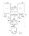
- FIG. 13 is a block diagram of an alternate embodiment of the tissue cavitation system which does not utilize a bubble generator.
- FIG. 14 is a section view of a transducer apparatus according to the present invention.
- One aspect of the present invention relates to a device for generating a microbubble solution and for a system using the device to selectively lyse tissue.
- the microbubble solution includes a fluid or mixture containing one or more of the following: active bubbles, partially dissolved bubbles, a saturated or supersaturated liquid containing fully dissolved bubbles or a material/chemical which generates bubbles in situ.
- the bubbles may be encapsulated within a lipid or the like, or may be unencapsulated (free) bubbles.
- Active bubbles refer to gaseous or vapor bubbles which may include encapsulated gas or unencapsulated gas. These active bubbles may or may not be visible to the naked eye.
- Dissolved bubbles refer to gas which has dissolved into the liquid at a given pressure and temperature but which will come out of solution when the temperature and/or pressure of the solution changes or in response to ultrasound insonation.
- the microbubbles may come out of solution in situ, i.e., after the solution is injected into the tissue. This may, for example, occur when the solution reaches the temperature of the tissue or when the tissue is subjected to ultrasound insonation. Alternatively, the microbubble may come out of solution before the solution is injected into the tissue when reaching atmospheric pressure. Thus, the bubbles may come out of solution before or after the solution is injected into the tissue.
- the solution includes a liquid (fluid) and a gas which may or may not be dissolved in the liquid.
- the liquid portion of enhancing agent may include an aqueous solution, isotonic saline, normal saline, hypotonic saline, hypotonic solution, or a hypertonic solution.
- the solution may optionally include one or more additives/agents to raise the pH (e.g., sodium bicarbonate) or a buffering agent such as known in the art.
- the gaseous portion of the solution may include air drawn from the room (“room air” or “ambient air”), oxygen, carbon dioxide, perfluoropropane, argon, hydrogen, or a mixture of one or more of these gases.
- the invention is not limited to any particular gas. There are a number of candidate gas and liquid combinations, the primary limitation being that both the gas and the liquid must be biocompatible, and the gas must be compatible with the liquid.
- the liquid portion of the microbubble solution includes hypotonic buffered saline and the gaseous portion includes air.
- the biocompatibility of overall solution depends on a variety of factors including the biocompatibility of the liquid and gas, the ratio of gas to liquid, and the size of the microbubbles. If the microbubbles are too large they may not reach the target tissue. Moreover, if the bubbles are too small they may go into solution before they can be used therapeutically.
- the microbubble solution of the present invention may include a distribution of different sized microbubbles. Thus it is anticipated that the solution may contain at least some microbubbles which are too small to be therapeutically useful as well as some which are larger than the ideal size. It is anticipated that a filter, filtering mechanism or the like may be provided to ensure that bubbles larger than a threshold size are not injected into the tissue.
- biocompatible is a relative term in that living tissue may tolerate a small amount of a substance whereas a large amount of the same substance may be toxic with both dose and dosage as considerations.
- biocompatibility of the microbubble solution of the present invention should be interpreted in relation to the amount of solution being infused, the size of the microbubbles, and the ratio of gas to liquid.
- biocompatible should be understood to include a mixture or solution which may result in localized cell lysis alone or in conjunction with ultrasound insonation.
- the microbubble solution according to the present invention may include one or more additives such as a surfactant to stabilize the microbubbles, a local anesthetic, a vasodilator, and a vasoconstrictor.
- a surfactant to stabilize the microbubbles
- the local anesthetic may be lidocaine and the vasoconstrictor may be epinephrine.
- Table 1 is a non-exclusive list of other vasoconstrictors which may be included in the microbubble solution of the present invention.
- Table 2 is a non-exclusive list of other local anesthetics which may be included in the microbubble solution of the present invention.
- Table 3 is a non-exclusive list of gaseous anesthetics which may be included in the gaseous portion of the solution of the present invention.
- Table 4 is a non-exclusive list of surfactants which may be included in the solution of the present invention.
- CPC Cetylpyridinium chloride
- POEA Polyethoxylated tallow amine
- BAC Benzalkonium chloride
- BZT Benzethonium chloride
- Dodecyl betaine Dodecyl dimethylamine oxide
- Cocamidopropyl betaine Coco ampho glycinate
- the enhancing solution may further include a buffering agent such as sodium bicarbonate.
- a buffering agent such as sodium bicarbonate.
- Table 5 is a non-exclusive list of buffers which may be included in the solution of the present invention.
- FIG. 1A depicts a first embodiment of a device 100 for generating microbubbles in the enhancing solution.
- the device 100 consists of a liquid reservoir 102 , a gas vapor reservoir 104 (shown in dashed lines) and a bubble generator 106 .
- the bubble generator 106 is a vessel or vessels in which the fluid and gas are mixed. Fluid from the liquid reservoir 102 and gas/vapor from the gas reservoir 104 flow into the bubble generator 106 and are mixed to create microbubbles and/or supersaturate the fluid.
- the device 100 may include a fluid metering device 124 (shown in dashed lines) controlling the amount of fluid dispensed into the bubble generator 106 and/or a fluid metering device 126 (shown in dashed lines) controlling the amount of microbubble solution to be injected into the tissue.
- the device 100 may further include a gas metering device 128 (shown in dashed lines) used to control the amount of gas dispensed into the bubble generator 106 .
- the device 100 depicted in FIG. 1A includes both of the fluid metering devices 124 and 126 and the gas metering device 128 ; however, in practice one or more of these devices may be eliminated. As noted previously, two or more components may be integrated together.
- the fluid metering device 124 may be integrated into the fluid injection device 202 .
- FIG. 1B is a more detailed illustration of a first embodiment of the bubble generator 106 and includes a housing 108 , a pair of cylinders 116 interconnected by a pathway 118 . At least one of the cylinders 116 is in fluid communication with the liquid reservoir 102 , and at least one of the cylinders 116 is in fluid communication with the gas reservoir 104 (which may be ambient environment).
- the fluid pathway 118 provides fluid communication between the cylinders 116 .
- One or more of the cylinder(s) 116 may be provided with a reciprocating piston 120 driven by an external power source 122 such as a source of compressed air, spring, elastomeric member, motor, stepper motor or the like.
- the reciprocating piston 120 is a pneumatic piston manufactured by the Bimba Corporation.
- Liquid from the liquid reservoir 102 may be pushed into the bubble generator 106 under positive pressure from an external pressurization source 110 (shown in dashed lines); it can be drawn into the bubble generator 106 under partial pressure which may for example be generated by the reciprocating piston 120 ; or it can flow into the generator 106 under gravity.
- gas from the gas reservoir 104 may be pushed into the bubble generator 106 under positive pressure from an external pressurization source 112 (shown in dashed lines) or it can be drawn into the bubble generator 106 under partial pressure.
- the piston 120 may also serve a dual purpose as a fluid pressurization mechanism for injecting the fluid into the tissue.
- the bubble generator 106 may or may not be pressurized to enhance the saturation of the gas in the solution or prevent dissolved gas from coming out of solution.
- An optional fluid pressurization mechanism 110 (shown in dashed lines) may be used to maintain the fluid at a desired pressurization. As will be described in further detail below, the fluid may be chilled to further enhance solubility/saturation of the gas in the solution.
- FIG. 1C is an alternate embodiment of the microbubble generator 106 , which utilizes a member 120 ′ (rotor) such as a blade, paddle, whisk, semi-permeable membrane or the like driven by an external power source 122 to generate the microbubbles within a cylinder or mixing chamber (stator) 116 .
- a member 120 ′ rotor
- the member 120 ′ is rotationally driven by the external power source 122 within a cylinder 116 or the like.
- An optional fluid pressurization mechanism 130 may be used for injecting the fluid into the tissue.
- the fluid in the reservoir 102 may be at ambient temperature. Alternatively, the fluid may be chilled slightly to enhance gas solubility (super saturation).
- the fluid reservoir 102 may be thermally insulated to maintain the fluid at its present temperature and or/the fluid reservoir 102 may include a heating/cooling mechanism (not illustrated) to maintain the fluid at a predetermined temperature.
- the gas reservoir 104 may be eliminated in favor of simply drawing air from the environment, i.e., the room housing the device 100 (“room air”).
- the device 100 may include an air filter 114 (shown in dashed lines) such as a HEPA filter or the like.
- FIG. 1D is an alternate embodiment of the microbubble generator 106 , which utilizes an agitator 133 to agitate or shake a container or cartridge 132 containing measured amounts of liquid and gas and generate the microbubbles within the cartridge 132 .
- the microbubble solution is dispensed from the cartridge 132 to fluid injection device 202 ( FIG. 2 ).
- this cartridge 132 may incorporate an active heating/cooling mechanism to control the temperature of the fluid at a predetermined setting.
- the cartridge 132 may be pressurized, such as by compressed air or mechanical mechanism to allow dispensation of the contents at a predetermined rate and pressure.
- FIG. 2 is a block diagram of a liposculpture system 200 according to the present invention.
- the system 200 includes device 100 , a fluid injection device 202 , an ultrasound transducer apparatus 204 , an ultrasound generator 206 , an ultrasound control unit 208 , and an injection control unit 210 .
- Device 100 may include the bubble generator 106 depicted in FIGS. 1A-1D or may be one of the alternative embodiments disclosed herein below.
- the fluid injection device 202 may include a needle array 214 which may include one or more needles 218 .
- the fluid injection device 202 may, for example, include one or more hypodermic syringes.
- the fluid injection device 202 further includes or is operably connected to a fluid pressurization mechanism 110 for pushing the solution into the tissue.
- a fluid pressurization mechanism 110 for pushing the solution into the tissue.
- the piston 120 or the like used to express fluid between the cylinders 116 may serve as the fluid pressurization mechanism 210 .
- system 200 may be combined.
- the fluid injection device 202 may be integrated as a single component with the ultrasound transducer apparatus 204 and/or the fluid injection control unit 210 .
- the ultrasound control unit 208 can be integrated as a single component with the ultrasound generator 206 . Such integration of components is contemplated and falls within the scope of the present invention.
- the fluid injection control unit 210 may control the amount of fluid and gas dispensed into the bubble generator 106 and/or the amount of solution injected into the tissue.
- the control unit 210 may be interfaced directly or indirectly with the fluid metering device(s) 124 , 126 and the gas metering device 128 .
- the fluid injection control unit 210 may control the mixing or agitation (if any) of the solution within the bubble generator 106 .
- the fluid injection control unit 210 may control the injection of solution into the tissue 220 by the injection device 202 , including the deployment of a needle array 214 , the depth to which the needle array 214 is deployed, and the amount of solution injected.
- the fluid injection control unit 210 may control the individual deployment and retraction of one more needles (or hypodermic syringes) of the needle array 214 .
- the control unit 210 may deploy or retract the needles 218 (or hypodermic syringes) one at a time, may deploy or retract two or more needles 218 at a time, or may deploy or retract all of the needles simultaneously.
- the fluid injection control unit 210 may individually control the amount of solution delivered to each needle 218 .
- One of ordinary skill in the art will appreciate that there are many ways to control the amount of solution delivered to each needle 218 . For example, it may be desirable to deliver more solution in the center of the treatment area and less to the peripheral portion of the treatment area or vice-versa.
- the fluid injection control unit 210 may control the amount of fluid distributed to each syringe. As noted above it may be desirable to provide differing amounts of solution to different areas of the treatment area, and this may be achieved by varying the amount of solution in each syringe.
- the fluid injection device 202 may include a manifold or fluid distribution pathway 212 (shown in dashed lines) in fluid connection with device 100 and needle array 214 , and a needle deployment mechanism 216 operably connected to the needle array 214 .
- the manifold 212 is the fluid pathway used to transport the microbubble solution from the microbubble generator 106 to the needle array 214 .
- One or more flow control devices 222 may be provided in the fluid pathway 212 to enable individualized control of the amount of fluid dispensed to each of the needles or syringes 218 .
- the manifold 212 alone or in combination with the flow control devices 222 controls the distribution of the microbubble solution among the needles 218 .
- the manifold 212 may be configured to deliver a uniform amount of solution to each of the needles 218 (or hypodermic syringes), or it may be configured to deliver differing amounts of solution to different needles 218 .
- the flow control devices 222 may be manually adjustable and/or may be controlled by the injection control unit 210 .
- An alternate embodiment may include infinitely variable volume control at each needle or hypodermic through active means, such as with an electronic flow meter and controller.
- the injection depth may be manually determined by selecting an appropriate needle length or setting a desired injection depth.
- the needle deployment mechanism 216 ( FIGS. 2 and 3A ) deploys one or more needles 218 (or hypodermic syringes) of the needle array 214 such that needles 218 penetrate a desired distance into the tissue.
- the needle deployment mechanism 216 may be configured to deploy the needle(s) 218 to a fixed predetermined depth or may include means for adjusting the depth that the needle(s) 218 are deployed.
- One way to adjust the injection depth is to provide needle arrays 214 of varying length needles. According to this embodiment, the user simply selects an array 214 having shorter/longer needles 218 to achieve a desired injection depth. Moreover, the different length needles 218 may be used within a given array 214 .
- the needle array 214 is displaced vertically in order to adjust the injection depth.
- FIG. 3A shows aspects of an adjusting means, which may include a flange 244 A and a groove 244 B arrangement for vertically adjusting the needle array in discrete intervals.
- FIG. 3D shows aspects of an adjusting means, which may include mating screw threads 240 formed on the needle array 214 and the fluid injection device 202 or housing 108 which enable the user to vertically adjust the needle array 214 thereby altering the injection depth.
- the injection depth may be continuously adjusted within a given range of injection depths.
- the user may be able to continually adjust the injection depth between 5 and 12 millimeters by rotating the needle array 214 .
- the injection depth may be adjusted in discrete intervals.
- the user may be able to adjust the injection depth between 3 and 15 millimeters in 1 millimeter increments.
- the needle depth may be controlled electronically whereby the user enters a specified depth on the control unit 210 .
- the injection depth adjustment described above may specify the injection depth for the entire needle array 214 . However, according to yet another approach it may be desirable to facilitate the individualized adjustment of one or more needles 218 of the needle array 214 .
- the needle deployment mechanism 216 may allow for the independent adjustment of the injection depth for one or more of the needles 218 or syringes.
- One or more of the needles 218 or syringes may be displaced vertically in order to adjust the injection depth of individual needles.
- the adjustment of the injection depth may be continuous or in discrete intervals, and may be manual or may be adjusted via the injection control unit 210 .
- the injection depth may be adjusted by providing mating screw threads 246 to dial in the desired injection depth ( FIG. 4A ), a standoff 248 to provide a means for adjusting the injection depth in discrete intervals ( FIG. 4B ), or the like on the needle array 214 to adjust the vertical height of the needles 218 relative to the tissue apposition surface 226 A.
- Yet another approach to individualized injection depth control is to deploy individual needles or syringes 218 as opposed to deploying the entire needle array 214 .
- the injection control unit 210 or needle deployment mechanism 216 selects the injection depth of each individual needle or syringe 218 ( FIG. 4C ).
- the needle deployment mechanism 216 deploys the needles 218 in response to a signal from the fluid injection control unit 210 .
- the deployment mechanism 216 may include a spring, pneumatic ram, or the like which deploys the needles 218 with sufficient force to penetrate the tissue 220 .
- the fluid injection control unit 210 synchronizes the deployment mechanism 216 with the injection of the microbubble solution into the tissue.
- a predetermined amount of the solution may be injected at a single injection depth.
- the fluid injection control unit 210 in synchronism with the deployment mechanism 216 may inject solution at each of plural injection depths, or may inject continuously as the needle array 214 on either the forward (penetration) or rearward (withdrawal) strokes. It may be desirable to deploy the needles to a first depth within the tissue and then retract the needles to a slightly shallower injection depth before injecting the solution.
- FIG. 5 is an enlarged view of the needle array 214 including at least one hypodermic needle or micro-needle 218 .
- the invention is not limited to any particular length or gauge needle, and needles 218 are selected in accordance with the depth of the tissue to be treated and to accommodate patient comfort. Moreover, it may be desirable for the needle array 214 to include needles of varying length and/or needles of varying gauge.
- the embodiment depicted in FIG. 5 includes a plurality of uniformly spaced needles 218 .
- the scope of the invention is not limited to any particular number of needles 218 ; moreover, the invention is not limited to any particular geometric arrangement or configuration of needles 218 .
- the use of additional needles 218 may facilitate uniform distribution of the microbubble solution in the tissue 220 and/or reduce the number of distinct injection cycles needed to treat a given area.
- FIG. 6A depicts a needle 218 having a single injection orifice 242 , which is linearly aligned with the needle shaft 224 .
- the hypodermic needle 218 is a tubular member having a lumen configured for injection of the solution through the needle and into the tissue.
- the lumen may include a textured surface for promoting the generation of microbubbles.
- FIG. 6B depicts an alternative needle 218 A having one or more side firing orifice(s) 242 A which are generally orthogonal to longitudinal axis of the shaft 224 A.
- the side firing orifice(s) may be formed at different heights along the length of the needle shaft such that solution is injected at varying injection depths.
- These orifice(s) may also be arranged in a specific radial pattern to preferentially direct the flow distribution.
- needle 218 is preferable over needle 218 A or vice versa.
- Reference to the needles 218 should be understood to refer generally to both the needles 218 ( FIG. 6A ) and the needles 218 A ( FIG. 6B ).
- some embodiments of the invention may include a mechanism 256 for selectively rotating one or more of the needles 218 in situ. This feature may facilitate the uniform distribution of solution in the tissue.
- the needle deployment mechanism 216 may ultrasonically vibrate one or more of the needles 218 .
- This feature may facilitate tissue penetration and/or bringing dissolved gas out of solution.
- an ultrasound transducer 258 may be operably coupled to the needles 218 and/or the needle array 214 .
- the ultrasound transducer 258 is shown for the sake of convenience in FIG. 7 however, the transducer 258 may be used in a device which does not include the needle rotation mechanism 256 and vice versa.
- the hypodermic needle 218 has a proximal end connected to the fluid distribution pathway 212 and a distal end configured for penetrating into the tissue 220 to be treated.
- the needles 218 may include micro-needles.
- the fluid injection device 202 includes needle deployment mechanism 216 for moving the hypodermic needle 218 from a fully retracted position ( FIG. 8A ) in which the distal end of the needle 218 is housed inside the solution injection member 202 to a fully extended position ( FIG. 8B ).
- the fluid injection device 202 may optionally be provided with a tissue apposition mechanism which urges the device 202 into firm apposition with the tissue 220 undergoing treatment.
- the tissue apposition mechanism includes at least one vacuum port 228 and a vacuum source 230 in fluid communication with the vacuum port 228 .
- the vacuum port 228 may be defined in the needle array 214 and/or the housing 108 . In operation the tissue apposition surface 226 A is pulled into apposition with the tissue 220 when vacuum from the vacuum source 230 is transmitted through the vacuum port 228 to the tissue 220 .
- the needle(s) 218 may be positioned within the vacuum port(s) 228 .
- the vacuum port 228 may define a recess or receptacle 229 such that the tissue 220 is at least partially pulled (sucked) into the recess 229 by the vacuum force.
- the needles 218 may be at least partially housed within and deployed through the recess 229 .
- An optional flange 232 may surround (skirt) the periphery of the needles 218 (or 218 A) to channel/contain the suction force.
- a separate flange 232 A may surround (skirt) each of the needles 218 (or 218 A) to channel/contain the suction force.
- liquid reservoir 102 and gas reservoir 104 in each of the aforementioned embodiments may be replaced with a cartridge 132 ( FIG. 1D ) containing a pre-measured amount of liquid and gas.
- the gas may be fully or partially dissolved in the fluid.
- the cartridge 132 is simply a sealed container filled with a predetermined amount of gas and liquid, e.g., a soda can.
- FIG. 10A shows an enhanced cartridge 106 A (“Guinness can”), which may be used to replace the liquid reservoir 102 , gas reservoir 104 , and bubble generator 106 in each of the aforementioned embodiments.
- the cartridge 106 A includes a hollow pressurized pod 134 such as disclosed in U.S. Pat. No. 4,832,968, which is hereby incorporated by reference. Both the cartridge 106 A and the pod 134 contain a solution of gas and liquid under greater than ambient pressure which may for example be achieved by providing or introducing a dose of liquid nitrogen into the solution before sealing the cartridge 106 A.
- the cartridge 106 A includes a headspace 136 , which is bounded between a top inner surface 138 and a gas-liquid interface 140 .
- the pod 134 includes a similar headspace 142 , which is bounded between a top inner surface 144 and a gas-liquid interface 146 .
- the pod 134 includes a small opening or orifice 148 , which enables the pressure within the headspace 136 of the cartridge 106 A to reach equilibrium with the pressure within the headspace 142 of the pod 134 .
- a seal 150 of the cartridge 106 A is pierced the pressure within the headspace 136 rapidly reaches equilibrium with the ambient pressure.
- the pressure within the pod 134 is greater than the pressure in the headspace 136 of the cartridge 106 A because the orifice 148 restricts the rate of flow of solution out of the pod 134 .
- a jet of solution forcefully streams out of the orifice 148 into the solution within the cartridge 106 A, which agitates and/or shears the solution within the cartridge causing some of the dissolved bubbles to come out of solution thereby generating microbubbles in the solution.
- the pod 134 is preferably situated at or near the bottom of the cartridge 106 A such that the orifice 148 is maintained below the liquid gas interface 140 .
- FIG. 10B is a block diagram showing the system 200 including cartridge 106 A in place of bubble generator 106 .
- the microbubble generator 106 may be mounted on (integrated with) the fluid injection device 202 thereby minimizing the distance that the solution travels before being injected into the tissue.
- the liquid reservoir 102 and gas reservoir 104 (if provided) may be removably connected to the microbubble generator 106 as needed to generate microbubble solution.
- the injection device 202 may be manually supported by the operator. Alternatively, the injection device 202 may be supported on an arm 302 ( FIG. 11 ) which may include a counterbalance to facilitate manipulation of the injection device 202 .
- FIG. 12A depicts a handpiece 300 which includes fluid injection device 202 and which is coupled to the microbubble generator 106 (not illustrated) by a flexible conduit 236 .
- This design minimizes the size and weight of handpiece 300 being handled by the operator since the handpiece 300 does not include the microbubble generator 106 .
- FIG. 12B depicts a handpiece 300 using the cartridge 106 A mounted on the fluid injection device 202 . This embodiment minimizes the distance that the microbubble solution travels before being injected into the tissue.
- the system of the invention includes a container which may be an enclosed or sealed cartridge 106 A or it may be an open container. If the container is sealed it includes a measured amount of a solution. Obviously, if the container is not sealed then solution may be freely added as needed.
- the system includes a needle array including at least one needle.
- the needle array 214 being in fluid connection with the container.
- the solution includes any of the solutions disclosed herein.
- the solution includes a liquid.
- the solution may further include a gas which may be partially or fully dissolved within the solution.
- the container may be enclosed and the solution may be maintained at greater than atmospheric pressure.
- the needle array 214 includes at least one needle 218 which may be any of the needles disclosed herein.
- the aforementioned gas may include one or more gases selected from the group of air, oxygen, carbon dioxide, carbon dioxide, perfluoropropane, argon, hydrogen, Halothane, Desflurane, Sevoflurane, Isoflurane, and Enflurane.
- the solution may include one or more of a vasoconstrictor, a surfactant, and an anesthetic.
- the vasoconstrictor may include one or more of Norepinephrine, Epinephrine, Angiotensin II, Vasopressin and Endothelin.
- the system may include refrigeration means for maintaining the container at a predefined temperature range.
- the container may be thermally insulated.
- the system may further include an ultrasound transducer apparatus 204 for transmitting ultrasound waves to the tissue.
- the transducer apparatus 204 is operated in synchronism with the injection of solution into the tissue.
- the transducer apparatus 204 may transmit ultrasound energy at a first setting to facilitate the distribution, absorption and/or uptake of solution by the tissue, i.e., sonoporation.
- Ultrasound parameters that enhance the distribution of the solution include those conditions which create microstreaming, such as large duty cycle pulsed ultrasound (>10% duty cycle) or continuous wave ultrasound at a range of frequencies from 500 kHz to 15 MHz, focused or unfocused, and a mechanical index less than 4.
- the mechanical index (MI) falls within the range 0.5 ⁇ MI ⁇ 4.
- the mechanical index falls within the range 0.5 ⁇ MI ⁇ 1.9.
- Sonoporation leading to increased absorption and/or uptake of the solution in the tissue can be generated by pulsed wave or continuous wave ultrasound, at a range of frequencies from 500 kHz to 15 MHz, focused or unfocused and medium to high mechanical index (MI>1.0).
- the preferred embodiment is pulsed wave ultrasound at a frequency of 500 kHz, unfocused, with high mechanical index (MI>1.9) in order to reproducibly create pores that are temporary or longer lasting pores.
- the transducer apparatus 204 may transmit ultrasound energy at a second setting to facilitate the generation of bubbles by bringing dissolved gas out of solution, i.e., stable cavitation.
- Ultrasound parameters for stable cavitation such as large duty cycle pulsed ultrasound (>10% duty cycle) or continuous wave ultrasound at a range of frequencies from 2 MHz to 15 MHz, focused or unfocused, and a mechanical index (MI) 0.05 ⁇ MI ⁇ 2.0.
- the transducer apparatus 204 may transmit ultrasound energy at a third setting to facilitate transient cavitation, i.e., popping bubbles.
- the duty cycle required for transient cavitation may be very low, and the preferred embodiment is a wideband pulse (1 to 20 cycles) transmitted at a duty cycle less than 5%.
- the transducer apparatus 204 may include any of the transducers disclosed herein, and may be operably connected to the needle array 214 .
- the transducer apparatus 204 may transmit ultrasound energy at a fourth frequency range to facilitate the pushing of bubbles within the tissue by acoustic streaming and/or acoustic radiation force.
- Sound propagating through a medium produces a force on particles suspended in the medium, and also upon the medium itself.
- Ultrasound produces a radiation force that is exerted upon objects in a medium with an acoustic impedance different than that of the medium.
- An example is a nanoparticle in blood, although, as one of ordinary skill will recognize, ultrasound radiation forces also may be generated on non-liquid core carrier particles.
- the medium is a liquid, the fluid translation resulting from application of ultrasound is called acoustic streaming.
- Acoustic streaming and optionally radiation force may be used to “push” or concentrate microbubbles injected into the tissue along a cell membrane.
- acoustic streaming has previously been used to push or concentrate carrier particles within a blood vessel.
- the present invention utilizes acoustic streaming to push bubbles within subcutaneous tissue to concentrate the bubble against the walls of cells to be treated.
- a solution containing microbubbles is injected into subcutaneous tissue or a solution containing dissolved gas is injected into subcutaneous tissue and insonated to bring the gas out of solution thereby generating bubbles within the subcutaneous tissue.
- the bubbles are pushed against the cell walls using acoustic streaming, and then insonated to induce transient cavitation to enhance the transport of the solution through the cell membrane and/or mechanically disrupt the cell membrane to selectively lyse cells.
- the ultrasound parameters useful for inducing acoustic streaming include ultrasound waves having center frequencies about 0.1-20 MHz, at an acoustic pressure about 100 kPa-20 MPa, a long cycle length (e.g., about >10 cycles and continuous-wave) OR a short cycle length (e.g., about ⁇ 10 cycle), and high pulse repetition frequency (e.g., about >500 Hz).
- the specific parameters will depend on the choice of carrier particle, as detailed further below, and can be readily determined by one of ordinary skill in the art.
- the transducer apparatus 204 includes a single transducer capable of operating a plurality of operating modes to facilitate stable cavitation, transient cavitation, acoustic streaming, and sonoporation.
- the transducer apparatus 204 includes first and second transducers with first transducer optimized for popping bubbles (transient cavitation) and the second transducer optimized for bringing dissolved gas out of solution (stable cavitation) and/or pushing the bubbles using acoustic radiation force.
- the transducer apparatus may produce focused, unfocused, or defocused ultrasound waves.
- Focused ultrasound refers to generally converging ultrasound waves
- unfocused ultrasound refers to generally parallel ultrasound waves
- defocused ultrasound wave refers to generally diverging ultrasound waves.
- the transducer apparatus 204 selectively produces unfocused and/or defocused ultrasound waves.
- the transducer apparatus may include a flat transducer, i.e., a transducer having a generally planar acoustic wear layer (acoustic window) for producing unfocused ultrasound waves (nonconverging waves) and/or a convex transducer, i.e., a transducer having a convex acoustic wear layer for producing defocused ultrasound waves (diverging waves).
- FIG. 14 depicts an embodiment in which the transducer apparatus 204 includes an inner transducer 204 A and an outer transducer 204 B.
- the inner transducer 204 A has a convex shaped acoustic wear layer for producing defocused waves 205 A
- the outer transducer 204 B has a planar shaped acoustic wear layer for producing unfocused waves 205 B.
- both of the inner and outer transducers 204 A and 204 B may be planar or both may be convex.
- one or both of the inner and outer transducers may be concave, i.e., may have a concave acoustic wear layer for producing focused waves.
- the ultrasound apparatus 204 may include any combination of focused, unfocused, and defocused transducers.
- the inner and outer transducers depicted in FIG. 14 are both circular and the outer transducer surrounds (encircles) the inner transducer. However, other configurations are contemplated and fall within the scope of the invention. According to a presently preferred embodiment, the inner transducer is used to produce stable cavitation and the outer transducer is used to create transient cavitation. However, the relative positions may be swapped with the inner transducer producing transient cavitation and the outer transducer producing stable cavitation.
- the ultrasound apparatus 204 illustrated in FIG. 14 includes a needle array 214 of the type described elsewhere in this disclosure.
- the transducer apparatus 204 of FIG. 14 may be incorporated in any of the embodiments disclosed herein which include an ultrasound transducer. Notably, the transducer apparatus 204 may be incorporated in system 200 .
- the transducer apparatus 204 may include one or more arrays of transducers.
- the transducer apparatus may include an array of transducers for stable cavitation and/or an array of transducers for transient cavitation.
- a solution which may or may not include microbubbles is injected into subcutaneous tissue.
- the solution is pushed against the cell walls using acoustic streaming, and then the subcutaneous tissue is insonated to induce sonoporation and facilitate the uptake/absorption of solution by the tissue.
- Solution is injected an insonated using a system such as system 200 depicted in FIG. 13 which does not include a bubble generator 100 .
- Absorption of the solution preferably results in cell lysis.
- the ultrasound energy from ultrasound generator 206 is applied to the tissue 220 via ultrasound transducer 204 .
- Ultrasound control unit 208 controls the various ultrasound parameters and generally controls the supply of ultrasound by generator 206 .
- ultrasound control unit 208 communicates with the injection control unit 210 to synchronize the application or ultrasound energy with the injection of fluid. It may for example be desirable to quickly apply energy to the tissue before the microbubbles dissipate or are absorbed by the tissue.
- the ultrasound transducer 204 is preferably configured to deliver unfocused ultrasound at an intensity and pressure sufficient to noninvasively cavitate the microbubbles within tissue thereby causing cell lysis.
- the intensity and pressure of the ultrasound applied to the tissue is preferably selected to minimize the heating of tissue and in particular avoid burning the patient's skin.
- the transducer 204 may include a thermocouple 238 or the like to monitor the temperature of the transducer 204 .
- the liposculpture system 200 ( FIG. 2 ) includes an ID reader 250 (shown in dashed lines), and the needle array 214 includes an identifier 252 (shown in dashed lines), which uniquely identifies the needle array 214 .
- the ID reader 250 reads the identifier 252 , and preferably authenticates or verifies the needle array 214 .
- the identifier 252 may contain information identifying the characteristics of the needle array 214 such as length and gauge of needles.
- the identifier 252 may further include identifying information which may be used to track the number of injection cycles (needle deployments) or use time for a given array 214 .
- the reader 250 preferably communicates with the injection control unit 210 .
- the injection control unit 210 may count the number of injection cycles that a given needle array 214 has been used, and may warn the operator if the number exceeds a threshold number.
- the injection control unit 250 may use information stored on the identifier 252 to adjust the injection depth or injection flow rate.
- the injection control unit 210 may further inhibit usage of a needle array if it cannot authenticate, verify or read the identifier 252 .
- the identifier 252 may be a barcode label, a radio frequency tag, smart chip or other machine-readable medium such as known in the art.
- the ultrasound transducer 204 may also include an identifier 252 .
- the identifier 252 may be used to store information identifying the characteristics of the transducer 204 , which is used by the ultrasound control unit 208 in setting or verifying the treatment settings.
- the ultrasound control unit 208 may inhibit insonation if it cannot authenticate, verify or read the identifier 252 .
- the transducer 204 may be integrated with the needle array 214 in which case a single identifier 252 may store information describing characteristics of both the needle(s) 218 and the transducer 204 .
- the ultrasound control unit 208 may use information on the identifier 252 to track the amount of time the identified ultrasound transducer 204 has been operated and at what power levels, and may inhibit insonation if the accumulated insonation time exceeds a threshold value.
- the constituent components of the device 100 may be formed of any sterilizable, biocompatible material. Moreover, some or all of the components may be disposable, i.e., manufactured for single-patient use, to minimize potential cross-contamination of patients.
- the needle array 214 is preferably a disposable component, as the needles 218 will likely dull with use.
- One or more optical or pressure sensors 254 may be provided to measure pressure exerted on the handpiece 300 ( FIG. 12A ) when the handpiece is placed in abutment with the tissue.
- the pressure sensor(s) 254 may provide a safety interlock function to prevent inadvertent deployment of the needle array 214 and/or actuation of the transducer 204 unless pressure is detected as the handpiece 300 is placed in abutment with the tissue. If two or more pressure sensors 254 are provided the injection of solution and/or insonation may be inhibited unless each of the measured pressure values fall within a predefined window and/or so long as the difference between any given two measured pressure values is less than a threshold value.
- the pressure sensor(s) 254 may, for example, be provided on the needle array 214 ( FIG. 4 ) or on the fluid injection device 202 (not illustrated). Alternatively, other sensing means, possibly optical or capacitive, may be used to detect proper positioning of the needle array against the tissue to be treated.
- FIG. 13 is a block diagram of a system 200 for a fat lysing system according to the present invention.
- the system 200 is identical to the system 200 of FIG. 2 but excludes the bubble generator 100 .
- the ultrasound transducer 204 , ultrasound generator 206 , and ultrasound control unit 208 are shown in dashed lines to indicate that these are optional components.
- the system 500 may be used to inject a fat lysing solution (as will be described below in greater detail) with or without the use of ultrasound.
- the fat lysing solution includes epinephrine as its active ingredient.
- the epinephrine may be combined with an aqueous solution, isotonic saline, normal saline, hypotonic saline, hypotonic solution, or a hypertonic solution.
- the solution may optionally include one or more additives/agents to raise the pH (e.g., sodium bicarbonate) or a buffering agent such those listed in Table 5 above or other buffering agents such as known in the art.
- the fat lysing solution includes epinephrine in hypotonic buffered saline.
- the inclusion of ultrasound in system 200 may facilitate the absorption and/or distribution of the fat lysing solution.
- the inclusion of ultrasound in system 200 may facilitate the absorption and/or distribution of the fat lysing solution.
- the ultrasound may be used to enhance the distribution, absorption, and/or uptake of the solution in the tissue by permanently or temporarily opening pores in the cell membrane (sonoporation), generating microstreaming in the solution, or locally heating the solution or the tissue.
- the ultrasound generator 206 may be operated at a first setting to facilitate distribution of the solution and then it may be operated at a second setting to facilitate absorption.
- the sonoporation may be reversible or irreversible.
- the system 200 may include an optional ultrasound transducer 258 for vibrating the needles 218 to facilitate tissue penetration and/or a needle rotation mechanism 256 which may be used in conjunction with side-firing needles 218 to facilitate even distribution of the solution.
- the same transducer apparatus 204 used to facilitate absorption and/or distribution of the solution may be used to facilitate tissue penetration thereby eliminating the need for a separate transducer 258 .
- the system 200 may include any or all of the features described in this disclosure including means for selectively adjusting the amount of solution injected by each of the needles 218 and/or the rate or pressure at which the solution is injected into the tissue. Still further the system 200 may include the selective adjustment of the injection depth and/or the tissue apposition mechanism as described above.
- solution is percutaneously injected into subcutaneous tissue, and the tissue is insonated at a first ultrasound setting to distribute the solution. Once the solution has been distributed the tissue is insonated at a second setting to induce sonoporation thereby inducing cell lysis.
- the solution need not contain microbubbles as they do not contribute to cell lysis. To increase the efficacy of this mode of operation it is recommended to repeat the injection and insonation of the tissue through 10 to 50 cycles.
- a solution containing microbubbles is percutaneously injected into subcutaneous tissue, and the tissue is insonated at a first ultrasound setting to distribute the solution and/or push the microbubbles against the cell walls. Thereafter the tissue is insonated at a second setting (for between 1 millisecond and 1 second) to induce transient cavitation inducing cell lysis.
- a second setting for between 1 millisecond and 1 second
- the microbubbles will be absorbed by the tissue and/or go into solution within a relatively short period of time.
- the tissue is insonated to facilitate distribution of the microbubble solution through acoustic radiation force and/or microstreaming occurs simultaneously as the solution is injected into the tissue or within a very short amount of time afterward.
- the injection of a small amount of the microbubble solution takes approximately 200 milliseconds and insonation to induce distribution through acoustic radiation force takes between 1 millisecond and 1 second.
- the tissue is insonated to induce transient cavitation for approximately 400 milliseconds.
- a solution containing dissolved gas i.e., dissolved gas bubbles is percutaneously injected into subcutaneous tissue, and the tissue is insonated at a first ultrasound setting to bring the bubbles out of solution (for between 100 microseconds and 1 millisecond) followed immediately by insonation at a second setting (for between 1 millisecond and 1 second) to distribute the solution and/or push the microbubbles against the cell walls. Thereafter the tissue is insonated at a third setting (for between 100 microseconds and 1 second) to induce transient cavitation inducing cell lysis. To increase the efficacy of this mode of operation it is recommended to repeat the injection and insonation of the tissue through 10 to 50 cycles.
- the microbubbles will be absorbed by the tissue and/or go into solution within a relatively short period of time.
- the tissue is insonated to induce stable cavitation and bring the bubbles out of solution after the solution has been injected into the subcutaneous tissue. Satisfactory stable cavitation results have been achieved by insonating for approximately 100 microseconds. Thereafter the tissue is insonated to facilitate distribution of the microbubble solution through acoustic radiation force and/or microstreaming occurs. Insonating for between 1 millisecond and 1 second is required to distribute the microbubbles. Immediately thereafter the tissue is insonated to induce transient cavitation for approximately 400 milliseconds.
- the invention may be combined with other methods or apparatus for treating tissues.
- the invention may also include use of skin tightening procedures, for example, ThermaCoolTM available from Thermage Corporation located in Hayward, Calif., Cutera TitanTM available from Cutera, Inc. located in Brisbane, Calif., or AlumaTM available from Lumenis, Inc. located in Santa Clara, Calif.
Abstract
Description
| TABLE 1 |
| Vasoconstrictors |
| Norepinephrine |
| Epinephrine |
| Angiotensin II |
| Vasopressin |
| Endothelin |
| TABLE 2 |
| Anesthetics (Local) |
| Amino esters |
| Benzocaine |
| Chloroprocaine |
| Cocaine |
| Procaine |
| Tetracaine |
| Amino amides |
| Bupivacaine |
| Levobupivacaine |
| Lidocaine |
| Mepivacaine |
| Prilocaine |
| Ropivacaine |
| Articaine |
| Trimecaine |
| TABLE 3 |
| Anesthetics (gaseous) |
| Halothane |
| Desflurane |
| Sevoflurane |
| Isoflurane |
| Enflurane |
| TABLE 4 |
| Surfactants |
| Anionic (based on sulfate, sulfonate or carboxylate anions) |
| Sodium dodecyl sulfate (SDS), ammonium lauryl sulfate, and other alkyl | |
| sulfate salts | |
| Sodium laureth sulfate, also known as sodium lauryl ether sulfate (SLES) | |
| Alkyl benzene sulfonate | |
| Soaps, or fatty acid salts |
| Cationic (based on quaternary ammonium cations) |
| Cetyl trimethylammonium bromide (CTAB) a.k.a. hexadecyl trimethyl | |
| ammonium bromide, and other alkyltrimethylammonium salts | |
| Cetylpyridinium chloride (CPC) | |
| Polyethoxylated tallow amine (POEA) | |
| Benzalkonium chloride (BAC) | |
| Benzethonium chloride (BZT) |
| Zwitterionic (amphoteric) |
| Dodecyl betaine | |
| Dodecyl dimethylamine oxide | |
| Cocamidopropyl betaine | |
| Coco ampho glycinate |
| Nonionic | |
| Alkyl poly(ethylene oxide) called Poloxamers or Poloxamines) | |
| Alkyl polyglucosides, including: |
| Octyl glucoside | |
| Decyl maltoside |
| Fatty alcohols |
| Cetyl alcohol | |
| Oleyl alcohol |
| Cocamide MEA, cocamide DEA, cocamide TEA | |
| TABLE 5 |
| Buffer |
| H3PO4/NaH2PO4 (pKa1) | NaH2PO4/Na2HPO4 (pKa2) |
| 1,3-Diaza-2,4-cyclopentadiene and Glyoxaline | N-Tris(hydroxymethyl)methyl-2- |
| (Imidazole) | aminoethanesulfonic acid (TES) |
| ampholyte N-(2-hydroxyethyl) piperazine-N′-2- | N-2-Hydroxyethylpiperazine-N′-2-ethanesulfonic |
| hydroxypropanesulfonic acid (HEPPSO) | acid (HEPES) |
| Acetic acid | Citric acid (pKa1) |
| N-Tris(hydroxymethyl)methyl-3 - | Triethanolamine (2,2′,2″-Nitrilotriethanol |
| aminopropanesulfonic acid (TAPS) | Tris(2-hydroxyethyl)amine) |
| Bis(2- | N-[Tris(hydroxymethyl)methyl]glycine, 3-[(3- |
| hydroxyethyl)iminotris(hydroxymethyl)methane | Cholamidopropyl)dimethylammonio]propanesulfonic |
| (Bis-Tris) | acid (Tricine) |
| Cacodylic acid | 2-Amino-2-(hydroxymethyl)-1,3-propanediol (Tris) |
| H2CO3/NaHCO3 (pKa1) | Glycine amide |
| Citric acid (pKa3) | N,N-Bis(2-hydroxyethyl)glycine (Bicine) |
| 2-(N-Morpholino)ethanesulfonic Acid (MES) | Glycylglycine (pKa2) |
| N-(2-Acetamido)iminodiacetic Acid (ADA) | Citric acid (pKa2) |
| Bis-Tris Propane (pKa1) | Bis-Tris Propane (pKa2) |
| Piperazine-1,4-bis(2-ethanesulfonic acid) | N-(2-Acetamido)-2-aminoethanesulfonic acid |
| (PIPES) | (ACES) |
| Boric acid (H3BO3/Na2B4O7) | N-Cyclohexyl-2-aminoethanesulfonic acid (CHES |
| Glycine (pKa1) | Glycine (pKa2) |
| N,N-Bis(2-hydroxyethyl)-2- | NaHCO3/Na2CO3 (pKa2) |
| aminoethanesulfonic acid (BES) | |
| 3-Morpholinopropanesulfonic acid (MOPS) | N-Cyclohexyl-3-aminopropanesulfonic acid (CAPS) |
| Na2HPO4/Na3PO4 (pKa3) | Hexahydropyridine (Piperidine) |
| *The anhydrous molecular weight is reported in the table. Actual molecular weight will depend on the degree of hydration. | |
Claims (12)
Priority Applications (2)
| Application Number | Priority Date | Filing Date | Title |
|---|---|---|---|
| US11/771,932 US9248317B2 (en) | 2005-12-02 | 2007-06-29 | Devices and methods for selectively lysing cells |
| PCT/US2008/067137 WO2009006009A1 (en) | 2007-06-29 | 2008-06-16 | Devices and methods for selectively lysing cells |
Applications Claiming Priority (5)
| Application Number | Priority Date | Filing Date | Title |
|---|---|---|---|
| US11/292,950 US7967763B2 (en) | 2005-09-07 | 2005-12-02 | Method for treating subcutaneous tissues |
| US11/334,805 US7601128B2 (en) | 2005-09-07 | 2006-01-17 | Apparatus for treating subcutaneous tissues |
| US11/334,794 US7588547B2 (en) | 2005-09-07 | 2006-01-17 | Methods and system for treating subcutaneous tissues |
| US11/515,634 US20070060989A1 (en) | 2005-09-07 | 2006-09-05 | Apparatus and method for disrupting subcutaneous structures |
| US11/771,932 US9248317B2 (en) | 2005-12-02 | 2007-06-29 | Devices and methods for selectively lysing cells |
Related Parent Applications (1)
| Application Number | Title | Priority Date | Filing Date |
|---|---|---|---|
| US11/292,950 Continuation-In-Part US7967763B2 (en) | 2005-09-07 | 2005-12-02 | Method for treating subcutaneous tissues |
Publications (2)
| Publication Number | Publication Date |
|---|---|
| US20080248554A1 US20080248554A1 (en) | 2008-10-09 |
| US9248317B2 true US9248317B2 (en) | 2016-02-02 |
Family
ID=40226456
Family Applications (1)
| Application Number | Title | Priority Date | Filing Date |
|---|---|---|---|
| US11/771,932 Expired - Fee Related US9248317B2 (en) | 2005-12-02 | 2007-06-29 | Devices and methods for selectively lysing cells |
Country Status (2)
| Country | Link |
|---|---|
| US (1) | US9248317B2 (en) |
| WO (1) | WO2009006009A1 (en) |
Cited By (1)
| Publication number | Priority date | Publication date | Assignee | Title |
|---|---|---|---|---|
| US20140074197A1 (en) * | 2012-09-10 | 2014-03-13 | Ronald L. Moy | Non-surgical orbital fat reduction |
Families Citing this family (36)
| Publication number | Priority date | Publication date | Assignee | Title |
|---|---|---|---|---|
| BR0312430A (en) | 2002-06-19 | 2005-04-26 | Palomar Medical Tech Inc | Method and apparatus for treating skin and subcutaneous conditions |
| US7856985B2 (en) | 2005-04-22 | 2010-12-28 | Cynosure, Inc. | Method of treatment body tissue using a non-uniform laser beam |
| US7586957B2 (en) | 2006-08-02 | 2009-09-08 | Cynosure, Inc | Picosecond laser apparatus and methods for its operation and use |
| US9132031B2 (en) | 2006-09-26 | 2015-09-15 | Zeltiq Aesthetics, Inc. | Cooling device having a plurality of controllable cooling elements to provide a predetermined cooling profile |
| US8192474B2 (en) | 2006-09-26 | 2012-06-05 | Zeltiq Aesthetics, Inc. | Tissue treatment methods |
| US20080287839A1 (en) | 2007-05-18 | 2008-11-20 | Juniper Medical, Inc. | Method of enhanced removal of heat from subcutaneous lipid-rich cells and treatment apparatus having an actuator |
| US8523927B2 (en) | 2007-07-13 | 2013-09-03 | Zeltiq Aesthetics, Inc. | System for treating lipid-rich regions |
| US8285390B2 (en) | 2007-08-21 | 2012-10-09 | Zeltiq Aesthetics, Inc. | Monitoring the cooling of subcutaneous lipid-rich cells, such as the cooling of adipose tissue |
| US8882753B2 (en) * | 2008-07-14 | 2014-11-11 | Syneron Medical Ltd | Devices and methods for percutaneous energy delivery |
| EP2346428B1 (en) | 2008-09-25 | 2019-11-06 | Zeltiq Aesthetics, Inc. | Treatment planning systems and methods for body contouring applications |
| US8603073B2 (en) | 2008-12-17 | 2013-12-10 | Zeltiq Aesthetics, Inc. | Systems and methods with interrupt/resume capabilities for treating subcutaneous lipid-rich cells |
| KR101701137B1 (en) | 2009-04-30 | 2017-02-01 | 젤티크 애스세틱스, 인코포레이티드. | Device, system and method of removing heat from subcutaneous lipid-rich cells |
| US9919168B2 (en) * | 2009-07-23 | 2018-03-20 | Palomar Medical Technologies, Inc. | Method for improvement of cellulite appearance |
| US9314368B2 (en) | 2010-01-25 | 2016-04-19 | Zeltiq Aesthetics, Inc. | Home-use applicators for non-invasively removing heat from subcutaneous lipid-rich cells via phase change coolants, and associates devices, systems and methods |
| BR112012029726A2 (en) * | 2010-05-27 | 2017-09-26 | Koninl Philips Electronics Nv | method for operating an ultrasonic transducer, ultrasonic transducer for selective generation of ultrasonic waves and heat, and sample analysis system |
| US8676338B2 (en) | 2010-07-20 | 2014-03-18 | Zeltiq Aesthetics, Inc. | Combined modality treatment systems, methods and apparatus for body contouring applications |
| KR102183581B1 (en) | 2012-04-18 | 2020-11-27 | 싸이노슈어, 엘엘씨 | Picosecond laser apparatus and methods for treating target tissues with same |
| US9545523B2 (en) | 2013-03-14 | 2017-01-17 | Zeltiq Aesthetics, Inc. | Multi-modality treatment systems, methods and apparatus for altering subcutaneous lipid-rich tissue |
| US9844460B2 (en) | 2013-03-14 | 2017-12-19 | Zeltiq Aesthetics, Inc. | Treatment systems with fluid mixing systems and fluid-cooled applicators and methods of using the same |
| EP2973894A2 (en) | 2013-03-15 | 2016-01-20 | Cynosure, Inc. | Picosecond optical radiation systems and methods of use |
| US10117892B2 (en) | 2013-08-29 | 2018-11-06 | Allergan, Inc. | Devices and methods for reducing the appearance of cellulite |
| US10575890B2 (en) | 2014-01-31 | 2020-03-03 | Zeltiq Aesthetics, Inc. | Treatment systems and methods for affecting glands and other targeted structures |
| US10675176B1 (en) | 2014-03-19 | 2020-06-09 | Zeltiq Aesthetics, Inc. | Treatment systems, devices, and methods for cooling targeted tissue |
| USD777338S1 (en) | 2014-03-20 | 2017-01-24 | Zeltiq Aesthetics, Inc. | Cryotherapy applicator for cooling tissue |
| US10952891B1 (en) | 2014-05-13 | 2021-03-23 | Zeltiq Aesthetics, Inc. | Treatment systems with adjustable gap applicators and methods for cooling tissue |
| US10935174B2 (en) | 2014-08-19 | 2021-03-02 | Zeltiq Aesthetics, Inc. | Stress relief couplings for cryotherapy apparatuses |
| US10568759B2 (en) | 2014-08-19 | 2020-02-25 | Zeltiq Aesthetics, Inc. | Treatment systems, small volume applicators, and methods for treating submental tissue |
| WO2017070112A1 (en) | 2015-10-19 | 2017-04-27 | Zeltiq Aesthetics, Inc. | Vascular treatment systems, cooling devices, and methods for cooling vascular structures |
| EP3399950A1 (en) | 2016-01-07 | 2018-11-14 | Zeltiq Aesthetics, Inc. | Temperature-dependent adhesion between applicator and skin during cooling of tissue |
| US10765552B2 (en) | 2016-02-18 | 2020-09-08 | Zeltiq Aesthetics, Inc. | Cooling cup applicators with contoured heads and liner assemblies |
| US11382790B2 (en) | 2016-05-10 | 2022-07-12 | Zeltiq Aesthetics, Inc. | Skin freezing systems for treating acne and skin conditions |
| US10555831B2 (en) | 2016-05-10 | 2020-02-11 | Zeltiq Aesthetics, Inc. | Hydrogel substances and methods of cryotherapy |
| US10682297B2 (en) | 2016-05-10 | 2020-06-16 | Zeltiq Aesthetics, Inc. | Liposomes, emulsions, and methods for cryotherapy |
| US11076879B2 (en) | 2017-04-26 | 2021-08-03 | Zeltiq Aesthetics, Inc. | Shallow surface cryotherapy applicators and related technology |
| SG11202008151QA (en) | 2018-02-26 | 2020-09-29 | Cynosure Inc | Q-switched cavity dumped sub-nanosecond laser |
| CA3107932A1 (en) | 2018-07-31 | 2020-02-06 | Zeltiq Aesthetics, Inc. | Methods, devices, and systems for improving skin characteristics |
Citations (510)
| Publication number | Priority date | Publication date | Assignee | Title |
|---|---|---|---|---|
| US2370529A (en) | 1942-04-21 | 1945-02-27 | Fuller Katherine | Golf ball teeing device |
| US2490409A (en) | 1947-10-03 | 1949-12-06 | Paul H Brown | Golf practice target element |
| US2738172A (en) | 1952-11-28 | 1956-03-13 | Nat Dairy Res Lab Inc | Apparatus for treatment of products with ultrasonic energy |
| US2945496A (en) | 1958-08-18 | 1960-07-19 | Fosdal Alfred | Dental instrument for immobilizing tissue |
| US2961382A (en) | 1957-07-25 | 1960-11-22 | Ortho Pharma Corp | Urokinase-a plasmiogen activator and methods of obtaining the same |
| US3129944A (en) | 1961-08-24 | 1964-04-21 | Fuller Brush Co | Golf mat composed of a plurality of parallel brush strips |
| US3324854A (en) | 1964-04-23 | 1967-06-13 | Harry Swartz | Apparatus for facilitating the insertion of a hypodermic syringe needle |
| GB1216813A (en) | 1969-02-21 | 1970-12-23 | Shozo Narusawa | Transcutaneous injection device |
| US3590808A (en) | 1968-09-04 | 1971-07-06 | Us Catheter & Instr Corp | Biopsy tool |
| US3735336A (en) | 1971-03-10 | 1973-05-22 | Ampex | Acoustic lens |
| US3991763A (en) | 1975-04-17 | 1976-11-16 | Arbrook, Inc. | Surgical evacuator |
| US4150669A (en) | 1977-03-16 | 1979-04-24 | Alvaro Latorre | Apparatus for effecting and enhancing an erection |
| US4188952A (en) | 1973-12-28 | 1980-02-19 | Loschilov Vladimir I | Surgical instrument for ultrasonic separation of biological tissue |
| US4211949A (en) | 1978-11-08 | 1980-07-08 | General Electric Company | Wear plate for piezoelectric ultrasonic transducer arrays |
| US4212206A (en) | 1977-10-07 | 1980-07-15 | Thomson-Csf | Ultrasonic system for the visual display of a thin section of a body |
| GB1577551A (en) | 1976-02-09 | 1980-10-22 | Fahim M | Medication for topical application by ultrasound |
| US4231368A (en) | 1978-03-23 | 1980-11-04 | Michael Becker | Device for intramuscular injections, especially of insulin |
| WO1980002365A1 (en) | 1979-05-04 | 1980-11-13 | Rasor Ass Inc | Ultrasonic image enhancement |
| US4248231A (en) | 1978-11-16 | 1981-02-03 | Corning Glass Works | Surgical cutting instrument |
| US4249923A (en) | 1979-07-10 | 1981-02-10 | Walda Kim L | Cardioplegic fluid refrigeration and delivery system |
| US4299219A (en) | 1979-12-17 | 1981-11-10 | Norris Jr George P | Intravenous needle insertion device |
| US4309989A (en) | 1976-02-09 | 1982-01-12 | The Curators Of The University Of Missouri | Topical application of medication by ultrasound with coupling agent |
| JPS57139358U (en) | 1981-02-24 | 1982-08-31 | ||
| US4382441A (en) | 1978-12-06 | 1983-05-10 | Svedman Paul | Device for treating tissues, for example skin |
| US4466442A (en) | 1981-10-16 | 1984-08-21 | Schering Aktiengesellschaft | Carrier liquid solutions for the production of gas microbubbles, preparation thereof, and use thereof as contrast medium for ultrasonic diagnostics |
| US4497325A (en) | 1982-07-15 | 1985-02-05 | Wedel Victor J | Ultrasound needle, biopsy instrument or catheter guide |
| EP0148116A1 (en) | 1983-12-02 | 1985-07-10 | Debiopharm S.A. | Coupling piece for an injection syringe |
| US4536180A (en) | 1983-07-22 | 1985-08-20 | Johnson Gerald W | Surgical instrument for suction lipolysis |
| US4549533A (en) | 1984-01-30 | 1985-10-29 | University Of Illinois | Apparatus and method for generating and directing ultrasound |
| US4608043A (en) | 1984-06-22 | 1986-08-26 | Abbott Laboratories | I.V. fluid storage and mixing system |
| US4641652A (en) | 1984-04-12 | 1987-02-10 | Richard Wolf Gmbh | Applicator for tying sewing threads |
| US4646754A (en) | 1985-02-19 | 1987-03-03 | Seale Joseph B | Non-invasive determination of mechanical characteristics in the body |
| US4657756A (en) | 1980-11-17 | 1987-04-14 | Schering Aktiengesellschaft | Microbubble precursors and apparatus for their production and use |
| US4673387A (en) | 1985-05-06 | 1987-06-16 | N. J. Phillips Pty. Limited | Pellet injector |
| US4681119A (en) | 1980-11-17 | 1987-07-21 | Schering Aktiengesellschaft | Method of production and use of microbubble precursors |
| US4684479A (en) | 1985-08-14 | 1987-08-04 | Arrigo Joseph S D | Surfactant mixtures, stable gas-in-liquid emulsions, and methods for the production of such emulsions from said mixtures |
| US4688570A (en) | 1981-03-09 | 1987-08-25 | The Regents Of The University Of California | Ophthalmologic surgical instrument |
| US4689986A (en) | 1985-03-13 | 1987-09-01 | The University Of Michigan | Variable frequency gas-bubble-manipulating apparatus and method |
| US4718433A (en) | 1983-01-27 | 1988-01-12 | Feinstein Steven B | Contrast agents for ultrasonic imaging |
| US4720075A (en) | 1986-07-28 | 1988-01-19 | Buell Industries, Inc. | Shock isolating mount |
| CA1232837A (en) | 1983-04-15 | 1988-02-16 | Jurgen Hilmann | Ultrasound contrast agent containing microparticles and gas micro-bubbles |
| US4751921A (en) | 1985-10-21 | 1988-06-21 | University Of Iowa Research Foundation | Bone cement syringe |
| US4762915A (en) | 1985-01-18 | 1988-08-09 | Liposome Technology, Inc. | Protein-liposome conjugates |
| US4796624A (en) | 1986-11-19 | 1989-01-10 | Concept, Inc. | Lashliner |
| US4797285A (en) | 1985-12-06 | 1989-01-10 | Yissum Research And Development Company Of The Hebrew University Of Jerusalem | Lipsome/anthraquinone drug composition and method |
| US4815462A (en) | 1987-04-06 | 1989-03-28 | Clark Vickie J | Lipectomy device |
| WO1989005160A1 (en) | 1987-12-02 | 1989-06-15 | Schering Aktiengesellschaft Berlin Und Bergkamen | Use of media containing bubbles in shock wave and ultrasonic therapy |
| WO1989005159A1 (en) | 1987-12-02 | 1989-06-15 | Schering Aktiengesellschaft Berlin Und Bergkamen | Ultrasonic or shock wave operating process and agent for implementing said process |
| US4844080A (en) | 1987-02-19 | 1989-07-04 | Michael Frass | Ultrasound contact medium dispenser |
| US4844882A (en) | 1987-12-29 | 1989-07-04 | Molecular Biosystems, Inc. | Concentrated stabilized microbubble-type ultrasonic imaging agent |
| US4844470A (en) | 1988-06-06 | 1989-07-04 | Ste-Mak, Inc. | Golf mat |
| EP0327490A1 (en) | 1988-02-05 | 1989-08-09 | Schering Aktiengesellschaft | Ultrasonic imaging agents, process for their preparation and their diagnostic and therapeutical use |
| WO1989009593A1 (en) | 1988-04-16 | 1989-10-19 | Schering Aktiengesellschaft Berlin Und Bergkamen | Process for producing liposomes and/or bilological cells, which have included ingredient substances, and their use |
| US4886491A (en) | 1988-02-29 | 1989-12-12 | Tulio Parisi | Liposuction procedure with ultrasonic probe |
| US4900540A (en) | 1983-06-20 | 1990-02-13 | Trustees Of The University Of Massachusetts | Lipisomes containing gas for ultrasound detection |
| WO1990001971A1 (en) | 1988-08-18 | 1990-03-08 | Massachusetts Institute Of Technology | Ultrasound enhancement of transbuccal drug delivery |
| US4919986A (en) | 1988-06-09 | 1990-04-24 | Daimler-Benz Ag | Plastic suction cup arrangement for laminatd glass pane repair |
| US4920954A (en) | 1988-08-05 | 1990-05-01 | Sonic Needle Corporation | Ultrasonic device for applying cavitation forces |
| DE3838530A1 (en) | 1988-11-14 | 1990-05-17 | Hilti Ag | Packaging for two-component compounds |
| US4936281A (en) | 1989-04-13 | 1990-06-26 | Everest Medical Corporation | Ultrasonically enhanced RF ablation catheter |
| US4936303A (en) | 1987-11-20 | 1990-06-26 | Ultrathermics | Ultrasonic heating apparatus and method |
| EP0384831A2 (en) | 1989-02-21 | 1990-08-29 | Technomed International | Apparatus for selective destruction of cells including soft tissues and bones inside a living being by implosing of gas bubbles |
| US4957656A (en) | 1988-09-14 | 1990-09-18 | Molecular Biosystems, Inc. | Continuous sonication method for preparing protein encapsulated microbubbles |
| US5022414A (en) | 1979-12-13 | 1991-06-11 | Joseph J. Berke | Tissue separator method |
| US5040537A (en) | 1987-11-24 | 1991-08-20 | Hitachi, Ltd. | Method and apparatus for the measurement and medical treatment using an ultrasonic wave |
| US5050537A (en) | 1990-05-01 | 1991-09-24 | Fox Harvey Z | Automatic animal feeding system |
| US5069664A (en) | 1990-01-25 | 1991-12-03 | Inter Therapy, Inc. | Intravascular ultrasonic angioplasty probe |
| US5083568A (en) | 1987-06-30 | 1992-01-28 | Yokogawa Medical Systems, Limited | Ultrasound diagnosing device |
| US5088499A (en) | 1989-12-22 | 1992-02-18 | Unger Evan C | Liposomes as contrast agents for ultrasonic imaging and methods for preparing the same |
| US5100390A (en) | 1990-10-22 | 1992-03-31 | Norma A. Lubeck | Lubeck spinal catheter needle |
| WO1992009238A1 (en) | 1990-11-30 | 1992-06-11 | Michele Zocchi | Method and apparatus for treating human fat tissue |
| US5131600A (en) | 1989-02-13 | 1992-07-21 | The Dow Chemical Company | Alkanol amine grinding aids |
| US5143063A (en) | 1988-02-09 | 1992-09-01 | Fellner Donald G | Method of removing adipose tissue from the body |
| US5149319A (en) | 1990-09-11 | 1992-09-22 | Unger Evan C | Methods for providing localized therapeutic heat to biological tissues and fluids |
| US5158071A (en) | 1988-07-01 | 1992-10-27 | Hitachi, Ltd. | Ultrasonic apparatus for therapeutical use |
| US5170604A (en) | 1991-08-21 | 1992-12-15 | Tamer Industries, Inc. | Wall panel system and fastener therefor |
| US5178433A (en) | 1987-06-12 | 1993-01-12 | Frank Wagner | Vehicle body mount |
| EP0278074B1 (en) | 1986-11-26 | 1993-04-14 | Shunro Tachibana | Apparatus for the endermic application of medicines |
| US5203785A (en) | 1990-05-10 | 1993-04-20 | Symbrosis Corporation | Laparoscopic hook scissors |
| US5209720A (en) | 1989-12-22 | 1993-05-11 | Unger Evan C | Methods for providing localized therapeutic heat to biological tissues and fluids using gas filled liposomes |
| US5215104A (en) | 1988-08-16 | 1993-06-01 | Steinert Roger F | Method for corneal modification |
| US5216130A (en) | 1990-05-17 | 1993-06-01 | Albany Medical College | Complex for in-vivo target localization |
| US5215680A (en) | 1990-07-10 | 1993-06-01 | Cavitation-Control Technology, Inc. | Method for the production of medical-grade lipid-coated microbubbles, paramagnetic labeling of such microbubbles and therapeutic uses of microbubbles |
| US5261922A (en) | 1992-02-20 | 1993-11-16 | Hood Larry L | Improved ultrasonic knife |
| US5310540A (en) | 1990-10-05 | 1994-05-10 | Sintetica Sa | Method for the preparation of stable suspensions of hollow gas-filled microspheres suitable for ultrasonic echography |
| US5312364A (en) | 1993-08-06 | 1994-05-17 | Pyng | Intraosseous infusion device |
| US5315998A (en) | 1991-03-22 | 1994-05-31 | Katsuro Tachibana | Booster for therapy of diseases with ultrasound and pharmaceutical liquid composition containing the same |
| US5316000A (en) | 1991-03-05 | 1994-05-31 | Technomed International (Societe Anonyme) | Use of at least one composite piezoelectric transducer in the manufacture of an ultrasonic therapy apparatus for applying therapy, in a body zone, in particular to concretions, to tissue, or to bones, of a living being and method of ultrasonic therapy |
| US5320607A (en) | 1992-02-13 | 1994-06-14 | Kabushiki Kaisya Advance | Simple blood sampling device |
| US5352436A (en) | 1992-11-02 | 1994-10-04 | Drexel University | Surfactant-stabilized microbubble mixtures, process for preparing and methods of using the same |
| US5354307A (en) | 1992-04-06 | 1994-10-11 | Porowski Jan S | Surgical means for removing a portion of a body |
| US5383858A (en) * | 1992-08-17 | 1995-01-24 | Medrad, Inc. | Front-loading medical injector and syringe for use therewith |
| US5409126A (en) | 1993-10-13 | 1995-04-25 | Demars; Robert A. | Storage container with reversible lid |
| US5413574A (en) | 1992-09-04 | 1995-05-09 | Fugo; Richard J. | Method of radiosurgery of the eye |
| US5415160A (en) | 1994-03-15 | 1995-05-16 | Ethicon, Inc. | Surgical lift method and apparatus |
| US5417654A (en) | 1994-02-02 | 1995-05-23 | Alcon Laboratories, Inc. | Elongated curved cavitation-generating tip for disintegrating tissue |
| US5419777A (en) | 1994-03-10 | 1995-05-30 | Bavaria Medizin Technologie Gmbh | Catheter for injecting a fluid or medicine |
| US5419761A (en) | 1993-08-03 | 1995-05-30 | Misonix, Inc. | Liposuction apparatus and associated method |
| WO1995015118A1 (en) | 1993-11-30 | 1995-06-08 | Imarx Pharmaceutical Corp. | Gas microspheres for topical and subcutaneous application |
| US5425580A (en) | 1990-12-28 | 1995-06-20 | Byk Gulden Lomberg Chemische Fabrik Gmbh | Dosage form for micro-bubble echo contrast agents |
| US5437640A (en) | 1994-01-31 | 1995-08-01 | Schwab; Louis | Apparatus and method for inserting hypodermic, tuberculin and other needles and for administering Mantoux tuberculin tests |
| US5441490A (en) | 1991-01-09 | 1995-08-15 | Principal Ab | Transdermal perfusion of fluids |
| US5457041A (en) | 1994-03-25 | 1995-10-10 | Science Applications International Corporation | Needle array and method of introducing biological substances into living cells using the needle array |
| US5476368A (en) | 1992-08-20 | 1995-12-19 | Ryder International Corporation | Sterile fluid pump diaphragm construction |
| US5478315A (en) | 1994-08-08 | 1995-12-26 | Brothers Family Investments, L.C. | Local anesthetic injection system |
| US5494038A (en) | 1995-04-25 | 1996-02-27 | Abbott Laboratories | Apparatus for ultrasound testing |
| US5507790A (en) | 1994-03-21 | 1996-04-16 | Weiss; William V. | Method of non-invasive reduction of human site-specific subcutaneous fat tissue deposits by accelerated lipolysis metabolism |
| US5522797A (en) | 1995-01-03 | 1996-06-04 | Ivy Laboratories, Inc. | Slide action veterinary implanter |
| US5533981A (en) | 1994-10-06 | 1996-07-09 | Baxter International Inc. | Syringe infusion pump having a syringe plunger sensor |
| US5556406A (en) | 1994-09-12 | 1996-09-17 | Medjet Inc. | Corneal template and surgical procedure for refractive vision correction |
| US5562693A (en) | 1995-08-11 | 1996-10-08 | Alcon Laboratories, Inc. | Cutting blade assembly for a surgical scissors |
| US5569242A (en) | 1994-05-06 | 1996-10-29 | Lax; Ronald G. | Method and apparatus for controlled contraction of soft tissue |
| US5571131A (en) | 1995-06-07 | 1996-11-05 | Smith & Nephew Endoscopy, Inc. | Back biting punch |
| US5573002A (en) | 1995-02-27 | 1996-11-12 | Micro Chemical, Inc. | Method and apparatus for measuring internal tissue characteristics in feed animals |
| US5573497A (en) | 1994-11-30 | 1996-11-12 | Technomed Medical Systems And Institut National | High-intensity ultrasound therapy method and apparatus with controlled cavitation effect and reduced side lobes |
| US5590657A (en) | 1995-11-06 | 1997-01-07 | The Regents Of The University Of Michigan | Phased array ultrasound system and method for cardiac ablation |
| US5601584A (en) | 1993-10-22 | 1997-02-11 | Zein E. Obagi | Scalpel with integrated visual control aperture |
| US5601526A (en) | 1991-12-20 | 1997-02-11 | Technomed Medical Systems | Ultrasound therapy apparatus delivering ultrasound waves having thermal and cavitation effects |
| US5607441A (en) | 1995-03-24 | 1997-03-04 | Ethicon Endo-Surgery, Inc. | Surgical dissector |
| US5639443A (en) | 1993-07-30 | 1997-06-17 | Alliance Pharmaceutical Corp. | Stabilized microbubble compositions |
| US5649947A (en) | 1990-11-09 | 1997-07-22 | Arthrotek, Inc. | Surgical instrument |
| WO1997029701A1 (en) | 1996-02-15 | 1997-08-21 | Biosense Inc. | Catheter based surgery |
| US5662646A (en) | 1992-11-04 | 1997-09-02 | Fumich; Robert Mark | Method and apparatus for laser surgery |
| US5681026A (en) | 1993-10-07 | 1997-10-28 | Lpg Systems | Solenoid valve and massage apparatus employing such a solenoid valve |
| US5690657A (en) | 1994-05-17 | 1997-11-25 | Koepnick; Russell G. | Universal automated keratectomy apparatus and method |
| US5695460A (en) | 1994-09-09 | 1997-12-09 | Coraje, Inc. | Enhancement of ultrasound thrombolysis |
| US5716326A (en) | 1995-08-14 | 1998-02-10 | Dannan; Patrick A. | Method for lifting tissue and apparatus for performing same |
| US5755753A (en) | 1995-05-05 | 1998-05-26 | Thermage, Inc. | Method for controlled contraction of collagen tissue |
| US5766198A (en) | 1996-06-10 | 1998-06-16 | Li; Bing | Surgical knife employing a vacuum to ensure precise depth of incisions |
| US5778894A (en) | 1996-04-18 | 1998-07-14 | Elizabeth Arden Co. | Method for reducing human body cellulite by treatment with pulsed electromagnetic energy |
| US5795311A (en) | 1993-06-01 | 1998-08-18 | Storz Medical Ag | Apparatus for the treatment of biological tissue and corporal concretions |
| US5797627A (en) | 1995-02-28 | 1998-08-25 | Salter Labs | Swivel |
| US5817115A (en) | 1995-12-04 | 1998-10-06 | Chiron Vision Corporation | Apparatus for resecting corneal tissue |
| US5817054A (en) | 1996-11-12 | 1998-10-06 | Ivy Laboratories, Inc. | Veterinary implanter with disinfectant dispenser |
| US5827216A (en) | 1995-06-07 | 1998-10-27 | Cormedics Corp. | Method and apparatus for accessing the pericardial space |
| US5827204A (en) | 1996-11-26 | 1998-10-27 | Grandia; Willem | Medical noninvasive operations using focused modulated high power ultrasound |
| US5865309A (en) | 1995-03-23 | 1999-02-02 | Nissho Corporation | Dual-chambered container and method of making same |
| GB2327614A (en) | 1997-07-30 | 1999-02-03 | Univ Dundee | Multiple Hypodermic Needle Arrangement |
| US5871524A (en) | 1995-05-05 | 1999-02-16 | Thermage, Inc. | Apparatus for controlled contraction of collagen tissue |
| US5885232A (en) | 1994-08-05 | 1999-03-23 | Lpg Systems | Roller massaging apparatus with suction function |
| US5884631A (en) | 1997-04-17 | 1999-03-23 | Silberg; Barry | Body contouring technique and apparatus |
| WO1999013936A1 (en) | 1997-09-19 | 1999-03-25 | Comedicus Incorporated | Direct pericardial access device and method |
| US5902272A (en) | 1992-01-07 | 1999-05-11 | Arthrocare Corporation | Planar ablation probe and method for electrosurgical cutting and ablation |
| US5911700A (en) | 1997-03-11 | 1999-06-15 | Microaire Surgical Instruments | Power assisted liposuction and lipoinjection equipment |
| US5911703A (en) | 1997-05-22 | 1999-06-15 | Avant Drug Delivery Systems, Inc. | Two-stage fluid medicament jet injector |
| US5918757A (en) | 1992-05-15 | 1999-07-06 | Mauser-Werke Gmbh | Plastic barrel |
| US5935143A (en) | 1992-02-20 | 1999-08-10 | Hood; Larry L. | Ultrasonic knife |
| US5942408A (en) | 1996-07-29 | 1999-08-24 | Christensen; Dennis E. | Process challenge device and method |
| WO1999042138A1 (en) | 1998-02-19 | 1999-08-26 | University Of Virginia Patent Foundation | The use of bjerknes forces to manipulate contrast agents |
| US5948011A (en) | 1995-05-05 | 1999-09-07 | Thermage, Inc. | Method for controlled contraction of collagen tissue via non-continuous energy delivery |
| US5961475A (en) | 1996-08-09 | 1999-10-05 | Lpg Systems | Massage apparatus with sucking and mobilising action on skin tissue |
| US5976153A (en) | 1997-03-31 | 1999-11-02 | Fischell; Robert E. | Stent delivery catheter system |
| EP0953432A1 (en) | 1998-04-28 | 1999-11-03 | Academisch Ziekenhuis Utrecht | Method and device for interconnecting two objects |
| US5980517A (en) | 1995-08-15 | 1999-11-09 | Rita Medical Systems, Inc. | Cell necrosis apparatus |
| US5984915A (en) | 1997-10-08 | 1999-11-16 | Trimedyne, Inc. | Percutaneous laser treatment |
| US5993423A (en) | 1998-08-18 | 1999-11-30 | Choi; Soo Bong | Portable automatic syringe device and injection needle unit thereof |
| US6035897A (en) | 1997-05-06 | 2000-03-14 | Kozyuk; Oleg Vyacheslavovich | Method and apparatus for conducting sonochemical reactions and processes using hydrodynamic cavitation |
| US6039048A (en) | 1998-04-08 | 2000-03-21 | Silberg; Barry | External ultrasound treatment of connective tissue |
| US6042539A (en) | 1999-03-26 | 2000-03-28 | Ethicon Endo-Surgery, Inc. | Vacuum-actuated tissue-lifting device and method |
| US6047215A (en) | 1998-03-06 | 2000-04-04 | Sonique Surgical Systems, Inc. | Method and apparatus for electromagnetically assisted liposuction |
| US6048337A (en) | 1992-01-07 | 2000-04-11 | Principal Ab | Transdermal perfusion of fluids |
| US6066131A (en) | 1996-07-03 | 2000-05-23 | Eclipse Surgical Technologies, Inc. | Contiguous, branched transmyocardial revascularization (TMR) channel, method and device |
| US6071239A (en) | 1997-10-27 | 2000-06-06 | Cribbs; Robert W. | Method and apparatus for lipolytic therapy using ultrasound energy |
| WO2000036982A1 (en) | 1998-12-22 | 2000-06-29 | The Regents Of The University Of Michigan | Method and assembly for performing ultrasound surgery using cavitation |
| US6083236A (en) | 1998-08-12 | 2000-07-04 | Feingold; Vladimir | Keratome method and apparatus |
| US6102887A (en) | 1998-08-11 | 2000-08-15 | Biocardia, Inc. | Catheter drug delivery system and method for use |
| US6113558A (en) | 1997-09-29 | 2000-09-05 | Angiosonics Inc. | Pulsed mode lysis method |
| US6117152A (en) | 1999-06-18 | 2000-09-12 | Ethicon Endo-Surgery, Inc. | Multi-function ultrasonic surgical instrument |
| US6128958A (en) | 1997-09-11 | 2000-10-10 | The Regents Of The University Of Michigan | Phased array system architecture |
| US6132755A (en) | 1995-07-14 | 2000-10-17 | Boehringer Ingelheim Kg | Transcorneal drug-release system |
| US6155989A (en) | 1999-06-25 | 2000-12-05 | The United States Of America As Represented By The United States Department Of Energy | Vacuum enhanced cutaneous biopsy instrument |
| US6162232A (en) | 1999-03-18 | 2000-12-19 | Shadduck; John H. | Instruments and techniques for high-velocity fluid abrasion of epidermal layers with skin cooling |
| US6176854B1 (en) | 1997-10-08 | 2001-01-23 | Robert Roy Cone | Percutaneous laser treatment |
| US6183442B1 (en) | 1998-03-02 | 2001-02-06 | Board Of Regents Of The University Of Texas System | Tissue penetrating device and methods for using same |
| US6193672B1 (en) | 1993-05-11 | 2001-02-27 | Mectra Labs, Inc. | Lavage |
| US6200313B1 (en) | 1994-03-31 | 2001-03-13 | Fuji Photo Optical Co., Ltd. | Puncture instrument for punctured high frequency treatments |
| US6200291B1 (en) | 1998-01-08 | 2001-03-13 | Antonio Di Pietro | Device for controlling the penetration depth of a needle, for application to an injection syringe |
| US6203540B1 (en) | 1998-05-28 | 2001-03-20 | Pearl I, Llc | Ultrasound and laser face-lift and bulbous lysing device |
| US6210393B1 (en) | 1997-12-31 | 2001-04-03 | Pharmasonics, Inc. | Methods and systems for the inhibition of vascular hyperplasia |
| US6237604B1 (en) | 1999-09-07 | 2001-05-29 | Scimed Life Systems, Inc. | Systems and methods for preventing automatic identification of re-used single use devices |
| US6254580B1 (en) | 1995-04-27 | 2001-07-03 | Pal Svedman | Suction blister sampling |
| US6254614B1 (en) | 1999-10-18 | 2001-07-03 | Jerry M. Jesseph | Device and method for improved diagnosis and treatment of cancer |
| US6258378B1 (en) | 1998-02-09 | 2001-07-10 | Bracco Research S.A. | Delivery of biologically active substance to target sites in the body of patients |
| US6258056B1 (en) | 1999-06-10 | 2001-07-10 | Mark L. Anderson | Implanter apparatus |
| US6261272B1 (en) | 1996-06-10 | 2001-07-17 | Elan Corporation, Plc | Needle for subcutaneous delivery of fluids |
| US20010014805A1 (en) | 1998-12-08 | 2001-08-16 | Fred Burbank | Devices for occlusion of the uterine arteries |
| US6277116B1 (en) | 1994-05-06 | 2001-08-21 | Vidaderm | Systems and methods for shrinking collagen in the dermis |
| US6280401B1 (en) | 1993-08-23 | 2001-08-28 | Sakharam D. Mahurkar | Hypodermic needle assembly |
| US6302863B1 (en) | 1998-06-16 | 2001-10-16 | Nikolai Tankovich | Method for removal of lipids via a perfluorocarbon tumescent solution |
| US6312439B1 (en) | 1999-01-15 | 2001-11-06 | Medjet, Inc. | Refraction correction with custom shaping by inner corneal tissue removal using a microjet beam |
| US6315777B1 (en) | 1998-07-07 | 2001-11-13 | Medtronic, Inc. | Method and apparatus for creating a virtual electrode used for the ablation of tissue |
| US6319230B1 (en) | 1999-05-07 | 2001-11-20 | Scimed Life Systems, Inc. | Lateral needle injection apparatus and method |
| US6325801B1 (en) | 1999-12-04 | 2001-12-04 | Karl Storz Gmbh & Co. Kg | Instrument for severing tissue with HF current |
| US20010053887A1 (en) | 2000-05-08 | 2001-12-20 | Joel Douglas | Micro infusion drug delivery device |
| US6338710B1 (en) | 1997-02-27 | 2002-01-15 | Medivas, Llc | Device for stabilizing a treatment site and method of use |
| US6350276B1 (en) | 1996-01-05 | 2002-02-26 | Thermage, Inc. | Tissue remodeling apparatus containing cooling fluid |
| US6366206B1 (en) | 1999-06-02 | 2002-04-02 | Ball Semiconductor, Inc. | Method and apparatus for attaching tags to medical and non-medical devices |
| US6375634B1 (en) | 1997-11-19 | 2002-04-23 | Oncology Innovations, Inc. | Apparatus and method to encapsulate, kill and remove malignancies, including selectively increasing absorption of x-rays and increasing free-radical damage to residual tumors targeted by ionizing and non-ionizing radiation therapy |
| US6391020B1 (en) | 1999-10-06 | 2002-05-21 | The Regents Of The Univerity Of Michigan | Photodisruptive laser nucleation and ultrasonically-driven cavitation of tissues and materials |
| US6391023B1 (en) | 1998-05-28 | 2002-05-21 | Pearl Technology Holdings, Llc | Thermal radiation facelift device |
| US6397098B1 (en) | 1994-09-21 | 2002-05-28 | Medrad, Inc. | Data communication and control for medical imaging systems |
| US6409665B1 (en) | 2000-06-01 | 2002-06-25 | Corey D. Scott | Apparatus for applying impedence matching fluid for ultrasonic imaging |
| US20020082589A1 (en) | 2000-12-27 | 2002-06-27 | Insightec - Image Guided Treatement Ltd. | Systems and methods for ultrasound assisted lipolysis |
| US6413255B1 (en) | 1999-03-09 | 2002-07-02 | Thermage, Inc. | Apparatus and method for treatment of tissue |
| US20020099356A1 (en) | 2001-01-19 | 2002-07-25 | Unger Evan C. | Transmembrane transport apparatus and method |
| US6425912B1 (en) | 1995-05-05 | 2002-07-30 | Thermage, Inc. | Method and apparatus for modifying skin surface and soft tissue structure |
| US6430466B1 (en) | 1999-08-23 | 2002-08-06 | General Electric Company | System for controlling clamp pressure in an automatic molding machine |
| US6432101B1 (en) | 1998-05-28 | 2002-08-13 | Pearl Technology Holdings, Llc | Surgical device for performing face-lifting using electromagnetic radiation |
| US6436078B1 (en) | 1994-12-06 | 2002-08-20 | Pal Svedman | Transdermal perfusion of fluids |
| US6438424B1 (en) | 1995-05-05 | 2002-08-20 | Thermage, Inc. | Apparatus for tissue remodeling |
| US6440121B1 (en) | 1998-05-28 | 2002-08-27 | Pearl Technology Holdings, Llc. | Surgical device for performing face-lifting surgery using radiofrequency energy |
| US20020120261A1 (en) | 2001-02-28 | 2002-08-29 | Morris David L. | Tissue surface treatment apparatus and method |
| US20020120238A1 (en) | 2001-02-28 | 2002-08-29 | Rex Medical | Apparatus for delivering ablation fluid to treat lesions |
| US6443914B1 (en) | 1998-08-10 | 2002-09-03 | Lysonix, Inc. | Apparatus and method for preventing and treating cellulite |
| US6450979B1 (en) | 1998-02-05 | 2002-09-17 | Miwa Science Laboratory Inc. | Ultrasonic wave irradiation apparatus |
| US20020130126A1 (en) | 2001-03-19 | 2002-09-19 | Rosenberg Mark L. | Variable-length, multi-sectioned container |
| US6454730B1 (en) | 1999-04-02 | 2002-09-24 | Misonix Incorporated | Thermal film ultrasonic dose indicator |
| US20020134733A1 (en) | 1995-05-05 | 2002-09-26 | Kerfoot William B. | Microporous diffusion apparatus |
| US20020137991A1 (en) | 1998-09-30 | 2002-09-26 | Scarantino Charles W. | Methods, systems, and associated implantable devices for dynamic monitoring of physiological and biological properties of tumors |
| US6461350B1 (en) | 1995-11-22 | 2002-10-08 | Arthrocare Corporation | Systems and methods for electrosurgical-assisted lipectomy |
| US6464680B1 (en) | 1998-07-29 | 2002-10-15 | Pharmasonics, Inc. | Ultrasonic enhancement of drug injection |
| US6470218B1 (en) | 1998-07-28 | 2002-10-22 | Radiotherapeutics, Inc. | Apparatus and method for treating tumors near the surface of an organ |
| US6479034B1 (en) * | 1989-12-22 | 2002-11-12 | Bristol-Myers Squibb Medical Imaging, Inc. | Method of preparing gas and gaseous precursor-filled microspheres |
| US20020169394A1 (en) | 1993-11-15 | 2002-11-14 | Eppstein Jonathan A. | Integrated tissue poration, fluid harvesting and analysis device, and method therefor |
| US20020177846A1 (en) | 2001-03-06 | 2002-11-28 | Mulier Peter M.J. | Vaporous delivery of thermal energy to tissue sites |
| US20020185557A1 (en) | 2001-06-06 | 2002-12-12 | Integrates Sensing Systems | Micromachined lysing device and method for performing cell lysis |
| US20020193784A1 (en) | 2001-03-07 | 2002-12-19 | Mchale Anthony Patrick | Ultrasound therapy for selective cell ablation |
| US20020193831A1 (en) | 2001-04-26 | 2002-12-19 | Smith Edward Dewey | Method and apparatus for the treatment of cosmetic skin conditions |
| US6500141B1 (en) | 1998-01-08 | 2002-12-31 | Karl Storz Gmbh & Co. Kg | Apparatus and method for treating body tissue, in particular soft surface tissue with ultrasound |
| US20030006677A1 (en) | 1998-05-29 | 2003-01-09 | Murata Manufacturing Co., Ltd. | Piezoelectric ceramic, method for producing piezoelectric ceramic, and piezoelectric oscillator |
| US20030009153A1 (en) | 1998-07-29 | 2003-01-09 | Pharmasonics, Inc. | Ultrasonic enhancement of drug injection |
| US6506611B2 (en) | 1998-08-07 | 2003-01-14 | Deutsches Resourcenzentrum Fur Genomforschung Gmbh | Metering head for parallel processing of a plurality of fluid samples |
| US6514220B2 (en) | 2001-01-25 | 2003-02-04 | Walnut Technologies | Non focussed method of exciting and controlling acoustic fields in animal body parts |
| US6517498B1 (en) | 1998-03-03 | 2003-02-11 | Senorx, Inc. | Apparatus and method for tissue capture |
| US6537242B1 (en) | 2000-06-06 | 2003-03-25 | Becton, Dickinson And Company | Method and apparatus for enhancing penetration of a member for the intradermal sampling or administration of a substance |
| US6537246B1 (en) | 1997-06-18 | 2003-03-25 | Imarx Therapeutics, Inc. | Oxygen delivery agents and uses for the same |
| US6544201B1 (en) | 1997-09-11 | 2003-04-08 | Lpg Systems | Massage apparatus operating by suction and mobilization of the skin tissue |
| US20030069502A1 (en) | 2001-05-29 | 2003-04-10 | Makin Inder Raj. S. | Ultrasound feedback in medically-treated patients |
| US20030074023A1 (en) | 2001-06-29 | 2003-04-17 | Andrew Kaplan | Suture method |
| WO2003030984A1 (en) | 2001-10-05 | 2003-04-17 | Becton, Dickinson And Company | Microdevice and method of delivering or withdrawing a substance through the skin of an animal |
| US20030083536A1 (en) | 2001-10-29 | 2003-05-01 | Ultrashape Inc. | Non-invasive ultrasonic body contouring |
| US6572839B2 (en) | 2000-03-09 | 2003-06-03 | Hitachi, Ltd. | Sensitizer for tumor treatment |
| US6575930B1 (en) | 1999-03-12 | 2003-06-10 | Medrad, Inc. | Agitation devices and dispensing systems incorporating such agitation devices |
| WO2003047689A1 (en) | 2001-11-29 | 2003-06-12 | Microlin Llc | Apparatus and methods for fluid delivery using electroactive needles and implantable electrochemical delivery devices |
| US6582442B2 (en) | 2000-02-28 | 2003-06-24 | Dynatronics Corporation | Method and system for performing microabrasion |
| US20030120269A1 (en) | 2001-11-08 | 2003-06-26 | Arthrocare Corporation | Methods and apparatus for skin treatment |
| US20030130655A1 (en) | 1995-06-07 | 2003-07-10 | Arthrocare Corporation | Electrosurgical systems and methods for removing and modifying tissue |
| US20030130628A1 (en) | 2002-01-08 | 2003-07-10 | Duffy David M. | Method and apparatus for the treatment of scar tissue and wrinkles |
| US20030130711A1 (en) | 2001-09-28 | 2003-07-10 | Pearson Robert M. | Impedance controlled tissue ablation apparatus and method |
| US20030139740A1 (en) | 2002-01-22 | 2003-07-24 | Syneron Medical Ltd. | System and method for treating skin |
| US6599305B1 (en) | 1998-08-12 | 2003-07-29 | Vladimir Feingold | Intracorneal lens placement method and apparatus |
| US6605080B1 (en) | 1998-03-27 | 2003-08-12 | The General Hospital Corporation | Method and apparatus for the selective targeting of lipid-rich tissues |
| US6605079B2 (en) | 2001-03-02 | 2003-08-12 | Erchonia Patent Holdings, Llc | Method for performing lipoplasty using external laser radiation |
| US20030153960A1 (en) | 2001-08-17 | 2003-08-14 | Chornenky Victor I. | Apparatus and method for reducing subcutaneous fat deposits by electroporation |
| US20030153905A1 (en) | 2002-01-25 | 2003-08-14 | Edwards Stuart Denzil | Selective ablation system |
| US6607498B2 (en) | 2001-01-03 | 2003-08-19 | Uitra Shape, Inc. | Method and apparatus for non-invasive body contouring by lysing adipose tissue |
| US6615166B1 (en) | 1999-05-27 | 2003-09-02 | Accenture Llp | Prioritizing components of a network framework required for implementation of technology |
| US20030171670A1 (en) | 2000-06-28 | 2003-09-11 | Lothar Gumb | Device for the injection of medical preparations with CT/MRI monitoring |
| US6623457B1 (en) | 1999-09-22 | 2003-09-23 | Becton, Dickinson And Company | Method and apparatus for the transdermal administration of a substance |
| US6626854B2 (en) | 2000-12-27 | 2003-09-30 | Insightec - Txsonics Ltd. | Systems and methods for ultrasound assisted lipolysis |
| US20030187371A1 (en) | 2002-03-27 | 2003-10-02 | Insightec-Txsonics Ltd. | Systems and methods for enhanced focused ultrasound ablation using microbubbles |
| US6638767B2 (en) | 1996-05-01 | 2003-10-28 | Imarx Pharmaceutical Corporation | Methods for delivering compounds into a cell |
| US20030212350A1 (en) | 2002-05-13 | 2003-11-13 | Mark Tadlock | Apparatus and method for treating scar tissue |
| US6662054B2 (en) | 2002-03-26 | 2003-12-09 | Syneron Medical Ltd. | Method and system for treating skin |
| US20030228254A1 (en) | 1996-10-28 | 2003-12-11 | Amersham Health As | Contrast agents |
| US6663616B1 (en) | 1997-02-24 | 2003-12-16 | Co.Don Aktiengesellschaft | Set of surgical instruments |
| US20030233083A1 (en) | 2002-06-12 | 2003-12-18 | Vincent Houwaert | Port, a container and a method for accessing a port |
| WO2004000116A1 (en) | 2002-06-25 | 2003-12-31 | Ultrashape Inc. | Devices and methodologies useful in body aesthetics |
| US20040006566A1 (en) | 2000-11-07 | 2004-01-08 | Matt Taylor | System and method for augmenting knowledge commerce |
| US20040019299A1 (en) | 1994-03-24 | 2004-01-29 | Ritchart Mark A. | Methods and devices for automated biopsy and collection of soft tissue |
| US20040019371A1 (en) | 2001-02-08 | 2004-01-29 | Ali Jaafar | Apparatus and method for reducing subcutaneous fat deposits, virtual face lift and body sculpturing by electroporation |
| US6685657B2 (en) | 1998-11-20 | 2004-02-03 | Joie P. Jones | Methods for selectively dissolving and removing materials using ultra-high frequency ultrasound |
| US6687537B2 (en) | 2001-04-06 | 2004-02-03 | Mattioli Engineering Ltd. | Method and apparatus for skin absorption enhancement and cellulite reduction |
| US20040023844A1 (en) | 2002-05-06 | 2004-02-05 | Pettis Ronald J. | Method and device for controlling drug pharmacokinetics |
| US20040030263A1 (en) | 2001-08-29 | 2004-02-12 | Artemis Medical, Inc. | Undamaged tissue collection assembly and method |
| US6695808B2 (en) | 2000-03-23 | 2004-02-24 | Scimed Life Systems, Inc. | Pressure sensor for therapeutic delivery device and method |
| US6695781B2 (en) | 1999-10-05 | 2004-02-24 | Omnisonics Medical Technologies, Inc. | Ultrasonic medical device for tissue remodeling |
| US20040039312A1 (en) | 2002-02-20 | 2004-02-26 | Liposonix, Inc. | Ultrasonic treatment and imaging of adipose tissue |
| US20040058882A1 (en) | 1995-05-19 | 2004-03-25 | Elof Eriksson | Microseeding device for gene delivery by microneedle injection |
| US20040073144A1 (en) | 2002-10-11 | 2004-04-15 | Carava Alma Delia | Devices and methods for applying negative pressure to body surfaces |
| US20040073206A1 (en) | 2000-02-11 | 2004-04-15 | Iotek, Inc. | Surgical devices and methods for use in tissue ablation procedures |
| US6725095B2 (en) | 2000-04-13 | 2004-04-20 | Celsion Corporation | Thermotherapy method for treatment and prevention of cancer in male and female patients and cosmetic ablation of tissue |
| US20040079371A1 (en) | 2002-09-09 | 2004-04-29 | Gray Nathan Lee | Limb for breathing circuit |
| US20040097967A1 (en) | 2000-10-27 | 2004-05-20 | Ignon Roger G. | Method for skin/surface abrasion |
| US6743215B2 (en) | 2001-04-06 | 2004-06-01 | Mattioli Engineering Ltd. | Method and apparatus for skin absorption enhancement and cellulite reduction |
| US6743211B1 (en) | 1999-11-23 | 2004-06-01 | Georgia Tech Research Corporation | Devices and methods for enhanced microneedle penetration of biological barriers |
| US20040106867A1 (en) | 2001-01-03 | 2004-06-03 | Yoram Eshel | Non-invasive ultrasonic body contouring |
| US20040122483A1 (en) | 2001-12-18 | 2004-06-24 | N.E.S.S. Neuromuscular Electrical Stimulation Systems Ltd. | Neuroprosthesis system for improved lower-limb function |
| US20040120861A1 (en) | 2002-10-11 | 2004-06-24 | Affymetrix, Inc. | System and method for high-throughput processing of biological probe arrays |
| US20040138712A1 (en) | 2002-11-29 | 2004-07-15 | Dov Tamarkin | Combination stimulating and exothermic heating device and method of use thereof |
| US20040158150A1 (en) | 1999-10-05 | 2004-08-12 | Omnisonics Medical Technologies, Inc. | Apparatus and method for an ultrasonic medical device for tissue remodeling |
| US20040162546A1 (en) | 2003-02-19 | 2004-08-19 | Liang Marc D. | Minimally invasive fat cavitation method |
| US20040162554A1 (en) | 2002-12-20 | 2004-08-19 | Manoa Medical, Inc., A Delaware Corporation | Systems and methods for cutting tissue |
| US6780171B2 (en) | 2002-04-02 | 2004-08-24 | Becton, Dickinson And Company | Intradermal delivery device |
| US20040167558A1 (en) | 2000-07-26 | 2004-08-26 | Igo Stephen R. | Method and apparatus for accessing the pericardial space |
| US6795727B2 (en) | 2001-10-17 | 2004-09-21 | Pedro Giammarusti | Devices and methods for promoting transcutaneous movement of substances |
| US20040186425A1 (en) | 1997-12-04 | 2004-09-23 | Michel Schneider | Automatic liquid injection system and method |
| US20040200909A1 (en) | 1999-05-28 | 2004-10-14 | Cepheid | Apparatus and method for cell disruption |
| JP2004283420A (en) | 2003-03-24 | 2004-10-14 | Medicos Hirata:Kk | Supporting device of biopsy needle |
| US20040206365A1 (en) | 2003-03-31 | 2004-10-21 | Knowlton Edward Wells | Method for treatment of tissue |
| US20040210214A1 (en) | 2003-03-31 | 2004-10-21 | Knowlton Edward Wells | Method for treatment of tissue |
| US20040215101A1 (en) | 2003-04-23 | 2004-10-28 | Robert Rioux | Method and assembly for breast immobilization |
| US20040215110A1 (en) | 2003-04-24 | 2004-10-28 | Syneron Medical Ltd. | Method and device for adipose tissue treatment |
| US6817988B2 (en) | 2000-05-15 | 2004-11-16 | Ares Trading S.A. | Injection device |
| US20040236252A1 (en) | 2001-07-13 | 2004-11-25 | Francesco Muzzi | Apparatus for treating cellulitis by combined methods |
| US6826429B2 (en) | 2001-01-19 | 2004-11-30 | Dynatronics Corporation | Interferential current treatment apparatus |
| US20040243159A1 (en) | 2003-05-27 | 2004-12-02 | Yichieh Shiuey | System for cutting the cornea of an eye |
| US20040251566A1 (en) | 2003-06-13 | 2004-12-16 | Kozyuk Oleg V. | Device and method for generating microbubbles in a liquid using hydrodynamic cavitation |
| US20040253148A1 (en) | 2003-06-16 | 2004-12-16 | Leaton John R. | Multiple probe expansion (MPX™) accessory device for manual, semi-automated and automated liquid handling equipment federally sponsored research |
| US20040253183A1 (en) | 2002-01-25 | 2004-12-16 | Uber Arthur E. | Apparatus, system and method for generating bubbles on demand |
| US20040264293A1 (en) | 1998-10-28 | 2004-12-30 | Covaris, Inc. | Apparatus and methods for controlling sonic treatment |
| US20050010197A1 (en) | 2003-07-08 | 2005-01-13 | Liming Lau | Organ manipulator apparatus |
| US20050015024A1 (en) | 2002-03-06 | 2005-01-20 | Eilaz Babaev | Ultrasonic method and device for lypolytic therapy |
| WO2005009865A1 (en) | 2003-07-28 | 2005-02-03 | Sara Lee/De N.V. | Package with partly foamable liquid by means of which a refreshment can be prepared |
| US20050033338A1 (en) | 2003-06-19 | 2005-02-10 | Ferree Bret A. | Surgical instruments particularly suited to severing ligaments and fibrous tissues |
| US20050049543A1 (en) | 2002-08-16 | 2005-03-03 | Anderson Robert S. | System and method for treating tissue |
| US20050055018A1 (en) | 2003-09-08 | 2005-03-10 | Michael Kreindel | Method and device for sub-dermal tissue treatment |
| JP2005087519A (en) | 2003-09-18 | 2005-04-07 | Terumo Corp | Liquid medicine injector |
| US20050080333A1 (en) | 2003-09-30 | 2005-04-14 | Piron Cameron Anthony | Hybrid imaging method to monitor medical device delivery and patient support for use in the method |
| US6882884B1 (en) | 2000-10-13 | 2005-04-19 | Soundskin, L.L.C. | Process for the stimulation of production of extracellular dermal proteins in human tissue |
| US20050085748A1 (en) | 2003-09-08 | 2005-04-21 | Culp William C. | Ultrasound apparatus and method for augmented clot lysis |
| US6883729B2 (en) | 2003-06-03 | 2005-04-26 | Archimedes Technology Group, Inc. | High frequency ultrasonic nebulizer for hot liquids |
| US6889090B2 (en) | 2001-11-20 | 2005-05-03 | Syneron Medical Ltd. | System and method for skin treatment using electrical current |
| US20050102009A1 (en) | 2003-07-31 | 2005-05-12 | Peter Costantino | Ultrasound treatment and imaging system |
| US6896666B2 (en) | 2002-11-08 | 2005-05-24 | Kochamba Family Trust | Cutaneous injection delivery under suction |
| US6896659B2 (en) | 1998-02-06 | 2005-05-24 | Point Biomedical Corporation | Method for ultrasound triggered drug delivery using hollow microbubbles with controlled fragility |
| US6896674B1 (en) | 1993-05-10 | 2005-05-24 | Arthrocare Corporation | Electrosurgical apparatus having digestion electrode and methods related thereto |
| US6905480B2 (en) | 2001-02-28 | 2005-06-14 | Rex Medical, L.P. | Apparatus for delivering ablation fluid to treat lesions |
| US20050131439A1 (en) | 2001-11-15 | 2005-06-16 | Expanding Concepts, L.L.C. | Percutaneous cellulite removal system |
| US20050136548A1 (en) | 2000-01-31 | 2005-06-23 | Board Of Regents, The University Of Texas System | System and method for the analysis of bodily fluids |
| US20050137525A1 (en) | 2003-06-04 | 2005-06-23 | Georgia Tech Research Corporation | Drilling microneedle device |
| US6910671B1 (en) | 2000-09-06 | 2005-06-28 | Illinois Tool Works Inc. | Shock mount assembly with polymeric thimble tube |
| US20050139142A1 (en) | 2002-02-06 | 2005-06-30 | Frank Kelley | Temperature indicator using thermochromic materials |
| US20050154314A1 (en) | 2003-12-30 | 2005-07-14 | Liposonix, Inc. | Component ultrasound transducer |
| US20050154443A1 (en) | 2004-01-09 | 2005-07-14 | Rubicon Medical, Inc. | Stent delivery device |
| US20050154309A1 (en) | 2003-12-30 | 2005-07-14 | Liposonix, Inc. | Medical device inline degasser |
| US6918907B2 (en) | 2003-03-13 | 2005-07-19 | Boston Scientific Scimed, Inc. | Surface electrode multiple mode operation |
| US6918908B2 (en) | 2003-01-15 | 2005-07-19 | Medtronic, Inc. | Methods and apparatus for accessing and stabilizing an area of the heart |
| US20050163711A1 (en) | 2003-06-13 | 2005-07-28 | Becton, Dickinson And Company, Inc. | Intra-dermal delivery of biologically active agents |
| US6926683B1 (en) | 1999-12-15 | 2005-08-09 | Tensor Technologies, Llc | Method for reducing the appearance of skin cellulites using vacuum radiant heat and mechanical manipulation |
| US6931277B1 (en) | 1999-06-09 | 2005-08-16 | The Procter & Gamble Company | Intracutaneous microneedle array apparatus |
| US20050182385A1 (en) | 2004-01-20 | 2005-08-18 | Epley John M. | Minimally invasive, sustained, intra-tympanic drug delivery system |
| US20050191252A1 (en) | 2004-03-01 | 2005-09-01 | Yukio Mitsui | Skin beautification cosmetic system using iontophoresis device, ultrasonic facial stimulator, and cosmetic additive |
| US20050203497A1 (en) | 2004-03-12 | 2005-09-15 | Trevor Speeg | Medical apparatus and method useful for positioning energy delivery device |
| US20050215987A1 (en) | 2001-12-10 | 2005-09-29 | Michael Slatkine | Method and apparatus for vacuum-assisted light-based treatments of the skin |
| US6957186B1 (en) | 1999-05-27 | 2005-10-18 | Accenture Llp | System method and article of manufacture for building, managing, and supporting various components of a system |
| US20050234527A1 (en) | 2001-12-10 | 2005-10-20 | Michael Slatkine | Method and apparatus for improving safety during exposure to a monochromatic light source |
| US6960205B2 (en) | 2000-04-27 | 2005-11-01 | Medtronic, Inc. | Suction stabilized epicardial ablation devices |
| WO2005105282A1 (en) | 2004-04-30 | 2005-11-10 | Mats Malmqvist | Closed reaction vessel system |
| WO2005105181A1 (en) | 2004-05-04 | 2005-11-10 | James Keenan | Medical device for generating transient bubbles |
| US20050256536A1 (en) | 2004-05-13 | 2005-11-17 | Grundeman Paul F | Device for making a cut in a tissue |
| US6971994B1 (en) | 1999-07-05 | 2005-12-06 | Michael John Radley Young | Method and apparatus for focussing ultrasonic energy |
| US20050268703A1 (en) | 2004-03-31 | 2005-12-08 | Theodor Funck | Sample receptacle for ultrasonic measurements |
| US6974450B2 (en) | 1999-12-30 | 2005-12-13 | Pearl Technology Holdings, Llc | Face-lifting device |
| US6994691B2 (en) | 2002-02-27 | 2006-02-07 | Precisense A/S | Injection apparatus |
| US6994705B2 (en) | 2003-09-29 | 2006-02-07 | Ethicon-Endo Surgery, Inc. | Endoscopic mucosal resection device with conductive tissue stop |
| US20060036300A1 (en) | 2004-08-16 | 2006-02-16 | Syneron Medical Ltd. | Method for lypolisis |
| US20060058678A1 (en) | 2004-08-26 | 2006-03-16 | Insightec - Image Guided Treatment Ltd. | Focused ultrasound system for surrounding a body tissue mass |
| US20060074314A1 (en) | 2004-10-06 | 2006-04-06 | Guided Therapy Systems, L.L.C. | Method and system for noninvasive mastopexy |
| US20060074313A1 (en) | 2004-10-06 | 2006-04-06 | Guided Therapy Systems, L.L.C. | Method and system for treating cellulite |
| US20060079921A1 (en) | 2002-09-10 | 2006-04-13 | Veresure, Inc. | Method of establishing pneumoperitoneum |
| US20060094988A1 (en) | 2004-10-28 | 2006-05-04 | Tosaya Carol A | Ultrasonic apparatus and method for treating obesity or fat-deposits or for delivering cosmetic or other bodily therapy |
| US20060100555A1 (en) | 2004-11-05 | 2006-05-11 | Cagle Merry M | Apparatus and system for treating cellulite |
| US20060111744A1 (en) | 2004-10-13 | 2006-05-25 | Guided Therapy Systems, L.L.C. | Method and system for treatment of sweat glands |
| WO2006053588A1 (en) | 2004-11-17 | 2006-05-26 | Agilent Technologies, Inc. | Supply arrangement with supply reservoir element and fluidic device |
| US20060122509A1 (en) | 2004-11-24 | 2006-06-08 | Liposonix, Inc. | System and methods for destroying adipose tissue |
| US7066922B2 (en) | 2001-10-26 | 2006-06-27 | Massachusetts Institute Of Technology | Transdermal transport device with suction |
| US7083580B2 (en) | 2001-04-06 | 2006-08-01 | Mattioli Engineering Ltd. | Method and apparatus for skin absorption enhancement and transdermal drug delivery |
| US20060206117A1 (en) | 2002-09-27 | 2006-09-14 | Harp Richard J | Surgical system with a blade |
| US20060206040A1 (en) | 2005-03-09 | 2006-09-14 | Greenberg Ronald A | aparatus and method of body contouring and skin conditioning using a mobile suction device |
| US20060211958A1 (en) | 2005-03-15 | 2006-09-21 | Syneron Medical Ltd. | Method and device for soft tissue treatment |
| US7115108B2 (en) | 2002-04-02 | 2006-10-03 | Becton, Dickinson And Company | Method and device for intradermally delivering a substance |
| US20060235732A1 (en) | 2001-12-07 | 2006-10-19 | Accenture Global Services Gmbh | Accelerated process improvement framework |
| US20060241673A1 (en) | 2005-04-21 | 2006-10-26 | Zadini Filiberto P | Subcision device |
| US20060241672A1 (en) | 2005-04-21 | 2006-10-26 | Zadini Filiberto P | Infra-epidermic subcision device for blunt dissection of sub-epidermic tissues |
| US20060259102A1 (en) | 2001-12-10 | 2006-11-16 | Michael Slatkine | Method and apparatus for vacuum-assisted light-based treatments of the skin |
| US20060264809A1 (en) | 2005-04-12 | 2006-11-23 | Hansmann Douglas R | Ultrasound catheter with cavitation promoting surface |
| US20060264926A1 (en) | 2002-11-08 | 2006-11-23 | Kochamba Gary S | Cutaneous stabilization by vacuum for delivery of micro-needle array |
| US7149698B2 (en) | 1999-05-27 | 2006-12-12 | Accenture, Llp | Business alliance identification in a web architecture Framework |
| US7153306B2 (en) | 2000-10-25 | 2006-12-26 | Kyphon Inc. | Systems and methods for reducing fractured bone using a fracture reduction cannula |
| US20060293722A1 (en) | 2002-08-02 | 2006-12-28 | Michael Slatkine | Apparatus and method for inhibiting pain signals transmitted during a skin related medical treatment |
| US20070005091A1 (en) | 2005-04-21 | 2007-01-04 | Zadini Filiberto P | Infra-epidermic subcision device for blunt dissection of sub-epidermic tissues |
| US7169115B2 (en) | 2003-09-29 | 2007-01-30 | Ethicon Endo-Surgery, Inc. | Endoscopic mucosal resection device with overtube and method of use |
| US20070027411A1 (en) | 2001-08-08 | 2007-02-01 | Engii (2001) Ltd. | System and method for face and body treatment |
| US20070031482A1 (en) | 2005-08-02 | 2007-02-08 | Ceramoptec Industries, Ind. | PDT treatment method for cellulites and cosmetic use |
| US20070035201A1 (en) | 2003-03-31 | 2007-02-15 | Liposonix, Inc. | Medical ultrasound transducer having non-ideal focal region |
| US20070041961A1 (en) | 2005-08-17 | 2007-02-22 | University Of Washington | Ultrasound target vessel occlusion using microbubbles |
| US20070043295A1 (en) | 2005-08-05 | 2007-02-22 | Siemens Medical Solutions Usa, Inc. | Contrast agent manipulation with medical ultrasound imaging |
| US7184614B2 (en) | 2001-12-10 | 2007-02-27 | Inolase 2002 Ltd. | Method and apparatus for improving safety during exposure to a monochromatic light source |
| US7186252B2 (en) | 2003-09-29 | 2007-03-06 | Ethicon Endo-Surgery, Inc. | Endoscopic mucosal resection device and method of use |
| US20070055179A1 (en) | 2005-09-07 | 2007-03-08 | Deem Mark E | Method for treating subcutaneous tissues |
| US20070055156A1 (en) | 2003-12-30 | 2007-03-08 | Liposonix, Inc. | Apparatus and methods for the destruction of adipose tissue |
| US20070060989A1 (en) | 2005-09-07 | 2007-03-15 | Deem Mark E | Apparatus and method for disrupting subcutaneous structures |
| US7217265B2 (en) | 2005-05-18 | 2007-05-15 | Cooltouch Incorporated | Treatment of cellulite with mid-infrared radiation |
| US20070118077A1 (en) | 2005-11-21 | 2007-05-24 | Becton, Dickinson And Company | Intradermal delivery device |
| US7226446B1 (en) | 1999-05-04 | 2007-06-05 | Dinesh Mody | Surgical microwave ablation assembly |
| US20070129708A1 (en) | 2005-02-01 | 2007-06-07 | Edwards Eric S | Devices, systems and methods for medicament delivery |
| US20070142881A1 (en) | 2005-05-18 | 2007-06-21 | Cooltouch Incorporated | Treatment of cellulite and adipose tissue with mid-infrared radiation |
| US20070156096A1 (en) | 2005-11-10 | 2007-07-05 | Terumo Kabushiki Kaisha | Puncture device |
| US20070179515A1 (en) | 2005-09-30 | 2007-08-02 | Kanji Matsutani | Medical knife |
| US7252641B2 (en) | 2003-02-25 | 2007-08-07 | Ethicon Endo-Surgery, Inc. | Method of operating a biopsy device |
| US20070191827A1 (en) | 2006-01-17 | 2007-08-16 | Endymion Medical Ltd. | Electrosurgical methods and devices employing phase-controlled radiofrequency energy |
| US20070197917A1 (en) | 2005-12-22 | 2007-08-23 | Bagge Jan P | Continuous-focus ultrasound lens |
| US20070197907A1 (en) | 2005-12-21 | 2007-08-23 | Herbert Bruder | Method and tomography unit for carrying out an analysis of a movement of an object |
| WO2007102161A2 (en) | 2006-03-09 | 2007-09-13 | Slender Medical Ltd. | A device for ultrasound monitored tissue treatment |
| US20070239075A1 (en) | 2006-02-16 | 2007-10-11 | Avner Rosenberg | Method and apparatus for treatment of adipose tissue |
| US20070255355A1 (en) | 2006-04-06 | 2007-11-01 | Palomar Medical Technologies, Inc. | Apparatus and method for skin treatment with compression and decompression |
| US20070270745A1 (en) | 2006-05-18 | 2007-11-22 | Camran Nezhat | Vacuum actuated tissue lifting device |
| US20070282318A1 (en) | 2006-05-16 | 2007-12-06 | Spooner Gregory J | Subcutaneous thermolipolysis using radiofrequency energy |
| US7306095B1 (en) | 2002-12-20 | 2007-12-11 | E. I. Du Pont De Nemours And Company | Multiple compartment pouch and beverage container with frangible seal |
| US20070293849A1 (en) | 2005-05-18 | 2007-12-20 | Cooltouch Incorporated | Treatment of cellulite and adipose tissue with mid-infrared radiation |
| US7315826B1 (en) | 1999-05-27 | 2008-01-01 | Accenture, Llp | Comparatively analyzing vendors of components required for a web-based architecture |
| US20080015624A1 (en) | 2005-10-31 | 2008-01-17 | Terumo Kabushiki Kaisha | Puncture device |
| US20080014627A1 (en) * | 2005-12-02 | 2008-01-17 | Cabochon Aesthetics, Inc. | Devices and methods for selectively lysing cells |
| US20080027328A1 (en) | 1997-12-29 | 2008-01-31 | Julia Therapeutics, Llc | Multi-focal treatment of skin with acoustic energy |
| US7335158B2 (en) | 1996-02-20 | 2008-02-26 | Cardiothoracic Systems, Inc. | Surgical devices for imposing a negative pressure to stabilize the cardiac tissue during surgery |
| US20080058603A1 (en) | 2006-09-01 | 2008-03-06 | Edelstein Peter S | Method and Apparatus for Assisting in the Introduction of Surgical Implements into a Body |
| US20080058851A1 (en) | 2006-09-01 | 2008-03-06 | Edelstein Peter Seth | Method and apparatus for assisting in the introduction of surgical implements into a body |
| US7351295B2 (en) | 2006-03-23 | 2008-04-01 | Pp6 Industries Ohio, Inc. | Cleaning and polishing rusted iron-containing surfaces |
| US20080091182A1 (en) | 2006-10-16 | 2008-04-17 | Primaeva Medical. Inc. | Methods and devices for treating tissue |
| US20080109023A1 (en) | 2006-11-03 | 2008-05-08 | Greer Steven E | Method and apparatus for altering the appearance of a patient's skin |
| US20080147084A1 (en) | 2006-12-07 | 2008-06-19 | Baxano, Inc. | Tissue removal devices and methods |
| US20080183164A1 (en) | 2005-05-20 | 2008-07-31 | Myoscience, Inc. | Subdermal cryogenic remodeling of muscles, nerves, connective tissue, and/or adipose tissue (fat) |
| US20080188835A1 (en) | 2005-05-18 | 2008-08-07 | Cooltouch Incorporated | Treatment of cellulite and adipose tissue with mid-infrared radiation |
| US20080195036A1 (en) | 2005-12-02 | 2008-08-14 | Cabochon Aesthetics, Inc. | Devices and methods for selectively lysing cells |
| US20080200845A1 (en) | 2005-06-07 | 2008-08-21 | Koninklijke Philips Electronics, N.V. | Method and Apparatus for Ultrasound Drug Delivery and Thermal Therapy with Phase-Convertible Fluids |
| US7419798B2 (en) | 2004-10-19 | 2008-09-02 | Zybac Llc | Rapid and sensitive detection of bacteria in blood products, urine, and other fluids |
| US20080215039A1 (en) | 2005-08-04 | 2008-09-04 | Inolase 2002 Ltd. | Method and Apparatus for Inhibiting Pain Signals During Vacuum-Assisted Medical Treatments of the Skin |
| US20080234609A1 (en) | 2007-03-19 | 2008-09-25 | Syneron Medical Ltd. | Method and system for soft tissue destruction |
| US20080249526A1 (en) | 2002-07-14 | 2008-10-09 | Knowlton Edward W | Method and apparatus for surgical dissection |
| CN201131982Y (en) | 2007-12-31 | 2008-10-15 | 邱兆芳 | Syringe needle for adding medicine |
| US20080262527A1 (en) | 2006-09-01 | 2008-10-23 | Joseph Charles Eder | Method and apparatus for assisting in the introduction of surgical implements into a body |
| US20080269668A1 (en) | 2006-10-27 | 2008-10-30 | Keenan James A | Medical Microbubble Generation |
| US20080269687A1 (en) | 2007-04-30 | 2008-10-30 | Medtronic Minimed, Inc. | Adhesive Patch Systems and Methods |
| US7452358B2 (en) | 1996-01-05 | 2008-11-18 | Thermage, Inc. | RF electrode assembly for handpiece |
| US20080306476A1 (en) | 2005-05-18 | 2008-12-11 | Cooltouch Incorporated | Thermally mediated tissue molding |
| US20080319358A1 (en) | 2005-12-02 | 2008-12-25 | Chuen Jeou Day Enterprise Co., Ltd | Massage Tool |
| US20080319356A1 (en) | 2005-09-22 | 2008-12-25 | Cain Charles A | Pulsed cavitational ultrasound therapy |
| US7470237B2 (en) | 2005-01-10 | 2008-12-30 | Ethicon Endo-Surgery, Inc. | Biopsy instrument with improved needle penetration |
| US7473251B2 (en) | 1996-01-05 | 2009-01-06 | Thermage, Inc. | Methods for creating tissue effect utilizing electromagnetic energy and a reverse thermal gradient |
| US20090012434A1 (en) | 2007-07-03 | 2009-01-08 | Anderson Robert S | Apparatus, method, and system to treat a volume of skin |
| US20090018522A1 (en) | 2007-07-10 | 2009-01-15 | Anima Ventures Ltd. | Tissue modification by targeted delivery of heat |
| US20090024192A1 (en) | 2007-07-16 | 2009-01-22 | Spamedica International Srl | Method and device for minimally invasive skin and fat treatment |
| US20090048544A1 (en) | 2007-08-16 | 2009-02-19 | Andrey Rybyanets | Apparatus and method for ultrasound treatment |
| US7494488B2 (en) | 1998-05-28 | 2009-02-24 | Pearl Technology Holdings, Llc | Facial tissue strengthening and tightening device and methods |
| US20090088823A1 (en) | 2007-09-28 | 2009-04-02 | Menashe Barak | Vacuum assisted treatment of the skin |
| US20090093864A1 (en) | 2007-10-08 | 2009-04-09 | Anderson Robert S | Methods and devices for applying energy to tissue |
| US7524318B2 (en) | 2004-10-28 | 2009-04-28 | Boston Scientific Scimed, Inc. | Ablation probe with flared electrodes |
| US20090125013A1 (en) | 2007-11-13 | 2009-05-14 | Roza Sypniewski | Fat retraction apparatus and method for using same |
| US7546918B2 (en) | 2002-10-17 | 2009-06-16 | Baxter International Inc. | Peelable seal |
| US20090156958A1 (en) | 2007-12-12 | 2009-06-18 | Mehta Bankim H | Devices and methods for percutaneous energy delivery |
| US20090171255A1 (en) | 2007-12-27 | 2009-07-02 | Andrey Rybyanets | Apparatus and method for ultrasound treatment |
| US7559905B2 (en) | 2006-09-21 | 2009-07-14 | Focus Surgery, Inc. | HIFU probe for treating tissue with in-line degassing of fluid |
| US7566318B2 (en) | 2003-04-11 | 2009-07-28 | Cardiac Pacemakers, Inc. | Ultrasonic subcutaneous dissection tool incorporating fluid delivery |
| US20090192441A1 (en) | 2008-01-25 | 2009-07-30 | Daniel Gelbart | Liposuction system |
| US20090198189A1 (en) | 2006-04-20 | 2009-08-06 | 3M Innovative Properties Company | Device for applying a microneedle array |
| US7588557B2 (en) | 2003-09-24 | 2009-09-15 | Granit-Medical Innovations, Llc | Medical instrument for fluid injection and related method |
| US20090248004A1 (en) | 2008-02-28 | 2009-10-01 | Palomar Medical Technologies, Inc. | Systems and methods for treatment of soft tissue |
| US20090275967A1 (en) | 2008-05-01 | 2009-11-05 | Ethicon Endo-Surgery, Inc. | Safe tissue puncture device |
| US7625354B2 (en) | 1998-04-10 | 2009-12-01 | Milestone Scientific, Inc. | Handpiece for fluid administration apparatus |
| WO2008139303A3 (en) | 2007-05-10 | 2009-12-30 | Cadila Pharmaceuticals Ltd. | A novel device for intradermal injection |
| US20090326439A1 (en) * | 2006-01-17 | 2009-12-31 | Cabochon Aesthetics, Inc. | High pressure pre-burst for improved fluid delivery |
| US20090326441A1 (en) | 2006-08-01 | 2009-12-31 | Agency For Science ,Technology And Research | Ultrasonic Enhanced Microneedles |
| US20100004536A1 (en) | 2008-07-03 | 2010-01-07 | Avner Rosenberg | Method and apparatus for ultrasound tissue treatment |
| US20100016761A1 (en) | 2008-07-16 | 2010-01-21 | Avner Rosenberg | Applicator for skin treatement with automatic regulation of skin protrusion magnitude |
| US20100022999A1 (en) | 2008-07-24 | 2010-01-28 | Gollnick David A | Symmetrical rf electrosurgical system and methods |
| WO2010020021A2 (en) | 2008-08-21 | 2010-02-25 | Samir Antonio Elias Arbache | Laser intradermal therapy procedure |
| US20100057056A1 (en) | 2008-09-02 | 2010-03-04 | Gurtner Geoffrey C | Methods and devices for improving the appearance of tissue |
| US7678097B1 (en) | 1999-11-12 | 2010-03-16 | Baxter International Inc. | Containers and methods for manufacturing same |
| US20100137799A1 (en) | 2007-04-27 | 2010-06-03 | Terumo Kabushiki Kaisha | Piercing tool |
| US7770611B2 (en) | 1999-11-12 | 2010-08-10 | Baxter International Inc. | Peelable seal closure assembly |
| US20100210915A1 (en) | 2009-02-13 | 2010-08-19 | Fondazione Istituto Italiano Di Tecnologia | Surgical lift device to assist in surgical access through skin, tissue and organs |
| US20100228182A1 (en) | 2005-09-07 | 2010-09-09 | Cabochon Aesthetics, Inc. | Dissection handpiece and method for reducing the appearance of cellulite |
| US20100228207A1 (en) | 2005-09-07 | 2010-09-09 | Cabochon Aesthetics, Inc. | Fluid-jet dissection system and method for reducing the appearance of cellulite |
| US7824348B2 (en) | 2004-09-16 | 2010-11-02 | Guided Therapy Systems, L.L.C. | System and method for variable depth ultrasound treatment |
| US20110028898A1 (en) | 2005-09-07 | 2011-02-03 | Cabochon Aesthetics, Inc. | Dissection handpiece and method for reducing the appearance of cellulite |
| WO2011017663A1 (en) | 2009-08-07 | 2011-02-10 | Cabochon Aesthetics, Inc. | Dissection handpiece and method for reducing the appearance of cellulite |
| US7935139B2 (en) | 2001-12-10 | 2011-05-03 | Candela Corporation | Eye safe dermatological phototherapy |
| US8086322B2 (en) | 2004-10-19 | 2011-12-27 | Meagan Medical Inc. | Method and means for electrical stimulation of cutaneous sensory receptors |
| US20120022504A1 (en) | 2008-09-11 | 2012-01-26 | Syneron Medical Ltd. | Device, apparatus, and method of adipose tissue treatment |
| US20120165725A1 (en) | 2010-12-22 | 2012-06-28 | Cabochon Aesthetics, Inc. | Dissection handpiece with aspiration means for reducing the appearance of cellulite |
| US20120277587A1 (en) | 2009-10-24 | 2012-11-01 | Adanny Yossef Ori | Method and apparatus for real time monitoring of tissue layers |
| US8406894B2 (en) | 2007-12-12 | 2013-03-26 | Miramar Labs, Inc. | Systems, apparatus, methods and procedures for the noninvasive treatment of tissue using microwave energy |
| US20130096596A1 (en) | 2011-10-17 | 2013-04-18 | Sound Surgical Technologies, Llc | Ultrasonic probe for treating cellulite |
| JP5215591B2 (en) | 2007-05-18 | 2013-06-19 | Ntn株式会社 | Grease-filled bearing for inverter drive motor |
| US20130197427A1 (en) | 2005-12-02 | 2013-08-01 | Cabochon Aesthetics, Inc. | Devices and methods for selectively lysing cells |
| US20130197315A1 (en) | 2010-04-12 | 2013-08-01 | Life Care Medical Devices, Ltd. | Device and method for lifting abdominal wall during medical procedure |
| US8535302B2 (en) | 2011-08-01 | 2013-09-17 | Miramar Labs, Inc. | Applicator and tissue interface module for dermatological device |
| US8540705B2 (en) | 2009-03-05 | 2013-09-24 | Syneron Medical Ltd. | Devices and methods for percutaneous energy delivery |
| US20130296744A1 (en) | 2010-12-01 | 2013-11-07 | Tapani Taskinen | Pulsation massage apparatus |
| US8608737B2 (en) | 2009-02-06 | 2013-12-17 | Syneron Medical Ltd. | Methods for applying energy to tissue using a graphical interface |
| US20140025050A1 (en) | 2012-07-18 | 2014-01-23 | Theradyme, Inc. | Vacuum suction pressure device equipped with heat enabled insert |
| US20140031803A1 (en) | 2008-01-24 | 2014-01-30 | Syneron Medical Ltd. | Device, apparatus, and method of adipose tissue treatment |
| US8663112B2 (en) | 2004-10-06 | 2014-03-04 | Guided Therapy Systems, Llc | Methods and systems for fat reduction and/or cellulite treatment |
| WO2014009826A3 (en) | 2012-07-09 | 2014-03-06 | Koninklijke Philips N.V. | Method and apparatus for treating a skin tissue. |
| WO2014009875A3 (en) | 2012-07-09 | 2014-03-06 | Koninklijke Philips N.V. | Skin treatment method and apparatus |
| US8676338B2 (en) | 2010-07-20 | 2014-03-18 | Zeltiq Aesthetics, Inc. | Combined modality treatment systems, methods and apparatus for body contouring applications |
| US8671622B2 (en) | 2010-12-06 | 2014-03-18 | Werner THOMAS | Device for the at least partial closing of an opening of a room |
| WO2014060977A1 (en) | 2012-10-19 | 2014-04-24 | L'oreal | Method for massaging the skin |
| WO2014097288A2 (en) | 2012-12-21 | 2014-06-26 | Syneron Medical Ltd. | Large area body shaping applicator |
| US20140228834A1 (en) | 2009-07-20 | 2014-08-14 | Syneron Medical Ltd. | Method and apparatus for fractional skin treatment |
| US20140249609A1 (en) | 2011-11-16 | 2014-09-04 | Btl Holdings Limited | Methods and systems for skin treatment |
| US20140257272A1 (en) | 2009-08-07 | 2014-09-11 | Ulthera, Inc. | Handpiece and methods for performing subcutaneous surgery |
| US8834547B2 (en) | 2002-03-15 | 2014-09-16 | The General Hospital Corporation | Treatment systems for removing heat from subcutaneous lipid-rich cells |
| US20140276693A1 (en) | 2013-03-15 | 2014-09-18 | Palomar Medical Technologies, Llc | Treatment of tissue |
| WO2014141229A1 (en) | 2013-03-14 | 2014-09-18 | Syneron Medical Ltd. | Skin treatment apparatus |
| US8868204B2 (en) | 2008-07-15 | 2014-10-21 | Venus Technologies LTD. | Esthetic device useful for increasing skin beautification and methods thereof |
| US8882758B2 (en) | 2009-01-09 | 2014-11-11 | Solta Medical, Inc. | Tissue treatment apparatus and systems with pain mitigation and methods for mitigating pain during tissue treatments |
| US8882753B2 (en) | 2008-07-14 | 2014-11-11 | Syneron Medical Ltd | Devices and methods for percutaneous energy delivery |
| WO2014108888A3 (en) | 2013-01-13 | 2015-11-12 | Syneron Medical Ltd | Apparatus for generation of a pressure pulse in tissue |
-
2007
- 2007-06-29 US US11/771,932 patent/US9248317B2/en not_active Expired - Fee Related
-
2008
- 2008-06-16 WO PCT/US2008/067137 patent/WO2009006009A1/en active Application Filing
Patent Citations (631)
| Publication number | Priority date | Publication date | Assignee | Title |
|---|---|---|---|---|
| US2370529A (en) | 1942-04-21 | 1945-02-27 | Fuller Katherine | Golf ball teeing device |
| US2490409A (en) | 1947-10-03 | 1949-12-06 | Paul H Brown | Golf practice target element |
| US2738172A (en) | 1952-11-28 | 1956-03-13 | Nat Dairy Res Lab Inc | Apparatus for treatment of products with ultrasonic energy |
| US2961382A (en) | 1957-07-25 | 1960-11-22 | Ortho Pharma Corp | Urokinase-a plasmiogen activator and methods of obtaining the same |
| US2945496A (en) | 1958-08-18 | 1960-07-19 | Fosdal Alfred | Dental instrument for immobilizing tissue |
| US3129944A (en) | 1961-08-24 | 1964-04-21 | Fuller Brush Co | Golf mat composed of a plurality of parallel brush strips |
| US3324854A (en) | 1964-04-23 | 1967-06-13 | Harry Swartz | Apparatus for facilitating the insertion of a hypodermic syringe needle |
| US3590808A (en) | 1968-09-04 | 1971-07-06 | Us Catheter & Instr Corp | Biopsy tool |
| GB1216813A (en) | 1969-02-21 | 1970-12-23 | Shozo Narusawa | Transcutaneous injection device |
| US3735336A (en) | 1971-03-10 | 1973-05-22 | Ampex | Acoustic lens |
| US4188952A (en) | 1973-12-28 | 1980-02-19 | Loschilov Vladimir I | Surgical instrument for ultrasonic separation of biological tissue |
| US3991763A (en) | 1975-04-17 | 1976-11-16 | Arbrook, Inc. | Surgical evacuator |
| GB1577551A (en) | 1976-02-09 | 1980-10-22 | Fahim M | Medication for topical application by ultrasound |
| US4309989A (en) | 1976-02-09 | 1982-01-12 | The Curators Of The University Of Missouri | Topical application of medication by ultrasound with coupling agent |
| US4150669A (en) | 1977-03-16 | 1979-04-24 | Alvaro Latorre | Apparatus for effecting and enhancing an erection |
| US4212206A (en) | 1977-10-07 | 1980-07-15 | Thomson-Csf | Ultrasonic system for the visual display of a thin section of a body |
| US4231368A (en) | 1978-03-23 | 1980-11-04 | Michael Becker | Device for intramuscular injections, especially of insulin |
| US4211949A (en) | 1978-11-08 | 1980-07-08 | General Electric Company | Wear plate for piezoelectric ultrasonic transducer arrays |
| US4248231A (en) | 1978-11-16 | 1981-02-03 | Corning Glass Works | Surgical cutting instrument |
| US4382441A (en) | 1978-12-06 | 1983-05-10 | Svedman Paul | Device for treating tissues, for example skin |
| WO1980002365A1 (en) | 1979-05-04 | 1980-11-13 | Rasor Ass Inc | Ultrasonic image enhancement |
| US4276885A (en) | 1979-05-04 | 1981-07-07 | Rasor Associates, Inc | Ultrasonic image enhancement |
| US4249923A (en) | 1979-07-10 | 1981-02-10 | Walda Kim L | Cardioplegic fluid refrigeration and delivery system |
| US5022414A (en) | 1979-12-13 | 1991-06-11 | Joseph J. Berke | Tissue separator method |
| US4299219A (en) | 1979-12-17 | 1981-11-10 | Norris Jr George P | Intravenous needle insertion device |
| US4657756A (en) | 1980-11-17 | 1987-04-14 | Schering Aktiengesellschaft | Microbubble precursors and apparatus for their production and use |
| US4681119A (en) | 1980-11-17 | 1987-07-21 | Schering Aktiengesellschaft | Method of production and use of microbubble precursors |
| JPS57139358U (en) | 1981-02-24 | 1982-08-31 | ||
| US4688570A (en) | 1981-03-09 | 1987-08-25 | The Regents Of The University Of California | Ophthalmologic surgical instrument |
| US4466442A (en) | 1981-10-16 | 1984-08-21 | Schering Aktiengesellschaft | Carrier liquid solutions for the production of gas microbubbles, preparation thereof, and use thereof as contrast medium for ultrasonic diagnostics |
| US4497325A (en) | 1982-07-15 | 1985-02-05 | Wedel Victor J | Ultrasound needle, biopsy instrument or catheter guide |
| US4718433A (en) | 1983-01-27 | 1988-01-12 | Feinstein Steven B | Contrast agents for ultrasonic imaging |
| CA1232837A (en) | 1983-04-15 | 1988-02-16 | Jurgen Hilmann | Ultrasound contrast agent containing microparticles and gas micro-bubbles |
| US4900540A (en) | 1983-06-20 | 1990-02-13 | Trustees Of The University Of Massachusetts | Lipisomes containing gas for ultrasound detection |
| US4536180A (en) | 1983-07-22 | 1985-08-20 | Johnson Gerald W | Surgical instrument for suction lipolysis |
| EP0148116A1 (en) | 1983-12-02 | 1985-07-10 | Debiopharm S.A. | Coupling piece for an injection syringe |
| US4549533A (en) | 1984-01-30 | 1985-10-29 | University Of Illinois | Apparatus and method for generating and directing ultrasound |
| US4641652A (en) | 1984-04-12 | 1987-02-10 | Richard Wolf Gmbh | Applicator for tying sewing threads |
| US4608043A (en) | 1984-06-22 | 1986-08-26 | Abbott Laboratories | I.V. fluid storage and mixing system |
| US4762915A (en) | 1985-01-18 | 1988-08-09 | Liposome Technology, Inc. | Protein-liposome conjugates |
| US4646754A (en) | 1985-02-19 | 1987-03-03 | Seale Joseph B | Non-invasive determination of mechanical characteristics in the body |
| US4689986A (en) | 1985-03-13 | 1987-09-01 | The University Of Michigan | Variable frequency gas-bubble-manipulating apparatus and method |
| US4673387A (en) | 1985-05-06 | 1987-06-16 | N. J. Phillips Pty. Limited | Pellet injector |
| US4684479A (en) | 1985-08-14 | 1987-08-04 | Arrigo Joseph S D | Surfactant mixtures, stable gas-in-liquid emulsions, and methods for the production of such emulsions from said mixtures |
| US4751921A (en) | 1985-10-21 | 1988-06-21 | University Of Iowa Research Foundation | Bone cement syringe |
| EP0224934B1 (en) | 1985-12-05 | 1992-01-29 | Steven B. Feinstein | Contrast agent, process for its preparation and its use for ultrasonic imaging |
| US4774958A (en) | 1985-12-05 | 1988-10-04 | Feinstein Steven B | Ultrasonic imaging agent and method of preparation |
| US4797285A (en) | 1985-12-06 | 1989-01-10 | Yissum Research And Development Company Of The Hebrew University Of Jerusalem | Lipsome/anthraquinone drug composition and method |
| US4720075A (en) | 1986-07-28 | 1988-01-19 | Buell Industries, Inc. | Shock isolating mount |
| US4796624A (en) | 1986-11-19 | 1989-01-10 | Concept, Inc. | Lashliner |
| EP0278074B1 (en) | 1986-11-26 | 1993-04-14 | Shunro Tachibana | Apparatus for the endermic application of medicines |
| US4844080A (en) | 1987-02-19 | 1989-07-04 | Michael Frass | Ultrasound contact medium dispenser |
| US4815462A (en) | 1987-04-06 | 1989-03-28 | Clark Vickie J | Lipectomy device |
| US5178433A (en) | 1987-06-12 | 1993-01-12 | Frank Wagner | Vehicle body mount |
| US5083568A (en) | 1987-06-30 | 1992-01-28 | Yokogawa Medical Systems, Limited | Ultrasound diagnosing device |
| US4936303A (en) | 1987-11-20 | 1990-06-26 | Ultrathermics | Ultrasonic heating apparatus and method |
| US5040537A (en) | 1987-11-24 | 1991-08-20 | Hitachi, Ltd. | Method and apparatus for the measurement and medical treatment using an ultrasonic wave |
| WO1989005160A1 (en) | 1987-12-02 | 1989-06-15 | Schering Aktiengesellschaft Berlin Und Bergkamen | Use of media containing bubbles in shock wave and ultrasonic therapy |
| US5380411A (en) | 1987-12-02 | 1995-01-10 | Schering Aktiengesellschaft | Ultrasound or shock wave work process and preparation for carrying out same |
| WO1989005159A1 (en) | 1987-12-02 | 1989-06-15 | Schering Aktiengesellschaft Berlin Und Bergkamen | Ultrasonic or shock wave operating process and agent for implementing said process |
| US4844882A (en) | 1987-12-29 | 1989-07-04 | Molecular Biosystems, Inc. | Concentrated stabilized microbubble-type ultrasonic imaging agent |
| EP0327490A1 (en) | 1988-02-05 | 1989-08-09 | Schering Aktiengesellschaft | Ultrasonic imaging agents, process for their preparation and their diagnostic and therapeutical use |
| US5143063A (en) | 1988-02-09 | 1992-09-01 | Fellner Donald G | Method of removing adipose tissue from the body |
| US4886491A (en) | 1988-02-29 | 1989-12-12 | Tulio Parisi | Liposuction procedure with ultrasonic probe |
| WO1989009593A1 (en) | 1988-04-16 | 1989-10-19 | Schering Aktiengesellschaft Berlin Und Bergkamen | Process for producing liposomes and/or bilological cells, which have included ingredient substances, and their use |
| US4844470A (en) | 1988-06-06 | 1989-07-04 | Ste-Mak, Inc. | Golf mat |
| US4919986A (en) | 1988-06-09 | 1990-04-24 | Daimler-Benz Ag | Plastic suction cup arrangement for laminatd glass pane repair |
| US5158071A (en) | 1988-07-01 | 1992-10-27 | Hitachi, Ltd. | Ultrasonic apparatus for therapeutical use |
| US4920954A (en) | 1988-08-05 | 1990-05-01 | Sonic Needle Corporation | Ultrasonic device for applying cavitation forces |
| US5215104A (en) | 1988-08-16 | 1993-06-01 | Steinert Roger F | Method for corneal modification |
| WO1990001971A1 (en) | 1988-08-18 | 1990-03-08 | Massachusetts Institute Of Technology | Ultrasound enhancement of transbuccal drug delivery |
| US4957656A (en) | 1988-09-14 | 1990-09-18 | Molecular Biosystems, Inc. | Continuous sonication method for preparing protein encapsulated microbubbles |
| DE3838530A1 (en) | 1988-11-14 | 1990-05-17 | Hilti Ag | Packaging for two-component compounds |
| US5131600A (en) | 1989-02-13 | 1992-07-21 | The Dow Chemical Company | Alkanol amine grinding aids |
| EP0384831A2 (en) | 1989-02-21 | 1990-08-29 | Technomed International | Apparatus for selective destruction of cells including soft tissues and bones inside a living being by implosing of gas bubbles |
| US5219401A (en) | 1989-02-21 | 1993-06-15 | Technomed Int'l | Apparatus for selective destruction of cells by implosion of gas bubbles |
| FR2643252B1 (en) | 1989-02-21 | 1991-06-07 | Technomed Int Sa | APPARATUS FOR THE SELECTIVE DESTRUCTION OF CELLS INCLUDING SOFT TISSUES AND BONES WITHIN THE BODY OF A LIVING BODY BY IMPLOSION OF GAS BUBBLES |
| US4936281A (en) | 1989-04-13 | 1990-06-26 | Everest Medical Corporation | Ultrasonically enhanced RF ablation catheter |
| US5209720A (en) | 1989-12-22 | 1993-05-11 | Unger Evan C | Methods for providing localized therapeutic heat to biological tissues and fluids using gas filled liposomes |
| US5733572A (en) | 1989-12-22 | 1998-03-31 | Imarx Pharmaceutical Corp. | Gas and gaseous precursor filled microspheres as topical and subcutaneous delivery vehicles |
| US5088499A (en) | 1989-12-22 | 1992-02-18 | Unger Evan C | Liposomes as contrast agents for ultrasonic imaging and methods for preparing the same |
| US6479034B1 (en) * | 1989-12-22 | 2002-11-12 | Bristol-Myers Squibb Medical Imaging, Inc. | Method of preparing gas and gaseous precursor-filled microspheres |
| US5069664A (en) | 1990-01-25 | 1991-12-03 | Inter Therapy, Inc. | Intravascular ultrasonic angioplasty probe |
| US5050537A (en) | 1990-05-01 | 1991-09-24 | Fox Harvey Z | Automatic animal feeding system |
| US5203785A (en) | 1990-05-10 | 1993-04-20 | Symbrosis Corporation | Laparoscopic hook scissors |
| US5216130A (en) | 1990-05-17 | 1993-06-01 | Albany Medical College | Complex for in-vivo target localization |
| US5215680A (en) | 1990-07-10 | 1993-06-01 | Cavitation-Control Technology, Inc. | Method for the production of medical-grade lipid-coated microbubbles, paramagnetic labeling of such microbubbles and therapeutic uses of microbubbles |
| US5149319A (en) | 1990-09-11 | 1992-09-22 | Unger Evan C | Methods for providing localized therapeutic heat to biological tissues and fluids |
| US5310540A (en) | 1990-10-05 | 1994-05-10 | Sintetica Sa | Method for the preparation of stable suspensions of hollow gas-filled microspheres suitable for ultrasonic echography |
| US5100390A (en) | 1990-10-22 | 1992-03-31 | Norma A. Lubeck | Lubeck spinal catheter needle |
| US5649947A (en) | 1990-11-09 | 1997-07-22 | Arthrotek, Inc. | Surgical instrument |
| WO1992009238A1 (en) | 1990-11-30 | 1992-06-11 | Michele Zocchi | Method and apparatus for treating human fat tissue |
| US5425580A (en) | 1990-12-28 | 1995-06-20 | Byk Gulden Lomberg Chemische Fabrik Gmbh | Dosage form for micro-bubble echo contrast agents |
| US5441490B1 (en) | 1991-01-09 | 1997-09-16 | Svedman Paul | Transdermal perfusion of fluids |
| US5441490A (en) | 1991-01-09 | 1995-08-15 | Principal Ab | Transdermal perfusion of fluids |
| US5316000A (en) | 1991-03-05 | 1994-05-31 | Technomed International (Societe Anonyme) | Use of at least one composite piezoelectric transducer in the manufacture of an ultrasonic therapy apparatus for applying therapy, in a body zone, in particular to concretions, to tissue, or to bones, of a living being and method of ultrasonic therapy |
| US6585678B1 (en) | 1991-03-22 | 2003-07-01 | Ekos Corporation | Booster for therapy of disease with ultrasound and pharmaceutical IDLIQU composition containing the same |
| USRE36939E (en) | 1991-03-22 | 2000-10-31 | Ekos Corporation | Composition for therapy of diseases with ultrasonic and pharmaceutical liquid composition containing the same |
| US5315998A (en) | 1991-03-22 | 1994-05-31 | Katsuro Tachibana | Booster for therapy of diseases with ultrasound and pharmaceutical liquid composition containing the same |
| US5170604A (en) | 1991-08-21 | 1992-12-15 | Tamer Industries, Inc. | Wall panel system and fastener therefor |
| US5601526A (en) | 1991-12-20 | 1997-02-11 | Technomed Medical Systems | Ultrasound therapy apparatus delivering ultrasound waves having thermal and cavitation effects |
| US6855133B2 (en) | 1992-01-07 | 2005-02-15 | Principal Ab | Transdermal perfusion of fluids |
| US20040236248A1 (en) | 1992-01-07 | 2004-11-25 | Pat Svedman | Transdermal perfusion of fluids |
| US6048337A (en) | 1992-01-07 | 2000-04-11 | Principal Ab | Transdermal perfusion of fluids |
| US5902272A (en) | 1992-01-07 | 1999-05-11 | Arthrocare Corporation | Planar ablation probe and method for electrosurgical cutting and ablation |
| US5320607A (en) | 1992-02-13 | 1994-06-14 | Kabushiki Kaisya Advance | Simple blood sampling device |
| US5261922A (en) | 1992-02-20 | 1993-11-16 | Hood Larry L | Improved ultrasonic knife |
| US5342380A (en) | 1992-02-20 | 1994-08-30 | Hood Larry L | Ultrasonic knife |
| US5935142A (en) | 1992-02-20 | 1999-08-10 | Hood; Larry L. | Cavitation-assisted method of material separation |
| US5935143A (en) | 1992-02-20 | 1999-08-10 | Hood; Larry L. | Ultrasonic knife |
| US5354307A (en) | 1992-04-06 | 1994-10-11 | Porowski Jan S | Surgical means for removing a portion of a body |
| US5918757A (en) | 1992-05-15 | 1999-07-06 | Mauser-Werke Gmbh | Plastic barrel |
| US5383858B1 (en) * | 1992-08-17 | 1996-10-29 | Medrad Inc | Front-loading medical injector and syringe for use therewith |
| US5383858A (en) * | 1992-08-17 | 1995-01-24 | Medrad, Inc. | Front-loading medical injector and syringe for use therewith |
| US5476368A (en) | 1992-08-20 | 1995-12-19 | Ryder International Corporation | Sterile fluid pump diaphragm construction |
| US5413574A (en) | 1992-09-04 | 1995-05-09 | Fugo; Richard J. | Method of radiosurgery of the eye |
| US5352436A (en) | 1992-11-02 | 1994-10-04 | Drexel University | Surfactant-stabilized microbubble mixtures, process for preparing and methods of using the same |
| US5662646A (en) | 1992-11-04 | 1997-09-02 | Fumich; Robert Mark | Method and apparatus for laser surgery |
| US6896674B1 (en) | 1993-05-10 | 2005-05-24 | Arthrocare Corporation | Electrosurgical apparatus having digestion electrode and methods related thereto |
| US6193672B1 (en) | 1993-05-11 | 2001-02-27 | Mectra Labs, Inc. | Lavage |
| US5795311A (en) | 1993-06-01 | 1998-08-18 | Storz Medical Ag | Apparatus for the treatment of biological tissue and corporal concretions |
| US5639443A (en) | 1993-07-30 | 1997-06-17 | Alliance Pharmaceutical Corp. | Stabilized microbubble compositions |
| US5419761A (en) | 1993-08-03 | 1995-05-30 | Misonix, Inc. | Liposuction apparatus and associated method |
| US5312364A (en) | 1993-08-06 | 1994-05-17 | Pyng | Intraosseous infusion device |
| US6280401B1 (en) | 1993-08-23 | 2001-08-28 | Sakharam D. Mahurkar | Hypodermic needle assembly |
| US5681026A (en) | 1993-10-07 | 1997-10-28 | Lpg Systems | Solenoid valve and massage apparatus employing such a solenoid valve |
| US5409126A (en) | 1993-10-13 | 1995-04-25 | Demars; Robert A. | Storage container with reversible lid |
| US5601584A (en) | 1993-10-22 | 1997-02-11 | Zein E. Obagi | Scalpel with integrated visual control aperture |
| US20020169394A1 (en) | 1993-11-15 | 2002-11-14 | Eppstein Jonathan A. | Integrated tissue poration, fluid harvesting and analysis device, and method therefor |
| WO1995015118A1 (en) | 1993-11-30 | 1995-06-08 | Imarx Pharmaceutical Corp. | Gas microspheres for topical and subcutaneous application |
| US5437640A (en) | 1994-01-31 | 1995-08-01 | Schwab; Louis | Apparatus and method for inserting hypodermic, tuberculin and other needles and for administering Mantoux tuberculin tests |
| US5417654A (en) | 1994-02-02 | 1995-05-23 | Alcon Laboratories, Inc. | Elongated curved cavitation-generating tip for disintegrating tissue |
| US5419777A (en) | 1994-03-10 | 1995-05-30 | Bavaria Medizin Technologie Gmbh | Catheter for injecting a fluid or medicine |
| US5415160A (en) | 1994-03-15 | 1995-05-16 | Ethicon, Inc. | Surgical lift method and apparatus |
| US5545123A (en) | 1994-03-15 | 1996-08-13 | Ethicon, Inc. | Surgical lift method and apparatus |
| US5507790A (en) | 1994-03-21 | 1996-04-16 | Weiss; William V. | Method of non-invasive reduction of human site-specific subcutaneous fat tissue deposits by accelerated lipolysis metabolism |
| US20040019299A1 (en) | 1994-03-24 | 2004-01-29 | Ritchart Mark A. | Methods and devices for automated biopsy and collection of soft tissue |
| US5457041A (en) | 1994-03-25 | 1995-10-10 | Science Applications International Corporation | Needle array and method of introducing biological substances into living cells using the needle array |
| US6200313B1 (en) | 1994-03-31 | 2001-03-13 | Fuji Photo Optical Co., Ltd. | Puncture instrument for punctured high frequency treatments |
| US6277116B1 (en) | 1994-05-06 | 2001-08-21 | Vidaderm | Systems and methods for shrinking collagen in the dermis |
| US5569242A (en) | 1994-05-06 | 1996-10-29 | Lax; Ronald G. | Method and apparatus for controlled contraction of soft tissue |
| US5690657A (en) | 1994-05-17 | 1997-11-25 | Koepnick; Russell G. | Universal automated keratectomy apparatus and method |
| US5885232A (en) | 1994-08-05 | 1999-03-23 | Lpg Systems | Roller massaging apparatus with suction function |
| US5478315A (en) | 1994-08-08 | 1995-12-26 | Brothers Family Investments, L.C. | Local anesthetic injection system |
| US5695460A (en) | 1994-09-09 | 1997-12-09 | Coraje, Inc. | Enhancement of ultrasound thrombolysis |
| US5556406A (en) | 1994-09-12 | 1996-09-17 | Medjet Inc. | Corneal template and surgical procedure for refractive vision correction |
| US6397098B1 (en) | 1994-09-21 | 2002-05-28 | Medrad, Inc. | Data communication and control for medical imaging systems |
| US5533981A (en) | 1994-10-06 | 1996-07-09 | Baxter International Inc. | Syringe infusion pump having a syringe plunger sensor |
| US5573497A (en) | 1994-11-30 | 1996-11-12 | Technomed Medical Systems And Institut National | High-intensity ultrasound therapy method and apparatus with controlled cavitation effect and reduced side lobes |
| US6436078B1 (en) | 1994-12-06 | 2002-08-20 | Pal Svedman | Transdermal perfusion of fluids |
| US5522797A (en) | 1995-01-03 | 1996-06-04 | Ivy Laboratories, Inc. | Slide action veterinary implanter |
| US5573002A (en) | 1995-02-27 | 1996-11-12 | Micro Chemical, Inc. | Method and apparatus for measuring internal tissue characteristics in feed animals |
| US5797627A (en) | 1995-02-28 | 1998-08-25 | Salter Labs | Swivel |
| US5865309A (en) | 1995-03-23 | 1999-02-02 | Nissho Corporation | Dual-chambered container and method of making same |
| US5607441A (en) | 1995-03-24 | 1997-03-04 | Ethicon Endo-Surgery, Inc. | Surgical dissector |
| US5494038A (en) | 1995-04-25 | 1996-02-27 | Abbott Laboratories | Apparatus for ultrasound testing |
| US6254580B1 (en) | 1995-04-27 | 2001-07-03 | Pal Svedman | Suction blister sampling |
| US6470216B1 (en) | 1995-05-05 | 2002-10-22 | Thermage, Inc. | Method for smoothing contour irregularities of skin surface |
| US6377855B1 (en) | 1995-05-05 | 2002-04-23 | Thermage, Inc. | Method and apparatus for controlled contraction of collagen tissue |
| US5919219A (en) | 1995-05-05 | 1999-07-06 | Thermage, Inc. | Method for controlled contraction of collagen tissue using RF energy |
| US6453202B1 (en) | 1995-05-05 | 2002-09-17 | Thermage, Inc. | Method and apparatus for controlled contraction of collagen tissue |
| US6438424B1 (en) | 1995-05-05 | 2002-08-20 | Thermage, Inc. | Apparatus for tissue remodeling |
| US5948011A (en) | 1995-05-05 | 1999-09-07 | Thermage, Inc. | Method for controlled contraction of collagen tissue via non-continuous energy delivery |
| US6241753B1 (en) | 1995-05-05 | 2001-06-05 | Thermage, Inc. | Method for scar collagen formation and contraction |
| US6425912B1 (en) | 1995-05-05 | 2002-07-30 | Thermage, Inc. | Method and apparatus for modifying skin surface and soft tissue structure |
| US6381497B1 (en) | 1995-05-05 | 2002-04-30 | Thermage, Inc. | Method for smoothing contour irregularity of skin surface by controlled contraction of collagen tissue |
| US5755753A (en) | 1995-05-05 | 1998-05-26 | Thermage, Inc. | Method for controlled contraction of collagen tissue |
| US6377854B1 (en) | 1995-05-05 | 2002-04-23 | Thermage, Inc. | Method for controlled contraction of collagen in fibrous septae in subcutaneous fat layers |
| US6311090B1 (en) | 1995-05-05 | 2001-10-30 | Thermage, Inc. | Method and apparatus for controlled contraction of collagen tissue |
| US6381498B1 (en) | 1995-05-05 | 2002-04-30 | Thermage, Inc. | Method and apparatus for controlled contraction of collagen tissue |
| US6405090B1 (en) | 1995-05-05 | 2002-06-11 | Thermage, Inc. | Method and apparatus for tightening skin by controlled contraction of collagen tissue |
| US20020134733A1 (en) | 1995-05-05 | 2002-09-26 | Kerfoot William B. | Microporous diffusion apparatus |
| US6461378B1 (en) | 1995-05-05 | 2002-10-08 | Thermage, Inc. | Apparatus for smoothing contour irregularities of skin surface |
| US6387380B1 (en) | 1995-05-05 | 2002-05-14 | Thermage, Inc. | Apparatus for controlled contraction of collagen tissue |
| US5871524A (en) | 1995-05-05 | 1999-02-16 | Thermage, Inc. | Apparatus for controlled contraction of collagen tissue |
| US20040058882A1 (en) | 1995-05-19 | 2004-03-25 | Elof Eriksson | Microseeding device for gene delivery by microneedle injection |
| US20030130655A1 (en) | 1995-06-07 | 2003-07-10 | Arthrocare Corporation | Electrosurgical systems and methods for removing and modifying tissue |
| US6770071B2 (en) | 1995-06-07 | 2004-08-03 | Arthrocare Corporation | Bladed electrosurgical probe |
| US5827216A (en) | 1995-06-07 | 1998-10-27 | Cormedics Corp. | Method and apparatus for accessing the pericardial space |
| US5571131A (en) | 1995-06-07 | 1996-11-05 | Smith & Nephew Endoscopy, Inc. | Back biting punch |
| US6132755A (en) | 1995-07-14 | 2000-10-17 | Boehringer Ingelheim Kg | Transcorneal drug-release system |
| US5562693A (en) | 1995-08-11 | 1996-10-08 | Alcon Laboratories, Inc. | Cutting blade assembly for a surgical scissors |
| US5716326A (en) | 1995-08-14 | 1998-02-10 | Dannan; Patrick A. | Method for lifting tissue and apparatus for performing same |
| US5980517A (en) | 1995-08-15 | 1999-11-09 | Rita Medical Systems, Inc. | Cell necrosis apparatus |
| US5590657A (en) | 1995-11-06 | 1997-01-07 | The Regents Of The University Of Michigan | Phased array ultrasound system and method for cardiac ablation |
| US6461350B1 (en) | 1995-11-22 | 2002-10-08 | Arthrocare Corporation | Systems and methods for electrosurgical-assisted lipectomy |
| US5817115A (en) | 1995-12-04 | 1998-10-06 | Chiron Vision Corporation | Apparatus for resecting corneal tissue |
| US5976163A (en) | 1995-12-04 | 1999-11-02 | Bausch & Lomb Surgical, Inc. | Apparatus for resecting corneal tissue |
| US7473251B2 (en) | 1996-01-05 | 2009-01-06 | Thermage, Inc. | Methods for creating tissue effect utilizing electromagnetic energy and a reverse thermal gradient |
| US6749624B2 (en) | 1996-01-05 | 2004-06-15 | Edward W. Knowlton | Fluid delivery apparatus |
| US6350276B1 (en) | 1996-01-05 | 2002-02-26 | Thermage, Inc. | Tissue remodeling apparatus containing cooling fluid |
| US7452358B2 (en) | 1996-01-05 | 2008-11-18 | Thermage, Inc. | RF electrode assembly for handpiece |
| US6321109B2 (en) | 1996-02-15 | 2001-11-20 | Biosense, Inc. | Catheter based surgery |
| WO1997029701A1 (en) | 1996-02-15 | 1997-08-21 | Biosense Inc. | Catheter based surgery |
| US7335158B2 (en) | 1996-02-20 | 2008-02-26 | Cardiothoracic Systems, Inc. | Surgical devices for imposing a negative pressure to stabilize the cardiac tissue during surgery |
| US5778894A (en) | 1996-04-18 | 1998-07-14 | Elizabeth Arden Co. | Method for reducing human body cellulite by treatment with pulsed electromagnetic energy |
| US6638767B2 (en) | 1996-05-01 | 2003-10-28 | Imarx Pharmaceutical Corporation | Methods for delivering compounds into a cell |
| US6261272B1 (en) | 1996-06-10 | 2001-07-17 | Elan Corporation, Plc | Needle for subcutaneous delivery of fluids |
| US5766198A (en) | 1996-06-10 | 1998-06-16 | Li; Bing | Surgical knife employing a vacuum to ensure precise depth of incisions |
| US6066131A (en) | 1996-07-03 | 2000-05-23 | Eclipse Surgical Technologies, Inc. | Contiguous, branched transmyocardial revascularization (TMR) channel, method and device |
| US5942408A (en) | 1996-07-29 | 1999-08-24 | Christensen; Dennis E. | Process challenge device and method |
| US5961475A (en) | 1996-08-09 | 1999-10-05 | Lpg Systems | Massage apparatus with sucking and mobilising action on skin tissue |
| US20030228254A1 (en) | 1996-10-28 | 2003-12-11 | Amersham Health As | Contrast agents |
| US5817054A (en) | 1996-11-12 | 1998-10-06 | Ivy Laboratories, Inc. | Veterinary implanter with disinfectant dispenser |
| US5827204A (en) | 1996-11-26 | 1998-10-27 | Grandia; Willem | Medical noninvasive operations using focused modulated high power ultrasound |
| US6139518A (en) | 1997-02-07 | 2000-10-31 | Microaire Surgical Instruments, Inc. | Powered assisted liposuction and lipoinjection equipment |
| US6663616B1 (en) | 1997-02-24 | 2003-12-16 | Co.Don Aktiengesellschaft | Set of surgical instruments |
| US6338710B1 (en) | 1997-02-27 | 2002-01-15 | Medivas, Llc | Device for stabilizing a treatment site and method of use |
| US5911700A (en) | 1997-03-11 | 1999-06-15 | Microaire Surgical Instruments | Power assisted liposuction and lipoinjection equipment |
| US5976153A (en) | 1997-03-31 | 1999-11-02 | Fischell; Robert E. | Stent delivery catheter system |
| US5884631A (en) | 1997-04-17 | 1999-03-23 | Silberg; Barry | Body contouring technique and apparatus |
| US6035897A (en) | 1997-05-06 | 2000-03-14 | Kozyuk; Oleg Vyacheslavovich | Method and apparatus for conducting sonochemical reactions and processes using hydrodynamic cavitation |
| US5911703A (en) | 1997-05-22 | 1999-06-15 | Avant Drug Delivery Systems, Inc. | Two-stage fluid medicament jet injector |
| US6537246B1 (en) | 1997-06-18 | 2003-03-25 | Imarx Therapeutics, Inc. | Oxygen delivery agents and uses for the same |
| US6730061B1 (en) | 1997-07-30 | 2004-05-04 | Alfred Cuschieri | Multiple hypodermic needle arrangement |
| GB2327614A (en) | 1997-07-30 | 1999-02-03 | Univ Dundee | Multiple Hypodermic Needle Arrangement |
| US6128958A (en) | 1997-09-11 | 2000-10-10 | The Regents Of The University Of Michigan | Phased array system architecture |
| US6544201B1 (en) | 1997-09-11 | 2003-04-08 | Lpg Systems | Massage apparatus operating by suction and mobilization of the skin tissue |
| WO1999013936A1 (en) | 1997-09-19 | 1999-03-25 | Comedicus Incorporated | Direct pericardial access device and method |
| JP2001516625A (en) | 1997-09-19 | 2001-10-02 | コメディカス・インコーポレーテッド | Direct pericardium access device and method |
| US20020111569A1 (en) | 1997-09-29 | 2002-08-15 | Uri Rosenschein | Lysis method and apparatus |
| US6113558A (en) | 1997-09-29 | 2000-09-05 | Angiosonics Inc. | Pulsed mode lysis method |
| US6176854B1 (en) | 1997-10-08 | 2001-01-23 | Robert Roy Cone | Percutaneous laser treatment |
| US5984915A (en) | 1997-10-08 | 1999-11-16 | Trimedyne, Inc. | Percutaneous laser treatment |
| US6071239A (en) | 1997-10-27 | 2000-06-06 | Cribbs; Robert W. | Method and apparatus for lipolytic therapy using ultrasound energy |
| US6375634B1 (en) | 1997-11-19 | 2002-04-23 | Oncology Innovations, Inc. | Apparatus and method to encapsulate, kill and remove malignancies, including selectively increasing absorption of x-rays and increasing free-radical damage to residual tumors targeted by ionizing and non-ionizing radiation therapy |
| US20040186425A1 (en) | 1997-12-04 | 2004-09-23 | Michel Schneider | Automatic liquid injection system and method |
| US20080027328A1 (en) | 1997-12-29 | 2008-01-31 | Julia Therapeutics, Llc | Multi-focal treatment of skin with acoustic energy |
| US6210393B1 (en) | 1997-12-31 | 2001-04-03 | Pharmasonics, Inc. | Methods and systems for the inhibition of vascular hyperplasia |
| US6500141B1 (en) | 1998-01-08 | 2002-12-31 | Karl Storz Gmbh & Co. Kg | Apparatus and method for treating body tissue, in particular soft surface tissue with ultrasound |
| US6200291B1 (en) | 1998-01-08 | 2001-03-13 | Antonio Di Pietro | Device for controlling the penetration depth of a needle, for application to an injection syringe |
| US6450979B1 (en) | 1998-02-05 | 2002-09-17 | Miwa Science Laboratory Inc. | Ultrasonic wave irradiation apparatus |
| US6896659B2 (en) | 1998-02-06 | 2005-05-24 | Point Biomedical Corporation | Method for ultrasound triggered drug delivery using hollow microbubbles with controlled fragility |
| US6258378B1 (en) | 1998-02-09 | 2001-07-10 | Bracco Research S.A. | Delivery of biologically active substance to target sites in the body of patients |
| WO1999042138A1 (en) | 1998-02-19 | 1999-08-26 | University Of Virginia Patent Foundation | The use of bjerknes forces to manipulate contrast agents |
| US6183442B1 (en) | 1998-03-02 | 2001-02-06 | Board Of Regents Of The University Of Texas System | Tissue penetrating device and methods for using same |
| US6517498B1 (en) | 1998-03-03 | 2003-02-11 | Senorx, Inc. | Apparatus and method for tissue capture |
| US6047215A (en) | 1998-03-06 | 2000-04-04 | Sonique Surgical Systems, Inc. | Method and apparatus for electromagnetically assisted liposuction |
| US6605080B1 (en) | 1998-03-27 | 2003-08-12 | The General Hospital Corporation | Method and apparatus for the selective targeting of lipid-rich tissues |
| US6039048A (en) | 1998-04-08 | 2000-03-21 | Silberg; Barry | External ultrasound treatment of connective tissue |
| US7625354B2 (en) | 1998-04-10 | 2009-12-01 | Milestone Scientific, Inc. | Handpiece for fluid administration apparatus |
| EP0953432A1 (en) | 1998-04-28 | 1999-11-03 | Academisch Ziekenhuis Utrecht | Method and device for interconnecting two objects |
| US6203540B1 (en) | 1998-05-28 | 2001-03-20 | Pearl I, Llc | Ultrasound and laser face-lift and bulbous lysing device |
| US6440121B1 (en) | 1998-05-28 | 2002-08-27 | Pearl Technology Holdings, Llc. | Surgical device for performing face-lifting surgery using radiofrequency energy |
| US6391023B1 (en) | 1998-05-28 | 2002-05-21 | Pearl Technology Holdings, Llc | Thermal radiation facelift device |
| US6432101B1 (en) | 1998-05-28 | 2002-08-13 | Pearl Technology Holdings, Llc | Surgical device for performing face-lifting using electromagnetic radiation |
| US6663618B2 (en) | 1998-05-28 | 2003-12-16 | Pearl Technology Holdings, Llc | Thermal radiation facelift device |
| US7494488B2 (en) | 1998-05-28 | 2009-02-24 | Pearl Technology Holdings, Llc | Facial tissue strengthening and tightening device and methods |
| US20030006677A1 (en) | 1998-05-29 | 2003-01-09 | Murata Manufacturing Co., Ltd. | Piezoelectric ceramic, method for producing piezoelectric ceramic, and piezoelectric oscillator |
| US6302863B1 (en) | 1998-06-16 | 2001-10-16 | Nikolai Tankovich | Method for removal of lipids via a perfluorocarbon tumescent solution |
| US6315756B1 (en) | 1998-06-16 | 2001-11-13 | Nikolai Tankovich | Tumescent solution |
| US6315777B1 (en) | 1998-07-07 | 2001-11-13 | Medtronic, Inc. | Method and apparatus for creating a virtual electrode used for the ablation of tissue |
| US6470218B1 (en) | 1998-07-28 | 2002-10-22 | Radiotherapeutics, Inc. | Apparatus and method for treating tumors near the surface of an organ |
| US20030009153A1 (en) | 1998-07-29 | 2003-01-09 | Pharmasonics, Inc. | Ultrasonic enhancement of drug injection |
| US6464680B1 (en) | 1998-07-29 | 2002-10-15 | Pharmasonics, Inc. | Ultrasonic enhancement of drug injection |
| US6506611B2 (en) | 1998-08-07 | 2003-01-14 | Deutsches Resourcenzentrum Fur Genomforschung Gmbh | Metering head for parallel processing of a plurality of fluid samples |
| US6443914B1 (en) | 1998-08-10 | 2002-09-03 | Lysonix, Inc. | Apparatus and method for preventing and treating cellulite |
| US6102887A (en) | 1998-08-11 | 2000-08-15 | Biocardia, Inc. | Catheter drug delivery system and method for use |
| US6599305B1 (en) | 1998-08-12 | 2003-07-29 | Vladimir Feingold | Intracorneal lens placement method and apparatus |
| US6083236A (en) | 1998-08-12 | 2000-07-04 | Feingold; Vladimir | Keratome method and apparatus |
| US5993423A (en) | 1998-08-18 | 1999-11-30 | Choi; Soo Bong | Portable automatic syringe device and injection needle unit thereof |
| US20020137991A1 (en) | 1998-09-30 | 2002-09-26 | Scarantino Charles W. | Methods, systems, and associated implantable devices for dynamic monitoring of physiological and biological properties of tumors |
| US20040264293A1 (en) | 1998-10-28 | 2004-12-30 | Covaris, Inc. | Apparatus and methods for controlling sonic treatment |
| US6685657B2 (en) | 1998-11-20 | 2004-02-03 | Joie P. Jones | Methods for selectively dissolving and removing materials using ultra-high frequency ultrasound |
| US6602251B2 (en) | 1998-12-08 | 2003-08-05 | Vascular Control Systems, Inc. | Device and methods for occlusion of the uterine artieries |
| US20010014805A1 (en) | 1998-12-08 | 2001-08-16 | Fred Burbank | Devices for occlusion of the uterine arteries |
| US6413216B1 (en) | 1998-12-22 | 2002-07-02 | The Regents Of The University Of Michigan | Method and assembly for performing ultrasound surgery using cavitation |
| WO2000036982A1 (en) | 1998-12-22 | 2000-06-29 | The Regents Of The University Of Michigan | Method and assembly for performing ultrasound surgery using cavitation |
| US6309355B1 (en) | 1998-12-22 | 2001-10-30 | The Regents Of The University Of Michigan | Method and assembly for performing ultrasound surgery using cavitation |
| US20020029053A1 (en) | 1999-01-15 | 2002-03-07 | Medjet, Inc. | Refraction correction with custom shaping by inner corneal tissue removal using a microjet beam |
| US6312439B1 (en) | 1999-01-15 | 2001-11-06 | Medjet, Inc. | Refraction correction with custom shaping by inner corneal tissue removal using a microjet beam |
| US6413255B1 (en) | 1999-03-09 | 2002-07-02 | Thermage, Inc. | Apparatus and method for treatment of tissue |
| US6575930B1 (en) | 1999-03-12 | 2003-06-10 | Medrad, Inc. | Agitation devices and dispensing systems incorporating such agitation devices |
| US6162232A (en) | 1999-03-18 | 2000-12-19 | Shadduck; John H. | Instruments and techniques for high-velocity fluid abrasion of epidermal layers with skin cooling |
| US6042539A (en) | 1999-03-26 | 2000-03-28 | Ethicon Endo-Surgery, Inc. | Vacuum-actuated tissue-lifting device and method |
| US6454730B1 (en) | 1999-04-02 | 2002-09-24 | Misonix Incorporated | Thermal film ultrasonic dose indicator |
| US7226446B1 (en) | 1999-05-04 | 2007-06-05 | Dinesh Mody | Surgical microwave ablation assembly |
| US6319230B1 (en) | 1999-05-07 | 2001-11-20 | Scimed Life Systems, Inc. | Lateral needle injection apparatus and method |
| US7315826B1 (en) | 1999-05-27 | 2008-01-01 | Accenture, Llp | Comparatively analyzing vendors of components required for a web-based architecture |
| US7149698B2 (en) | 1999-05-27 | 2006-12-12 | Accenture, Llp | Business alliance identification in a web architecture Framework |
| US6615166B1 (en) | 1999-05-27 | 2003-09-02 | Accenture Llp | Prioritizing components of a network framework required for implementation of technology |
| US6957186B1 (en) | 1999-05-27 | 2005-10-18 | Accenture Llp | System method and article of manufacture for building, managing, and supporting various components of a system |
| US20040200909A1 (en) | 1999-05-28 | 2004-10-14 | Cepheid | Apparatus and method for cell disruption |
| US6366206B1 (en) | 1999-06-02 | 2002-04-02 | Ball Semiconductor, Inc. | Method and apparatus for attaching tags to medical and non-medical devices |
| US6931277B1 (en) | 1999-06-09 | 2005-08-16 | The Procter & Gamble Company | Intracutaneous microneedle array apparatus |
| US6258056B1 (en) | 1999-06-10 | 2001-07-10 | Mark L. Anderson | Implanter apparatus |
| US6117152A (en) | 1999-06-18 | 2000-09-12 | Ethicon Endo-Surgery, Inc. | Multi-function ultrasonic surgical instrument |
| US6155989A (en) | 1999-06-25 | 2000-12-05 | The United States Of America As Represented By The United States Department Of Energy | Vacuum enhanced cutaneous biopsy instrument |
| US6971994B1 (en) | 1999-07-05 | 2005-12-06 | Michael John Radley Young | Method and apparatus for focussing ultrasonic energy |
| US6430466B1 (en) | 1999-08-23 | 2002-08-06 | General Electric Company | System for controlling clamp pressure in an automatic molding machine |
| US6237604B1 (en) | 1999-09-07 | 2001-05-29 | Scimed Life Systems, Inc. | Systems and methods for preventing automatic identification of re-used single use devices |
| US6623457B1 (en) | 1999-09-22 | 2003-09-23 | Becton, Dickinson And Company | Method and apparatus for the transdermal administration of a substance |
| US20040158150A1 (en) | 1999-10-05 | 2004-08-12 | Omnisonics Medical Technologies, Inc. | Apparatus and method for an ultrasonic medical device for tissue remodeling |
| US6695781B2 (en) | 1999-10-05 | 2004-02-24 | Omnisonics Medical Technologies, Inc. | Ultrasonic medical device for tissue remodeling |
| US6391020B1 (en) | 1999-10-06 | 2002-05-21 | The Regents Of The Univerity Of Michigan | Photodisruptive laser nucleation and ultrasonically-driven cavitation of tissues and materials |
| US6254614B1 (en) | 1999-10-18 | 2001-07-03 | Jerry M. Jesseph | Device and method for improved diagnosis and treatment of cancer |
| US6569176B2 (en) | 1999-10-18 | 2003-05-27 | Jerry M. Jesseph | Device and method for improved diagnosis and treatment of cancer |
| US20030233110A1 (en) | 1999-10-18 | 2003-12-18 | Jesseph Jerry M. | Device and method for improved diagnosis and treatment of cancer |
| US7678097B1 (en) | 1999-11-12 | 2010-03-16 | Baxter International Inc. | Containers and methods for manufacturing same |
| US7770611B2 (en) | 1999-11-12 | 2010-08-10 | Baxter International Inc. | Peelable seal closure assembly |
| US6743211B1 (en) | 1999-11-23 | 2004-06-01 | Georgia Tech Research Corporation | Devices and methods for enhanced microneedle penetration of biological barriers |
| US6325801B1 (en) | 1999-12-04 | 2001-12-04 | Karl Storz Gmbh & Co. Kg | Instrument for severing tissue with HF current |
| US6926683B1 (en) | 1999-12-15 | 2005-08-09 | Tensor Technologies, Llc | Method for reducing the appearance of skin cellulites using vacuum radiant heat and mechanical manipulation |
| US6974450B2 (en) | 1999-12-30 | 2005-12-13 | Pearl Technology Holdings, Llc | Face-lifting device |
| US20050136548A1 (en) | 2000-01-31 | 2005-06-23 | Board Of Regents, The University Of Texas System | System and method for the analysis of bodily fluids |
| US20040073206A1 (en) | 2000-02-11 | 2004-04-15 | Iotek, Inc. | Surgical devices and methods for use in tissue ablation procedures |
| US6582442B2 (en) | 2000-02-28 | 2003-06-24 | Dynatronics Corporation | Method and system for performing microabrasion |
| US6572839B2 (en) | 2000-03-09 | 2003-06-03 | Hitachi, Ltd. | Sensitizer for tumor treatment |
| US6695808B2 (en) | 2000-03-23 | 2004-02-24 | Scimed Life Systems, Inc. | Pressure sensor for therapeutic delivery device and method |
| US6725095B2 (en) | 2000-04-13 | 2004-04-20 | Celsion Corporation | Thermotherapy method for treatment and prevention of cancer in male and female patients and cosmetic ablation of tissue |
| US6960205B2 (en) | 2000-04-27 | 2005-11-01 | Medtronic, Inc. | Suction stabilized epicardial ablation devices |
| US20010053887A1 (en) | 2000-05-08 | 2001-12-20 | Joel Douglas | Micro infusion drug delivery device |
| US6817988B2 (en) | 2000-05-15 | 2004-11-16 | Ares Trading S.A. | Injection device |
| US6409665B1 (en) | 2000-06-01 | 2002-06-25 | Corey D. Scott | Apparatus for applying impedence matching fluid for ultrasonic imaging |
| US6537242B1 (en) | 2000-06-06 | 2003-03-25 | Becton, Dickinson And Company | Method and apparatus for enhancing penetration of a member for the intradermal sampling or administration of a substance |
| US20030171670A1 (en) | 2000-06-28 | 2003-09-11 | Lothar Gumb | Device for the injection of medical preparations with CT/MRI monitoring |
| US20040167558A1 (en) | 2000-07-26 | 2004-08-26 | Igo Stephen R. | Method and apparatus for accessing the pericardial space |
| US7938824B2 (en) | 2000-08-17 | 2011-05-10 | Angiodynamics, Inc. | Apparatus and method for reducing subcutaneous fat deposits, virtual face lift and body sculpturing by electroporation |
| US20050182462A1 (en) | 2000-08-17 | 2005-08-18 | Chornenky Victor I. | Apparatus and method for reducing subcutaneous fat deposits, virtual face lift and body sculpturing by electroporation |
| US6910671B1 (en) | 2000-09-06 | 2005-06-28 | Illinois Tool Works Inc. | Shock mount assembly with polymeric thimble tube |
| US6882884B1 (en) | 2000-10-13 | 2005-04-19 | Soundskin, L.L.C. | Process for the stimulation of production of extracellular dermal proteins in human tissue |
| US7153306B2 (en) | 2000-10-25 | 2006-12-26 | Kyphon Inc. | Systems and methods for reducing fractured bone using a fracture reduction cannula |
| US20040097967A1 (en) | 2000-10-27 | 2004-05-20 | Ignon Roger G. | Method for skin/surface abrasion |
| US20040006566A1 (en) | 2000-11-07 | 2004-01-08 | Matt Taylor | System and method for augmenting knowledge commerce |
| US6645162B2 (en) | 2000-12-27 | 2003-11-11 | Insightec - Txsonics Ltd. | Systems and methods for ultrasound assisted lipolysis |
| US20020082589A1 (en) | 2000-12-27 | 2002-06-27 | Insightec - Image Guided Treatement Ltd. | Systems and methods for ultrasound assisted lipolysis |
| US6626854B2 (en) | 2000-12-27 | 2003-09-30 | Insightec - Txsonics Ltd. | Systems and methods for ultrasound assisted lipolysis |
| US6607498B2 (en) | 2001-01-03 | 2003-08-19 | Uitra Shape, Inc. | Method and apparatus for non-invasive body contouring by lysing adipose tissue |
| US20040106867A1 (en) | 2001-01-03 | 2004-06-03 | Yoram Eshel | Non-invasive ultrasonic body contouring |
| US6826429B2 (en) | 2001-01-19 | 2004-11-30 | Dynatronics Corporation | Interferential current treatment apparatus |
| US20020099356A1 (en) | 2001-01-19 | 2002-07-25 | Unger Evan C. | Transmembrane transport apparatus and method |
| US6514220B2 (en) | 2001-01-25 | 2003-02-04 | Walnut Technologies | Non focussed method of exciting and controlling acoustic fields in animal body parts |
| US6892099B2 (en) | 2001-02-08 | 2005-05-10 | Minnesota Medical Physics, Llc | Apparatus and method for reducing subcutaneous fat deposits, virtual face lift and body sculpturing by electroporation |
| US20040019371A1 (en) | 2001-02-08 | 2004-01-29 | Ali Jaafar | Apparatus and method for reducing subcutaneous fat deposits, virtual face lift and body sculpturing by electroporation |
| US6905480B2 (en) | 2001-02-28 | 2005-06-14 | Rex Medical, L.P. | Apparatus for delivering ablation fluid to treat lesions |
| US20020120260A1 (en) | 2001-02-28 | 2002-08-29 | Morris David L. | Tissue surface treatment apparatus and method |
| US7278991B2 (en) * | 2001-02-28 | 2007-10-09 | Angiodynamics, Inc. | Tissue surface treatment apparatus and method |
| US20020120238A1 (en) | 2001-02-28 | 2002-08-29 | Rex Medical | Apparatus for delivering ablation fluid to treat lesions |
| US20020120261A1 (en) | 2001-02-28 | 2002-08-29 | Morris David L. | Tissue surface treatment apparatus and method |
| US7625371B2 (en) | 2001-02-28 | 2009-12-01 | Angiodynamics, Inc. | Tissue surface treatment apparatus and method |
| US6605079B2 (en) | 2001-03-02 | 2003-08-12 | Erchonia Patent Holdings, Llc | Method for performing lipoplasty using external laser radiation |
| US20020177846A1 (en) | 2001-03-06 | 2002-11-28 | Mulier Peter M.J. | Vaporous delivery of thermal energy to tissue sites |
| US20020193784A1 (en) | 2001-03-07 | 2002-12-19 | Mchale Anthony Patrick | Ultrasound therapy for selective cell ablation |
| US20020130126A1 (en) | 2001-03-19 | 2002-09-19 | Rosenberg Mark L. | Variable-length, multi-sectioned container |
| US6687537B2 (en) | 2001-04-06 | 2004-02-03 | Mattioli Engineering Ltd. | Method and apparatus for skin absorption enhancement and cellulite reduction |
| US6743215B2 (en) | 2001-04-06 | 2004-06-01 | Mattioli Engineering Ltd. | Method and apparatus for skin absorption enhancement and cellulite reduction |
| US7083580B2 (en) | 2001-04-06 | 2006-08-01 | Mattioli Engineering Ltd. | Method and apparatus for skin absorption enhancement and transdermal drug delivery |
| US7376460B2 (en) | 2001-04-06 | 2008-05-20 | Mattioli Engineering Ltd. | Method and apparatus for skin absorption enhancement and transdermal drug delivery |
| US20020193831A1 (en) | 2001-04-26 | 2002-12-19 | Smith Edward Dewey | Method and apparatus for the treatment of cosmetic skin conditions |
| US20030069502A1 (en) | 2001-05-29 | 2003-04-10 | Makin Inder Raj. S. | Ultrasound feedback in medically-treated patients |
| US20020185557A1 (en) | 2001-06-06 | 2002-12-12 | Integrates Sensing Systems | Micromachined lysing device and method for performing cell lysis |
| US20030074023A1 (en) | 2001-06-29 | 2003-04-17 | Andrew Kaplan | Suture method |
| US20040236252A1 (en) | 2001-07-13 | 2004-11-25 | Francesco Muzzi | Apparatus for treating cellulitis by combined methods |
| US20070027411A1 (en) | 2001-08-08 | 2007-02-01 | Engii (2001) Ltd. | System and method for face and body treatment |
| US6795728B2 (en) | 2001-08-17 | 2004-09-21 | Minnesota Medical Physics, Llc | Apparatus and method for reducing subcutaneous fat deposits by electroporation |
| US20030153960A1 (en) | 2001-08-17 | 2003-08-14 | Chornenky Victor I. | Apparatus and method for reducing subcutaneous fat deposits by electroporation |
| US20040030263A1 (en) | 2001-08-29 | 2004-02-12 | Artemis Medical, Inc. | Undamaged tissue collection assembly and method |
| US20030130711A1 (en) | 2001-09-28 | 2003-07-10 | Pearson Robert M. | Impedance controlled tissue ablation apparatus and method |
| WO2003030984A1 (en) | 2001-10-05 | 2003-04-17 | Becton, Dickinson And Company | Microdevice and method of delivering or withdrawing a substance through the skin of an animal |
| US6795727B2 (en) | 2001-10-17 | 2004-09-21 | Pedro Giammarusti | Devices and methods for promoting transcutaneous movement of substances |
| US7066922B2 (en) | 2001-10-26 | 2006-06-27 | Massachusetts Institute Of Technology | Transdermal transport device with suction |
| US20030083536A1 (en) | 2001-10-29 | 2003-05-01 | Ultrashape Inc. | Non-invasive ultrasonic body contouring |
| US7347855B2 (en) | 2001-10-29 | 2008-03-25 | Ultrashape Ltd. | Non-invasive ultrasonic body contouring |
| US6920883B2 (en) | 2001-11-08 | 2005-07-26 | Arthrocare Corporation | Methods and apparatus for skin treatment |
| US20030120269A1 (en) | 2001-11-08 | 2003-06-26 | Arthrocare Corporation | Methods and apparatus for skin treatment |
| US6916328B2 (en) | 2001-11-15 | 2005-07-12 | Expanding Concepts, L.L.C | Percutaneous cellulite removal system |
| US20050131439A1 (en) | 2001-11-15 | 2005-06-16 | Expanding Concepts, L.L.C. | Percutaneous cellulite removal system |
| US6889090B2 (en) | 2001-11-20 | 2005-05-03 | Syneron Medical Ltd. | System and method for skin treatment using electrical current |
| WO2003047689A1 (en) | 2001-11-29 | 2003-06-12 | Microlin Llc | Apparatus and methods for fluid delivery using electroactive needles and implantable electrochemical delivery devices |
| US20060235732A1 (en) | 2001-12-07 | 2006-10-19 | Accenture Global Services Gmbh | Accelerated process improvement framework |
| US7184614B2 (en) | 2001-12-10 | 2007-02-27 | Inolase 2002 Ltd. | Method and apparatus for improving safety during exposure to a monochromatic light source |
| US7771374B2 (en) | 2001-12-10 | 2010-08-10 | Candela Corporation | Method and apparatus for vacuum-assisted light-based treatments of the skin |
| US7935139B2 (en) | 2001-12-10 | 2011-05-03 | Candela Corporation | Eye safe dermatological phototherapy |
| US20060259102A1 (en) | 2001-12-10 | 2006-11-16 | Michael Slatkine | Method and apparatus for vacuum-assisted light-based treatments of the skin |
| US20050215987A1 (en) | 2001-12-10 | 2005-09-29 | Michael Slatkine | Method and apparatus for vacuum-assisted light-based treatments of the skin |
| US7762964B2 (en) | 2001-12-10 | 2010-07-27 | Candela Corporation | Method and apparatus for improving safety during exposure to a monochromatic light source |
| US20050234527A1 (en) | 2001-12-10 | 2005-10-20 | Michael Slatkine | Method and apparatus for improving safety during exposure to a monochromatic light source |
| US7762965B2 (en) | 2001-12-10 | 2010-07-27 | Candela Corporation | Method and apparatus for vacuum-assisted light-based treatments of the skin |
| US20040122483A1 (en) | 2001-12-18 | 2004-06-24 | N.E.S.S. Neuromuscular Electrical Stimulation Systems Ltd. | Neuroprosthesis system for improved lower-limb function |
| US20030130628A1 (en) | 2002-01-08 | 2003-07-10 | Duffy David M. | Method and apparatus for the treatment of scar tissue and wrinkles |
| US20030139740A1 (en) | 2002-01-22 | 2003-07-24 | Syneron Medical Ltd. | System and method for treating skin |
| US20040220512A1 (en) | 2002-01-22 | 2004-11-04 | Syneron Medical Ltd. | System and method for treating skin |
| US7238183B2 (en) | 2002-01-22 | 2007-07-03 | Syneron Medical Ltd. | System and method for treating skin |
| US20030153905A1 (en) | 2002-01-25 | 2003-08-14 | Edwards Stuart Denzil | Selective ablation system |
| US20040253183A1 (en) | 2002-01-25 | 2004-12-16 | Uber Arthur E. | Apparatus, system and method for generating bubbles on demand |
| US20050139142A1 (en) | 2002-02-06 | 2005-06-30 | Frank Kelley | Temperature indicator using thermochromic materials |
| US20040039312A1 (en) | 2002-02-20 | 2004-02-26 | Liposonix, Inc. | Ultrasonic treatment and imaging of adipose tissue |
| US7258674B2 (en) | 2002-02-20 | 2007-08-21 | Liposonix, Inc. | Ultrasonic treatment and imaging of adipose tissue |
| US20080015435A1 (en) | 2002-02-20 | 2008-01-17 | Liposonix, Inc. | Ultrasonic treatment and imaging of adipose tissue |
| US6994691B2 (en) | 2002-02-27 | 2006-02-07 | Precisense A/S | Injection apparatus |
| US20050015024A1 (en) | 2002-03-06 | 2005-01-20 | Eilaz Babaev | Ultrasonic method and device for lypolytic therapy |
| US8834547B2 (en) | 2002-03-15 | 2014-09-16 | The General Hospital Corporation | Treatment systems for removing heat from subcutaneous lipid-rich cells |
| US6662054B2 (en) | 2002-03-26 | 2003-12-09 | Syneron Medical Ltd. | Method and system for treating skin |
| US20030187371A1 (en) | 2002-03-27 | 2003-10-02 | Insightec-Txsonics Ltd. | Systems and methods for enhanced focused ultrasound ablation using microbubbles |
| US6780171B2 (en) | 2002-04-02 | 2004-08-24 | Becton, Dickinson And Company | Intradermal delivery device |
| US7115108B2 (en) | 2002-04-02 | 2006-10-03 | Becton, Dickinson And Company | Method and device for intradermally delivering a substance |
| US20040023844A1 (en) | 2002-05-06 | 2004-02-05 | Pettis Ronald J. | Method and device for controlling drug pharmacokinetics |
| US20030212350A1 (en) | 2002-05-13 | 2003-11-13 | Mark Tadlock | Apparatus and method for treating scar tissue |
| US20030233083A1 (en) | 2002-06-12 | 2003-12-18 | Vincent Houwaert | Port, a container and a method for accessing a port |
| WO2004000116A1 (en) | 2002-06-25 | 2003-12-31 | Ultrashape Inc. | Devices and methodologies useful in body aesthetics |
| US7331951B2 (en) | 2002-06-25 | 2008-02-19 | Ultrashape Inc. | Devices and methodologies useful in body aesthetics |
| US20080249526A1 (en) | 2002-07-14 | 2008-10-09 | Knowlton Edward W | Method and apparatus for surgical dissection |
| US7442192B2 (en) | 2002-07-14 | 2008-10-28 | Knowlton Edward W | Method and apparatus for surgical dissection |
| US20060293722A1 (en) | 2002-08-02 | 2006-12-28 | Michael Slatkine | Apparatus and method for inhibiting pain signals transmitted during a skin related medical treatment |
| US7740600B2 (en) | 2002-08-02 | 2010-06-22 | Candela Corporation | Apparatus and method for inhibiting pain signals transmitted during a skin related medical treatment |
| US7250047B2 (en) | 2002-08-16 | 2007-07-31 | Lumenis Ltd. | System and method for treating tissue |
| US20050049543A1 (en) | 2002-08-16 | 2005-03-03 | Anderson Robert S. | System and method for treating tissue |
| US20040079371A1 (en) | 2002-09-09 | 2004-04-29 | Gray Nathan Lee | Limb for breathing circuit |
| US7507209B2 (en) | 2002-09-10 | 2009-03-24 | Aragon Surgical, Inc. | Method of establishing pneumoperitoneum |
| US7585281B2 (en) | 2002-09-10 | 2009-09-08 | Aragon Surgical, Inc. | Vacuum-actuated tissue perforation device for establishing pneumoperitoneum |
| US20060079921A1 (en) | 2002-09-10 | 2006-04-13 | Veresure, Inc. | Method of establishing pneumoperitoneum |
| US20060206117A1 (en) | 2002-09-27 | 2006-09-14 | Harp Richard J | Surgical system with a blade |
| US20040120861A1 (en) | 2002-10-11 | 2004-06-24 | Affymetrix, Inc. | System and method for high-throughput processing of biological probe arrays |
| US20040073144A1 (en) | 2002-10-11 | 2004-04-15 | Carava Alma Delia | Devices and methods for applying negative pressure to body surfaces |
| US7546918B2 (en) | 2002-10-17 | 2009-06-16 | Baxter International Inc. | Peelable seal |
| US20060264926A1 (en) | 2002-11-08 | 2006-11-23 | Kochamba Gary S | Cutaneous stabilization by vacuum for delivery of micro-needle array |
| US20070010810A1 (en) | 2002-11-08 | 2007-01-11 | Kochamba Gary S | Ablation and micro-needles |
| US6896666B2 (en) | 2002-11-08 | 2005-05-24 | Kochamba Family Trust | Cutaneous injection delivery under suction |
| US20040138712A1 (en) | 2002-11-29 | 2004-07-15 | Dov Tamarkin | Combination stimulating and exothermic heating device and method of use thereof |
| US7306095B1 (en) | 2002-12-20 | 2007-12-11 | E. I. Du Pont De Nemours And Company | Multiple compartment pouch and beverage container with frangible seal |
| US20040162554A1 (en) | 2002-12-20 | 2004-08-19 | Manoa Medical, Inc., A Delaware Corporation | Systems and methods for cutting tissue |
| US6918908B2 (en) | 2003-01-15 | 2005-07-19 | Medtronic, Inc. | Methods and apparatus for accessing and stabilizing an area of the heart |
| WO2004069153A3 (en) | 2003-01-27 | 2006-11-09 | Medrad Inc | Apparatus, system and method for generating bubbles on demand |
| US20040162546A1 (en) | 2003-02-19 | 2004-08-19 | Liang Marc D. | Minimally invasive fat cavitation method |
| US7374551B2 (en) | 2003-02-19 | 2008-05-20 | Pittsburgh Plastic Surgery Research Associates | Minimally invasive fat cavitation method |
| US7252641B2 (en) | 2003-02-25 | 2007-08-07 | Ethicon Endo-Surgery, Inc. | Method of operating a biopsy device |
| US6918907B2 (en) | 2003-03-13 | 2005-07-19 | Boston Scientific Scimed, Inc. | Surface electrode multiple mode operation |
| JP2004283420A (en) | 2003-03-24 | 2004-10-14 | Medicos Hirata:Kk | Supporting device of biopsy needle |
| US20040210214A1 (en) | 2003-03-31 | 2004-10-21 | Knowlton Edward Wells | Method for treatment of tissue |
| US20040206365A1 (en) | 2003-03-31 | 2004-10-21 | Knowlton Edward Wells | Method for treatment of tissue |
| US20070035201A1 (en) | 2003-03-31 | 2007-02-15 | Liposonix, Inc. | Medical ultrasound transducer having non-ideal focal region |
| US7566318B2 (en) | 2003-04-11 | 2009-07-28 | Cardiac Pacemakers, Inc. | Ultrasonic subcutaneous dissection tool incorporating fluid delivery |
| US20040215101A1 (en) | 2003-04-23 | 2004-10-28 | Robert Rioux | Method and assembly for breast immobilization |
| US20040215110A1 (en) | 2003-04-24 | 2004-10-28 | Syneron Medical Ltd. | Method and device for adipose tissue treatment |
| US7223275B2 (en) | 2003-05-27 | 2007-05-29 | Yichieh Shiuey | System for cutting the cornea of an eye |
| US7901421B2 (en) | 2003-05-27 | 2011-03-08 | Yichieh Shiuey | System for cutting the cornea of an eye |
| US20040243159A1 (en) | 2003-05-27 | 2004-12-02 | Yichieh Shiuey | System for cutting the cornea of an eye |
| US6883729B2 (en) | 2003-06-03 | 2005-04-26 | Archimedes Technology Group, Inc. | High frequency ultrasonic nebulizer for hot liquids |
| US20080027384A1 (en) | 2003-06-04 | 2008-01-31 | Wang Ping M | Drilling microneedle device |
| US20050137525A1 (en) | 2003-06-04 | 2005-06-23 | Georgia Tech Research Corporation | Drilling microneedle device |
| US20050163711A1 (en) | 2003-06-13 | 2005-07-28 | Becton, Dickinson And Company, Inc. | Intra-dermal delivery of biologically active agents |
| US7338551B2 (en) | 2003-06-13 | 2008-03-04 | Five Star Technologies, Inc. | Device and method for generating micro bubbles in a liquid using hydrodynamic cavitation |
| US20040251566A1 (en) | 2003-06-13 | 2004-12-16 | Kozyuk Oleg V. | Device and method for generating microbubbles in a liquid using hydrodynamic cavitation |
| US20040253148A1 (en) | 2003-06-16 | 2004-12-16 | Leaton John R. | Multiple probe expansion (MPX™) accessory device for manual, semi-automated and automated liquid handling equipment federally sponsored research |
| US20050033338A1 (en) | 2003-06-19 | 2005-02-10 | Ferree Bret A. | Surgical instruments particularly suited to severing ligaments and fibrous tissues |
| US20050010197A1 (en) | 2003-07-08 | 2005-01-13 | Liming Lau | Organ manipulator apparatus |
| US7479104B2 (en) | 2003-07-08 | 2009-01-20 | Maquet Cardiovascular, Llc | Organ manipulator apparatus |
| WO2005009865A1 (en) | 2003-07-28 | 2005-02-03 | Sara Lee/De N.V. | Package with partly foamable liquid by means of which a refreshment can be prepared |
| US20050102009A1 (en) | 2003-07-31 | 2005-05-12 | Peter Costantino | Ultrasound treatment and imaging system |
| US20050055018A1 (en) | 2003-09-08 | 2005-03-10 | Michael Kreindel | Method and device for sub-dermal tissue treatment |
| US6945937B2 (en) | 2003-09-08 | 2005-09-20 | Board Of Trustees University Of Arkansas | Ultrasound apparatus and method for augmented clot lysis |
| US20050085748A1 (en) | 2003-09-08 | 2005-04-21 | Culp William C. | Ultrasound apparatus and method for augmented clot lysis |
| JP2005087519A (en) | 2003-09-18 | 2005-04-07 | Terumo Corp | Liquid medicine injector |
| US7588557B2 (en) | 2003-09-24 | 2009-09-15 | Granit-Medical Innovations, Llc | Medical instrument for fluid injection and related method |
| US7186252B2 (en) | 2003-09-29 | 2007-03-06 | Ethicon Endo-Surgery, Inc. | Endoscopic mucosal resection device and method of use |
| US20070118166A1 (en) | 2003-09-29 | 2007-05-24 | Rudolph Nobis | Endoscopic Mucosal Resection Device with Overtube and Method of Use |
| US7169115B2 (en) | 2003-09-29 | 2007-01-30 | Ethicon Endo-Surgery, Inc. | Endoscopic mucosal resection device with overtube and method of use |
| US6994705B2 (en) | 2003-09-29 | 2006-02-07 | Ethicon-Endo Surgery, Inc. | Endoscopic mucosal resection device with conductive tissue stop |
| US20050080333A1 (en) | 2003-09-30 | 2005-04-14 | Piron Cameron Anthony | Hybrid imaging method to monitor medical device delivery and patient support for use in the method |
| US20050154309A1 (en) | 2003-12-30 | 2005-07-14 | Liposonix, Inc. | Medical device inline degasser |
| US20050154314A1 (en) | 2003-12-30 | 2005-07-14 | Liposonix, Inc. | Component ultrasound transducer |
| US20070055156A1 (en) | 2003-12-30 | 2007-03-08 | Liposonix, Inc. | Apparatus and methods for the destruction of adipose tissue |
| US20050154443A1 (en) | 2004-01-09 | 2005-07-14 | Rubicon Medical, Inc. | Stent delivery device |
| US20050182385A1 (en) | 2004-01-20 | 2005-08-18 | Epley John M. | Minimally invasive, sustained, intra-tympanic drug delivery system |
| US20050191252A1 (en) | 2004-03-01 | 2005-09-01 | Yukio Mitsui | Skin beautification cosmetic system using iontophoresis device, ultrasonic facial stimulator, and cosmetic additive |
| US20050203497A1 (en) | 2004-03-12 | 2005-09-15 | Trevor Speeg | Medical apparatus and method useful for positioning energy delivery device |
| US20050268703A1 (en) | 2004-03-31 | 2005-12-08 | Theodor Funck | Sample receptacle for ultrasonic measurements |
| WO2005105282A1 (en) | 2004-04-30 | 2005-11-10 | Mats Malmqvist | Closed reaction vessel system |
| WO2005105181A1 (en) | 2004-05-04 | 2005-11-10 | James Keenan | Medical device for generating transient bubbles |
| US20050256536A1 (en) | 2004-05-13 | 2005-11-17 | Grundeman Paul F | Device for making a cut in a tissue |
| US20060036300A1 (en) | 2004-08-16 | 2006-02-16 | Syneron Medical Ltd. | Method for lypolisis |
| US20060058678A1 (en) | 2004-08-26 | 2006-03-16 | Insightec - Image Guided Treatment Ltd. | Focused ultrasound system for surrounding a body tissue mass |
| US7824348B2 (en) | 2004-09-16 | 2010-11-02 | Guided Therapy Systems, L.L.C. | System and method for variable depth ultrasound treatment |
| US8672848B2 (en) | 2004-10-06 | 2014-03-18 | Guided Therapy Systems, Llc | Method and system for treating cellulite |
| US8663112B2 (en) | 2004-10-06 | 2014-03-04 | Guided Therapy Systems, Llc | Methods and systems for fat reduction and/or cellulite treatment |
| US8636665B2 (en) | 2004-10-06 | 2014-01-28 | Guided Therapy Systems, Llc | Method and system for ultrasound treatment of fat |
| US20060074313A1 (en) | 2004-10-06 | 2006-04-06 | Guided Therapy Systems, L.L.C. | Method and system for treating cellulite |
| US20060074314A1 (en) | 2004-10-06 | 2006-04-06 | Guided Therapy Systems, L.L.C. | Method and system for noninvasive mastopexy |
| US20060111744A1 (en) | 2004-10-13 | 2006-05-25 | Guided Therapy Systems, L.L.C. | Method and system for treatment of sweat glands |
| US7419798B2 (en) | 2004-10-19 | 2008-09-02 | Zybac Llc | Rapid and sensitive detection of bacteria in blood products, urine, and other fluids |
| US8086322B2 (en) | 2004-10-19 | 2011-12-27 | Meagan Medical Inc. | Method and means for electrical stimulation of cutaneous sensory receptors |
| US7524318B2 (en) | 2004-10-28 | 2009-04-28 | Boston Scientific Scimed, Inc. | Ablation probe with flared electrodes |
| US20060094988A1 (en) | 2004-10-28 | 2006-05-04 | Tosaya Carol A | Ultrasonic apparatus and method for treating obesity or fat-deposits or for delivering cosmetic or other bodily therapy |
| US20060100555A1 (en) | 2004-11-05 | 2006-05-11 | Cagle Merry M | Apparatus and system for treating cellulite |
| WO2006053588A1 (en) | 2004-11-17 | 2006-05-26 | Agilent Technologies, Inc. | Supply arrangement with supply reservoir element and fluidic device |
| US20060122509A1 (en) | 2004-11-24 | 2006-06-08 | Liposonix, Inc. | System and methods for destroying adipose tissue |
| US7470237B2 (en) | 2005-01-10 | 2008-12-30 | Ethicon Endo-Surgery, Inc. | Biopsy instrument with improved needle penetration |
| US20070129708A1 (en) | 2005-02-01 | 2007-06-07 | Edwards Eric S | Devices, systems and methods for medicament delivery |
| US20060206040A1 (en) | 2005-03-09 | 2006-09-14 | Greenberg Ronald A | aparatus and method of body contouring and skin conditioning using a mobile suction device |
| US20060211958A1 (en) | 2005-03-15 | 2006-09-21 | Syneron Medical Ltd. | Method and device for soft tissue treatment |
| US20060264809A1 (en) | 2005-04-12 | 2006-11-23 | Hansmann Douglas R | Ultrasound catheter with cavitation promoting surface |
| US20060241672A1 (en) | 2005-04-21 | 2006-10-26 | Zadini Filiberto P | Infra-epidermic subcision device for blunt dissection of sub-epidermic tissues |
| US20060241673A1 (en) | 2005-04-21 | 2006-10-26 | Zadini Filiberto P | Subcision device |
| US20070005091A1 (en) | 2005-04-21 | 2007-01-04 | Zadini Filiberto P | Infra-epidermic subcision device for blunt dissection of sub-epidermic tissues |
| US20130023855A1 (en) | 2005-05-18 | 2013-01-24 | Cooltouch Incorporated | Thermally mediated tissue molding |
| US8357146B2 (en) | 2005-05-18 | 2013-01-22 | Cooltouch Incorporated | Treatment of cellulite and adipose tissue with mid-infrared radiation |
| US8573227B2 (en) | 2005-05-18 | 2013-11-05 | Cooltouch Incorporated | Treatment of cellulite and adipose tissue with mid-infrared radiation |
| US20070142881A1 (en) | 2005-05-18 | 2007-06-21 | Cooltouch Incorporated | Treatment of cellulite and adipose tissue with mid-infrared radiation |
| US20080188835A1 (en) | 2005-05-18 | 2008-08-07 | Cooltouch Incorporated | Treatment of cellulite and adipose tissue with mid-infrared radiation |
| US8127771B2 (en) | 2005-05-18 | 2012-03-06 | Cooltouch Incorporated | Treatment of cellulite and adipose tissue with mid-infrared radiation |
| US8685012B2 (en) | 2005-05-18 | 2014-04-01 | New Star Lasers, Inc. | Treatment of cellulite and adipose tissue with mid-infrared radiation |
| US7217265B2 (en) | 2005-05-18 | 2007-05-15 | Cooltouch Incorporated | Treatment of cellulite with mid-infrared radiation |
| US20120316547A1 (en) | 2005-05-18 | 2012-12-13 | Cooltouch Incorporated | Treatment of Cellulite and Adipose Tissue with Mid-Infrared Radiation |
| US8256429B2 (en) | 2005-05-18 | 2012-09-04 | Cooltouch Incorporated | Treatment of cellulite and adipose tissue with mid-infrared radiation |
| US20080306476A1 (en) | 2005-05-18 | 2008-12-11 | Cooltouch Incorporated | Thermally mediated tissue molding |
| US20070293849A1 (en) | 2005-05-18 | 2007-12-20 | Cooltouch Incorporated | Treatment of cellulite and adipose tissue with mid-infrared radiation |
| US20120116375A1 (en) | 2005-05-18 | 2012-05-10 | Cooltouch Incorporated | Treatment of Cellulite and Adipose Tissue with Mid-Infrared Radiation |
| US20080183164A1 (en) | 2005-05-20 | 2008-07-31 | Myoscience, Inc. | Subdermal cryogenic remodeling of muscles, nerves, connective tissue, and/or adipose tissue (fat) |
| US20080200845A1 (en) | 2005-06-07 | 2008-08-21 | Koninklijke Philips Electronics, N.V. | Method and Apparatus for Ultrasound Drug Delivery and Thermal Therapy with Phase-Convertible Fluids |
| US20070031482A1 (en) | 2005-08-02 | 2007-02-08 | Ceramoptec Industries, Ind. | PDT treatment method for cellulites and cosmetic use |
| US20080215039A1 (en) | 2005-08-04 | 2008-09-04 | Inolase 2002 Ltd. | Method and Apparatus for Inhibiting Pain Signals During Vacuum-Assisted Medical Treatments of the Skin |
| US20070043295A1 (en) | 2005-08-05 | 2007-02-22 | Siemens Medical Solutions Usa, Inc. | Contrast agent manipulation with medical ultrasound imaging |
| US20070041961A1 (en) | 2005-08-17 | 2007-02-22 | University Of Washington | Ultrasound target vessel occlusion using microbubbles |
| US20070055179A1 (en) | 2005-09-07 | 2007-03-08 | Deem Mark E | Method for treating subcutaneous tissues |
| US7601128B2 (en) * | 2005-09-07 | 2009-10-13 | Cabochon Aesthetics, Inc. | Apparatus for treating subcutaneous tissues |
| US20110028898A1 (en) | 2005-09-07 | 2011-02-03 | Cabochon Aesthetics, Inc. | Dissection handpiece and method for reducing the appearance of cellulite |
| US8366643B2 (en) | 2005-09-07 | 2013-02-05 | Cabochon Aesthetics, Inc. | System and method for treating subcutaneous tissues |
| US8753339B2 (en) | 2005-09-07 | 2014-06-17 | Ulthera, Inc. | Dissection handpiece and method for reducing the appearance of cellulite |
| US20090275879A1 (en) | 2005-09-07 | 2009-11-05 | Cabochon Aesthetics, Inc. | Method for treating subcutaneous tissues |
| US8348867B2 (en) | 2005-09-07 | 2013-01-08 | Cabochon Aesthetics, Inc. | Method for treating subcutaneous tissues |
| US7967763B2 (en) | 2005-09-07 | 2011-06-28 | Cabochon Aesthetics, Inc. | Method for treating subcutaneous tissues |
| US20090275899A1 (en) | 2005-09-07 | 2009-11-05 | Cabochon Aesthetics, Inc. | Apparatus for treating subcutaneous tissues |
| US20070060989A1 (en) | 2005-09-07 | 2007-03-15 | Deem Mark E | Apparatus and method for disrupting subcutaneous structures |
| US20100228207A1 (en) | 2005-09-07 | 2010-09-09 | Cabochon Aesthetics, Inc. | Fluid-jet dissection system and method for reducing the appearance of cellulite |
| US7588547B2 (en) * | 2005-09-07 | 2009-09-15 | Cabochon Aesthetics, Inc. | Methods and system for treating subcutaneous tissues |
| US8518069B2 (en) | 2005-09-07 | 2013-08-27 | Cabochon Aesthetics, Inc. | Dissection handpiece and method for reducing the appearance of cellulite |
| US20100228182A1 (en) | 2005-09-07 | 2010-09-09 | Cabochon Aesthetics, Inc. | Dissection handpiece and method for reducing the appearance of cellulite |
| US20080319356A1 (en) | 2005-09-22 | 2008-12-25 | Cain Charles A | Pulsed cavitational ultrasound therapy |
| US20070179515A1 (en) | 2005-09-30 | 2007-08-02 | Kanji Matsutani | Medical knife |
| US8083715B2 (en) | 2005-10-31 | 2011-12-27 | Terumo Kabushiki Kaisha | Puncture device |
| US20080015624A1 (en) | 2005-10-31 | 2008-01-17 | Terumo Kabushiki Kaisha | Puncture device |
| US20100331875A1 (en) | 2005-10-31 | 2010-12-30 | Terumo Kabushiki Kaisha | Puncture device |
| US20070156096A1 (en) | 2005-11-10 | 2007-07-05 | Terumo Kabushiki Kaisha | Puncture device |
| US20070118077A1 (en) | 2005-11-21 | 2007-05-24 | Becton, Dickinson And Company | Intradermal delivery device |
| US7842008B2 (en) | 2005-11-21 | 2010-11-30 | Becton, Dickinson And Company | Intradermal delivery device |
| US20080014627A1 (en) * | 2005-12-02 | 2008-01-17 | Cabochon Aesthetics, Inc. | Devices and methods for selectively lysing cells |
| US20080319358A1 (en) | 2005-12-02 | 2008-12-25 | Chuen Jeou Day Enterprise Co., Ltd | Massage Tool |
| US20080195036A1 (en) | 2005-12-02 | 2008-08-14 | Cabochon Aesthetics, Inc. | Devices and methods for selectively lysing cells |
| US20130197427A1 (en) | 2005-12-02 | 2013-08-01 | Cabochon Aesthetics, Inc. | Devices and methods for selectively lysing cells |
| US20070197907A1 (en) | 2005-12-21 | 2007-08-23 | Herbert Bruder | Method and tomography unit for carrying out an analysis of a movement of an object |
| US20070197917A1 (en) | 2005-12-22 | 2007-08-23 | Bagge Jan P | Continuous-focus ultrasound lens |
| US20090326439A1 (en) * | 2006-01-17 | 2009-12-31 | Cabochon Aesthetics, Inc. | High pressure pre-burst for improved fluid delivery |
| US20070191827A1 (en) | 2006-01-17 | 2007-08-16 | Endymion Medical Ltd. | Electrosurgical methods and devices employing phase-controlled radiofrequency energy |
| US20090221938A1 (en) | 2006-02-16 | 2009-09-03 | Syneron Medical Ltd | Method and Apparatus for Treatment of Adipose Tissue |
| US8133191B2 (en) | 2006-02-16 | 2012-03-13 | Syneron Medical Ltd. | Method and apparatus for treatment of adipose tissue |
| US20070239075A1 (en) | 2006-02-16 | 2007-10-11 | Avner Rosenberg | Method and apparatus for treatment of adipose tissue |
| US20120136282A1 (en) | 2006-02-16 | 2012-05-31 | Syneron Medical Ltd. | Method and apparatus for treatment of adipose tissue |
| US20120136280A1 (en) | 2006-02-16 | 2012-05-31 | Syneron Medical Ltd. | Method and apparatus for treatment of adipose tissue |
| WO2007102161A2 (en) | 2006-03-09 | 2007-09-13 | Slender Medical Ltd. | A device for ultrasound monitored tissue treatment |
| US7351295B2 (en) | 2006-03-23 | 2008-04-01 | Pp6 Industries Ohio, Inc. | Cleaning and polishing rusted iron-containing surfaces |
| US20070255355A1 (en) | 2006-04-06 | 2007-11-01 | Palomar Medical Technologies, Inc. | Apparatus and method for skin treatment with compression and decompression |
| US20090198189A1 (en) | 2006-04-20 | 2009-08-06 | 3M Innovative Properties Company | Device for applying a microneedle array |
| US20070282318A1 (en) | 2006-05-16 | 2007-12-06 | Spooner Gregory J | Subcutaneous thermolipolysis using radiofrequency energy |
| US20070270745A1 (en) | 2006-05-18 | 2007-11-22 | Camran Nezhat | Vacuum actuated tissue lifting device |
| US20090326441A1 (en) | 2006-08-01 | 2009-12-31 | Agency For Science ,Technology And Research | Ultrasonic Enhanced Microneedles |
| US20080058603A1 (en) | 2006-09-01 | 2008-03-06 | Edelstein Peter S | Method and Apparatus for Assisting in the Introduction of Surgical Implements into a Body |
| US20080058851A1 (en) | 2006-09-01 | 2008-03-06 | Edelstein Peter Seth | Method and apparatus for assisting in the introduction of surgical implements into a body |
| US20080262527A1 (en) | 2006-09-01 | 2008-10-23 | Joseph Charles Eder | Method and apparatus for assisting in the introduction of surgical implements into a body |
| US7559905B2 (en) | 2006-09-21 | 2009-07-14 | Focus Surgery, Inc. | HIFU probe for treating tissue with in-line degassing of fluid |
| US20080091182A1 (en) | 2006-10-16 | 2008-04-17 | Primaeva Medical. Inc. | Methods and devices for treating tissue |
| US20140107742A1 (en) | 2006-10-16 | 2014-04-17 | Syneron Medical Ltd. | Methods and devices for treating tissue |
| US20080269668A1 (en) | 2006-10-27 | 2008-10-30 | Keenan James A | Medical Microbubble Generation |
| US20080109023A1 (en) | 2006-11-03 | 2008-05-08 | Greer Steven E | Method and apparatus for altering the appearance of a patient's skin |
| US20080147084A1 (en) | 2006-12-07 | 2008-06-19 | Baxano, Inc. | Tissue removal devices and methods |
| US20080234609A1 (en) | 2007-03-19 | 2008-09-25 | Syneron Medical Ltd. | Method and system for soft tissue destruction |
| US20100137799A1 (en) | 2007-04-27 | 2010-06-03 | Terumo Kabushiki Kaisha | Piercing tool |
| US20080269687A1 (en) | 2007-04-30 | 2008-10-30 | Medtronic Minimed, Inc. | Adhesive Patch Systems and Methods |
| WO2008139303A3 (en) | 2007-05-10 | 2009-12-30 | Cadila Pharmaceuticals Ltd. | A novel device for intradermal injection |
| JP5215591B2 (en) | 2007-05-18 | 2013-06-19 | Ntn株式会社 | Grease-filled bearing for inverter drive motor |
| US20090012434A1 (en) | 2007-07-03 | 2009-01-08 | Anderson Robert S | Apparatus, method, and system to treat a volume of skin |
| US20090018522A1 (en) | 2007-07-10 | 2009-01-15 | Anima Ventures Ltd. | Tissue modification by targeted delivery of heat |
| US8103355B2 (en) | 2007-07-16 | 2012-01-24 | Invasix Ltd | Method and device for minimally invasive skin and fat treatment |
| US20090024192A1 (en) | 2007-07-16 | 2009-01-22 | Spamedica International Srl | Method and device for minimally invasive skin and fat treatment |
| US20090048544A1 (en) | 2007-08-16 | 2009-02-19 | Andrey Rybyanets | Apparatus and method for ultrasound treatment |
| US20090088823A1 (en) | 2007-09-28 | 2009-04-02 | Menashe Barak | Vacuum assisted treatment of the skin |
| US20090093864A1 (en) | 2007-10-08 | 2009-04-09 | Anderson Robert S | Methods and devices for applying energy to tissue |
| US20090125013A1 (en) | 2007-11-13 | 2009-05-14 | Roza Sypniewski | Fat retraction apparatus and method for using same |
| US8406894B2 (en) | 2007-12-12 | 2013-03-26 | Miramar Labs, Inc. | Systems, apparatus, methods and procedures for the noninvasive treatment of tissue using microwave energy |
| US20090156958A1 (en) | 2007-12-12 | 2009-06-18 | Mehta Bankim H | Devices and methods for percutaneous energy delivery |
| US8825176B2 (en) | 2007-12-12 | 2014-09-02 | Miramar Labs, Inc. | Apparatus for the noninvasive treatment of tissue using microwave energy |
| US20090171255A1 (en) | 2007-12-27 | 2009-07-02 | Andrey Rybyanets | Apparatus and method for ultrasound treatment |
| CN201131982Y (en) | 2007-12-31 | 2008-10-15 | 邱兆芳 | Syringe needle for adding medicine |
| US8771263B2 (en) | 2008-01-24 | 2014-07-08 | Syneron Medical Ltd | Device, apparatus, and method of adipose tissue treatment |
| US20140031803A1 (en) | 2008-01-24 | 2014-01-30 | Syneron Medical Ltd. | Device, apparatus, and method of adipose tissue treatment |
| US20090192441A1 (en) | 2008-01-25 | 2009-07-30 | Daniel Gelbart | Liposuction system |
| US20090248004A1 (en) | 2008-02-28 | 2009-10-01 | Palomar Medical Technologies, Inc. | Systems and methods for treatment of soft tissue |
| US20090275967A1 (en) | 2008-05-01 | 2009-11-05 | Ethicon Endo-Surgery, Inc. | Safe tissue puncture device |
| US20100004536A1 (en) | 2008-07-03 | 2010-01-07 | Avner Rosenberg | Method and apparatus for ultrasound tissue treatment |
| US8882753B2 (en) | 2008-07-14 | 2014-11-11 | Syneron Medical Ltd | Devices and methods for percutaneous energy delivery |
| US8868204B2 (en) | 2008-07-15 | 2014-10-21 | Venus Technologies LTD. | Esthetic device useful for increasing skin beautification and methods thereof |
| US20120197242A1 (en) | 2008-07-16 | 2012-08-02 | Avner Rosenberg | Applicator for skin treatement with automatic regulation of skin protrusion magnitude |
| US20100017750A1 (en) | 2008-07-16 | 2010-01-21 | Avner Rosenberg | User interface |
| US20100016761A1 (en) | 2008-07-16 | 2010-01-21 | Avner Rosenberg | Applicator for skin treatement with automatic regulation of skin protrusion magnitude |
| US20100022999A1 (en) | 2008-07-24 | 2010-01-28 | Gollnick David A | Symmetrical rf electrosurgical system and methods |
| WO2010020021A2 (en) | 2008-08-21 | 2010-02-25 | Samir Antonio Elias Arbache | Laser intradermal therapy procedure |
| US20100057056A1 (en) | 2008-09-02 | 2010-03-04 | Gurtner Geoffrey C | Methods and devices for improving the appearance of tissue |
| US8652123B2 (en) | 2008-09-02 | 2014-02-18 | Geoffrey C. GURTNER | Methods and devices for improving the appearance of tissue |
| US20120022504A1 (en) | 2008-09-11 | 2012-01-26 | Syneron Medical Ltd. | Device, apparatus, and method of adipose tissue treatment |
| US8882758B2 (en) | 2009-01-09 | 2014-11-11 | Solta Medical, Inc. | Tissue treatment apparatus and systems with pain mitigation and methods for mitigating pain during tissue treatments |
| US8608737B2 (en) | 2009-02-06 | 2013-12-17 | Syneron Medical Ltd. | Methods for applying energy to tissue using a graphical interface |
| US20100210915A1 (en) | 2009-02-13 | 2010-08-19 | Fondazione Istituto Italiano Di Tecnologia | Surgical lift device to assist in surgical access through skin, tissue and organs |
| US8540705B2 (en) | 2009-03-05 | 2013-09-24 | Syneron Medical Ltd. | Devices and methods for percutaneous energy delivery |
| US20140228834A1 (en) | 2009-07-20 | 2014-08-14 | Syneron Medical Ltd. | Method and apparatus for fractional skin treatment |
| US20140277025A1 (en) | 2009-08-07 | 2014-09-18 | Ulthera, Inc. | Apparatus for reducing the appearance of cellulite |
| US20140277047A1 (en) | 2009-08-07 | 2014-09-18 | Ulthera, Inc. | Methods of tissue release to reduce the appearance of cellulite |
| US20140277048A1 (en) | 2009-08-07 | 2014-09-18 | Ulthera, Inc. | Methods and handpiece for use in tissue dissection |
| US8900262B2 (en) | 2009-08-07 | 2014-12-02 | Ulthera, Inc. | Device for dissection of subcutaneous tissue |
| US8900261B2 (en) | 2009-08-07 | 2014-12-02 | Ulthera, Inc. | Tissue treatment system for reducing the appearance of cellulite |
| US20140257272A1 (en) | 2009-08-07 | 2014-09-11 | Ulthera, Inc. | Handpiece and methods for performing subcutaneous surgery |
| US8894678B2 (en) | 2009-08-07 | 2014-11-25 | Ulthera, Inc. | Cellulite treatment methods |
| WO2011017663A1 (en) | 2009-08-07 | 2011-02-10 | Cabochon Aesthetics, Inc. | Dissection handpiece and method for reducing the appearance of cellulite |
| US20120277587A1 (en) | 2009-10-24 | 2012-11-01 | Adanny Yossef Ori | Method and apparatus for real time monitoring of tissue layers |
| US20130197315A1 (en) | 2010-04-12 | 2013-08-01 | Life Care Medical Devices, Ltd. | Device and method for lifting abdominal wall during medical procedure |
| US20140316393A1 (en) | 2010-07-20 | 2014-10-23 | Zeltiq Aesthetics, Inc. | Combined modality treatment systems, methods and apparatus for body contouring applications |
| US8676338B2 (en) | 2010-07-20 | 2014-03-18 | Zeltiq Aesthetics, Inc. | Combined modality treatment systems, methods and apparatus for body contouring applications |
| US20130296744A1 (en) | 2010-12-01 | 2013-11-07 | Tapani Taskinen | Pulsation massage apparatus |
| US8671622B2 (en) | 2010-12-06 | 2014-03-18 | Werner THOMAS | Device for the at least partial closing of an opening of a room |
| US8439940B2 (en) | 2010-12-22 | 2013-05-14 | Cabochon Aesthetics, Inc. | Dissection handpiece with aspiration means for reducing the appearance of cellulite |
| US20120165725A1 (en) | 2010-12-22 | 2012-06-28 | Cabochon Aesthetics, Inc. | Dissection handpiece with aspiration means for reducing the appearance of cellulite |
| US8535302B2 (en) | 2011-08-01 | 2013-09-17 | Miramar Labs, Inc. | Applicator and tissue interface module for dermatological device |
| US20130096596A1 (en) | 2011-10-17 | 2013-04-18 | Sound Surgical Technologies, Llc | Ultrasonic probe for treating cellulite |
| WO2013059263A1 (en) | 2011-10-17 | 2013-04-25 | Sound Surgical Technologies, Llc | Ultrasonic probe for treating cellulite |
| US20140249609A1 (en) | 2011-11-16 | 2014-09-04 | Btl Holdings Limited | Methods and systems for skin treatment |
| WO2014009875A3 (en) | 2012-07-09 | 2014-03-06 | Koninklijke Philips N.V. | Skin treatment method and apparatus |
| WO2014009826A3 (en) | 2012-07-09 | 2014-03-06 | Koninklijke Philips N.V. | Method and apparatus for treating a skin tissue. |
| US20140025050A1 (en) | 2012-07-18 | 2014-01-23 | Theradyme, Inc. | Vacuum suction pressure device equipped with heat enabled insert |
| WO2014060977A1 (en) | 2012-10-19 | 2014-04-24 | L'oreal | Method for massaging the skin |
| WO2014097288A2 (en) | 2012-12-21 | 2014-06-26 | Syneron Medical Ltd. | Large area body shaping applicator |
| WO2014108888A3 (en) | 2013-01-13 | 2015-11-12 | Syneron Medical Ltd | Apparatus for generation of a pressure pulse in tissue |
| WO2014141229A1 (en) | 2013-03-14 | 2014-09-18 | Syneron Medical Ltd. | Skin treatment apparatus |
| US20140276693A1 (en) | 2013-03-15 | 2014-09-18 | Palomar Medical Technologies, Llc | Treatment of tissue |
Non-Patent Citations (42)
| Title |
|---|
| Albrecht, T., et al., Guidelines for the Use of Contrast Agents in Ultrasound, Ultraschall in Med 2004, Jan. 2004, pp. 249-256, vol. 25. |
| Bindal, Dr. V. V., et al., Environmental Health Criteria for Ultrasound, International Programme on Chemical Safety, 1982, pp. 1-153, World Health Organization. |
| Boyer, J. et al., Undermining in Cutaneous Surgery, Dermatol Surg 27:1, Jan. 2001, pp. 75-78, Blackwell Science, Inc. |
| Cartensen, E.L., Allerton Conference for Ultrasonics in Biophysics and Bioengineering: Cavitation, Ultrasound in Med. & Biol., 1987, pp. 687-688, vol. 13, Pergamon Journals, Ltd. |
| Chang, Peter P., et al., Thresholds for Inertial Cavitation in Albunex Suspensions Under Pulsed Ultrasound Conditions, IEEE Transactions on Ultrasonics, Ferroelectrics, and Frequency Control, Jan. 2001, pp. 161-170, vol. 48, No. 1. |
| Chen, Wen-Shiang, Ultrasound Contrast Agent Behavior near the Fragmentation Threshold, 2000 IEEE Ultrasonics Symposium, 2000, pp. 1935-1938. |
| Dijkmans, P.A., et al., Microbubbles and Ultrasound: From Diagnosis to Therapy, Eur J Echocardiography, 2004, pp. 245-256, vol. 5, Elsevier Ltd., The Netherlands. |
| Farlex "Chamber" , retrieved Jun. 16, 2013, 4 pages. * |
| Farlex "Chamber" <URL: http://www.thefreedictionary.com/chamber>, retrieved Jun. 16, 2013, 4 pages. * |
| Ferik, L.B., et al., Enhanced Ultrasound-Induced Apoptosis and Cell Lysis by a Hypnotic Medium, International Journal of Radiation Biology, Feb. 2004, pp. 165-175, vol. 2, Taylor & Francis Ltd., United Kingdom. |
| Feril, Jr., Loreto B., et al., Biological Effects of Low Intensity Ultrasound: The Mechanism Involved, and its Implications on Therapy and on Biosafety of Ultrasound, J. Radiat. Res., 2004, pp. 479-489, vol. 45. |
| Forsberg, Ph.D., F., et al., On the Usefulness of the Mechanical Index Displayed on Clinical Ultrasound Scanners for Predicting Contrast Microbubble Destruction, J Ultrasound Med, 2005, pp. 443-450, vol. 24, American Institute of Ultrasound in Medicine. |
| Hanscom, D.R., Infringement Search Report prepared for K. Angela Macfarlane, Esq., Chief Technology Counsel, The Foundry, Nov. 15, 2005, 3 pages. |
| Hexsel, D. et al, Side-By-Side Comparison of Areas with and without Cellulite Depressions Using Magnetic Resonance Imaging, American Society for Dermatologic Surgery, Inc., 2009, pp. 1-7,Wiley Periodicals, Inc. |
| Hexsel, M.D., Doris Maria, et al., Subcision: a Treatment for Cellulite, International journal of Dermatology 2000, pp. 539-544, vol. 39. |
| Holland, Christy K., et al., In Vitro Detection of Cavitation Induced by a Diagnostic Ultrasound System, IEEE Transactions on Ultrasonics, Ferroelectrics, and Frequency Control, Jan. 1992, pp. 95-101, vol. 39, No. 1. |
| Internet Web Site-www.icin.nl/read/project-21, The Interuniversity Cardiology Institute of the Netherlands, 3 pgs., visited Dec. 22, 2005. |
| Internet Web Site-www.turnwoodinternational.com/Cellulite.htm, Acthyderm Treating Cellulite, Aug. 5, 2005, 4 pgs., visited Jan. 12, 2006. |
| Khan, M. et al., Treatment of cellulite-Part I. Pathophysiology, J Am Acad Dermatol, 2009, vol. 62, No. 3, pp. 361-370. |
| Lawrence, M.D., N., et al., The Efficacy of External Ultrasound-Assisted Liposuction: A Randomized Controlled Trial, Dermatol Surg, Apr. 2000, pp. 329-332, vol. 26, Blackwell Science, Inc. |
| Letters to the Editor re On the Thermal Motions of Small Bubbles, Ultrasound in Med. & Biol., 1984, pp. L377-L379, Pergamon Press Ltd., U.S.A. |
| Michaelson, Solomon M., et al., Fundamental and Applied Aspects of Nonionizing Radiation, Rochester International Conference on Environmental Toxicity, 75h, 1974, pp. 275-299, Plenum Press, New York and London. |
| Miller, Douglas L., A Review of the Ultrasonic Bioeffects of Microsonation, Gas-Body Activiation, andRelated Cavitation-Like Phenomena, Ultrasound in Med. & Biol., 1987, pp. 443-470, vol. 13, Pergamon Journals Ltd. |
| Miller, Douglas L., et al., Further Investigations of ATP Release From Human Erythrocytes Exposed to Ultrasonically Activated Gas-Filled Pores, Ultrasound in Med. & Biol., 1983, pp. 297-307, vol. 9, No. 3, Pergamon Press Ltd., Great Britain. |
| Miller, Douglas L., et al., On the Oscillation Mode of Gas-filled Micropores, J. Acoust. Soc. Am., 1985, pp. 946-953, vol. 77 (3). |
| Miller, Douglas L., Gas Body Activation, Ultrasonics, Nov. 1984, pp. 261-269, vol. 22, No. 6, Butterworth & Co. Ltd. |
| Miller, Douglas L., Microstreaming Shear As a Mechanism of Cell Death in Elodea Leaves Exposed to Ultrasound, Ultrasound in Med. & Biol., 1985, pp. 285-292, vol. 11, No. 2, Pergamon Press, U.S.A. |
| Miller, Morton W., et al., A Review of In Vitro Bioeffects of Inertial Ultrasonic Cavitation From a Mechanistic Perspective, Ultrasound in Med. & Biol., 1996, pp. 1131-1154, vol. 22, No. 9. |
| Notification of Transmittal of the International Search Report and the Written Opinion of the International Searching Authority, or the Declaration, Patent Cooperation Treaty, European Patent Office, Apr. 2, 2010 (7 pages). |
| Notification of Transmittal of the International Search Report and the Written Opinion of the International Searching Authority, or the Declaration, Patent Cooperation Treaty, European Patent Office, Jan. 26, 2010 (8 pages). |
| Nyborg, Dr. Wesley L., Physical Mechanisms for Biological Effects of Ultrasound, HEW Publicaton (FDA) 78/8062, Sep. 1977, pp. 1-59, U.S. Department of Health, Education, and Welfare, Rockville, Maryland. |
| Nyborg, Dr. Wesley L., Physical Mechanisms for Biological Effects of Ultrasound, HEW Publicaton (FDA) 78-8062, Sep. 1977, pp. 1-59, U.S. Department of Health, Education, and Welfare, Rockville, Maryland. |
| Patent Search, CTX System Microbubble Cavitation, Nov. 11, 2005, 2 pages. |
| Report, Carstensen, E.L., Biological Effects of Acoustic Cavitation, University of Rochester, Rochester, New York, May 13-16, 1985 , 1 page. |
| Rohrich, M.D., R.J., et al., Comparative Lipoplasty Analysis of in Vivo-Treated Adipose Tissue, Plastic and Reconstructive Surgery, May 2000, pp. 2152-2158, vol. 105, No. 6. |
| Rohrich, R.J., et al., "Comparative Lipoplasty Analysis of in vivo-Treated Adipose Tissue", Plastic and Reconstructive Surgery, May 2000, 105(6), pp. 2152-2158. * |
| Scheinfeld, M.D., J.D. FAAD, N. S., Liposuction Techniques: External Ultrasound-Assisted, eMedicine.com, Inc., 2005, retrieved from <URL: http: //www.emedicine.com/plastic/topic487.htm> 9 pages. |
| Scheinfeld, M.D., J.D. FAAD, N. S., Liposuction Techniques: External Ultrasound-Assisted, eMedicine.com, Inc., 2005, retrieved from 9 pages. |
| Villarraga, M.D., H.R., et al., Destruction of Contrast Microbubbles During Ultrasound Imaging at Conventional Power Output, Journal of the American Society of Echocardiography, Oct. 1997, pp. 783-791. |
| Vivino, Alfred A., et al., Stable Cavitation at low Ultrasonic Intensities Induces Cell Death and Inhibits 3H-TdR Incorporation by Con-A-Stimulated Murine Lymphocytes In Vitro, Ultrasound in Med. & Biol., 1985, pp. 751-759, vol. 11, No. 5, Pergamon Press Ltd. |
| Vivino, Alfred A., et al., Stable Cavitation at low Ultrasonic Intensities Induces Cell Death and Inhibits H-TdR Incorporation by Con-A-Stimulated Murine Lymphocytes In Vitro, Ultrasound in Med. & Biol., 1985, pp. 751-759, vol. 11, No. 5, Pergamon Press Ltd. |
| Weaver, James C. Electroporation; a general phenomenon for manipulating cells and tissues. J Cell Biochem. Apr. 1993; 51(4):426-35. |
Cited By (2)
| Publication number | Priority date | Publication date | Assignee | Title |
|---|---|---|---|---|
| US20140074197A1 (en) * | 2012-09-10 | 2014-03-13 | Ronald L. Moy | Non-surgical orbital fat reduction |
| US20180064483A1 (en) * | 2012-09-10 | 2018-03-08 | Ronald L. Moy | Non-surgical orbital fat reduction |
Also Published As
| Publication number | Publication date |
|---|---|
| WO2009006009A1 (en) | 2009-01-08 |
| US20080248554A1 (en) | 2008-10-09 |
Similar Documents
| Publication | Publication Date | Title |
|---|---|---|
| US9272124B2 (en) | Systems and devices for selective cell lysis and methods of using same | |
| US9248317B2 (en) | Devices and methods for selectively lysing cells | |
| US20080014627A1 (en) | Devices and methods for selectively lysing cells | |
| US20080200863A1 (en) | Devices and methods for selectively lysing cells | |
| US20080195036A1 (en) | Devices and methods for selectively lysing cells | |
| US20080200864A1 (en) | Devices and methods for selectively lysing cells | |
| US7967763B2 (en) | Method for treating subcutaneous tissues | |
| US10603066B2 (en) | Fluid-jet dissection system and method for reducing the appearance of cellulite | |
| JP2009506873A (en) | Apparatus and method for disrupting subcutaneous structures | |
| CA2521117A1 (en) | Ultrasnoic method and device for wound treatment | |
| BR112013002988B1 (en) | tissue treatment device | |
| CA2949047A1 (en) | Apparatus and method for transdermal fluid delivery | |
| US20080197517A1 (en) | Devices and methods for selectively lysing cells | |
| Izadifar et al. | Applications and safety of therapeutic ultrasound: current trends and future potential | |
| van Blokland | Instigating and monitoring transdermal drug delivery using ultrasound-mediated cavitation | |
| WO2009146054A1 (en) | Method and apparatus for heating biological tissue | |
| KR20200100897A (en) | Multi-function skin care device |
Legal Events
| Date | Code | Title | Description |
|---|---|---|---|
| AS | Assignment |
Owner name: CABOCHON AESTHETICS, INC., CALIFORNIA Free format text: ASSIGNMENT OF ASSIGNORS INTEREST;ASSIGNORS:MERCHANT, ADNAN I;DEEM, MARK E;REEL/FRAME:019500/0735 Effective date: 20070629 |
|
| AS | Assignment |
Owner name: ULTHERA, INC., ARIZONA Free format text: MERGER;ASSIGNOR:CABOCHON AESTHETICS, INC.;REEL/FRAME:032795/0126 Effective date: 20140203 |
|
| ZAAA | Notice of allowance and fees due |
Free format text: ORIGINAL CODE: NOA |
|
| ZAAB | Notice of allowance mailed |
Free format text: ORIGINAL CODE: MN/=. |
|
| STCF | Information on status: patent grant |
Free format text: PATENTED CASE |
|
| MAFP | Maintenance fee payment |
Free format text: PAYMENT OF MAINTENANCE FEE, 4TH YEAR, LARGE ENTITY (ORIGINAL EVENT CODE: M1551); ENTITY STATUS OF PATENT OWNER: LARGE ENTITY Year of fee payment: 4 |
|
| FEPP | Fee payment procedure |
Free format text: MAINTENANCE FEE REMINDER MAILED (ORIGINAL EVENT CODE: REM.); ENTITY STATUS OF PATENT OWNER: LARGE ENTITY |
|
| LAPS | Lapse for failure to pay maintenance fees |
Free format text: PATENT EXPIRED FOR FAILURE TO PAY MAINTENANCE FEES (ORIGINAL EVENT CODE: EXP.); ENTITY STATUS OF PATENT OWNER: LARGE ENTITY |
|
| STCH | Information on status: patent discontinuation |
Free format text: PATENT EXPIRED DUE TO NONPAYMENT OF MAINTENANCE FEES UNDER 37 CFR 1.362 |
|
| FP | Lapsed due to failure to pay maintenance fee |
Effective date: 20240202 |