US8628480B2 - Methods and systems for monitoring respiratory data - Google Patents
Methods and systems for monitoring respiratory data Download PDFInfo
- Publication number
- US8628480B2 US8628480B2 US12/976,080 US97608010A US8628480B2 US 8628480 B2 US8628480 B2 US 8628480B2 US 97608010 A US97608010 A US 97608010A US 8628480 B2 US8628480 B2 US 8628480B2
- Authority
- US
- United States
- Prior art keywords
- subject
- computer
- data
- volume
- respiratory
- Prior art date
- Legal status (The legal status is an assumption and is not a legal conclusion. Google has not performed a legal analysis and makes no representation as to the accuracy of the status listed.)
- Active, expires
Links
Images


Classifications
-
- A—HUMAN NECESSITIES
- A61—MEDICAL OR VETERINARY SCIENCE; HYGIENE
- A61B—DIAGNOSIS; SURGERY; IDENTIFICATION
- A61B5/00—Measuring for diagnostic purposes; Identification of persons
- A61B5/08—Detecting, measuring or recording devices for evaluating the respiratory organs
- A61B5/091—Measuring volume of inspired or expired gases, e.g. to determine lung capacity
-
- A—HUMAN NECESSITIES
- A61—MEDICAL OR VETERINARY SCIENCE; HYGIENE
- A61B—DIAGNOSIS; SURGERY; IDENTIFICATION
- A61B5/00—Measuring for diagnostic purposes; Identification of persons
- A61B5/0002—Remote monitoring of patients using telemetry, e.g. transmission of vital signals via a communication network
- A61B5/0015—Remote monitoring of patients using telemetry, e.g. transmission of vital signals via a communication network characterised by features of the telemetry system
- A61B5/0022—Monitoring a patient using a global network, e.g. telephone networks, internet
-
- A—HUMAN NECESSITIES
- A61—MEDICAL OR VETERINARY SCIENCE; HYGIENE
- A61B—DIAGNOSIS; SURGERY; IDENTIFICATION
- A61B5/00—Measuring for diagnostic purposes; Identification of persons
- A61B5/08—Detecting, measuring or recording devices for evaluating the respiratory organs
- A61B5/0806—Detecting, measuring or recording devices for evaluating the respiratory organs by whole-body plethysmography
-
- A—HUMAN NECESSITIES
- A61—MEDICAL OR VETERINARY SCIENCE; HYGIENE
- A61B—DIAGNOSIS; SURGERY; IDENTIFICATION
- A61B5/00—Measuring for diagnostic purposes; Identification of persons
- A61B5/103—Detecting, measuring or recording devices for testing the shape, pattern, colour, size or movement of the body or parts thereof, for diagnostic purposes
- A61B5/11—Measuring movement of the entire body or parts thereof, e.g. head or hand tremor, mobility of a limb
- A61B5/113—Measuring movement of the entire body or parts thereof, e.g. head or hand tremor, mobility of a limb occurring during breathing
- A61B5/1135—Measuring movement of the entire body or parts thereof, e.g. head or hand tremor, mobility of a limb occurring during breathing by monitoring thoracic expansion
-
- A—HUMAN NECESSITIES
- A61—MEDICAL OR VETERINARY SCIENCE; HYGIENE
- A61B—DIAGNOSIS; SURGERY; IDENTIFICATION
- A61B5/00—Measuring for diagnostic purposes; Identification of persons
- A61B5/41—Detecting, measuring or recording for evaluating the immune or lymphatic systems
- A61B5/411—Detecting or monitoring allergy or intolerance reactions to an allergenic agent or substance
-
- A—HUMAN NECESSITIES
- A61—MEDICAL OR VETERINARY SCIENCE; HYGIENE
- A61B—DIAGNOSIS; SURGERY; IDENTIFICATION
- A61B5/00—Measuring for diagnostic purposes; Identification of persons
- A61B5/68—Arrangements of detecting, measuring or recording means, e.g. sensors, in relation to patient
- A61B5/6801—Arrangements of detecting, measuring or recording means, e.g. sensors, in relation to patient specially adapted to be attached to or worn on the body surface
- A61B5/6802—Sensor mounted on worn items
- A61B5/6804—Garments; Clothes
- A61B5/6805—Vests
-
- A—HUMAN NECESSITIES
- A61—MEDICAL OR VETERINARY SCIENCE; HYGIENE
- A61B—DIAGNOSIS; SURGERY; IDENTIFICATION
- A61B5/00—Measuring for diagnostic purposes; Identification of persons
- A61B5/68—Arrangements of detecting, measuring or recording means, e.g. sensors, in relation to patient
- A61B5/6801—Arrangements of detecting, measuring or recording means, e.g. sensors, in relation to patient specially adapted to be attached to or worn on the body surface
- A61B5/683—Means for maintaining contact with the body
- A61B5/6831—Straps, bands or harnesses
-
- G—PHYSICS
- G16—INFORMATION AND COMMUNICATION TECHNOLOGY [ICT] SPECIALLY ADAPTED FOR SPECIFIC APPLICATION FIELDS
- G16H—HEALTHCARE INFORMATICS, i.e. INFORMATION AND COMMUNICATION TECHNOLOGY [ICT] SPECIALLY ADAPTED FOR THE HANDLING OR PROCESSING OF MEDICAL OR HEALTHCARE DATA
- G16H40/00—ICT specially adapted for the management or administration of healthcare resources or facilities; ICT specially adapted for the management or operation of medical equipment or devices
- G16H40/60—ICT specially adapted for the management or administration of healthcare resources or facilities; ICT specially adapted for the management or operation of medical equipment or devices for the operation of medical equipment or devices
- G16H40/67—ICT specially adapted for the management or administration of healthcare resources or facilities; ICT specially adapted for the management or operation of medical equipment or devices for the operation of medical equipment or devices for remote operation
-
- G—PHYSICS
- G16—INFORMATION AND COMMUNICATION TECHNOLOGY [ICT] SPECIALLY ADAPTED FOR SPECIFIC APPLICATION FIELDS
- G16Z—INFORMATION AND COMMUNICATION TECHNOLOGY [ICT] SPECIALLY ADAPTED FOR SPECIFIC APPLICATION FIELDS, NOT OTHERWISE PROVIDED FOR
- G16Z99/00—Subject matter not provided for in other main groups of this subclass
-
- A—HUMAN NECESSITIES
- A61—MEDICAL OR VETERINARY SCIENCE; HYGIENE
- A61B—DIAGNOSIS; SURGERY; IDENTIFICATION
- A61B5/00—Measuring for diagnostic purposes; Identification of persons
- A61B5/02—Detecting, measuring or recording pulse, heart rate, blood pressure or blood flow; Combined pulse/heart-rate/blood pressure determination; Evaluating a cardiovascular condition not otherwise provided for, e.g. using combinations of techniques provided for in this group with electrocardiography or electroauscultation; Heart catheters for measuring blood pressure
- A61B5/0205—Simultaneously evaluating both cardiovascular conditions and different types of body conditions, e.g. heart and respiratory condition
-
- A—HUMAN NECESSITIES
- A61—MEDICAL OR VETERINARY SCIENCE; HYGIENE
- A61B—DIAGNOSIS; SURGERY; IDENTIFICATION
- A61B5/00—Measuring for diagnostic purposes; Identification of persons
- A61B5/08—Detecting, measuring or recording devices for evaluating the respiratory organs
- A61B5/0816—Measuring devices for examining respiratory frequency
-
- A—HUMAN NECESSITIES
- A61—MEDICAL OR VETERINARY SCIENCE; HYGIENE
- A61B—DIAGNOSIS; SURGERY; IDENTIFICATION
- A61B5/00—Measuring for diagnostic purposes; Identification of persons
- A61B5/24—Detecting, measuring or recording bioelectric or biomagnetic signals of the body or parts thereof
- A61B5/316—Modalities, i.e. specific diagnostic methods
- A61B5/318—Heart-related electrical modalities, e.g. electrocardiography [ECG]
Definitions
- the present invention relates to determination of pulmonary parameters of individuals, especially pulmonary parameters of patients with obstructive pulmonary diseases. More particularly, this invention provides systems and methods that measure dynamic hyperinflation using methods that require little if any patient attention.
- COPD chronic obstructive pulmonary disease
- other diseases with a similar physiological defects e.g., acute and chronic asthma
- Cardinal symptoms of these diseases include sensations of dyspnea or breathlessness as well as other respiratory discomforts. These occur on exertion, and in advanced disease also at rest. Briefly, these symptoms are due to progressive loss of lung volume available for active breathing as the lung becomes filled with more and more air trapped (“hyperinflation”) behind airways that have increasing expiratory flow limitations.
- the airway expiratory flow limitations result from the pathology underlying these diseases that, for example, causes blockages within airways (e.g., by increased mucus) or partial airway collapse (e.g., by decreased tethering due to parenchymal destruction).
- the increase in lung volume changes the pressure-volume relationship of the chest-wall, reducing the efficiency of the respiratory musculature.
- COPD chronic bronchitis
- expiratory flow limitations usually due to narrow, easily collapsed airways.
- emphysema or chronic bronchitis parenchymal and vascular destruction reduces lung recoil and airway tethering leading to expiratory collapse of small and large airways.
- Acute and chronic asthma, along with chronic bronchitis can also cause expiratory flow limitation by airway narrowing due to bronchial hypertrophy, bronchial spasm, and increased viscid secretions into the bronchi.
- Pulmonary diseases characterized by prominent expiratory air flow limitations are generically referred to herein as “obstructive pulmonary diseases” (OPD).
- Dynamic hyperinflation is associated with periods of increased drive to breathe which can be due to exercise (“exercise dyspnea”), excitement, pulmonary infections, waking in the morning, and numerous other factors.
- exercise dyspnea exercise
- pulmonary infections waking in the morning
- numerous other factors exercise
- the additional hyperinflation caused by DH can even further decrease lung capacity available for active breathing, and therefore can be a substantial factor in the experience of patients with COPD and similar diseases, negatively impacting their functional capacity and quality of life.
- DH has usually been tracked by serial measurements of inspiratory capacity (abbreviated herein as “IC”) requiring a patient to perform a specific breathing maneuver at rest while, for example, breathing into a spirometer or breathing while inside a calibrated pneumo-tachographic chamber.
- IC inspiratory capacity
- the specific maneuver requires that the patient must, first, repeatedly inspire and expire in a relaxed manner, and then must inspire maximally and resume normal breathing.
- the IC is difference between the last inspiratory volume and the last tidal expiratory volume. Preferably, this maneuver is repeated until two or more consistent IC values are obtained.
- the prior art lacks systems and methods for measuring DH that require little or no attention by a patient. Such methods and system would be useful for, e.g., assessing and managing COPD and other lung diseases.
- the objects of this invention include methods and systems that assess dynamic hyperinflation (“DH”) in a patient unobtrusively, that is with little or no attention by the patient, and also preferably non-invasively, that is permitting the patient to perform normal daily activities. Tracking and managing DH using this invention can be useful in improving the quality of life of patients with obstructive pulmonary diseases (“OPD”), because DH can be a substantial factor in their disease experience.
- OPD patients include patients with obstructive pulmonary diseases, e.g., COPD, chronic bronchitis, emphysema, chronic or acute asthma, and other diseases with similar physiological effects.
- This invention is based on the inventor's discovery that the presence or absence of DH and its amount (e.g., the volume of dynamically retained air) can be assessed by a novel combination of respiratory parameters.
- DH e.g., the volume of dynamically retained air
- patterns of changes in the median rib cage contribution to tidal volume (M % RC when measured in percent) and the median absolute value of changes in end-expiratory lung volume (MqdEELV) can reliably detect DH.
- M % RC median absolute value of changes in end-expiratory lung volume
- MqdEELV median absolute value of changes in end-expiratory lung volume
- a parameter “changes” if its values, or if an average, or median, or mode, or other statistical measure of its values, observed in two conditions differ to a statistically meaningful degree.
- This invention provides methods and programmed computer systems that implement this discovery. These methods and systems receive respiratory data sufficient to determine MqdEELV and M % RC, process this data, and output assessments of the presence or absence of DH and optionally of its amount.
- Various preferred embodiments of this inventor are more or less specifically directed to different patient measurement environments, e.g., hospital environments, clinical environments, ambulatory environments, laboratory environments, and the like.
- the various embodiments are adapted to accept respiratory data from the different respiratory sensors found in these different environments, and are implemented on the various types of computers also found in these different environments, from computers with limited portability to portable computers that can be carried by a patient.
- ambulatory environment is taken to means an environment that permits patients to engage their normal daily activities in a substantially unconstrained manner.
- respiratory data is measured using sensors configured on and/or carried by a comfortable wearable item.
- Preferred respiratory sensors measure sizes of the patient's torso at one or more levels, e.g., at a rib cage level and/or and abdominal level, using plethysmographic technologies, particularly inductive plethysmographic technologies.
- Data is processed either by portable processing devices that can be carried by the patient or by remote computer systems at least to extract tidal volume (V T ) from sensor data and then to determine MqdEELV and M % RC from V T data. DH is then assessed in dependence one the latter two parameters.
- Processed and/or raw respiratory data is preferably transmitted from local devices to remote systems using means that permit a patient to carry out their normal activities with little or no significant constraint. For example, data can be transmitted wirelessly, or physically transferred on computer readable media.
- plethysmography and its derivative words, as used herein, refer to an externally-determined (non-invasive) measurement of a size of a body part.
- inductive plethysmography is a plethysmographic measurement based on determination of an inductance or a mutual inductance of conductive elements arranged on the body part.
- a “plethysmographic signal” is a signal generated by plethysmography, and usually by inductive plethysmography.
- the part of the body measured by plethysmography may include, singly or in combination, one or more portions of the chest, abdomen, neck, arm, or the like.
- the present invention also includes computer readable mediums, both for long term storage and for portable storage, which are configured with encoded instructions for causing a processor to perform the methods of this invention and/or with raw or processed data used by these methods.
- FIGS. 1 A 1 - 1 A 4 illustrate aspects or respiratory anatomy
- FIGS. 1B-D illustrate aspects of respiratory function
- FIG. 2 illustrates calibrated respiratory data
- FIG. 3 illustrates the measurement of EELV in this invention
- FIG. 4 illustrates respiratory data from a normal patient
- FIG. 6 illustrates ambulatory monitoring devices
- FIG. 7 illustrates methods of this invention.
- FIGS. 1 A 1 - 4 schematically illustrate relevant aspects of respiratory anatomy and mechanics.
- FIGS. 1 A 1 and 1 A 2 illustrate a side view and a cross section of a rib cage in inspiration.
- FIGS. 1 A 3 and 1 A 4 illustrate a side view and a cross section of a rib cage in expiration. Respiratory muscles acting directly on the rib cage are not illustrated; only diaphragm 23 is illustrated. Referring first to FIGS. 1 A 1 and 1 A 2 , the respiratory muscles during inspiration act to lift and expand rib cage to position 11 and to lower the diaphragm to position 15 . Referring now to FIGS.
- rib cage size increases during inspiration and decrease during expiration. This can be appreciated from the relation of the rib cage to equal length arrows 13 (FIGS. 1 A 1 and 1 A 3 ).
- measures of abdomen (AB) size increase during inspiration and decrease during expiration. This can also be appreciated from the relation of the abdomen to equal length arrows 17 (FIGS. 1 A 2 and 1 A 4 ).
- RC and AB size measurements can be linearly combined according to a two compartment breathing model in order to determine the various lung volumes, e.g., tidal volume, to within 5-10% of these volumes determined using a spirometer (a current measurement standard). Furthermore, comparing measurements of changes in rib cage and abdominal sizes to determined lung volumes, it can be determined how much of an individuals is due to rib cage motion and how much is due to diaphragmatic motion.
- FIGS. 1B-C illustrate schematically (and not to scale) relevant aspects of functional lung volumes.
- total lung capacity is the total volume of air in the lung
- residual volume is the total volume of air remaining in the lung after a maximum expiratory effort
- function residual capacity is the total volume of air remaining in the lung after a tidal expiration.
- TLC is often defined to include dead volume where there is no significant air exchange.
- Measurement of these volumes commonly requires cumbersome techniques, for example, body plethysmography or gas dilution measurements.
- Vital capacity is the expiratory volume from a maximal inspiration down to a maximal expiration.
- Normal breathing defines tidal end inspiratory lung volume (EILV) and tidal end expiratory lung volume (EELV), and the difference of these volumes defines tidal volume (V T ).
- Inspiratory capacity (IC), inspiratory reserve volume (IRV), and expiratory reserve volume can then be determined from VC and V T (either resting or exercise) as illustrated.
- IC is the inspiratory volume from a regular expiration up to a maximal inspiration, and will vary proportionately with the EELV.
- FIG. 1C illustrates a normal subject's response to increased respiratory demand, such as occurs during exercise.
- the principal response is to increase V T ; the secondary response is to increase respiratory frequency, but usually only at high levels of respiratory demand.
- V T is easily increased by simply taking deeper inspirations without changing the EELV. Healthy subjects may also demonstrate a decreasing EELV as permitted by their VC and ERV.
- These inspiratory volume increases occur in the mid-range of TLC, e.g., between 20% and 60% of TLC. In this range, the respiratory system's compliance is largely constant, and increased respiratory effort leads to substantially linear and proportional increases in V T and VE.
- FIG. 1D illustrates a patient with advanced COPD.
- This patent's ventilatory reserve volumes are dramatically decreased by air flow limitations, incomplete expiratory lung, and static trapping of air not expired in lung segments with little of no ventilation in such patients leads.
- At rest i.e. for ventilation near 10 L/min in FIG. 1D , approximately 70% of the lung is no longer available for active respiration and gas exchange.
- Only approximately 20% of TLC remains available as ventilatory reserve volumes, VC, IC, and ERV, which can be used to increase V T when needed.
- the active and ventilated lung volumes are displaced upward and closer to total lung capacity, respiratory compliance decreases.
- FIGS. 1C and 1D illustrate the course of EELV and IC in FIGS. 1C and 1D .
- FIG. 1C illustrates that EELV and IC are not substantially altered when VE increases.
- ventilatory residual volumes, or the lung volumes available for increased ventilation are not compromised and remain available for increasing V T .
- FIG. 1D illustrates that, in an OPD patient, EELV can further increase and IC can further decrease during even modest increases in VE.
- ventilatory residual volumes are compromised and are not available for increasing V T .
- V T is quite limited.
- EELV and IC were substantially constant as VE increased, the patient's ventilatory reserve volumes, although already quite limited, are at least not further compromised and can be fully used to increase V T and thus VE.
- DH is dynamic because after precipitating factors cease, lung volumes return to their previous values.
- M % RC is a parameter available for each breath and measures the relative portion of a breath that is due to expansion and contraction of the rib cage. The remaining portion of the breath is due, as explained above, to contraction and relaxation of the diaphragm.
- FIG. 2 illustrates exemplary data.
- Graph 45 represents the tidal volumes (V T ) a series of breaths, each breath has a rising inspiratory portion and falling expiratory portion.
- Graph 47 represents concurrent relative changes in rib cage volumes, and graph 49 concurrent relative changes in abdomen volumes.
- the % RC (percent RC) is the ratio of the amplitude in graph 47 to the corresponding amplitude in graph 45 .
- changes in EELV are determined breath-by-breath by comparing the inspiratory volume of each breath to its expiratory volume.
- FIG. 3 illustrates a preferred method.
- Graph 31 schematically (not to scale) represents V T and includes four illustrative breaths, breaths 36 , 40 and 42 being specifically identified.
- Breath 36 has inspiratory volume 35 , which is measured from the end expiration of the previous breath to peak inspiration of breath 36 , and expiratory volume 37 , which is measured from peak inspiration of breath 36 to end expiration of breath 36 .
- EELV is represented by graph 33 , and this graph depicts the EELV increase due to breath 36 by step 39 .
- Breath 40 is similar: inspiratory volume 39 is greater than expiratory volume 41 ; and graph 33 represents this EELV increase by step 43 .
- graph 31 of EELV steps up by amount 39 at breath 36 , by amount 43 at breath 40 , and by a further amount at breath 42 . Over the course of these three breaths, EELV cumulatively stepped up by amount 45 . In actual respiratory data, EELV can both increase and decrease, and the cumulative change in EELV for a period of time cumulates all increases and decreases during that period. Finally, MqdEELV is the statistical median of the absolute value of a number of cumulative changes in EELV determinations made during sequential 30 sec., or 1 min, or 2 min., or other periods.
- changes in MqdEELV can be determined by linear or non-linear combinations of inspiratory and expiratory volumes from two or more breaths; EELV changes can be cumulated by running averages and the like; and alternate statistical means, e.g., an average, can be used to characterize changes in EELV during sequential time periods.
- the methods of this invention identify a patient who dynamically traps air in response to an inciting event (e.g., exercise, infection, etc.) because of an increasing MqdEELV together with a decreasing M % RC.
- an inciting event e.g., exercise, infection, etc.
- Such patients are referred to herein as “+DH”.
- ⁇ DH MqdEELV and M % RC do not exhibit this pattern.
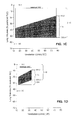
- FIGS. 4 and 5 illustrate these two patterns.
- median values are determined for a time equal to approximately one and one-half time the vertical grid line spacing.
- FIG. 4 illustrates a healthy, ⁇ DH, subject who exhibits a normal pattern of joint changes in MqdEELV and M % RC during exercise. This patient has a significant exercise capability as evidence by a heart rate increase from approximately 70 beats per minute (bpm) to over 170 bpm. During the exercise period, the MqdEELV is stable, while the M % RC increases a small amount, from approximately 40% to approximately 50%.
- This pattern reflects a normal exercise response in which minute ventilation is increased by increasing tidal volume using rib cage muscles and the diaphragm in approximately equal proportions.
- FIG. 5 illustrates a subject with an obstructive pulmonary disease (here, COPD).
- COPD obstructive pulmonary disease
- Table 1 further illustrates how the novel pattern of an increasing MqdEELV and a decreasing M % RC separates +DH from ⁇ DH patients.
- values of MqdEELV and M % RC are combined using a model developed according to statistical pattern recognition techniques for particular patient populations into a discriminant variables that clearly distinguish +DH from ⁇ DH in each population.
- these discriminant variables can be chosen so that amount of DH (in ml) correlates with the value of the variables so that both presence and amount of DH can be determined.
- Other embodiments use models developed by other than statistical techniques and can incorporate further variables (such as intensity of exercise or of other inciting cause).
- This invention can be practiced in many different patient monitoring environments as long as respiratory data is available from which at least moment-by-moment V T data and rib cage contribution to V T can be determined.
- this invention can be practiced in hospital, clinic, or laboratory environments and use data from respiratory sensors available in these environments.
- sensors include, e.g., spirometeric measuring arrangements, body plethysmography, and the like and are often less portable, can limit or prevent patient motion, but offer greater measurement accuracies.
- These environments also often provide, e.g., exercise treadmills and the like which can provide graded stimuli to precipitate DH.
- This invention can also be practiced in a patient's day-to-day environment while the patient is performing day-to-day activities (referred to herein as “ambulatory environments”).
- this invention usually processes data from respiratory sensors that are portable, light weight, non-invasive, and is arranged and configured so as not to limit patient motion or activity.
- respiratory sensors that are portable, light weight, non-invasive, and is arranged and configured so as not to limit patient motion or activity.
- respiratory sensors preferably respond to indicia of body sizes, such as lengths, circumferences, diameters, or geometrically similar measures of at least the rib cage and the abdominal and of their moment-by-moment changes during respiration.
- body sizes such as lengths, circumferences, diameters, or geometrically similar measures of at least the rib cage and the abdominal and of their moment-by-moment changes during respiration.
- moment-by-moment V T and rib cage contribution to V T can be determined.
- Such sensors at one or more additional torso or limb cross-sections can provide additional data responsive to cardiac or aortic pulsations, venous or arterial pulsations, and the like.
- Size sensors can be based on diverse technologies, including magnetometers; strain gauges using magnetic, mechanical or optical means; optical techniques including interferometry; electrical impedance; surface electrical or magnetic activity; body plethysmography, ultrasonic and doppler measurements of body wall motions or body diameters; and so forth.
- Preferred size sensors are based on inductive plethysmographic (IP) technology, which is responsive to anatomic sizes by measuring the self-inductance of one or more conductive elements (metallic or non-metallic) arranged on the anatomic portion to be measured.
- IP inductive plethysmographic
- IP sensor self-inductance varies as its size changes in response to an underlying body part; the varying self-inductance is sensed by variable frequency oscillator/demodulator modules; these modules output digital signals responsive to oscillator frequencies and ultimately to sensor size.
- Respiratory measurements obtained by IP technology are routinely within 5-7% (or 10 s of ml) of measurements obtained by spirometry, a current clinical standard.
- IP technology has been described in patent applications and issued patents assigned to the assignee of the present application including: U.S. Pat. Nos. 6,551,252; 6,413,225; 6,047,203; 6,341,504; 5,331,968; 5,301,678; 4,807,640; 4,373,534; and 4,834,209, and also U.S. application Ser. No. 10/822,260. All patents and published U.S. application cited herein are incorporated herein by reference in their entireties for all purposes.
- accelerometers mechanically coupled to a monitored patient can provide data reflecting activity level and posture; sensors for blood oxygen saturation can provide data reflecting any de-saturation accompanying DH.
- Other sensors can provide data reflecting skin conductance, electrical impedances, temperature, sensors; ultrasound, and the like.
- Respiratory and other sensor data is conveyed from the monitored patient to processing devices, or computers, or computer systems for processing and analysis by programmed implementations of this invention's methods.
- sensors can be linked directly to processing systems, e.g., by cable.
- a portable processing device or computer referred to as a “portable data unit” or “PDU” carried by a subject receive sensor data.
- the PDU also temporarily stores and/or transmits it to remote computers for analysis.
- data transmission should not limit a subject and can be by, e.g., wireless transmission, or physical transport of computer readable media, or the like.
- the PDU also perform the methods of this invention.
- respiratory and other sensors are preferably configured unobtrusively and comfortably on the patient so as not to substantially limit motion or activity.
- sensors can be configured into one or more wearable items, e.g., shirts, jackets, bands, patches, and the like.
- FIGS. 6A-C illustrate exemplary ambulatory monitoring systems having sensors configured into wearable items.
- FIG. 6A The subject of FIG. 6A is actively exercising unconstrained by concurrent monitoring with a single chest band 71 and PDU 73 configured as a wrist watch.
- the single band preferably incorporates a size sensor sensitive to respiration and can also incorporate accelerometers, ECG sensors, temperature sensors, and so forth.
- FIG. 6B illustrates shirt-like garment 75 having several types of sensors, including two (or more) size sensors 77 preferably sensitive to rib cage (RC) and abdomen (AB) sizes, two ECG leads, and optionally additional sensors (not illustrated).
- PDU 81 can displays data and accept user input.
- FIG. 6C illustrates a body-suit-like garment 83 equipped with a more extensive array of size sensors 85 for measuring respiration rate and volume, venous and arterial pulsations, cardiac pulsations, individual lung function, and the like.
- PDU 87 is attached to the garment and retrieves and wirelessly transmits sensor data to storage and analysis systems. Monitoring garments and systems are available from VivoMetrics, Inc., Ventura, Calif.
- FIG. 6D illustrates an exemplary analysis system including PC-type or workstation-type computer 91 with an attached monitor for viewing unprocessed and processing sensor data.
- Data is 89 conveyed to system 91 by, e.g., wireless connection, physical transfer, or wired connection.
- Local or remote online computer readable memory 93 and removable computer readable memories 95 e.g., optical ROM holds unprocessed and/or processed data and/or programs, and the like.
- the methods of this invention are generally performed on a computer or other processing device. Accordingly, these methods are programmed in a convenient computer language, such as assembly language, C, or C++, compiled into an executable form and stored on a computer readable medium for loading into a program memory of a computer or the configuration registers of a programmable device, or the like.
- FIG. 7 illustrates an exemplary implementation of these methods. These illustrated steps can be repeated on demand, or intermittently, or periodically to make multiple DH assessments.
- next step 103 measures and/or retrieves and/or inputs current monitoring data and optionally comparative measurement data.
- DH is additional hyperinflation acutely superimposed on chronic, baseline (BL) hyperinflation.
- Comparative data can include values or combinations of MqdEELV and M % RC during past bouts of DH, statistical distributions of multiple observations of MqdEELV and M % RC; the values of MqdEELV and M % RC resulting from specific precipitation factors, the amount of air retained, and the like.
- step 105 next extracts tidal volume (V T ) and the rib cage contribution to tidal volume from these raw methods accordingly to methods known for IP.
- step 107 perform implementations of the previously methods for finding M % RC and MqdEELV.
- step 109 assesses DH, its presence or absence and optionally its amount or volume, from the determined M % RC and MqdEELV parameters accordingly to the discrimination already described.
- the severity of DH can be estimated from the increases in lung volume or other measured in view of past values provided in the comparative data.
- Validation step 111 is optional but preferred to insure and/or improve the reliability of DH assessments.
- the M % RC and MqdEELV parameters are made from sufficiently long measurement periods, e.g., at least 30 sec, or at least 60 sec, or at least 120 sec. long or longer.
- DH assessment uses M % RC and MqdEELV values that are determined by statistically combining two or more independent measurements of these parameters.
- a final DH assessment is determined by statistically combining values from two or more independent episodes of DH precipitated by separate occurrences of a precipitating factor, e.g., exercise. Values can be combined using modes, medians, averages, and the like in order to statistically improve accuracies and limit errors. Prior DH episodes can be provided in the comparative data.
Abstract
Description
V T =α*AB+β*RC (1)
M % RC is a statistical median of % RC determined for breaths occurring during sequential 30 sec., or 1 min, or 2 min., or other periods. In other embodiments, VT can be alternately determined from a linear or non-linear function of AB and RC, and an alternate statistical measure, e.g., an average, can be used to represent values of % RC during sequential periods.
| TABLE 1 | |||||
| Patients | |||||
| without | Patients | ||||
| Parameter | DH on | with DH on | Difference | ||
| (base line - | exercise | exercise | (+DH | ||
| end exercise) | (−DH) | (+DH) | minus −DH) | ||
| MqdEELV | −81.36 | 66.50 | −147.86 | ||
| M % RC | 1.09 | −12.50 | 13.59 | ||
This table summarizes data from a study of fifteen patients, some with DH confirmed by standard measurement techniques and some without DH also as confirmed by standard techniques. Changes in MqdEELV and M % RC were measured for each patient. It is readily apparent that +DH patients exhibit the pattern of increasing MqdEELV and decreasing M % RC, while −DH patients exhibit other patterns.
Claims (18)
Priority Applications (1)
| Application Number | Priority Date | Filing Date | Title |
|---|---|---|---|
| US12/976,080 US8628480B2 (en) | 2005-05-20 | 2010-12-22 | Methods and systems for monitoring respiratory data |
Applications Claiming Priority (3)
| Application Number | Priority Date | Filing Date | Title |
|---|---|---|---|
| US68287605P | 2005-05-20 | 2005-05-20 | |
| US11/437,335 US7878979B2 (en) | 2005-05-20 | 2006-05-19 | Methods and systems for determining dynamic hyperinflation |
| US12/976,080 US8628480B2 (en) | 2005-05-20 | 2010-12-22 | Methods and systems for monitoring respiratory data |
Related Parent Applications (1)
| Application Number | Title | Priority Date | Filing Date |
|---|---|---|---|
| US11/437,335 Continuation US7878979B2 (en) | 2005-05-20 | 2006-05-19 | Methods and systems for determining dynamic hyperinflation |
Publications (2)
| Publication Number | Publication Date |
|---|---|
| US20110092795A1 US20110092795A1 (en) | 2011-04-21 |
| US8628480B2 true US8628480B2 (en) | 2014-01-14 |
Family
ID=37452682
Family Applications (2)
| Application Number | Title | Priority Date | Filing Date |
|---|---|---|---|
| US11/437,335 Active 2029-05-12 US7878979B2 (en) | 2005-05-20 | 2006-05-19 | Methods and systems for determining dynamic hyperinflation |
| US12/976,080 Active 2027-06-23 US8628480B2 (en) | 2005-05-20 | 2010-12-22 | Methods and systems for monitoring respiratory data |
Family Applications Before (1)
| Application Number | Title | Priority Date | Filing Date |
|---|---|---|---|
| US11/437,335 Active 2029-05-12 US7878979B2 (en) | 2005-05-20 | 2006-05-19 | Methods and systems for determining dynamic hyperinflation |
Country Status (6)
| Country | Link |
|---|---|
| US (2) | US7878979B2 (en) |
| EP (1) | EP1887933B1 (en) |
| JP (1) | JP5340727B2 (en) |
| AU (1) | AU2006251609B2 (en) |
| CA (1) | CA2606699C (en) |
| WO (1) | WO2006127573A2 (en) |
Cited By (14)
| Publication number | Priority date | Publication date | Assignee | Title |
|---|---|---|---|---|
| US20150148699A1 (en) * | 2013-11-26 | 2015-05-28 | Cardiac Pacemakers, Inc. | Detection of chronic obstructive pulmonary disease exacerbations from breathing patterns |
| US10141675B2 (en) | 2010-06-25 | 2018-11-27 | Nox Medical | Biometric belt connector |
| US10478065B2 (en) | 2004-06-18 | 2019-11-19 | Adidas Ag | Systems and methods for monitoring subjects in potential physiological distress |
| US10548497B2 (en) | 2009-05-15 | 2020-02-04 | Nox Medical | Systems and methods using flexible capacitive electrodes for measuring biosignals |
| US10588550B2 (en) | 2013-11-06 | 2020-03-17 | Nox Medical | Method, apparatus, and system for measuring respiratory effort |
| US10869619B2 (en) | 2016-08-19 | 2020-12-22 | Nox Medical | Method, apparatus, and system for measuring respiratory effort of a subject |
| US11273283B2 (en) | 2017-12-31 | 2022-03-15 | Neuroenhancement Lab, LLC | Method and apparatus for neuroenhancement to enhance emotional response |
| US11364361B2 (en) | 2018-04-20 | 2022-06-21 | Neuroenhancement Lab, LLC | System and method for inducing sleep by transplanting mental states |
| US11452839B2 (en) | 2018-09-14 | 2022-09-27 | Neuroenhancement Lab, LLC | System and method of improving sleep |
| US11602282B2 (en) | 2017-09-08 | 2023-03-14 | Nox Medical Ehf | System and method for non-invasively determining an internal component of respiratory effort |
| US11717686B2 (en) | 2017-12-04 | 2023-08-08 | Neuroenhancement Lab, LLC | Method and apparatus for neuroenhancement to facilitate learning and performance |
| US11723579B2 (en) | 2017-09-19 | 2023-08-15 | Neuroenhancement Lab, LLC | Method and apparatus for neuroenhancement |
| US11786694B2 (en) | 2019-05-24 | 2023-10-17 | NeuroLight, Inc. | Device, method, and app for facilitating sleep |
| US11896386B2 (en) | 2017-06-02 | 2024-02-13 | Nox Medical Ehf | Coherence-based method, apparatus, and system for identifying corresponding signals of a physiological study |
Families Citing this family (36)
| Publication number | Priority date | Publication date | Assignee | Title |
|---|---|---|---|---|
| AU5359901A (en) | 2000-04-17 | 2001-10-30 | Vivometrics Inc | Systems and methods for ambulatory monitoring of physiological signs |
| US20080082018A1 (en) * | 2003-04-10 | 2008-04-03 | Sackner Marvin A | Systems and methods for respiratory event detection |
| US9504410B2 (en) * | 2005-09-21 | 2016-11-29 | Adidas Ag | Band-like garment for physiological monitoring |
| WO2006113804A2 (en) * | 2005-04-20 | 2006-10-26 | Vivometrics, Inc. | Systems and methods for non-invasive physiological monitoring of non-human animals |
| CA2606699C (en) * | 2005-05-20 | 2017-04-18 | Vivometrics, Inc. | Methods and systems for determining dynamic hyperinflation |
| US8033996B2 (en) | 2005-07-26 | 2011-10-11 | Adidas Ag | Computer interfaces including physiologically guided avatars |
| US8762733B2 (en) * | 2006-01-30 | 2014-06-24 | Adidas Ag | System and method for identity confirmation using physiologic biometrics to determine a physiologic fingerprint |
| US8475387B2 (en) * | 2006-06-20 | 2013-07-02 | Adidas Ag | Automatic and ambulatory monitoring of congestive heart failure patients |
| US9833184B2 (en) * | 2006-10-27 | 2017-12-05 | Adidas Ag | Identification of emotional states using physiological responses |
| EP2375977B1 (en) * | 2009-01-14 | 2014-12-24 | St. Michael's Hospital | Detection of dynamic hyperinflation in spontaneously breathing mechanically ventilated patients |
| US9545222B2 (en) | 2009-09-01 | 2017-01-17 | Adidas Ag | Garment with noninvasive method and system for monitoring physiological characteristics and athletic performance |
| KR20110024205A (en) * | 2009-09-01 | 2011-03-09 | 한국전자통신연구원 | The non-intrusive wearable tidal volume measurement apparatus, system and method thereof |
| US9326705B2 (en) | 2009-09-01 | 2016-05-03 | Adidas Ag | Method and system for monitoring physiological and athletic performance characteristics of a subject |
| EP2517131A2 (en) * | 2009-12-21 | 2012-10-31 | Koninklijke Philips Electronics N.V. | Bode index measurement |
| WO2011117862A2 (en) * | 2010-03-24 | 2011-09-29 | Haim Melman | Wearable sensors |
| EP2397074B1 (en) * | 2010-06-19 | 2012-10-24 | M Stenqvist AB | A system and computer readable medium for determination of transpulmonary pressure in a patient connected to a breathing apparatus |
| JP6441572B2 (en) | 2011-01-25 | 2018-12-19 | アペリス・ホールディングス,エルエルシー | Apparatus and method for assisting breathing |
| US10363453B2 (en) | 2011-02-07 | 2019-07-30 | New Balance Athletics, Inc. | Systems and methods for monitoring athletic and physiological performance |
| CN106418870B (en) | 2011-02-07 | 2019-10-22 | 新平衡运动公司 | System and method for monitoring athletic performance |
| CN104768455B (en) | 2012-09-11 | 2018-01-02 | L.I.F.E.公司 | Wearable communications platform |
| US10462898B2 (en) | 2012-09-11 | 2019-10-29 | L.I.F.E. Corporation S.A. | Physiological monitoring garments |
| US11246213B2 (en) | 2012-09-11 | 2022-02-08 | L.I.F.E. Corporation S.A. | Physiological monitoring garments |
| US8948839B1 (en) | 2013-08-06 | 2015-02-03 | L.I.F.E. Corporation S.A. | Compression garments having stretchable and conductive ink |
| US8945328B2 (en) | 2012-09-11 | 2015-02-03 | L.I.F.E. Corporation S.A. | Methods of making garments having stretchable and conductive ink |
| US10159440B2 (en) | 2014-03-10 | 2018-12-25 | L.I.F.E. Corporation S.A. | Physiological monitoring garments |
| US9817440B2 (en) | 2012-09-11 | 2017-11-14 | L.I.F.E. Corporation S.A. | Garments having stretchable and conductive ink |
| US10201310B2 (en) | 2012-09-11 | 2019-02-12 | L.I.F.E. Corporation S.A. | Calibration packaging apparatuses for physiological monitoring garments |
| EP2931340B1 (en) | 2012-12-13 | 2020-08-12 | Koninklijke Philips N.V. | Handheld pressure support system for treating hyperinflation |
| EP3091864B8 (en) | 2014-01-06 | 2018-12-19 | L.I.F.E. Corporation S.A. | Systems and methods to automatically determine garment fit |
| NL2013551B1 (en) * | 2014-10-01 | 2016-10-03 | Medwear B V | Device and method for assessing respiratory data in a monitored subject. |
| EP3324831A1 (en) | 2015-07-20 | 2018-05-30 | L.I.F.E. Corporation S.A. | Flexible fabric ribbon connectors for garments with sensors and electronics |
| GB201519985D0 (en) * | 2015-11-12 | 2015-12-30 | Respinor As | Ultrasonic method and apparatus for respiration monitoring |
| JP2019524204A (en) | 2016-07-01 | 2019-09-05 | エル.アイ.エフ.イー. コーポレーション エス.エー.L.I.F.E. Corporation S.A. | Biometric identification by clothing with multiple sensors |
| JP6396981B2 (en) * | 2016-12-27 | 2018-09-26 | Ami株式会社 | Biological monitoring device |
| JP7296313B2 (en) | 2019-12-20 | 2023-06-22 | 株式会社豊田中央研究所 | Height distribution measuring device and height distribution measuring method |
| US11877863B2 (en) * | 2020-03-27 | 2024-01-23 | Inkwell Health Ltd. | Apparatus, systems, and methods for monitoring respiratory function |
Citations (230)
| Publication number | Priority date | Publication date | Assignee | Title |
|---|---|---|---|---|
| US3534727A (en) | 1967-03-24 | 1970-10-20 | Nasa | Biomedical electrode arrangement |
| US3731184A (en) | 1948-12-21 | 1973-05-01 | H Goldberg | Deformable pick up coil and cooperating magnet for measuring physical quantities, with means for rendering coil output independent of orientation |
| US3874368A (en) | 1973-04-19 | 1975-04-01 | Manfred Asrican | Impedance plethysmograph having blocking system |
| US3926177A (en) | 1972-09-11 | 1975-12-16 | Cavitron Corp | Activity and respiration monitor |
| US4016868A (en) | 1975-11-25 | 1977-04-12 | Allison Robert D | Garment for impedance plethysmograph use |
| US4033332A (en) | 1972-09-11 | 1977-07-05 | Cavitron Corporation | Activity and respiration monitor |
| US4102331A (en) | 1976-09-21 | 1978-07-25 | Datascope Corporation | Device for transmitting electrical energy |
| US4258718A (en) | 1979-04-16 | 1981-03-31 | Goldman Michael D | Measuring respiratory air volume |
| US4267845A (en) | 1978-10-05 | 1981-05-19 | Robertson Jr Charles H | Method and apparatus for measuring pulmonary ventilation |
| GB1596298A (en) | 1977-04-07 | 1981-08-26 | Morgan Ltd P K | Method of and apparatus for detecting or measuring changes in the cross-sectional area of a non-magnetic object |
| US4289142A (en) | 1978-11-24 | 1981-09-15 | Kearns Kenneth L | Physiological occurrence, such as apnea, monitor and X-ray triggering device |
| US4306567A (en) | 1977-12-22 | 1981-12-22 | Krasner Jerome L | Detection and monitoring device |
| US4373534A (en) | 1981-04-14 | 1983-02-15 | Respitrace Corporation | Method and apparatus for calibrating respiration monitoring system |
| US4387722A (en) | 1978-11-24 | 1983-06-14 | Kearns Kenneth L | Respiration monitor and x-ray triggering apparatus |
| US4433693A (en) | 1979-09-27 | 1984-02-28 | Hochstein Peter A | Method and assembly for monitoring respiration and detecting apnea |
| US4446872A (en) | 1977-09-08 | 1984-05-08 | Avl Ag | Method and apparatus for determining systolic time intervals |
| US4452252A (en) | 1981-05-26 | 1984-06-05 | Respitrace Corporation | Non-invasive method for monitoring cardiopulmonary parameters |
| US4456015A (en) | 1981-05-26 | 1984-06-26 | Respitrace Corporation | Non-invasive method for semiquantitative measurement of neck volume changes |
| US4463764A (en) | 1981-09-29 | 1984-08-07 | Medical Graphics Corporation | Cardiopulmonary exercise system |
| US4494553A (en) | 1981-04-01 | 1985-01-22 | F. William Carr | Vital signs monitor |
| GB2116725B (en) | 1982-03-15 | 1985-07-03 | Scotland The Secretary Of Stat | Respiration monitor |
| US4537196A (en) | 1981-12-21 | 1985-08-27 | American Home Products Corporation (Del.) | Systems and methods for processing physiological signals |
| US4545376A (en) | 1982-03-09 | 1985-10-08 | Werner Beiter | Blood lancet |
| US4546777A (en) | 1981-03-06 | 1985-10-15 | Siemens Gammasonics, Inc. | Heart sound detector and synchronization for diagnostics |
| US4548204A (en) | 1981-03-06 | 1985-10-22 | Siemens Gammasonics, Inc. | Apparatus for monitoring cardiac activity via ECG and heart sound signals |
| US4549552A (en) | 1981-03-06 | 1985-10-29 | Siemens Gammasonics, Inc. | Heart sound detector and cardiac cycle data are combined for diagnostic reliability |
| US4572197A (en) | 1982-07-01 | 1986-02-25 | The General Hospital Corporation | Body hugging instrumentation vest having radioactive emission detection for ejection fraction |
| US4580572A (en) | 1983-06-01 | 1986-04-08 | Bio-Stimu Trend Corp. | Garment apparatus for delivering or receiving electric impulses |
| US4648407A (en) | 1985-07-08 | 1987-03-10 | Respitrace Corporation | Method for detecting and differentiating central and obstructive apneas in newborns |
| US4672975A (en) | 1985-06-17 | 1987-06-16 | Vladimir Sirota | Stethoscope with image of periodically expanding and contracting heart |
| US4753088A (en) | 1986-10-14 | 1988-06-28 | Collins & Aikman Corporation | Mesh knit fabrics having electrically conductive filaments for use in manufacture of anti-static garments and accessories |
| US4777962A (en) | 1986-05-09 | 1988-10-18 | Respitrace Corporation | Method and apparatus for distinguishing central obstructive and mixed apneas by external monitoring devices which measure rib cage and abdominal compartmental excursions during respiration |
| US4796639A (en) | 1987-11-05 | 1989-01-10 | Medical Graphics Corporation | Pulmonary diagnostic system |
| US4800495A (en) | 1986-08-18 | 1989-01-24 | Physio-Control Corporation | Method and apparatus for processing signals used in oximetry |
| US4807640A (en) | 1986-11-19 | 1989-02-28 | Respitrace Corporation | Stretchable band-type transducer particularly suited for respiration monitoring apparatus |
| US4817625A (en) | 1987-04-24 | 1989-04-04 | Laughton Miles | Self-inductance sensor |
| US4819752A (en) | 1987-10-02 | 1989-04-11 | Datascope Corp. | Blood constituent measuring device and method |
| US4834109A (en) | 1986-01-21 | 1989-05-30 | Respitrace Corporation | Single position non-invasive calibration technique |
| US4860766A (en) | 1983-11-18 | 1989-08-29 | Respitrace Corp. | Noninvasive method for measuring and monitoring intrapleural pressure in newborns |
| US4863265A (en) | 1987-10-16 | 1989-09-05 | Mine Safety Appliances Company | Apparatus and method for measuring blood constituents |
| US4867571A (en) | 1986-09-26 | 1989-09-19 | Sensormedics Corporation | Wave form filter pulse detector and method for modulated signal |
| US4889131A (en) | 1987-12-03 | 1989-12-26 | American Health Products, Inc. | Portable belt monitor of physiological functions and sensors therefor |
| US4909260A (en) | 1987-12-03 | 1990-03-20 | American Health Products, Inc. | Portable belt monitor of physiological functions and sensors therefor |
| US4911167A (en) | 1985-06-07 | 1990-03-27 | Nellcor Incorporated | Method and apparatus for detecting optical pulses |
| US4920969A (en) | 1985-10-08 | 1990-05-01 | Capintec, Inc. | Ambulatory physiological evaluation system including cardiac monitoring |
| US4928692A (en) | 1985-04-01 | 1990-05-29 | Goodman David E | Method and apparatus for detecting optical pulses |
| US4934372A (en) | 1985-04-01 | 1990-06-19 | Nellcor Incorporated | Method and apparatus for detecting optical pulses |
| US4955379A (en) | 1987-08-14 | 1990-09-11 | National Research Development Corporation | Motion artefact rejection system for pulse oximeters |
| US4960118A (en) | 1989-05-01 | 1990-10-02 | Pennock Bernard E | Method and apparatus for measuring respiratory flow |
| US4966155A (en) | 1985-01-31 | 1990-10-30 | The University Of Strathclyde | Apparatus for monitoring physiological parameters |
| US4972842A (en) | 1988-06-09 | 1990-11-27 | Vital Signals, Inc. | Method and apparatus for precision monitoring of infants on assisted ventilation |
| US4981139A (en) | 1983-08-11 | 1991-01-01 | Pfohl Robert L | Vital signs monitoring and communication system |
| US4986277A (en) | 1988-08-24 | 1991-01-22 | Sackner Marvin A | Method and apparatus for non-invasive monitoring of central venous pressure |
| US5007427A (en) | 1987-05-07 | 1991-04-16 | Capintec, Inc. | Ambulatory physiological evaluation system including cardiac monitoring |
| US5025791A (en) | 1989-06-28 | 1991-06-25 | Colin Electronics Co., Ltd. | Pulse oximeter with physical motion sensor |
| US5036857A (en) | 1989-10-26 | 1991-08-06 | Rutgers, The State University Of New Jersey | Noninvasive diagnostic system for coronary artery disease |
| US5040540A (en) | 1988-08-24 | 1991-08-20 | Nims, Inc. | Method and apparatus for non-invasive monitoring of central venous pressure, and improved transducer therefor |
| US5074129A (en) | 1989-12-26 | 1991-12-24 | Novtex | Formable fabric |
| US5076801A (en) | 1990-06-22 | 1991-12-31 | Xerox Corporation | Electronic component including insulation displacement interconnect means |
| US5099841A (en) | 1989-02-06 | 1992-03-31 | Instrumentarium Corporation | Measurement of the composition of blood |
| US5099855A (en) | 1989-11-09 | 1992-03-31 | State Of Oregon, Acting By And Through The Oregon State Board Of Higher Education, Acting For And On Behalf Of The Oregon Health Sciences University | Methods of and apparatus for monitoring respiration and conductive gel used therewith |
| US5111817A (en) | 1988-12-29 | 1992-05-12 | Medical Physics, Inc. | Noninvasive system and method for enhanced arterial oxygen saturation determination and arterial blood pressure monitoring |
| US5131399A (en) | 1990-08-06 | 1992-07-21 | Sciarra Michael J | Patient monitoring apparatus and method |
| US5143089A (en) | 1989-05-03 | 1992-09-01 | Eckhard Alt | Assembly and method of communicating electrical signals between electrical therapeutic systems and body tissue |
| US5159935A (en) | 1990-03-08 | 1992-11-03 | Nims, Inc. | Non-invasive estimation of individual lung function |
| US5173151A (en) | 1990-07-30 | 1992-12-22 | Seiko Epson Corporation | Method of dry etching in semiconductor device processing |
| US5178151A (en) | 1988-04-20 | 1993-01-12 | Sackner Marvin A | System for non-invasive detection of changes of cardiac volumes and aortic pulses |
| US5224479A (en) | 1991-06-21 | 1993-07-06 | Topy Enterprises Limited | ECG diagnostic pad |
| US5241300A (en) | 1992-04-24 | 1993-08-31 | Johannes Buschmann | SIDS detection apparatus and methods |
| DE4214263A1 (en) | 1992-05-03 | 1993-11-04 | Maximilian Dr Moser | Garment has sensors coming into contact with body parts - where values received by sensors are led to central collector |
| US5271551A (en) | 1988-11-18 | 1993-12-21 | Gustav Roepke | Container |
| US5295490A (en) | 1993-01-21 | 1994-03-22 | Dodakian Wayne S | Self-contained apnea monitor |
| US5299120A (en) | 1989-09-15 | 1994-03-29 | Hewlett-Packard Company | Method for digitally processing signals containing information regarding arterial blood flow |
| US5301678A (en) | 1986-11-19 | 1994-04-12 | Non-Invasive Monitoring System, Inc. | Stretchable band - type transducer particularly suited for use with respiration monitoring apparatus |
| US5333106A (en) | 1992-10-09 | 1994-07-26 | Circadian, Inc. | Apparatus and visual display method for training in the power use of aerosol pharmaceutical inhalers |
| US5331968A (en) | 1990-10-19 | 1994-07-26 | Gerald Williams | Inductive plethysmographic transducers and electronic circuitry therefor |
| US5348008A (en) | 1991-11-25 | 1994-09-20 | Somnus Corporation | Cardiorespiratory alert system |
| US5416961A (en) | 1994-01-26 | 1995-05-23 | Schlegel Corporation | Knitted wire carrier having bonded warp threads and method for forming same |
| US5447164A (en) | 1993-11-08 | 1995-09-05 | Hewlett-Packard Company | Interactive medical information display system and method for displaying user-definable patient events |
| USRE35122E (en) | 1985-04-01 | 1995-12-19 | Nellcor Incorporated | Method and apparatus for detecting optical pulses |
| US5520192A (en) | 1991-12-23 | 1996-05-28 | Imperial College Of Science, Technology And Medicine | Apparatus for the monitoring and control of respiration |
| US5533511A (en) | 1994-01-05 | 1996-07-09 | Vital Insite, Incorporated | Apparatus and method for noninvasive blood pressure measurement |
| US5535738A (en) | 1994-06-03 | 1996-07-16 | Respironics, Inc. | Method and apparatus for providing proportional positive airway pressure to treat sleep disordered breathing |
| US5544661A (en) | 1994-01-13 | 1996-08-13 | Charles L. Davis | Real time ambulatory patient monitor |
| US5577510A (en) | 1995-08-18 | 1996-11-26 | Chittum; William R. | Portable and programmable biofeedback system with switching circuit for voice-message recording and playback |
| US5582337A (en) | 1995-06-20 | 1996-12-10 | Mcpherson; Mathew A. | Strap system for carrying skates and shoes and method of use |
| US5584295A (en) | 1995-09-01 | 1996-12-17 | Analogic Corporation | System for measuring the period of a quasi-periodic signal |
| US5588425A (en) | 1993-05-21 | 1996-12-31 | Nims, Incorporated | Method and apparatus for discriminating between valid and artifactual pulse waveforms in pulse oximetry |
| US5601088A (en) | 1995-02-17 | 1997-02-11 | Ep Technologies, Inc. | Systems and methods for filtering artifacts from composite signals |
| US5611085A (en) | 1992-11-02 | 1997-03-18 | Rasmussen; Verner | Garment for holding an electrocardiographic monitoring unit and cables |
| US5617847A (en) | 1995-10-12 | 1997-04-08 | Howe; Stephen L. | Assisted breathing apparatus and tubing therefore |
| US5694939A (en) | 1995-10-03 | 1997-12-09 | The United States Of America As Represented By The Administrator Of The National Aeronautics And Space Administration | Autogenic-feedback training exercise (AFTE) method and system |
| US5718234A (en) | 1996-09-30 | 1998-02-17 | Northrop Grumman Corporation | Physiological data communication system |
| US5719950A (en) | 1994-03-24 | 1998-02-17 | Minnesota Mining And Manufacturing Company | Biometric, personal authentication system |
| US5720709A (en) | 1995-10-25 | 1998-02-24 | S.M.C. Sleep Medicine Center | Apparatus and method for measuring respiratory airway resistance and airway collapsibility in patients |
| US5724025A (en) | 1993-10-21 | 1998-03-03 | Tavori; Itzchak | Portable vital signs monitor |
| US5749365A (en) | 1991-11-07 | 1998-05-12 | Magill; Alan | Health monitoring |
| US5820567A (en) | 1995-11-02 | 1998-10-13 | Healthcare Technology Limited | Heart rate sensing apparatus adapted for chest or earlobe mounted sensor |
| US5825293A (en) | 1996-09-20 | 1998-10-20 | Ahmed; Adel A. | Apparatus and method for monitoring breathing magnetically |
| US5848027A (en) | 1995-11-22 | 1998-12-08 | Biometrics, Inc. | Method for processing personal data |
| US5882307A (en) | 1994-08-05 | 1999-03-16 | Acuson Corporation | Method and apparatus for receive beamformer system |
| US5899855A (en) | 1992-11-17 | 1999-05-04 | Health Hero Network, Inc. | Modular microprocessor-based health monitoring system |
| US5913830A (en) | 1997-08-20 | 1999-06-22 | Respironics, Inc. | Respiratory inductive plethysmography sensor |
| US5921920A (en) | 1996-12-12 | 1999-07-13 | The Trustees Of The University Of Pennsylvania | Intensive care information graphical display |
| US5937854A (en) | 1998-01-06 | 1999-08-17 | Sensormedics Corporation | Ventilator pressure optimization method and apparatus |
| US5989193A (en) | 1995-05-19 | 1999-11-23 | Somed Pty Limited | Device and method for detecting and recording snoring |
| US5991922A (en) | 1996-12-26 | 1999-11-30 | Banks; David L. | Monitored static electricity dissipation garment |
| US6002952A (en) | 1997-04-14 | 1999-12-14 | Masimo Corporation | Signal processing apparatus and method |
| US6015388A (en) | 1997-03-17 | 2000-01-18 | Nims, Inc. | Method for analyzing breath waveforms as to their neuromuscular respiratory implications |
| US6018677A (en) | 1997-11-25 | 2000-01-25 | Tectrix Fitness Equipment, Inc. | Heart rate monitor and method |
| US6035154A (en) | 1997-11-28 | 2000-03-07 | Seiko Epson Corporation | Image forming apparatus |
| US6047203A (en) | 1997-03-17 | 2000-04-04 | Nims, Inc. | Physiologic signs feedback system |
| US6066093A (en) | 1995-07-28 | 2000-05-23 | Unilead International Inc. | Disposable electrodeless electro-dermal devices |
| US6068568A (en) | 1996-12-12 | 2000-05-30 | Tsubakimoto Chain Co. | Silent chain |
| US6070098A (en) | 1997-01-11 | 2000-05-30 | Circadian Technologies, Inc. | Method of and apparatus for evaluation and mitigation of microsleep events |
| US6120441A (en) | 1995-10-16 | 2000-09-19 | Map Medizintechnik Fur Arzt Und Patient Gmbh | Method and device for quantitative analysis of sleep disturbances |
| US6142953A (en) | 1999-07-08 | 2000-11-07 | Compumedics Sleep Pty Ltd | Respiratory inductive plethysmography band transducer |
| US6145551A (en) | 1997-09-22 | 2000-11-14 | Georgia Tech Research Corp. | Full-fashioned weaving process for production of a woven garment with intelligence capability |
| US6179786B1 (en) | 1998-10-02 | 2001-01-30 | Profemme Ltd. | System for thermometry-based breast cancer risk-assessment |
| US6198394B1 (en) | 1996-12-05 | 2001-03-06 | Stephen C. Jacobsen | System for remote monitoring of personnel |
| JP2001104259A (en) | 1999-08-27 | 2001-04-17 | Laerdal Medical As | System for reducing outer disturbance of signal in electrocardiogram induced by cardiopulmonary resuscitation |
| US6223072B1 (en) | 1999-06-08 | 2001-04-24 | Impulse Dynamics N.V. | Apparatus and method for collecting data useful for determining the parameters of an alert window for timing delivery of ETC signals to a heart under varying cardiac conditions |
| US6254551B1 (en) | 1997-02-05 | 2001-07-03 | Instrumentarium Corp. | Apparatus for monitoring a mechanically transmitted signal based on the organs or vital functions and for processing the results |
| US6261238B1 (en) | 1996-10-04 | 2001-07-17 | Karmel Medical Acoustic Technologies, Ltd. | Phonopneumograph system |
| US6273859B1 (en) | 1995-11-01 | 2001-08-14 | University Technologies International, Inc. | Adaptively controlled mandibular positioning device and method of using the device |
| US6287264B1 (en) | 1999-04-23 | 2001-09-11 | The Trustees Of Tufts College | System for measuring respiratory function |
| US6302844B1 (en) | 1999-03-31 | 2001-10-16 | Walker Digital, Llc | Patient care delivery system |
| US6306088B1 (en) | 1998-10-03 | 2001-10-23 | Individual Monitoring Systems, Inc. | Ambulatory distributed recorders system for diagnosing medical disorders |
| US6341504B1 (en) | 2001-01-31 | 2002-01-29 | Vivometrics, Inc. | Composite elastic and wire fabric for physiological monitoring apparel |
| US20020032386A1 (en) | 2000-04-17 | 2002-03-14 | Sackner Marvin A. | Systems and methods for ambulatory monitoring of physiological signs |
| US6361501B1 (en) | 1997-08-26 | 2002-03-26 | Seiko Epson Corporation | Pulse wave diagnosing device |
| US6381482B1 (en) | 1998-05-13 | 2002-04-30 | Georgia Tech Research Corp. | Fabric or garment with integrated flexible information infrastructure |
| US6413225B1 (en) * | 1999-06-18 | 2002-07-02 | Vivometrics, Inc. | Quantitative calibration of breathing monitors with transducers placed on both rib cage and abdomen |
| US20020084130A1 (en) | 2000-04-12 | 2002-07-04 | Viken Der Ghazarian | Breathalyzer with voice recognition |
| US20020090667A1 (en) | 2000-07-14 | 2002-07-11 | Hypoguard Limited | Detection of Helicobacter Pylori |
| US6419636B1 (en) | 1998-10-02 | 2002-07-16 | David Ernest Young | System for thermometry-based breast assessment including cancer risk |
| US6436057B1 (en) | 1999-04-22 | 2002-08-20 | The United States Of America As Represented By The Department Of Health And Human Services, Centers For Disease Control And Prevention | Method and apparatus for cough sound analysis |
| US6443890B1 (en) | 2000-03-01 | 2002-09-03 | I-Medik, Inc. | Wireless internet bio-telemetry monitoring system |
| US20020123701A1 (en) | 1998-10-30 | 2002-09-05 | Volusense As | Volumetric physiological measuring system and method |
| US6449504B1 (en) | 1999-08-20 | 2002-09-10 | Cardiac Pacemakers, Inc. | Arrhythmia display |
| US6454719B1 (en) | 2000-08-01 | 2002-09-24 | Pacesetter, Inc. | Apparatus and method for diagnosis of cardiac disease using a respiration monitor |
| US6461307B1 (en) | 2000-09-13 | 2002-10-08 | Flaga Hf | Disposable sensor for measuring respiration |
| US6463385B1 (en) | 1996-11-01 | 2002-10-08 | William R. Fry | Sports computer with GPS receiver and performance tracking capabilities |
| US6478736B1 (en) | 1999-10-08 | 2002-11-12 | Healthetech, Inc. | Integrated calorie management system |
| US6483929B1 (en) | 2000-06-08 | 2002-11-19 | Tarian Llc | Method and apparatus for histological and physiological biometric operation and authentication |
| US6485431B1 (en) | 1999-11-24 | 2002-11-26 | Duncan Campbell Patents Pty. Ltd. | Method and apparatus for determining cardiac output or total peripheral resistance |
| US6506153B1 (en) | 1998-09-02 | 2003-01-14 | Med-Dev Limited | Method and apparatus for subject monitoring |
| US6513532B2 (en) | 2000-01-19 | 2003-02-04 | Healthetech, Inc. | Diet and activity-monitoring device |
| US20030100843A1 (en) | 1999-04-23 | 2003-05-29 | The Trustees Of Tufts College | System for measuring respiratory function |
| US6579231B1 (en) | 1998-03-27 | 2003-06-17 | Mci Communications Corporation | Personal medical monitoring unit and system |
| US20030135097A1 (en) | 2001-06-25 | 2003-07-17 | Science Applications International Corporation | Identification by analysis of physiometric variation |
| US6604115B1 (en) | 1999-11-05 | 2003-08-05 | Ge Marquette Medical Systems, Inc. | Method and apparatus for storing data |
| US20030185408A1 (en) | 2002-03-29 | 2003-10-02 | Elvir Causevic | Fast wavelet estimation of weak bio-signals using novel algorithms for generating multiple additional data frames |
| US20030187341A1 (en) | 2002-03-26 | 2003-10-02 | Sackner Marvin A. | Method and system for extracting cardiac parameters from plethysmographic signals |
| US6633772B2 (en) | 2000-08-18 | 2003-10-14 | Cygnus, Inc. | Formulation and manipulation of databases of analyte and associated values |
| US6647252B2 (en) | 2002-01-18 | 2003-11-11 | General Instrument Corporation | Adaptive threshold algorithm for real-time wavelet de-noising applications |
| US6656127B1 (en) | 1999-06-08 | 2003-12-02 | Oridion Breathid Ltd. | Breath test apparatus and methods |
| US20040010420A1 (en) | 2001-08-30 | 2004-01-15 | Rooks Daniel S | System for developing implementing and monitoring a health management program |
| US20040019289A1 (en) | 2002-03-01 | 2004-01-29 | Christine Ross | Novel utilization of heart rate variability in animals |
| US6687523B1 (en) | 1997-09-22 | 2004-02-03 | Georgia Tech Research Corp. | Fabric or garment with integrated flexible information infrastructure for monitoring vital signs of infants |
| US20040030224A1 (en) | 2002-08-07 | 2004-02-12 | Apneos Corporation | Service center system and method as a component of a population diagnostic for sleep disorders |
| US6702752B2 (en) | 2002-02-22 | 2004-03-09 | Datex-Ohmeda, Inc. | Monitoring respiration based on plethysmographic heart rate signal |
| EP0875199B1 (en) | 1996-09-10 | 2004-03-10 | Seiko Epson Corporation | Organism state measuring device and relaxation state indicator device |
| US6709402B2 (en) | 2002-02-22 | 2004-03-23 | Datex-Ohmeda, Inc. | Apparatus and method for monitoring respiration with a pulse oximeter |
| US6721594B2 (en) | 1999-08-24 | 2004-04-13 | Cardiac Pacemakers, Inc. | Arrythmia display |
| US6723055B2 (en) | 1999-04-23 | 2004-04-20 | Trustees Of Tufts College | System for measuring respiratory function |
| US6727197B1 (en) | 1999-11-18 | 2004-04-27 | Foster-Miller, Inc. | Wearable transmission device |
| US6747561B1 (en) | 2000-06-20 | 2004-06-08 | Med-Datanet, Llc | Bodily worn device for digital storage and retrieval of medical records and personal identification |
| US20040111040A1 (en) | 2002-12-04 | 2004-06-10 | Quan Ni | Detection of disordered breathing |
| US20040117204A1 (en) | 2002-12-17 | 2004-06-17 | Cardiac Pacemakers, Inc. | Repeater device for communications with an implantable medical device |
| US20040122334A1 (en) | 2002-09-19 | 2004-06-24 | Alfred E. Mann Institute For Biomedical Engineering At The University Of Southern | Vantilation and volume change measurements using permanent magnet and magnet sensor affixed to body |
| US20040143194A1 (en) | 2001-03-02 | 2004-07-22 | Norio Kihara | Respiratory function measuring system and application thereof |
| US6775389B2 (en) | 2001-08-10 | 2004-08-10 | Advanced Bionics Corporation | Ear auxiliary microphone for behind the ear hearing prosthetic |
| US6801916B2 (en) | 1998-04-01 | 2004-10-05 | Cyberpulse, L.L.C. | Method and system for generation of medical reports from data in a hierarchically-organized database |
| US20040204636A1 (en) | 1991-03-07 | 2004-10-14 | Diab Mohamed K. | Signal processing apparatus |
| US20040210147A1 (en) | 2003-04-16 | 2004-10-21 | Houben Richard P.M. | Biomedical signal analysis techniques using wavelets |
| US20040225227A1 (en) | 2001-06-22 | 2004-11-11 | Newman Richard Gerard | Asymmetric inductive band |
| US6817979B2 (en) | 2002-06-28 | 2004-11-16 | Nokia Corporation | System and method for interacting with a user's virtual physiological model via a mobile terminal |
| US20040249299A1 (en) | 2003-06-06 | 2004-12-09 | Cobb Jeffrey Lane | Methods and systems for analysis of physiological signals |
| US20050027207A1 (en) | 2000-12-29 | 2005-02-03 | Westbrook Philip R. | Sleep apnea risk evaluation |
| US6858006B2 (en) | 2000-09-08 | 2005-02-22 | Wireless Medical, Inc. | Cardiopulmonary monitoring |
| US20050054941A1 (en) | 2003-08-22 | 2005-03-10 | Joseph Ting | Physiological monitoring garment |
| US20050076908A1 (en) | 2003-09-18 | 2005-04-14 | Kent Lee | Autonomic arousal detection system and method |
| US6881192B1 (en) | 2002-06-12 | 2005-04-19 | Pacesetter, Inc. | Measurement of sleep apnea duration and evaluation of response therapies using duration metrics |
| US20050119586A1 (en) | 2003-04-10 | 2005-06-02 | Vivometrics, Inc. | Systems and methods for respiratory event detection |
| US20050125970A1 (en) | 2003-12-16 | 2005-06-16 | Graco Children's Products Inc. | Buckle assembly |
| US6941775B2 (en) | 2002-04-05 | 2005-09-13 | Electronic Textile, Inc. | Tubular knit fabric and system |
| US20050211247A1 (en) | 2004-03-25 | 2005-09-29 | Sanyo Electric Co., Ltd. | Method of and device for snore detection |
| US20050228234A1 (en) | 2002-05-17 | 2005-10-13 | Chang-Ming Yang | Method and device for monitoring physiologic signs and implementing emergency disposals |
| US20050240087A1 (en) | 2003-11-18 | 2005-10-27 | Vivometrics Inc. | Method and system for processing data from ambulatory physiological monitoring |
| US6961448B2 (en) | 1999-12-30 | 2005-11-01 | Medtronic, Inc. | User authentication in medical device systems |
| US6970731B1 (en) | 1998-09-21 | 2005-11-29 | Georgia Tech Research Corp. | Fabric-based sensor for monitoring vital signs |
| US20060000420A1 (en) | 2004-05-24 | 2006-01-05 | Martin Davies Michael A | Animal instrumentation |
| US7001337B2 (en) | 2002-02-22 | 2006-02-21 | Datex-Ohmeda, Inc. | Monitoring physiological parameters based on variations in a photoplethysmographic signal |
| US20060074334A1 (en) | 2004-06-24 | 2006-04-06 | Michael Coyle | Systems and methods for monitoring cough |
| US20060122528A1 (en) | 2004-09-21 | 2006-06-08 | Yoav Gal | Sensors for inductive plethysmographic monitoring applications and apparel using same |
| US7073129B1 (en) | 1998-12-18 | 2006-07-04 | Tangis Corporation | Automated selection of appropriate information based on a computer user's context |
| US7077810B2 (en) | 2004-02-05 | 2006-07-18 | Earlysense Ltd. | Techniques for prediction and monitoring of respiration-manifested clinical episodes |
| US7081095B2 (en) | 2001-05-17 | 2006-07-25 | Lynn Lawrence A | Centralized hospital monitoring system for automatically detecting upper airway instability and for preventing and aborting adverse drug reactions |
| US20060178591A1 (en) | 2004-11-19 | 2006-08-10 | Hempfling Ralf H | Methods and systems for real time breath rate determination with limited processor resources |
| US7099714B2 (en) | 2003-03-31 | 2006-08-29 | Medtronic, Inc. | Biomedical signal denoising techniques |
| US7104962B2 (en) | 2003-06-24 | 2006-09-12 | Buxco Electronics, Inc. | Cough/sneeze analyzer and method |
| US20060258914A1 (en) | 2005-04-20 | 2006-11-16 | Derchak P A | Systems and methods for non-invasive physiological monitoring of non-human animals |
| US7154398B2 (en) | 2003-01-06 | 2006-12-26 | Chen Thomas C H | Wireless communication and global location enabled intelligent health monitoring system |
| US20060293609A1 (en) | 2005-05-11 | 2006-12-28 | Cardiac Pacemakers, Inc. | Sensitivity and specificity of pulmonary edema detection when using transthoracic impedance |
| US20070027368A1 (en) | 2005-07-14 | 2007-02-01 | Collins John P | 3D anatomical visualization of physiological signals for online monitoring |
| US20070050715A1 (en) | 2005-07-26 | 2007-03-01 | Vivometrics, Inc. | Computer interfaces including physiologically guided avatars |
| US20070100622A1 (en) | 2005-10-31 | 2007-05-03 | Hitachi, Ltd. | Adaptation method for inter-person biometrics variability |
| US20070150006A1 (en) | 2005-12-28 | 2007-06-28 | Imad Libbus | Neural stimulator to treat sleep disordered breathing |
| US20070177770A1 (en) | 2006-01-30 | 2007-08-02 | Derchak P A | System and method for identity confirmation using physiologic biometrics to determine a physiologic fingerprint |
| US7254516B2 (en) | 2004-12-17 | 2007-08-07 | Nike, Inc. | Multi-sensor monitoring of athletic performance |
| US20070208262A1 (en) | 2006-03-03 | 2007-09-06 | Kovacs Gregory T | Dual-mode physiologic monitoring systems and methods |
| US20070209669A1 (en) | 2006-03-09 | 2007-09-13 | Derchak P Alexander | Monitoring and quantification of smoking behaviors |
| US20070270671A1 (en) | 2006-04-10 | 2007-11-22 | Vivometrics, Inc. | Physiological signal processing devices and associated processing methods |
| US7319385B2 (en) | 2004-09-17 | 2008-01-15 | Nokia Corporation | Sensor data sharing |
| US20080015454A1 (en) | 2005-09-21 | 2008-01-17 | Yoav Gal | Band-like garment for physiological monitoring |
| US20080027341A1 (en) | 2002-03-26 | 2008-01-31 | Marvin Sackner | Method and system for extracting cardiac parameters from plethysmographic signals |
| US20080045815A1 (en) | 2006-06-20 | 2008-02-21 | Derchak P A | Automatic and ambulatory monitoring of congestive heart failure patients |
| US20080051839A1 (en) | 2006-08-25 | 2008-02-28 | Imad Libbus | System for abating neural stimulation side effects |
| US20080082018A1 (en) | 2003-04-10 | 2008-04-03 | Sackner Marvin A | Systems and methods for respiratory event detection |
| US20080221401A1 (en) | 2006-10-27 | 2008-09-11 | Derchak P Alexander | Identification of emotional states using physiological responses |
| US20080269644A1 (en) | 2007-04-26 | 2008-10-30 | Ray Gregory C | Precision Athletic Aptitude and Performance Data Analysis System |
| US20090131759A1 (en) | 2003-11-04 | 2009-05-21 | Nathaniel Sims | Life sign detection and health state assessment system |
| WO2009074973A1 (en) | 2007-12-13 | 2009-06-18 | Vincze Jozsef | Arrangement and method for respiration detection and/or measurement |
| WO2010027515A1 (en) | 2008-09-05 | 2010-03-11 | Vivometrics, Inc. | Noninvasive method and system for measuring pulmonary ventilation |
| US7727161B2 (en) | 2003-04-10 | 2010-06-01 | Vivometrics, Inc. | Systems and methods for monitoring cough |
| US7762953B2 (en) | 2005-04-20 | 2010-07-27 | Adidas Ag | Systems and methods for non-invasive physiological monitoring of non-human animals |
| US7809433B2 (en) | 2005-08-09 | 2010-10-05 | Adidas Ag | Method and system for limiting interference in electroencephalographic signals |
| US20100274100A1 (en) | 2004-06-18 | 2010-10-28 | Andrew Behar | Systems and methods for monitoring subjects in potential physiological distress |
| US7878979B2 (en) * | 2005-05-20 | 2011-02-01 | Adidas Ag | Methods and systems for determining dynamic hyperinflation |
Family Cites Families (2)
| Publication number | Priority date | Publication date | Assignee | Title |
|---|---|---|---|---|
| US4753988A (en) | 1987-02-18 | 1988-06-28 | The Dow Chemical Company | High gloss acrylate rubber-modified weatherable resins |
| US6254552B1 (en) | 1997-10-03 | 2001-07-03 | E.I. Du Pont De Nemours And Company | Intra-coronary radiation devices containing Ce-144 or Ru-106 |
-
2006
- 2006-05-19 CA CA2606699A patent/CA2606699C/en active Active
- 2006-05-19 WO PCT/US2006/019683 patent/WO2006127573A2/en active Application Filing
- 2006-05-19 AU AU2006251609A patent/AU2006251609B2/en not_active Ceased
- 2006-05-19 JP JP2008512586A patent/JP5340727B2/en not_active Expired - Fee Related
- 2006-05-19 US US11/437,335 patent/US7878979B2/en active Active
- 2006-05-19 EP EP06784447.2A patent/EP1887933B1/en active Active
-
2010
- 2010-12-22 US US12/976,080 patent/US8628480B2/en active Active
Patent Citations (254)
| Publication number | Priority date | Publication date | Assignee | Title |
|---|---|---|---|---|
| US3731184A (en) | 1948-12-21 | 1973-05-01 | H Goldberg | Deformable pick up coil and cooperating magnet for measuring physical quantities, with means for rendering coil output independent of orientation |
| US3534727A (en) | 1967-03-24 | 1970-10-20 | Nasa | Biomedical electrode arrangement |
| US3926177A (en) | 1972-09-11 | 1975-12-16 | Cavitron Corp | Activity and respiration monitor |
| US4033332A (en) | 1972-09-11 | 1977-07-05 | Cavitron Corporation | Activity and respiration monitor |
| US3874368A (en) | 1973-04-19 | 1975-04-01 | Manfred Asrican | Impedance plethysmograph having blocking system |
| US4016868A (en) | 1975-11-25 | 1977-04-12 | Allison Robert D | Garment for impedance plethysmograph use |
| US4102331A (en) | 1976-09-21 | 1978-07-25 | Datascope Corporation | Device for transmitting electrical energy |
| US4308872A (en) | 1977-04-07 | 1982-01-05 | Respitrace Corporation | Method and apparatus for monitoring respiration |
| GB1596298A (en) | 1977-04-07 | 1981-08-26 | Morgan Ltd P K | Method of and apparatus for detecting or measuring changes in the cross-sectional area of a non-magnetic object |
| US4815473A (en) | 1977-04-07 | 1989-03-28 | Respitrace Corporation | Method and apparatus for monitoring respiration |
| US4446872A (en) | 1977-09-08 | 1984-05-08 | Avl Ag | Method and apparatus for determining systolic time intervals |
| US4306567A (en) | 1977-12-22 | 1981-12-22 | Krasner Jerome L | Detection and monitoring device |
| US4267845A (en) | 1978-10-05 | 1981-05-19 | Robertson Jr Charles H | Method and apparatus for measuring pulmonary ventilation |
| US4289142A (en) | 1978-11-24 | 1981-09-15 | Kearns Kenneth L | Physiological occurrence, such as apnea, monitor and X-ray triggering device |
| US4387722A (en) | 1978-11-24 | 1983-06-14 | Kearns Kenneth L | Respiration monitor and x-ray triggering apparatus |
| US4258718A (en) | 1979-04-16 | 1981-03-31 | Goldman Michael D | Measuring respiratory air volume |
| US4433693A (en) | 1979-09-27 | 1984-02-28 | Hochstein Peter A | Method and assembly for monitoring respiration and detecting apnea |
| US4548204A (en) | 1981-03-06 | 1985-10-22 | Siemens Gammasonics, Inc. | Apparatus for monitoring cardiac activity via ECG and heart sound signals |
| US4549552A (en) | 1981-03-06 | 1985-10-29 | Siemens Gammasonics, Inc. | Heart sound detector and cardiac cycle data are combined for diagnostic reliability |
| US4546777A (en) | 1981-03-06 | 1985-10-15 | Siemens Gammasonics, Inc. | Heart sound detector and synchronization for diagnostics |
| US4494553A (en) | 1981-04-01 | 1985-01-22 | F. William Carr | Vital signs monitor |
| US4373534A (en) | 1981-04-14 | 1983-02-15 | Respitrace Corporation | Method and apparatus for calibrating respiration monitoring system |
| US4456015A (en) | 1981-05-26 | 1984-06-26 | Respitrace Corporation | Non-invasive method for semiquantitative measurement of neck volume changes |
| US4452252A (en) | 1981-05-26 | 1984-06-05 | Respitrace Corporation | Non-invasive method for monitoring cardiopulmonary parameters |
| US4463764A (en) | 1981-09-29 | 1984-08-07 | Medical Graphics Corporation | Cardiopulmonary exercise system |
| US4537196A (en) | 1981-12-21 | 1985-08-27 | American Home Products Corporation (Del.) | Systems and methods for processing physiological signals |
| US4545376A (en) | 1982-03-09 | 1985-10-08 | Werner Beiter | Blood lancet |
| GB2116725B (en) | 1982-03-15 | 1985-07-03 | Scotland The Secretary Of Stat | Respiration monitor |
| US4572197A (en) | 1982-07-01 | 1986-02-25 | The General Hospital Corporation | Body hugging instrumentation vest having radioactive emission detection for ejection fraction |
| US4580572A (en) | 1983-06-01 | 1986-04-08 | Bio-Stimu Trend Corp. | Garment apparatus for delivering or receiving electric impulses |
| US4981139A (en) | 1983-08-11 | 1991-01-01 | Pfohl Robert L | Vital signs monitoring and communication system |
| US4860766A (en) | 1983-11-18 | 1989-08-29 | Respitrace Corp. | Noninvasive method for measuring and monitoring intrapleural pressure in newborns |
| US4966155A (en) | 1985-01-31 | 1990-10-30 | The University Of Strathclyde | Apparatus for monitoring physiological parameters |
| USRE35122E (en) | 1985-04-01 | 1995-12-19 | Nellcor Incorporated | Method and apparatus for detecting optical pulses |
| US4934372A (en) | 1985-04-01 | 1990-06-19 | Nellcor Incorporated | Method and apparatus for detecting optical pulses |
| US4928692A (en) | 1985-04-01 | 1990-05-29 | Goodman David E | Method and apparatus for detecting optical pulses |
| US4911167A (en) | 1985-06-07 | 1990-03-27 | Nellcor Incorporated | Method and apparatus for detecting optical pulses |
| US4672975A (en) | 1985-06-17 | 1987-06-16 | Vladimir Sirota | Stethoscope with image of periodically expanding and contracting heart |
| US4648407A (en) | 1985-07-08 | 1987-03-10 | Respitrace Corporation | Method for detecting and differentiating central and obstructive apneas in newborns |
| US4920969A (en) | 1985-10-08 | 1990-05-01 | Capintec, Inc. | Ambulatory physiological evaluation system including cardiac monitoring |
| US4834109A (en) | 1986-01-21 | 1989-05-30 | Respitrace Corporation | Single position non-invasive calibration technique |
| US4777962A (en) | 1986-05-09 | 1988-10-18 | Respitrace Corporation | Method and apparatus for distinguishing central obstructive and mixed apneas by external monitoring devices which measure rib cage and abdominal compartmental excursions during respiration |
| US4800495A (en) | 1986-08-18 | 1989-01-24 | Physio-Control Corporation | Method and apparatus for processing signals used in oximetry |
| EP0262778B1 (en) | 1986-08-18 | 1993-06-16 | Physio-Control Corporation | Method and apparatus for processing signals used in oximetry |
| US4867571A (en) | 1986-09-26 | 1989-09-19 | Sensormedics Corporation | Wave form filter pulse detector and method for modulated signal |
| US4753088A (en) | 1986-10-14 | 1988-06-28 | Collins & Aikman Corporation | Mesh knit fabrics having electrically conductive filaments for use in manufacture of anti-static garments and accessories |
| US4807640A (en) | 1986-11-19 | 1989-02-28 | Respitrace Corporation | Stretchable band-type transducer particularly suited for respiration monitoring apparatus |
| US5301678A (en) | 1986-11-19 | 1994-04-12 | Non-Invasive Monitoring System, Inc. | Stretchable band - type transducer particularly suited for use with respiration monitoring apparatus |
| US4817625A (en) | 1987-04-24 | 1989-04-04 | Laughton Miles | Self-inductance sensor |
| US5007427A (en) | 1987-05-07 | 1991-04-16 | Capintec, Inc. | Ambulatory physiological evaluation system including cardiac monitoring |
| US4955379A (en) | 1987-08-14 | 1990-09-11 | National Research Development Corporation | Motion artefact rejection system for pulse oximeters |
| US4819752A (en) | 1987-10-02 | 1989-04-11 | Datascope Corp. | Blood constituent measuring device and method |
| US4863265A (en) | 1987-10-16 | 1989-09-05 | Mine Safety Appliances Company | Apparatus and method for measuring blood constituents |
| US4796639A (en) | 1987-11-05 | 1989-01-10 | Medical Graphics Corporation | Pulmonary diagnostic system |
| US4889131A (en) | 1987-12-03 | 1989-12-26 | American Health Products, Inc. | Portable belt monitor of physiological functions and sensors therefor |
| US4909260A (en) | 1987-12-03 | 1990-03-20 | American Health Products, Inc. | Portable belt monitor of physiological functions and sensors therefor |
| US5178151A (en) | 1988-04-20 | 1993-01-12 | Sackner Marvin A | System for non-invasive detection of changes of cardiac volumes and aortic pulses |
| US4972842A (en) | 1988-06-09 | 1990-11-27 | Vital Signals, Inc. | Method and apparatus for precision monitoring of infants on assisted ventilation |
| US4986277A (en) | 1988-08-24 | 1991-01-22 | Sackner Marvin A | Method and apparatus for non-invasive monitoring of central venous pressure |
| US5040540A (en) | 1988-08-24 | 1991-08-20 | Nims, Inc. | Method and apparatus for non-invasive monitoring of central venous pressure, and improved transducer therefor |
| US5271551A (en) | 1988-11-18 | 1993-12-21 | Gustav Roepke | Container |
| US5111817A (en) | 1988-12-29 | 1992-05-12 | Medical Physics, Inc. | Noninvasive system and method for enhanced arterial oxygen saturation determination and arterial blood pressure monitoring |
| US5099841A (en) | 1989-02-06 | 1992-03-31 | Instrumentarium Corporation | Measurement of the composition of blood |
| US4960118A (en) | 1989-05-01 | 1990-10-02 | Pennock Bernard E | Method and apparatus for measuring respiratory flow |
| US5143089A (en) | 1989-05-03 | 1992-09-01 | Eckhard Alt | Assembly and method of communicating electrical signals between electrical therapeutic systems and body tissue |
| US5025791A (en) | 1989-06-28 | 1991-06-25 | Colin Electronics Co., Ltd. | Pulse oximeter with physical motion sensor |
| US5299120A (en) | 1989-09-15 | 1994-03-29 | Hewlett-Packard Company | Method for digitally processing signals containing information regarding arterial blood flow |
| US5036857A (en) | 1989-10-26 | 1991-08-06 | Rutgers, The State University Of New Jersey | Noninvasive diagnostic system for coronary artery disease |
| US5099855A (en) | 1989-11-09 | 1992-03-31 | State Of Oregon, Acting By And Through The Oregon State Board Of Higher Education, Acting For And On Behalf Of The Oregon Health Sciences University | Methods of and apparatus for monitoring respiration and conductive gel used therewith |
| US5329932A (en) | 1989-11-09 | 1994-07-19 | Oregon Health Sciences University | Methods of and apparatus for monitoring respiration and conductive composition used therewith |
| US5074129A (en) | 1989-12-26 | 1991-12-24 | Novtex | Formable fabric |
| US5159935A (en) | 1990-03-08 | 1992-11-03 | Nims, Inc. | Non-invasive estimation of individual lung function |
| US5076801A (en) | 1990-06-22 | 1991-12-31 | Xerox Corporation | Electronic component including insulation displacement interconnect means |
| US5173151A (en) | 1990-07-30 | 1992-12-22 | Seiko Epson Corporation | Method of dry etching in semiconductor device processing |
| US5131399A (en) | 1990-08-06 | 1992-07-21 | Sciarra Michael J | Patient monitoring apparatus and method |
| US5331968A (en) | 1990-10-19 | 1994-07-26 | Gerald Williams | Inductive plethysmographic transducers and electronic circuitry therefor |
| US20040204636A1 (en) | 1991-03-07 | 2004-10-14 | Diab Mohamed K. | Signal processing apparatus |
| US20050256385A1 (en) | 1991-03-07 | 2005-11-17 | Diab Mohamed K | Signal processing apparatus |
| US5224479A (en) | 1991-06-21 | 1993-07-06 | Topy Enterprises Limited | ECG diagnostic pad |
| US5749365A (en) | 1991-11-07 | 1998-05-12 | Magill; Alan | Health monitoring |
| US5564429A (en) | 1991-11-25 | 1996-10-15 | Vitalscan, Inc. | Method of identifying valid signal-carrying channels in a cardiorespiratory alert system |
| US5348008A (en) | 1991-11-25 | 1994-09-20 | Somnus Corporation | Cardiorespiratory alert system |
| US5353793A (en) | 1991-11-25 | 1994-10-11 | Oishi-Kogyo Company | Sensor apparatus |
| US5520192A (en) | 1991-12-23 | 1996-05-28 | Imperial College Of Science, Technology And Medicine | Apparatus for the monitoring and control of respiration |
| US5241300A (en) | 1992-04-24 | 1993-08-31 | Johannes Buschmann | SIDS detection apparatus and methods |
| US5241300B1 (en) | 1992-04-24 | 1995-10-31 | Johannes Buschmann | Sids detection apparatus and methods |
| DE4214263A1 (en) | 1992-05-03 | 1993-11-04 | Maximilian Dr Moser | Garment has sensors coming into contact with body parts - where values received by sensors are led to central collector |
| US5333106A (en) | 1992-10-09 | 1994-07-26 | Circadian, Inc. | Apparatus and visual display method for training in the power use of aerosol pharmaceutical inhalers |
| US5611085A (en) | 1992-11-02 | 1997-03-18 | Rasmussen; Verner | Garment for holding an electrocardiographic monitoring unit and cables |
| US5899855A (en) | 1992-11-17 | 1999-05-04 | Health Hero Network, Inc. | Modular microprocessor-based health monitoring system |
| US5295490A (en) | 1993-01-21 | 1994-03-22 | Dodakian Wayne S | Self-contained apnea monitor |
| US5588425A (en) | 1993-05-21 | 1996-12-31 | Nims, Incorporated | Method and apparatus for discriminating between valid and artifactual pulse waveforms in pulse oximetry |
| US5724025A (en) | 1993-10-21 | 1998-03-03 | Tavori; Itzchak | Portable vital signs monitor |
| US5447164A (en) | 1993-11-08 | 1995-09-05 | Hewlett-Packard Company | Interactive medical information display system and method for displaying user-definable patient events |
| US5533511A (en) | 1994-01-05 | 1996-07-09 | Vital Insite, Incorporated | Apparatus and method for noninvasive blood pressure measurement |
| US5544661A (en) | 1994-01-13 | 1996-08-13 | Charles L. Davis | Real time ambulatory patient monitor |
| US5416961A (en) | 1994-01-26 | 1995-05-23 | Schlegel Corporation | Knitted wire carrier having bonded warp threads and method for forming same |
| US5719950A (en) | 1994-03-24 | 1998-02-17 | Minnesota Mining And Manufacturing Company | Biometric, personal authentication system |
| US5535738A (en) | 1994-06-03 | 1996-07-16 | Respironics, Inc. | Method and apparatus for providing proportional positive airway pressure to treat sleep disordered breathing |
| US5882307A (en) | 1994-08-05 | 1999-03-16 | Acuson Corporation | Method and apparatus for receive beamformer system |
| US5601088A (en) | 1995-02-17 | 1997-02-11 | Ep Technologies, Inc. | Systems and methods for filtering artifacts from composite signals |
| US5989193A (en) | 1995-05-19 | 1999-11-23 | Somed Pty Limited | Device and method for detecting and recording snoring |
| US5582337A (en) | 1995-06-20 | 1996-12-10 | Mcpherson; Mathew A. | Strap system for carrying skates and shoes and method of use |
| US6066093A (en) | 1995-07-28 | 2000-05-23 | Unilead International Inc. | Disposable electrodeless electro-dermal devices |
| US5577510A (en) | 1995-08-18 | 1996-11-26 | Chittum; William R. | Portable and programmable biofeedback system with switching circuit for voice-message recording and playback |
| US5584295A (en) | 1995-09-01 | 1996-12-17 | Analogic Corporation | System for measuring the period of a quasi-periodic signal |
| US5694939A (en) | 1995-10-03 | 1997-12-09 | The United States Of America As Represented By The Administrator Of The National Aeronautics And Space Administration | Autogenic-feedback training exercise (AFTE) method and system |
| US5617847A (en) | 1995-10-12 | 1997-04-08 | Howe; Stephen L. | Assisted breathing apparatus and tubing therefore |
| US6120441A (en) | 1995-10-16 | 2000-09-19 | Map Medizintechnik Fur Arzt Und Patient Gmbh | Method and device for quantitative analysis of sleep disturbances |
| US5720709A (en) | 1995-10-25 | 1998-02-24 | S.M.C. Sleep Medicine Center | Apparatus and method for measuring respiratory airway resistance and airway collapsibility in patients |
| US6273859B1 (en) | 1995-11-01 | 2001-08-14 | University Technologies International, Inc. | Adaptively controlled mandibular positioning device and method of using the device |
| US5820567A (en) | 1995-11-02 | 1998-10-13 | Healthcare Technology Limited | Heart rate sensing apparatus adapted for chest or earlobe mounted sensor |
| US5848027A (en) | 1995-11-22 | 1998-12-08 | Biometrics, Inc. | Method for processing personal data |
| EP0875199B1 (en) | 1996-09-10 | 2004-03-10 | Seiko Epson Corporation | Organism state measuring device and relaxation state indicator device |
| US5825293A (en) | 1996-09-20 | 1998-10-20 | Ahmed; Adel A. | Apparatus and method for monitoring breathing magnetically |
| US5718234A (en) | 1996-09-30 | 1998-02-17 | Northrop Grumman Corporation | Physiological data communication system |
| US6261238B1 (en) | 1996-10-04 | 2001-07-17 | Karmel Medical Acoustic Technologies, Ltd. | Phonopneumograph system |
| US6463385B1 (en) | 1996-11-01 | 2002-10-08 | William R. Fry | Sports computer with GPS receiver and performance tracking capabilities |
| US6198394B1 (en) | 1996-12-05 | 2001-03-06 | Stephen C. Jacobsen | System for remote monitoring of personnel |
| US6068568A (en) | 1996-12-12 | 2000-05-30 | Tsubakimoto Chain Co. | Silent chain |
| US5921920A (en) | 1996-12-12 | 1999-07-13 | The Trustees Of The University Of Pennsylvania | Intensive care information graphical display |
| US5991922A (en) | 1996-12-26 | 1999-11-30 | Banks; David L. | Monitored static electricity dissipation garment |
| US6070098A (en) | 1997-01-11 | 2000-05-30 | Circadian Technologies, Inc. | Method of and apparatus for evaluation and mitigation of microsleep events |
| US6511424B1 (en) | 1997-01-11 | 2003-01-28 | Circadian Technologies, Inc. | Method of and apparatus for evaluation and mitigation of microsleep events |
| US6254551B1 (en) | 1997-02-05 | 2001-07-03 | Instrumentarium Corp. | Apparatus for monitoring a mechanically transmitted signal based on the organs or vital functions and for processing the results |
| US6047203A (en) | 1997-03-17 | 2000-04-04 | Nims, Inc. | Physiologic signs feedback system |
| US6015388A (en) | 1997-03-17 | 2000-01-18 | Nims, Inc. | Method for analyzing breath waveforms as to their neuromuscular respiratory implications |
| US6067462A (en) | 1997-04-14 | 2000-05-23 | Masimo Corporation | Signal processing apparatus and method |
| US6699194B1 (en) | 1997-04-14 | 2004-03-02 | Masimo Corporation | Signal processing apparatus and method |
| US6002952A (en) | 1997-04-14 | 1999-12-14 | Masimo Corporation | Signal processing apparatus and method |
| US5913830A (en) | 1997-08-20 | 1999-06-22 | Respironics, Inc. | Respiratory inductive plethysmography sensor |
| US6361501B1 (en) | 1997-08-26 | 2002-03-26 | Seiko Epson Corporation | Pulse wave diagnosing device |
| US6145551A (en) | 1997-09-22 | 2000-11-14 | Georgia Tech Research Corp. | Full-fashioned weaving process for production of a woven garment with intelligence capability |
| US6687523B1 (en) | 1997-09-22 | 2004-02-03 | Georgia Tech Research Corp. | Fabric or garment with integrated flexible information infrastructure for monitoring vital signs of infants |
| US6018677A (en) | 1997-11-25 | 2000-01-25 | Tectrix Fitness Equipment, Inc. | Heart rate monitor and method |
| US6035154A (en) | 1997-11-28 | 2000-03-07 | Seiko Epson Corporation | Image forming apparatus |
| US5937854A (en) | 1998-01-06 | 1999-08-17 | Sensormedics Corporation | Ventilator pressure optimization method and apparatus |
| US6579231B1 (en) | 1998-03-27 | 2003-06-17 | Mci Communications Corporation | Personal medical monitoring unit and system |
| US6801916B2 (en) | 1998-04-01 | 2004-10-05 | Cyberpulse, L.L.C. | Method and system for generation of medical reports from data in a hierarchically-organized database |
| US6381482B1 (en) | 1998-05-13 | 2002-04-30 | Georgia Tech Research Corp. | Fabric or garment with integrated flexible information infrastructure |
| US6506153B1 (en) | 1998-09-02 | 2003-01-14 | Med-Dev Limited | Method and apparatus for subject monitoring |
| US6970731B1 (en) | 1998-09-21 | 2005-11-29 | Georgia Tech Research Corp. | Fabric-based sensor for monitoring vital signs |
| US6179786B1 (en) | 1998-10-02 | 2001-01-30 | Profemme Ltd. | System for thermometry-based breast cancer risk-assessment |
| US6419636B1 (en) | 1998-10-02 | 2002-07-16 | David Ernest Young | System for thermometry-based breast assessment including cancer risk |
| US6306088B1 (en) | 1998-10-03 | 2001-10-23 | Individual Monitoring Systems, Inc. | Ambulatory distributed recorders system for diagnosing medical disorders |
| US20020123701A1 (en) | 1998-10-30 | 2002-09-05 | Volusense As | Volumetric physiological measuring system and method |
| US7073129B1 (en) | 1998-12-18 | 2006-07-04 | Tangis Corporation | Automated selection of appropriate information based on a computer user's context |
| US6302844B1 (en) | 1999-03-31 | 2001-10-16 | Walker Digital, Llc | Patient care delivery system |
| US6436057B1 (en) | 1999-04-22 | 2002-08-20 | The United States Of America As Represented By The Department Of Health And Human Services, Centers For Disease Control And Prevention | Method and apparatus for cough sound analysis |
| US6723055B2 (en) | 1999-04-23 | 2004-04-20 | Trustees Of Tufts College | System for measuring respiratory function |
| US20030100843A1 (en) | 1999-04-23 | 2003-05-29 | The Trustees Of Tufts College | System for measuring respiratory function |
| US6287264B1 (en) | 1999-04-23 | 2001-09-11 | The Trustees Of Tufts College | System for measuring respiratory function |
| US6656127B1 (en) | 1999-06-08 | 2003-12-02 | Oridion Breathid Ltd. | Breath test apparatus and methods |
| US6223072B1 (en) | 1999-06-08 | 2001-04-24 | Impulse Dynamics N.V. | Apparatus and method for collecting data useful for determining the parameters of an alert window for timing delivery of ETC signals to a heart under varying cardiac conditions |
| US6413225B1 (en) * | 1999-06-18 | 2002-07-02 | Vivometrics, Inc. | Quantitative calibration of breathing monitors with transducers placed on both rib cage and abdomen |
| US6142953A (en) | 1999-07-08 | 2000-11-07 | Compumedics Sleep Pty Ltd | Respiratory inductive plethysmography band transducer |
| US6449504B1 (en) | 1999-08-20 | 2002-09-10 | Cardiac Pacemakers, Inc. | Arrhythmia display |
| US6721594B2 (en) | 1999-08-24 | 2004-04-13 | Cardiac Pacemakers, Inc. | Arrythmia display |
| JP2001104259A (en) | 1999-08-27 | 2001-04-17 | Laerdal Medical As | System for reducing outer disturbance of signal in electrocardiogram induced by cardiopulmonary resuscitation |
| US6478736B1 (en) | 1999-10-08 | 2002-11-12 | Healthetech, Inc. | Integrated calorie management system |
| US6604115B1 (en) | 1999-11-05 | 2003-08-05 | Ge Marquette Medical Systems, Inc. | Method and apparatus for storing data |
| US6727197B1 (en) | 1999-11-18 | 2004-04-27 | Foster-Miller, Inc. | Wearable transmission device |
| US6485431B1 (en) | 1999-11-24 | 2002-11-26 | Duncan Campbell Patents Pty. Ltd. | Method and apparatus for determining cardiac output or total peripheral resistance |
| US6961448B2 (en) | 1999-12-30 | 2005-11-01 | Medtronic, Inc. | User authentication in medical device systems |
| US6513532B2 (en) | 2000-01-19 | 2003-02-04 | Healthetech, Inc. | Diet and activity-monitoring device |
| US6443890B1 (en) | 2000-03-01 | 2002-09-03 | I-Medik, Inc. | Wireless internet bio-telemetry monitoring system |
| US6726636B2 (en) | 2000-04-12 | 2004-04-27 | Loran Technologies, Inc. | Breathalyzer with voice recognition |
| US20020084130A1 (en) | 2000-04-12 | 2002-07-04 | Viken Der Ghazarian | Breathalyzer with voice recognition |
| US6551252B2 (en) | 2000-04-17 | 2003-04-22 | Vivometrics, Inc. | Systems and methods for ambulatory monitoring of physiological signs |
| US20030135127A1 (en) | 2000-04-17 | 2003-07-17 | Vivometrics, Inc. | Systems and methods for ambulatory monitoring of physiological signs |
| US7670295B2 (en) | 2000-04-17 | 2010-03-02 | Vivometrics, Inc. | Systems and methods for ambulatory monitoring of physiological signs |
| US20020032386A1 (en) | 2000-04-17 | 2002-03-14 | Sackner Marvin A. | Systems and methods for ambulatory monitoring of physiological signs |
| US6483929B1 (en) | 2000-06-08 | 2002-11-19 | Tarian Llc | Method and apparatus for histological and physiological biometric operation and authentication |
| US6747561B1 (en) | 2000-06-20 | 2004-06-08 | Med-Datanet, Llc | Bodily worn device for digital storage and retrieval of medical records and personal identification |
| US20020090667A1 (en) | 2000-07-14 | 2002-07-11 | Hypoguard Limited | Detection of Helicobacter Pylori |
| US6454719B1 (en) | 2000-08-01 | 2002-09-24 | Pacesetter, Inc. | Apparatus and method for diagnosis of cardiac disease using a respiration monitor |
| US6633772B2 (en) | 2000-08-18 | 2003-10-14 | Cygnus, Inc. | Formulation and manipulation of databases of analyte and associated values |
| US6858006B2 (en) | 2000-09-08 | 2005-02-22 | Wireless Medical, Inc. | Cardiopulmonary monitoring |
| US6461307B1 (en) | 2000-09-13 | 2002-10-08 | Flaga Hf | Disposable sensor for measuring respiration |
| US20050027207A1 (en) | 2000-12-29 | 2005-02-03 | Westbrook Philip R. | Sleep apnea risk evaluation |
| US6341504B1 (en) | 2001-01-31 | 2002-01-29 | Vivometrics, Inc. | Composite elastic and wire fabric for physiological monitoring apparel |
| US20040143194A1 (en) | 2001-03-02 | 2004-07-22 | Norio Kihara | Respiratory function measuring system and application thereof |
| US7081095B2 (en) | 2001-05-17 | 2006-07-25 | Lynn Lawrence A | Centralized hospital monitoring system for automatically detecting upper airway instability and for preventing and aborting adverse drug reactions |
| US20040225227A1 (en) | 2001-06-22 | 2004-11-11 | Newman Richard Gerard | Asymmetric inductive band |
| US6993378B2 (en) | 2001-06-25 | 2006-01-31 | Science Applications International Corporation | Identification by analysis of physiometric variation |
| US20030135097A1 (en) | 2001-06-25 | 2003-07-17 | Science Applications International Corporation | Identification by analysis of physiometric variation |
| US6775389B2 (en) | 2001-08-10 | 2004-08-10 | Advanced Bionics Corporation | Ear auxiliary microphone for behind the ear hearing prosthetic |
| US20040010420A1 (en) | 2001-08-30 | 2004-01-15 | Rooks Daniel S | System for developing implementing and monitoring a health management program |
| US6647252B2 (en) | 2002-01-18 | 2003-11-11 | General Instrument Corporation | Adaptive threshold algorithm for real-time wavelet de-noising applications |
| US7001337B2 (en) | 2002-02-22 | 2006-02-21 | Datex-Ohmeda, Inc. | Monitoring physiological parameters based on variations in a photoplethysmographic signal |
| US6709402B2 (en) | 2002-02-22 | 2004-03-23 | Datex-Ohmeda, Inc. | Apparatus and method for monitoring respiration with a pulse oximeter |
| US6702752B2 (en) | 2002-02-22 | 2004-03-09 | Datex-Ohmeda, Inc. | Monitoring respiration based on plethysmographic heart rate signal |
| US20040019289A1 (en) | 2002-03-01 | 2004-01-29 | Christine Ross | Novel utilization of heart rate variability in animals |
| US20030187341A1 (en) | 2002-03-26 | 2003-10-02 | Sackner Marvin A. | Method and system for extracting cardiac parameters from plethysmographic signals |
| US7604603B2 (en) | 2002-03-26 | 2009-10-20 | Vivometrics, Inc. | Method and system for extracting cardiac parameters from plethysmographic signals |
| US20060036183A1 (en) | 2002-03-26 | 2006-02-16 | Vivometrics Inc. | Method and system for extracting cardiac parameters from plethysmographic signals |
| US20080027341A1 (en) | 2002-03-26 | 2008-01-31 | Marvin Sackner | Method and system for extracting cardiac parameters from plethysmographic signals |
| US6783498B2 (en) | 2002-03-26 | 2004-08-31 | Vivometrics, Inc. | Method and system for extracting cardiac parameters from plethysmographic signals |
| US20030185408A1 (en) | 2002-03-29 | 2003-10-02 | Elvir Causevic | Fast wavelet estimation of weak bio-signals using novel algorithms for generating multiple additional data frames |
| US6941775B2 (en) | 2002-04-05 | 2005-09-13 | Electronic Textile, Inc. | Tubular knit fabric and system |
| US20050228234A1 (en) | 2002-05-17 | 2005-10-13 | Chang-Ming Yang | Method and device for monitoring physiologic signs and implementing emergency disposals |
| US6881192B1 (en) | 2002-06-12 | 2005-04-19 | Pacesetter, Inc. | Measurement of sleep apnea duration and evaluation of response therapies using duration metrics |
| US6817979B2 (en) | 2002-06-28 | 2004-11-16 | Nokia Corporation | System and method for interacting with a user's virtual physiological model via a mobile terminal |
| US20040030224A1 (en) | 2002-08-07 | 2004-02-12 | Apneos Corporation | Service center system and method as a component of a population diagnostic for sleep disorders |
| US20040122334A1 (en) | 2002-09-19 | 2004-06-24 | Alfred E. Mann Institute For Biomedical Engineering At The University Of Southern | Vantilation and volume change measurements using permanent magnet and magnet sensor affixed to body |
| US20040111040A1 (en) | 2002-12-04 | 2004-06-10 | Quan Ni | Detection of disordered breathing |
| US20040117204A1 (en) | 2002-12-17 | 2004-06-17 | Cardiac Pacemakers, Inc. | Repeater device for communications with an implantable medical device |
| US7154398B2 (en) | 2003-01-06 | 2006-12-26 | Chen Thomas C H | Wireless communication and global location enabled intelligent health monitoring system |
| US7099714B2 (en) | 2003-03-31 | 2006-08-29 | Medtronic, Inc. | Biomedical signal denoising techniques |
| US7267652B2 (en) | 2003-04-10 | 2007-09-11 | Vivometrics, Inc. | Systems and methods for respiratory event detection |
| US7727161B2 (en) | 2003-04-10 | 2010-06-01 | Vivometrics, Inc. | Systems and methods for monitoring cough |
| US20050119586A1 (en) | 2003-04-10 | 2005-06-02 | Vivometrics, Inc. | Systems and methods for respiratory event detection |
| US20080082018A1 (en) | 2003-04-10 | 2008-04-03 | Sackner Marvin A | Systems and methods for respiratory event detection |
| US20040210147A1 (en) | 2003-04-16 | 2004-10-21 | Houben Richard P.M. | Biomedical signal analysis techniques using wavelets |
| US7082327B2 (en) | 2003-04-16 | 2006-07-25 | Medtronic, Inc. | Biomedical signal analysis techniques using wavelets |
| US20040249299A1 (en) | 2003-06-06 | 2004-12-09 | Cobb Jeffrey Lane | Methods and systems for analysis of physiological signals |
| US7104962B2 (en) | 2003-06-24 | 2006-09-12 | Buxco Electronics, Inc. | Cough/sneeze analyzer and method |
| US20050054941A1 (en) | 2003-08-22 | 2005-03-10 | Joseph Ting | Physiological monitoring garment |
| US7559902B2 (en) | 2003-08-22 | 2009-07-14 | Foster-Miller, Inc. | Physiological monitoring garment |
| US20050076908A1 (en) | 2003-09-18 | 2005-04-14 | Kent Lee | Autonomic arousal detection system and method |
| US20090131759A1 (en) | 2003-11-04 | 2009-05-21 | Nathaniel Sims | Life sign detection and health state assessment system |
| US20050240087A1 (en) | 2003-11-18 | 2005-10-27 | Vivometrics Inc. | Method and system for processing data from ambulatory physiological monitoring |
| US20050125970A1 (en) | 2003-12-16 | 2005-06-16 | Graco Children's Products Inc. | Buckle assembly |
| US7077810B2 (en) | 2004-02-05 | 2006-07-18 | Earlysense Ltd. | Techniques for prediction and monitoring of respiration-manifested clinical episodes |
| US20050211247A1 (en) | 2004-03-25 | 2005-09-29 | Sanyo Electric Co., Ltd. | Method of and device for snore detection |
| US20060000420A1 (en) | 2004-05-24 | 2006-01-05 | Martin Davies Michael A | Animal instrumentation |
| US20100274100A1 (en) | 2004-06-18 | 2010-10-28 | Andrew Behar | Systems and methods for monitoring subjects in potential physiological distress |
| US20060074334A1 (en) | 2004-06-24 | 2006-04-06 | Michael Coyle | Systems and methods for monitoring cough |
| US7207948B2 (en) | 2004-06-24 | 2007-04-24 | Vivometrics, Inc. | Systems and methods for monitoring cough |
| US7319385B2 (en) | 2004-09-17 | 2008-01-15 | Nokia Corporation | Sensor data sharing |
| US20060122528A1 (en) | 2004-09-21 | 2006-06-08 | Yoav Gal | Sensors for inductive plethysmographic monitoring applications and apparel using same |
| US20060178591A1 (en) | 2004-11-19 | 2006-08-10 | Hempfling Ralf H | Methods and systems for real time breath rate determination with limited processor resources |
| US7254516B2 (en) | 2004-12-17 | 2007-08-07 | Nike, Inc. | Multi-sensor monitoring of athletic performance |
| US7762953B2 (en) | 2005-04-20 | 2010-07-27 | Adidas Ag | Systems and methods for non-invasive physiological monitoring of non-human animals |
| US20060258914A1 (en) | 2005-04-20 | 2006-11-16 | Derchak P A | Systems and methods for non-invasive physiological monitoring of non-human animals |
| US20060293609A1 (en) | 2005-05-11 | 2006-12-28 | Cardiac Pacemakers, Inc. | Sensitivity and specificity of pulmonary edema detection when using transthoracic impedance |
| US7878979B2 (en) * | 2005-05-20 | 2011-02-01 | Adidas Ag | Methods and systems for determining dynamic hyperinflation |
| US20070027368A1 (en) | 2005-07-14 | 2007-02-01 | Collins John P | 3D anatomical visualization of physiological signals for online monitoring |
| US20070050715A1 (en) | 2005-07-26 | 2007-03-01 | Vivometrics, Inc. | Computer interfaces including physiologically guided avatars |
| US7809433B2 (en) | 2005-08-09 | 2010-10-05 | Adidas Ag | Method and system for limiting interference in electroencephalographic signals |
| US20080015454A1 (en) | 2005-09-21 | 2008-01-17 | Yoav Gal | Band-like garment for physiological monitoring |
| US20070100622A1 (en) | 2005-10-31 | 2007-05-03 | Hitachi, Ltd. | Adaptation method for inter-person biometrics variability |
| US20070150006A1 (en) | 2005-12-28 | 2007-06-28 | Imad Libbus | Neural stimulator to treat sleep disordered breathing |
| US20070177770A1 (en) | 2006-01-30 | 2007-08-02 | Derchak P A | System and method for identity confirmation using physiologic biometrics to determine a physiologic fingerprint |
| US20070208262A1 (en) | 2006-03-03 | 2007-09-06 | Kovacs Gregory T | Dual-mode physiologic monitoring systems and methods |
| US20070209669A1 (en) | 2006-03-09 | 2007-09-13 | Derchak P Alexander | Monitoring and quantification of smoking behaviors |
| US20070270671A1 (en) | 2006-04-10 | 2007-11-22 | Vivometrics, Inc. | Physiological signal processing devices and associated processing methods |
| US20080045815A1 (en) | 2006-06-20 | 2008-02-21 | Derchak P A | Automatic and ambulatory monitoring of congestive heart failure patients |
| US20080051839A1 (en) | 2006-08-25 | 2008-02-28 | Imad Libbus | System for abating neural stimulation side effects |
| US20080221401A1 (en) | 2006-10-27 | 2008-09-11 | Derchak P Alexander | Identification of emotional states using physiological responses |
| US20080269644A1 (en) | 2007-04-26 | 2008-10-30 | Ray Gregory C | Precision Athletic Aptitude and Performance Data Analysis System |
| WO2009074973A1 (en) | 2007-12-13 | 2009-06-18 | Vincze Jozsef | Arrangement and method for respiration detection and/or measurement |
| WO2010027515A1 (en) | 2008-09-05 | 2010-03-11 | Vivometrics, Inc. | Noninvasive method and system for measuring pulmonary ventilation |
| US20110009766A1 (en) | 2008-09-05 | 2011-01-13 | Mccool Franklin Dennis | Noninvasive method and system for measuring pulmonary ventilation |
Non-Patent Citations (76)
| Title |
|---|
| 6th Portuguese Conference on Biomedical Engineering, "BioEng' 2001 Conference Papers", (Jun. 2001) 6 pages. |
| Aliverti, et al., "Chronic Obstructive Pulmonary Disease: Regional Chest Wall Volumes During Exercise in Chronic Obstructive Pulmonary Disease." Thorax, 59:210-216, 7 pages, 2004. |
| Almeida et al., "Wavelet Transform Based Matlab System for the Detection and Delineation of QRS Complexes in Ambulatory ECG Recordingd", 6th Portuguese Conference on Biomedical Engineering (Jun. 2001), 2 pages. |
| Anderer et al., "Artifact Processing in Computerized Analysis of Sleep EEG-A Review" Neuropsychobiology, 40:150-157 (1999), 8 pages. |
| Bianchi et al., "Extraction of the Respiration Influence From the Heart Rate Variability Signal by Means of Lattice Adaptive Filter", IEEE Transactions on Biomedical Engineering, pp. 121-122 (1994), 2 pages. |
| Blechert et al., "Identifying Anxiety States Using Broad Sampling and Advance Processing of Peripheral Physiological Information," Psychosom Med Dec. 2007; 935-43 Epub Nov. 8, 2007, 6 pages. |
| Bloch et al., "Specific respiratory patterns distinguish among human basic emotions," International Journal of Psychophysiology, 11:141-154 (1991), 14 pages. |
| Bonnet et al., "EEG Arousals: Scoring Rules and Examples, A Preliminary Report from the Sleep Disorders Atlas Task Force of the American Sleep Disorders Assocation," Sleep, 152(2): 173-184 (1992), 12 pages. |
| Brack, "Cheyne-Stokes respiration in patients with congestive heart failure," Swiss Med Weekly 133:605-610 (2003), 7 pages. |
| Costa et al., "Multiscale Entropy Analysis of Complex Physiologic Time Series," Physical Review Letters 89(6):068102-1-4 Aug. 5, 2002, 4 pages. |
| Coyle et al., "Home Measurement of Cough Indicates Circadian Frequency Pattern and Abnormal Distribution During Sleep," LifeShirt System, study sponsored by Pfizer, Inc., Jun. 2004, 1 page. |
| Extended European Search Report for Application No. EP 07798146.2, Applicant: adidas AG, mailed Oct. 19, 2010. |
| Extended European Search Report for Application No. EP 10174680.8, Applicant: adidas AG, mailed Dec. 9, 2010. |
| Extended European Search Report for Application No. EP 10174683.2, Applicant: adidas AG, mailed Dec. 27, 2010. |
| Extended European Search Report for Application No. EP 10174873.9, Applicant: adidas AG, mailed Dec. 8, 2010. |
| Extended European Search Report for Application No. EP 10174876.2, Applicant: adidas AG, mailed Dec. 9, 2010. |
| Extended European Search Report for Application No. EP 10174881.2, Applicant: adidas AG, mailed Dec. 9, 2010. |
| Fahrenberg, "Origins and Developments of Ambulatory Monitoring and Assessment," (2001), 30 pages. |
| Gore Electronic Products, "Expanded PTFE Insulation Material", www.goreelectronics.com (visited Aug. 2005), 4 pages. |
| Grossman et al., "A Comparison of Three Quantification Methods for Estimation of Respiratory Sinus Arrhythmia", Psychophycology, 27(6):702-714 (1900), 17 pages. |
| Grossman et al., "Reliability of Respiratory Tidal Volume Estimation by Means of Ambulatory Inductive Plethysmography," Biomed Sci Instrum 42:193-8 (2006), 6 pages. |
| International Search Report and the Written Opinion of the International Searching Authority, application No. PCT/US06/60264, dated Jan. 15, 2008, 8 pages. |
| International Search Report and the Written Opinion of the International Searching Authority, application No. PCT/US2007/82688, dated May 8, 2008, 7 pages. |
| International Search Report and the Written Opinion of the International Searching Authority, application No. PCT/US2008/061171, dated Nov. 14, 2008, 10 pages. |
| International Search Report and the Written Opinion of the International Searching Authority, application No. PCT/US2008/072414, dated Nov. 12, 2008, 7 pages. |
| Istepanian et al., "Microcontroller-Based Underwater Acoustic ECG Telemetry System", IEEE Transactions on Information Technology in Biomedicine, 1(2):150-154 (Jun. 1997), 5 pages. |
| Keenan et al., "Adaptive Filtering of Heart Rate Signals for an Improved Measure of Sympathovagal Balance," Jan. 1, 2005, 8 pages. |
| Klabunde, "Electrocardiogram (EKG, ECG)", Cardiovasular Physiology Concepts, www.cvphysiology.com (visited Mar. 2005),3 pages. |
| Lake et al., "Sample entropy analysis of neonatal heart rate variability," Am J Physiol Regul Integr Comp 283:R789-97 (2002), 10 pages. |
| Marin et al., "Inspiratory Capacity, Dynamic Hyperinflation, Breathlessness, and Exercise Performance During the 6-Minute-Walk Test in Chronic Obstructive Pulmonary Disease", Am. J. Respir. Crit. Care Med., vol. 163., pp. 1395-1399, (2001), 5 pages. |
| McCool et al., "Estimates of ventilation from body surface measurements in unstricted subjects," J. Appl. Physiol. 61(3):1114-9 (1986), 6 pages. |
| McCool et al., "Tidal Volume and Respiratory Timing Derived From a Portable Ventilation Monitor," Chest 122:684-91 (2002), 10 pages. |
| McNaughton et al., "Metallized Polymer Fibers as Leadwires and Intrafascicular Microelectrodes", J. Neurosci. Methods, 70(1):103-10 (1996), 2 pages. |
| Micro-Coax, "About Micro-Coax", www.micro-coax.com (visited Aug. 2004), 9 pages. |
| National Biometric Test Center, "Biometric Technology-Testing, Evaluation, Results", San Jose State University Biometrics Publications, www.engr.sjsu.edu/biometrics/publications-tech.html (printed Jul. 28, 2005), 13 pages. |
| National Biometric Test Center, "The Functions of Biometric Identification Devices", San Jose State University Biometrics Publications, www.engr.sjsu.edu/biometrics/publications-tech.html (printed Jul. 28, 2005), 25 pages. |
| Niskanen et al., "Software for Advanced HRV Analysis", University of Kuopio Department of Applied Physics Report Series, pp. 1-11 (Feb. 2002), 12 pages. |
| O'Donnell et al., "Dynamic Hyperinflation and Exercise Intolerance in Chronic Obstructive Pulmonary Disease", Am. J. Respir. Crit. Care Med., 164:770-777 (2001), 8 pages. |
| O'Donnell, "Ventilatory Limitations in Chronic Obstructive Pulmonary Disease", Medicine & Science in Sports & Exercise, pp. S647-S655, (2001), 9 pages. |
| Office Action dated Aug. 2, 2010 from U.S. Appl. No. 11/373,822, Sackner, System and Methods for Ambulatory Monitoring of Physiological Signs, filed Mar. 9, 2006. |
| Office Action dated Feb. 2, 2011 from U.S. Appl. No. 11/373,822, Sackner, Systems and methods for ambulatory monitoring of physiological signs, Mar. 9, 2006. |
| Office Action dated Jan. 27, 2011 from U.S. Appl. No. 10/991,877, Keenan, Method and system for processing data from ambulatory physiological monitoring, Nov. 18, 2004. |
| Office Action dated Jan. 4, 2011 from U.S. Appl. No. 11/233,317,Gal, Improved Sensors for Inductive Plethysmographic Monitoring Applications and Apparel Using Same, Sep. 21, 2005. |
| Office Action dated Nov. 18, 2010 from U.S. Appl. No. 11/492,484, Behar, Computer Interfaces Including Physiologically Guided Avatars, filed Jul. 24, 2006. |
| Office Action dated Nov. 30, 2010 from Japanese Appl. No. 2006-509897, Adidas AG, Systems and Methods for Respiratory Event Detection, with translation. |
| Office Action dated Oct. 15, 2010 from U.S. Appl. No. 11/627,198, Derchak, System and Method for Identity Confirmation Using Physiologic Biometrics to Deteimine a Physiologic Fingerprint, filed Jan. 25, 2007. |
| Office Action dated Sep. 28, 2010 from U.S. Appl. No. 11/503,350, Behar, Systems and Methods for Monitoring Subjects in Potential Physiological Distress, Aug. 10, 2006. |
| Park et al., "Automated Detection and Elimination of Periodic ECG Artifacts in EEG Using the Energy Interval Histogram Method", IEEE Transactions on Biomedical Engineering 49(12):1526-1533 (2002),8 pages. |
| Partial European Search Report for Application No. EP 10174885.3, Applicant: adidas AG, mailed Jan. 4, 2011. |
| Pietraszek et al., "Simple Telemetry System for ECG Recording", Polish J. Med. Phys. & Eng. 2002; 8(3): 193-198, 4 pages. |
| Rampil, "A Primer for EEG Signal Processing in Anesthesia," Anesthesiology 89(4):980-1002 Oct. 1998, 15 pages. |
| Richman et al., "Physiological time-series analysis using approximate entropy and sample entropy," Am. J. Physiol Circ Physiol 278:H2039-49 (2000), 11 pages. |
| Signal Consulting Inc., "Inductance of Circular Loop", www.sigcon.com (visited Aug. 2005), 2 pages. |
| Sijbers et al., "Reduction of ECG and gradient related artifacts in simultaneously recorded human EEG/MRI data,", Magnetic Resonance Imaging 18:881-6 (2000), 6 pages. |
| Snyder et al., "Ventilatory Responses to Hypoxia and High Altitude During Sleep in Aconcagua Climbers," Wilderness and Environmental Medicine 18:138-145 (2007), 8 pages. |
| Supplementary Partial European Search Report of the European Patent Office, Application No. EP 04759405.6, dated Jan. 25, 2011, 4 pages. |
| Supplementary Partial European Search Report of the European Patent Office, Application No. EP 06784447.2, dated Jan. 20, 2010, 9 pages. |
| Szabo et al., "Prognostic Value of Heart Rate Variability in Chronic Congestive Heart Failure Secondary to Idiopathic or Ischemic Dilated Cardiomypathy," Am J Cardiol. 79:978-980 (1997), 3 pages. |
| U.S. Appl. No. 11/373,822, Sackner, System and Methods for Ambulatory Monitoring of Physiological Signs, filed Mar. 9, 2006. |
| U.S. Appl. No. 12/869,576, Stone, Method and System for Limiting Interference in Magnetometer Fields, filed Aug. 26, 2010. |
| U.S. Appl. No. 12/869,578, Derchak, Noninvasive Method and System for Monitoring Physiological Characterisitics, filed Aug. 26, 2010. |
| U.S. Appl. No. 12/869,582, Derchak, Noninvasive Method and System for Monitoring Physiological Characteristics and Athletic Performance, filed Aug. 26, 2010. |
| U.S. Appl. No. 12/869,585, Derchak, Noninvasive Method and System for Monitoring Physiological and Athletic Performance Characteristics of a Subject, filed Aug. 26, 2010. |
| U.S. Appl. No. 12/869,586, Derchak, Physiological Database and System for Population Modeling and Method of Population Modeling, filed Aug. 26, 2010. |
| U.S. Appl. No. 12/869,592, Derchak, Multimodal Method and System for Transmitting Information About a Subject, filed Aug. 26, 2010. |
| U.S. Appl. No. 12/869,625, Derchak, Method and System for Interpretation and Analysis of Physiological, Performance, and Contextual Information, filed Aug. 26, 2010. |
| U.S. Appl. No. 12/869,627, Derchak, Physiological Monitoring Garment, filed Aug. 26, 2010. |
| U.S. Appl. No. 12/872,174, Derchak, Physiological Monitoring Garment, filed Aug. 31, 2010. |
| U.S. Appl. No. 12/971,193, Sackner, Systems and Methods for Ambulaory Monitoring of Physiological Signs, filed Dec. 17, 2010. |
| U.S. Appl. No. 12/976,080, Derchak, Methods and Systems for Monitoring Respiratory Data, filed Dec. 22, 2010. |
| U.S. Appl. No. 13/357,772, Sackner, System and Methods for Ambulatory Monitoring of Physiological Signs, filed Feb. 17, 2006. |
| van Dijk et al., "Determinants of Brachial Artery mean 24 h PulsePressure in Individuals with Type II diabetes mellitus and untreated mild hypertension", Clinical Science (2002), 102, pp. 177-186, 10 pages. |
| Vogiatzis, et al., "Respiratory Kinematics by Optoelectronic Plethysmography During Exercise in Men and Women.", Eur J of App Physiol, 581-587, 7 pages, 2004, 7 pages. |
| Wachowski, Andy and Larry, The Matrix, released Mar. 31, 1999 by Warner Bros. Pictures, see 1:26:29, 2:03:10, and 2:04:41, 13 pages. |
| Wilhelm et al., "Distinguishing Emotional From Physical Activation in Ambulatory Psychophysiological Monitoring," Biomed Sci Instrum 42:458-63 (2006), 6 pages. |
| Wilhelm et al., "Taking the laboratory to the skies: Ambulatory assessment of self-report, autonomic, and respiratory responses in flying phobia," Psychophysiology 35:596-606 (1998), 11 pages. |
Cited By (17)
| Publication number | Priority date | Publication date | Assignee | Title |
|---|---|---|---|---|
| US10478065B2 (en) | 2004-06-18 | 2019-11-19 | Adidas Ag | Systems and methods for monitoring subjects in potential physiological distress |
| US10548497B2 (en) | 2009-05-15 | 2020-02-04 | Nox Medical | Systems and methods using flexible capacitive electrodes for measuring biosignals |
| US10141675B2 (en) | 2010-06-25 | 2018-11-27 | Nox Medical | Biometric belt connector |
| US10588550B2 (en) | 2013-11-06 | 2020-03-17 | Nox Medical | Method, apparatus, and system for measuring respiratory effort |
| US20150148699A1 (en) * | 2013-11-26 | 2015-05-28 | Cardiac Pacemakers, Inc. | Detection of chronic obstructive pulmonary disease exacerbations from breathing patterns |
| US9814409B2 (en) * | 2013-11-26 | 2017-11-14 | Cardiac Pacemakers, Inc. | Detection of chronic obstructive pulmonary disease exacerbations from breathing patterns |
| US10869619B2 (en) | 2016-08-19 | 2020-12-22 | Nox Medical | Method, apparatus, and system for measuring respiratory effort of a subject |
| US11896386B2 (en) | 2017-06-02 | 2024-02-13 | Nox Medical Ehf | Coherence-based method, apparatus, and system for identifying corresponding signals of a physiological study |
| US11602282B2 (en) | 2017-09-08 | 2023-03-14 | Nox Medical Ehf | System and method for non-invasively determining an internal component of respiratory effort |
| US11723579B2 (en) | 2017-09-19 | 2023-08-15 | Neuroenhancement Lab, LLC | Method and apparatus for neuroenhancement |
| US11717686B2 (en) | 2017-12-04 | 2023-08-08 | Neuroenhancement Lab, LLC | Method and apparatus for neuroenhancement to facilitate learning and performance |
| US11478603B2 (en) | 2017-12-31 | 2022-10-25 | Neuroenhancement Lab, LLC | Method and apparatus for neuroenhancement to enhance emotional response |
| US11318277B2 (en) | 2017-12-31 | 2022-05-03 | Neuroenhancement Lab, LLC | Method and apparatus for neuroenhancement to enhance emotional response |
| US11273283B2 (en) | 2017-12-31 | 2022-03-15 | Neuroenhancement Lab, LLC | Method and apparatus for neuroenhancement to enhance emotional response |
| US11364361B2 (en) | 2018-04-20 | 2022-06-21 | Neuroenhancement Lab, LLC | System and method for inducing sleep by transplanting mental states |
| US11452839B2 (en) | 2018-09-14 | 2022-09-27 | Neuroenhancement Lab, LLC | System and method of improving sleep |
| US11786694B2 (en) | 2019-05-24 | 2023-10-17 | NeuroLight, Inc. | Device, method, and app for facilitating sleep |
Also Published As
| Publication number | Publication date |
|---|---|
| WO2006127573A3 (en) | 2007-03-01 |
| EP1887933A2 (en) | 2008-02-20 |
| JP2008540062A (en) | 2008-11-20 |
| US20070049843A1 (en) | 2007-03-01 |
| AU2006251609B2 (en) | 2012-09-20 |
| CA2606699C (en) | 2017-04-18 |
| EP1887933B1 (en) | 2017-01-18 |
| US7878979B2 (en) | 2011-02-01 |
| AU2006251609A1 (en) | 2006-11-30 |
| WO2006127573A2 (en) | 2006-11-30 |
| CA2606699A1 (en) | 2006-11-30 |
| JP5340727B2 (en) | 2013-11-13 |
| US20110092795A1 (en) | 2011-04-21 |
| EP1887933A4 (en) | 2010-02-17 |
Similar Documents
| Publication | Publication Date | Title |
|---|---|---|
| US8628480B2 (en) | Methods and systems for monitoring respiratory data | |
| US11931131B2 (en) | Apparatus, system and method for chronic disease monitoring | |
| US9414771B2 (en) | Noninvasive method and system for measuring pulmonary ventilation | |
| US9681838B2 (en) | Monitoring a condition of a subject | |
| US8840564B2 (en) | Monitoring a condition of a subject | |
| US9002427B2 (en) | Apparatus and method for continuous noninvasive measurement of respiratory function and events | |
| US8790274B2 (en) | Non-invasive method for measuring changes in body position and respiration | |
| US20080045815A1 (en) | Automatic and ambulatory monitoring of congestive heart failure patients | |
| US8128575B2 (en) | System and method for the automatic detection of the expiratory flow limitation | |
| CA2963471C (en) | Device and method for assessing respiratory data in a monitored subject | |
| Ullal | Non-invasive monitoring of vital signs using recliner chair and respiratory pattern analysis |
Legal Events
| Date | Code | Title | Description |
|---|---|---|---|
| STCF | Information on status: patent grant |
Free format text: PATENTED CASE |
|
| FEPP | Fee payment procedure |
Free format text: MAINTENANCE FEE REMINDER MAILED (ORIGINAL EVENT CODE: REM.) |
|
| FEPP | Fee payment procedure |
Free format text: SURCHARGE FOR LATE PAYMENT, LARGE ENTITY (ORIGINAL EVENT CODE: M1554) |
|
| MAFP | Maintenance fee payment |
Free format text: PAYMENT OF MAINTENANCE FEE, 4TH YEAR, LARGE ENTITY (ORIGINAL EVENT CODE: M1551) Year of fee payment: 4 |
|
| MAFP | Maintenance fee payment |
Free format text: PAYMENT OF MAINTENANCE FEE, 8TH YEAR, LARGE ENTITY (ORIGINAL EVENT CODE: M1552); ENTITY STATUS OF PATENT OWNER: LARGE ENTITY Year of fee payment: 8 |