US7589057B2 - Oil-in-water method for making alpha-2 agonist polymeric drug delivery systems - Google Patents

Oil-in-water method for making alpha-2 agonist polymeric drug delivery systems Download PDF

Info

Publication number
US7589057B2
US7589057B2 US11/395,019 US39501906A US7589057B2 US 7589057 B2 US7589057 B2 US 7589057B2 US 39501906 A US39501906 A US 39501906A US 7589057 B2 US7589057 B2 US 7589057B2
Authority
US
United States
Prior art keywords
microspheres
bimatoprost
microparticles
ocular
eye
Prior art date
Legal status (The legal status is an assumption and is not a legal conclusion. Google has not performed a legal analysis and makes no representation as to the accuracy of the status listed.)
Active, expires
Application number
US11/395,019
Other versions
US20060173060A1 (en
Inventor
James Chang
Patrick Hughes
Current Assignee (The listed assignees may be inaccurate. Google has not performed a legal analysis and makes no representation or warranty as to the accuracy of the list.)
Allergan Inc
Original Assignee
Allergan Inc
Priority date (The priority date is an assumption and is not a legal conclusion. Google has not performed a legal analysis and makes no representation as to the accuracy of the date listed.)
Filing date
Publication date
Priority claimed from US10/837,260 external-priority patent/US7799336B2/en
Priority claimed from US10/837,143 external-priority patent/US8425929B2/en
Priority claimed from US11/303,462 external-priority patent/US7993634B2/en
Priority claimed from US11/368,845 external-priority patent/US20060182781A1/en
Priority claimed from US11/371,117 external-priority patent/US20070212395A1/en
Priority to US11/395,019 priority Critical patent/US7589057B2/en
Application filed by Allergan Inc filed Critical Allergan Inc
Assigned to ALLERGAN, INC. reassignment ALLERGAN, INC. ASSIGNMENT OF ASSIGNORS INTEREST (SEE DOCUMENT FOR DETAILS). Assignors: CHANG, JAMES, HUGHES, PATRICK
Publication of US20060173060A1 publication Critical patent/US20060173060A1/en
Publication of US7589057B2 publication Critical patent/US7589057B2/en
Application granted granted Critical
Active legal-status Critical Current
Adjusted expiration legal-status Critical

Links

Images

Classifications

    • AHUMAN NECESSITIES
    • A61MEDICAL OR VETERINARY SCIENCE; HYGIENE
    • A61KPREPARATIONS FOR MEDICAL, DENTAL OR TOILETRY PURPOSES
    • A61K31/00Medicinal preparations containing organic active ingredients
    • A61K31/33Heterocyclic compounds
    • A61K31/395Heterocyclic compounds having nitrogen as a ring hetero atom, e.g. guanethidine or rifamycins
    • A61K31/41Heterocyclic compounds having nitrogen as a ring hetero atom, e.g. guanethidine or rifamycins having five-membered rings with two or more ring hetero atoms, at least one of which being nitrogen, e.g. tetrazole
    • A61K31/41641,3-Diazoles
    • A61K31/41661,3-Diazoles having oxo groups directly attached to the heterocyclic ring, e.g. phenytoin

Definitions

  • the present invention generally relates to processes for making polymeric microparticles or microspheres containing an alpha-2 adrenergic receptor agonist and use thereof to treat an ocular condition.
  • Ocular hypotensive agents are useful in the treatment of a number of various ocular hypertensive conditions, such as post-surgical and post-laser trabeculectomy ocular hypertensive episodes, glaucoma, and as presurgical adjuncts.
  • Glaucoma is a disease of the eye characterized by increased intraocular pressure. On the basis of its etiology, glaucoma has been classified as primary or secondary. For example, primary glaucoma in adults (congenital glaucoma) may be either open-angle or acute or chronic angle-closure. Secondary glaucoma results from pre-existing ocular diseases such as uveitis, intraocular tumor or an enlarged cataract.
  • the underlying causes of primary glaucoma are not yet known.
  • the increased intraocular tension is due to the obstruction of aqueous humor outflow.
  • chronic open-angle glaucoma the anterior chamber and its anatomic structures appear normal, but drainage of the aqueous humor is impeded.
  • acute or chronic angle-closure glaucoma the anterior chamber is shallow, the filtration angle is narrowed, and the iris may obstruct the trabecular meshwork at the entrance of the canal of Schlemm. Dilation of the pupil may push the root of the iris forward against the angle, and may produce pupillary block and thus precipitate an acute attack. Eyes with narrow anterior chamber angles are predisposed to acute angle-closure glaucoma attacks of various degrees of severity.
  • Secondary glaucoma is caused by any interference with the flow of aqueous humor from the posterior chamber into the anterior chamber and subsequently, into the canal of Schlemm.
  • Inflammatory disease of the anterior segment may prevent aqueous escape by causing complete posterior synechia in iris bombe and may plug the drainage channel with exudates.
  • Other common causes are intraocular tumors, enlarged cataracts, central retinal vein occlusion, trauma to the eye, operative procedures and intraocular hemorrhage.
  • glaucoma occurs in about 2% of all persons over the age of 40 and may be asymptotic for years before progressing to rapid loss of vision.
  • topical beta-adrenoreceptor antagonists have traditionally been the drugs of choice for treating glaucoma.
  • Prostaglandins were earlier regarded as potent ocular hypertensives; however, evidence accumulated in the last two decades shows that some prostaglandins are highly effective ocular hypotensive agents and are ideally suited for the long-term medical management of glaucoma.
  • Some prostaglandins are highly effective ocular hypotensive agents and are ideally suited for the long-term medical management of glaucoma.
  • Such prostaglandins include PGF 2 ⁇ , PGF 1 ⁇ , PGE 2 , and certain lipid-soluble esters, such as C 1 to C 5 alkyl esters, e.g. 1-isopropyl ester, of such compounds.
  • the isopropyl ester of PGF 2 ⁇ has been shown to have significantly greater hypotensive potency than the parent compound, which was attributed to its more effective penetration through the cornea. In 1987, this compound was described as “the most potent ocular hypotensive agent ever reported.” [See, for example, Bito, L. Z., Arch. Ophthalmol. 105, 1036 (1987), and Siebold et al., Prodrug 5, 3 (1989)].
  • prostaglandins appear to be devoid of significant intraocular side effects
  • ocular surface (conjunctival) hyperemia and foreign-body sensation have been consistently associated with the topical ocular use of such compounds, in particular PGF 2 ⁇ and its prodrugs, e.g. its 1-isopropyl ester, in humans.
  • the clinical potential of prostaglandins in the management of conditions associated with increased ocular pressure, e.g. glaucoma, is greatly limited by these side effects.
  • prostaglandins and their analogs and derivatives such as the PGF 2 ⁇ derivative latanoprost, sold under the trademark Xalatan®, have been established as compounds useful in treating ocular hypertension and glaucoma.
  • latanoprost the first prostaglandin approved by the United States Food And Drug Administration for this indication, is a prostaglandin derivative possessing the undesirable side effect of producing an increase in brown pigment in the iris of 5-15% of human eyes. The change in color results from an increased number of melanosomes (pigment granules) within iridial melanocytes. See e.g., Watson et al., Ophthalmology 103:126 (1996). While it is still unclear whether this effect has additional and deleterious clinical ramifications, from a cosmetic standpoint alone such side effects are undesirable.
  • 11,15-9,15- and 9,11-diesters of prostaglandins for example 11,15-dipivaloyl PGF 2 ⁇ are known to have ocular hypotensive activity. See U.S. Ser. No. 385,645 filed Jul. 27, 1990,. now U.S. Pat. No. 4,494,274; 584,370 which is a continuation of U.S. Ser. No. 386,312, and U.S. Ser. No. 585,284, now U.S. Pat. No. 5,034,413 which is a continuation of U.S. Ser. No. 386,834, where the parent applications were filed on Jul. 27, 1989.
  • Woodward et al U.S. Pat. Nos. 5,688,819 and 6,403,649 disclose certain cyclopentane heptanoic acid, 2-cycloalkyl or arylalkyl compounds as ocular hypotensives. These compounds, which can properly be characterized as hypotensive lipids, are effective in treating ocular hypertension.
  • the prostamide analog, bimatoprost has been discovered to be effective in reducing intraocular pressure possibly by increasing the aqueous humour outflow of an eye
  • Bimatoprost is an analog (e.g., a structural derivative) of a naturally occurring prostamide.
  • Bimatoprost's chemical name is (Z)-7-[(1R,2R,3R,5S)-3,5-Dihydroxy-2-[1E,3S)-3-hydroxy-5-phenyl-1-pentenyl]cyclopentyl]-5-N-ethylheptenamide, and it has a molecular weight of 415.58. Its molecular formula is C 25 H 37 NO 4 .
  • Bimatoprost is available in a topical ophthalmic solution under the tradename Lumigan® (Allergan, Inc.).
  • Each mL of the solution contains 0.3 mg of bimatoprost as the active agent, 0.05 mg of benzalkonium chloride (BAK) as a preservative, and sodium chloride, sodium phosphate, dibasic; citric acid; and purified water as inactive agents.
  • BAK benzalkonium chloride
  • Biocompatible implants for placement in the eye have been disclosed in a number of patents, such as U.S. Pat. Nos. 4,521,210; 4,853,224; 4,997,652; 5,164,188; 5,443,505; 5,501,856; 5,766,242; 5,824,072; 5,869,079; 6,074,661; 6,331,313; 6,369,116; and 6,699,493.
  • Yamada et al. US Patent Publication 2006/0013859 discuss the subconjunctival injection of gels or liquids capable of forming gels in situ in which a sparingly soluble drug-containing component is contained, in order to form a depot for ocular drug delivery, including delivery of such drugs to the posterior segment of the eye.
  • eye implantable drug delivery systems such as microparticles that are capable of releasing a therapeutic agent, such as a cyclic lipid component, preferably at a sustained or controlled rate for extended periods of time and in amounts with few or no negative side effects.
  • a therapeutic agent such as a cyclic lipid component
  • the present invention provides new drug delivery methods for drug release, advantageously extended and/or sustained and/or controlled drug release, into an eye, for example, to achieve one or more desired therapeutic effects.
  • the present methods employ drug delivery systems in the form of microparticles that are administered subconjuctivally to an eye.
  • the present methods -advantageously provide for extended release times of one or more therapeutic agents, such as one or more ophthalmically active cyclic lipid components.
  • one or more therapeutic agents such as one or more ophthalmically active cyclic lipid components.
  • the patient has a therapeutically active agent available for treatment of the eye over a relatively long period of time, for example, on the order of at least about one week, such as between about two and about six months after administering the microparticles.
  • a therapeutically active agent available for treatment of the eye over a relatively long period of time, for example, on the order of at least about one week, such as between about two and about six months after administering the microparticles.
  • Such extended release times facilitate obtaining successful treatment results.
  • administering such microparticles subconjunctivally preferably reduces the occurrence and/or severity of at least one side effect, for example, hyperemia, relative to administering an identical amount of the cyclic lipid component to the eye in the form of a topical composition.
  • subconjunctival administration of microparticles comprising cyclic lipid components has unexpectedly been found to be highly effective in providing such cyclic lipid components to the retina of the eye. In Laedwif M.
  • the present invention would provide a particularly advantageous method of delivering the drug to ocular tissue without the possibility of side effects which may occur in the systemic administration of certain such cyclic lipids.
  • Microparticles in accordance with the disclosure herein comprise a therapeutic component and a drug release sustaining component associated with the therapeutic component.
  • the therapeutic component comprises, consists essentially of, or consists of, a cyclic lipid component, such as, without limitation, a prostaglandin, prostaglandin analog, prostaglandin derivative, prostamide, prostamide analog, and a prostamide derivative that is effective in providing an ophthalmic therapeutic effect, such as, without limitation, reducing or maintaining a reduced intraocular pressure in a hypertensive eye, or providing to the retina of an eye an effective amount of a cyclic lipid component having neuroprotective activities.
  • a cyclic lipid component such as, without limitation, a prostaglandin, prostaglandin analog, prostaglandin derivative, prostamide, prostamide analog, and a prostamide derivative that is effective in providing an ophthalmic therapeutic effect, such as, without limitation, reducing or maintaining a reduced intraocular pressure in a hypertensive eye, or providing to the retina of
  • the microparticles are associated with the therapeutic component to sustain release of an amount of the cyclic lipid component into an eye in which the microparticles are placed.
  • the cyclic lipid component is released into the eye for an extended period of time after the microparticles are administered subconjunctivally and is effective in treating or reducing at least one symptom of an ocular condition of an eye.
  • the present microparticles may be effective in relieving a hypertensive eye by reducing the intraocular pressure of the eye or maintaining the intraocular pressure at a reduced level without substantial amounts of ocular hyperemia.
  • the present microparticles may be effective in relieving disorders of the posterior segment of the eye, particularly, a retinal condition such as exudative or non-exudative age-related macular degeneration, by delivering cyclic lipid components via the sclera to the tissues of the posterior segment, in particular, the retina.
  • a retinal condition such as exudative or non-exudative age-related macular degeneration
  • Embodiments of the present cyclic lipid-containing microparticles can be understood from the following description and claims.
  • the microparticles comprise a cyclic lipid component and a biodegradable polymer matrix.
  • the cyclic lipid component is associated with a biodegradable polymer matrix that releases drug at a rate effective to sustain release of an amount of the cyclic lipid component from the microparticles effective to treat an ocular condition.
  • the microparticles are biodegradable or bioerodible and provide a sustained release of the cyclic lipid component to either or both the anterior and posterior segments of the eye for extended periods of time, such as for more than one week, for example for about three months or more and up to about six months or more.
  • the biodegradable polymer component of the foregoing microparticles may be a mixture of biodegradable polymers, wherein at least one of the biodegradable polymers is a polylactic acid polymer having a molecular weight less than 64 kiloDaltons (kD). Additionally or alternatively, the foregoing microparticles may comprise a first biodegradable polymer of a polylactic acid, and a different second biodegradable polymer of a polylactic acid. Furthermore, the foregoing microparticles may comprise a mixture of different biodegradable polymers, each biodegradable polymer having an inherent viscosity in a range of about 0.2 deciliters/gram (dl/g) to about 1.0 dl/g.
  • dl/g deciliters/gram
  • the cyclic lipid component of the implants disclosed herein may include a therapeutic component comprising a prostaglandin, prostaglandin analog, prostaglandin derivative, prostamide, prostamide analog, or a prostamide derivative, that is effective in treating ocular conditions.
  • a suitable prostamide derivative is bimatoprost.
  • Other examples of cyclic lipid components of the present invention include, without limitation, latanoprost, travoprost and unoprostone and salts derivatives, and analogs of these.
  • the therapeutic component of the present microspheres may include one or more additional and different therapeutic agents that may be effective in treating an ocular condition.
  • a method of making the present microspheres involves combining or mixing the cyclic lipid component with a biodegradable polymer or polymers. The mixture may then be extruded or compressed to form a single composition. The single composition may then be processed to form microspheres suitable for placement subconjunctivally.
  • a method of making the present microspheres may also include using an oil-in-oil emulsion process to form the microspheres. Such methods may be particularly useful in forming microparticles, nanoparticles and the like.
  • an embodiment of the present invention relates to methods of making microparticles using an oil-in-oil emulsion process and microparticles so produced, as described herein.
  • the microspheres which may include a population of microparticles or nanoparticles, may be placed in an ocular region such as, without limitation, subconjunctivally, to treat a variety of ocular conditions of the anterior or posterior segment.
  • the microspheres may be effective in delivering a therapeutic component comprising a cyclic lipid to tissues of the anterior segment, thereby reducing ocular hypertension, and thus may be effective in reducing at least one symptom of an ocular condition associated with an increased intraocular pressure.
  • ARMD age related macular degeneration
  • Kits in accordance with the present invention may comprise one or more of the present microspheres, and instructions for using the microspheres.
  • the instructions may explain how to administer the microspheres to a patient, and types of conditions that may be treated with the microspheres.
  • the present invention also encompasses the use of the present microspheres in treating a patient, such as in treating one or more of the conditions or diseases set forth herein, as well as medicaments, which are oil-in-oil emulsified microparticles, for treating an ocular condition of a patient.
  • the invention also encompasses the use of a cyclic lipid component and a polymeric component, as described herein, in the manufacture of a medicament for treating a patient.
  • the drug containing microspheres disclosed herein can be placed onto the surface of the eye by injection through a hollow dome sutured onto the eye, as set forth for example in U.S. patent application 2005 113806A1 and in international patent application WO 03/020172A1, to thereby prevent or reduce the leaching or wash out of the drug from the site of its ocular administration.
  • Our invention also includes a method for making biodegradable polymeric microspheres for treating an ocular condition using an oil in water process for associating an alpha-2 adrenergic receptor agonist with a biodegradable polymer to thereby make biodegradable polymeric microspheres comprising an alpha-2 adrenergic receptor agonist.
  • This oil-in-water emulsion process can have the steps of: (a) combining a polylactide co-glycolide (PLGA) polymer with an alpha-2 adrenergic receptor agonist in an organic solvent to form an oil phase; b) adding an excess amount of the alpha-2 adrenergic receptor agonist to water to form a saturated aqueous phase (an emulsifying agent such as polyvinyl alcohol can be added to the water); c) emulsifying the oil phase in the saturated aqueous phase by mixing the two phases together to form an oil in water emulsion; and; (d) removing the organic solvent from the oil phase of the oil in water emulsion, thereby making PLGA microspheres comprising the alpha-2 adrenergic receptor agonist.
  • PLGA polylactide co-glycolide
  • the alpha-2 adrenergic receptor agonist used in our oil in water process can be selected from the group consisting of clonidine, apraclonidine, and brimonidine, and salts, derivatives and mixtures thereof and the alpha-2 adrenergic receptor agonist of the microparticles so produced is encapsulated by the biodegradable polymeric so as to preserve therapeutic activity of the alpha-2 adrenergic receptor agonist after a terminal sterilization procedure.
  • Our invention also includes a population of microparticles comprising a polymeric component encapsulating a alpha-2 adrenergic receptor agonist in the form of oil-in-water emulsified microparticles.
  • the polymeric component can be a poly (lactide-co-glycolide) copolymer, and the alpha-2 adrenergic receptor agonist can be brimonidine.
  • the alpha-2 adrenergic receptor agonist can remain stable after a terminal sterilization.
  • Our invention also includes a method of treating an ocular condition in an eye of a patient by placing the population of microparticles in an eye of a patient to treat an ocular condition such as glaucoma or elevated intraocular pressure.
  • FIG. 1 is flow chart of a method for making microspheres.
  • FIG. 2A is the chemical structure of bimatoprost.
  • FIG. 2B is the chemical structure of 15 ⁇ bimatoprost.
  • FIG. 2C is the chemical structure of 5,6,-trans bimatoprost isomer.
  • FIG. 2D is the chemical structure of the C1 acid of bimatoprost.
  • FIG. 2E is the chemical structure of triphenyphosphine oxide.
  • FIG. 2F is the chemical structure of 15-Keto bimatoprost.
  • FIG. 3A is a chromatogram of a bimatoprost standard.
  • FIG. 3B is a chromatogram of non-sterilized bimatoprost microparticles.
  • FIG. 3C is a chromatogram of sterilized bimatoprost microparticles.
  • FIG. 3D is a chromatogram of non-sterilized placebo compositions.
  • FIG. 3E is a chromatogram of sterilized placebo compositions.
  • FIG. 4A is a graph of volume % as a function of particle diameter.
  • FIG. 4B is a graph of number % as a function of particle diameter.
  • FIG. 4C is a graph of volume % as a function of particle diameter for a different batch of microparticles than shown in FIG. 4A .
  • FIG. 4D is a graph of number % as a function of particle diameter for the batch of FIG. 4C .
  • FIG. 5A is a photograph of one batch of sterile microspheres.
  • FIG. 5B is a photograph of a different batch of sterile microspheres other than the batch of FIG. 5A .
  • FIG. 5C is a photograph of non-sterile microspheres.
  • FIG. 5D is a photograph of sterile microspheres.
  • FIG. 5E is a photograph of a non-sterile batch of microspheres.
  • FIG. 5F is a photograph of sterile microspheres.
  • FIG. 6 is a graph percent bimatoprost released as a function of time.
  • FIG. 7 is a plot of-the in vitro release of drug from irradiated and non-irradiated microspheres.
  • FIG. 8 is a graph showing the cumulative average percent of brimonidine free base released in vitro (Y-axis) versus time in days on the X-axis.
  • controlled and sustained administration of a therapeutic agent through the subconjunctival administration of one or more microparticles may improve treatment of undesirable ocular conditions of the anterior or posterior segment.
  • the microparticles comprise a pharmaceutically acceptable polymeric composition and are formulated to release one or more pharmaceutically active agents, such as a cyclic lipid, or other intraocular pressure lowering or neuroprotective agent, over an extended period of time.
  • the microspheres are effective to provide a therapeutically effective dosage of the agent or agents to a region of the eye to treat or prevent one or more undesirable ocular conditions.
  • cyclic lipids will be made available at the site where they are needed and will be maintained for an extended period of time, rather than subjecting the patient to repeated injections or repeated administration of topical drops.
  • the microparticles of the present invention comprise a therapeutic component and a drug release-sustaining component associated with the therapeutic component.
  • the therapeutic component comprises, consists essentially of, or consists of, a cyclic lipid component.
  • the drug release sustaining component is associated with the therapeutic component to sustain release of an effective amount of the cyclic lipid component into an eye in which the microparticles are placed.
  • the amount of the cyclic lipid component is released into the eye for a period of time greater than about one week after the microparticles are placed in the eye, and is effective in treating or reducing a symptom of an ocular condition, such as ocular hypertension of retinal degeneration.
  • microspheres or microparticles are interchangeably used to refer to a device or element that is structured, sized, or otherwise configured to be administered subconjunctivally. It will be understood that the term microspheres or microparticles includes particles, micro or nanospheres, small fragments, microparticles, nanoparticles, fine powders and the like comprising a biocompatible matrix encapsulating or incorporating a therapeutic component. Microspheres are generally biocompatible with physiological conditions of an eye and do not cause adverse side effects. Microspheres administered subconjunctivally may be used safely without disrupting vision of the eye. Microspheres have a maximum dimension, such as diameter or length, less than 1 mm.
  • microparticles can have a maximum dimension less than about 500 ⁇ m. Microparticles may also have a maximum dimension no greater than about 200 ⁇ m, or may have a maximum dimension from about 30 ⁇ m to about 50 ⁇ m, among other sizes.
  • a “therapeutic component” refers to that portion of an microsphere other than the polymer matrix comprising one or more therapeutic agents or substances used to treat a medical condition of the eye.
  • the therapeutic component may be a discrete region of a microsphere, or it may be homogenously distributed throughout the microsphere.
  • the therapeutic agents of the therapeutic component comprise at least one cyclic lipid and are typically ophthalmically acceptable, and are provided in a form that does not cause significant adverse reactions when the microsphere is placed in an eye.
  • a “cyclic lipid component” refers to a portion of an intraocular implant that comprises one or more cyclic lipids having ocular therapeutic activity, including, without limitation, a prostaglandin, prostaglandin analog, prostaglandin derivative, prostamide, prostamide analog, and a prostamide derivative that is effective in providing an ophthalmic therapeutic effect, such as, without limitation, reducing or maintaining a reduced intraocular pressure in a hypertensive eye, or providing to the retina of an eye an effective amount of a cyclic lipid component having neuroprotective activities.
  • Cyclic lipids having anti-glaucoma activity can be identified by applying the cyclic lipid to an eye with increased intraocular pressure, and evaluating whether the intraocular pressure decreases after the application.
  • Cyclic lipids having neuroprotective activity may be identified by, for example, intravitreal administration of the cyclic lipid to an eye having a neurodegenerative disorder such as ARMD, and evaluating whether the neurodegeneration is slowed or halted, or whether visual acuity has increased.
  • a “drug release sustaining component” refers to a portion of the microsphere that is effective to provide a sustained release of the therapeutic agents from the microsphere.
  • a drug release sustaining component may be a biodegradable polymer matrix, or it may be a coating covering a core region of the microsphere that comprises a therapeutic component.
  • association with means mixed with, dispersed within, coupled to, covering, or surrounding.
  • an “ocular region” or “ocular site” refers generally to any area of the eyeball, including the anterior and posterior segment of the eye, and which generally includes, but is not limited to, any functional (e.g., for vision) or structural tissues found in the eyeball, or tissues or cellular layers that partly or completely line the interior or exterior of the eyeball.
  • areas of the eyeball in an ocular region include the anterior chamber, the posterior chamber, the vitreous cavity, the choroid, the suprachoroidal space, the conjunctiva, the subconjunctival space, the episcleral space, the intracorneal space, the epicorneal space, the sclera, the pars plana, surgically-induced avascular regions, the macula, and the retina.
  • an “ocular condition” is a disease, ailment or condition which affects or involves the eye or one of the parts or regions of the eye.
  • the eye includes the eyeball and the tissues and fluids which constitute the eyeball, the periocular muscles (such as the oblique and rectus muscles) and the portion of the optic nerve which is within or adjacent to the eyeball.
  • An anterior ocular condition is a disease, ailment or condition which affects or which involves an anterior (i.e. front of the eye) ocular region or site, such as a periocular muscle, an eye lid or an eye ball tissue or fluid which is located anterior to the posterior wall of the lens capsule or ciliary muscles.
  • an anterior ocular condition primarily affects or involves the conjunctiva, the cornea, the anterior chamber, the iris, the posterior chamber (behind the retina but in front of the posterior wall of the lens capsule), the lens or the lens capsule and blood vessels and nerve which vascularize or innervate an anterior ocular region or site.
  • an anterior ocular condition can include a disease, ailment or condition, such as for example, aphakia; pseudophakia; astigmatism; blepharospasm; cataract; conjunctival diseases; conjunctivitis; corneal diseases;, corneal ulcer; dry eye syndromes; eyelid diseases; lacrimal apparatus diseases; lacrimal duct obstruction; myopia; presbyopia; pupil disorders; refractive disorders and strabismus.
  • Glaucoma can also be considered to be an anterior ocular condition because a clinical goal of glaucoma treatment can be to reduce a hypertension of aqueous fluid in the anterior chamber of the eye (i.e. reduce intraocular pressure).
  • a posterior ocular condition is a disease, ailment or condition which primarily affects or involves a posterior ocular region or site such as choroid or sclera (in a position posterior to a plane through the posterior wall of the lens capsule), vitreous, vitreous chamber, retina, optic nerve (i.e. the optic disc), and blood vessels and nerves which vascularize or innervate a posterior ocular region or site.
  • a posterior ocular region or site such as choroid or sclera (in a position posterior to a plane through the posterior wall of the lens capsule), vitreous, vitreous chamber, retina, optic nerve (i.e. the optic disc), and blood vessels and nerves which vascularize or innervate a posterior ocular region or site.
  • a posterior ocular condition can include a disease, ailment or condition, such as for example, acute macular neuroretinopathy; Behcet's disease; choroidal neovascularization; diabetic uveitis; histoplasmosis; infections, such as fungal or viral-caused infections; macular degeneration, such as acute macular degeneration, non-exudative age related macular degeneration and exudative age related macular degeneration; edema, such as macular edema, cystoid macular edema and diabetic macular edema; multifocal choroiditis; ocular trauma which affects a posterior ocular site or location; ocular tumors; retinal disorders, such as central retinal vein occlusion, diabetic retinopathy (including proliferative diabetic retinopathy), proliferative vitreoretinopathy (PVR), retinal arterial occlusive disease, retinal detachment, uveitic retinal
  • biodegradable polymer refers to a polymer or polymers which degrade in vivo, and wherein erosion of the polymer or polymers over time occurs concurrent with or subsequent to release of the therapeutic agent.
  • hydrogels such as methylcellulose which act to release drug through polymer swelling are specifically excluded from the term “biodegradable polymer”.
  • biodegradable and “bioerodible” are equivalent and are used interchangeably herein.
  • a biodegradable polymer may be a homopolymer, a copolymer, or a polymer comprising more than two different polymeric units.
  • treat refers to reduction or resolution or prevention of an ocular condition, ocular injury or damage, or to promote healing of injured or damaged ocular tissue.
  • a treatment is usually effective to reduce at least one symptom of an ocular condition, ocular injury or damage.
  • a therapeutically effective amount refers to the level or amount of agent needed to treat an ocular condition, or reduce or prevent ocular injury or damage without causing significant negative or adverse side effects to the eye or a region of the eye.
  • a therapeutically effective amount of a therapeutic agent such as a cyclic lipid, is an amount that is effective in reducing at least one symptom of an ocular condition.
  • Microspheres have been developed which can release drug loads over various time periods. These microspheres, which when inserted into the subconjunctival space of an eye provide therapeutic levels of a cyclic lipid for extended periods of time (e.g., for about 1 week or more).
  • the disclosed microspheres are effective in treating ocular conditions, such as ocular conditions associated with elevated intraocular pressure, and more specifically in reducing at least one symptom of glaucoma.
  • the present invention encompasses therapeutic polymeric microparticles and methods of making and using such microparticles.
  • the microparticles may be oil-in-oil emulsified microparticles.
  • a microsphere comprises a biodegradable polymer matrix.
  • the biodegradable polymer matrix is one type of a drug release sustaining component.
  • the biodegradable polymer matrix is effective in forming a biodegradable microsphere.
  • the biodegradable microsphere comprises a cyclic lipid component associated with the biodegradable polymer matrix.
  • the matrix degrades at a rate effective to sustain release of an amount of the cyclic lipid component for a time greater than about one week from the time in which the microsphere is placed in ocular region or ocular site, such as the subconjunctival space of an eye.
  • the cyclic lipid component of the microsphere may include one or more types of prostaglandin, prostaglandin analog, prostaglandin derivative, prostamide, prostamide analog, and a prostamide derivative, and any salts thereof, and mixtures thereof.
  • the cyclic lipid component may comprise a compound having the following formula (VI)
  • A is an alkyene or alkenylene radical having from two to six carbon atoms, which radical may be interrupted by one or more oxide radicals and substituted with one or more hydroxy, oxo, alkoxy or alkycarboxyl groups wherein said alkyl radical comprises from one to six carbon atoms;
  • D is a branched or unbranched alkyl or heteroalkyl radical of from two to 10 carbon atoms, a cycloalkyl radical having from three to seven carbon atoms, or an aryl radical, selected from the group consisting of hydrocarbyl aryl and heteroaryl radicals having from four to ten carbon atoms wherein the heteroatom is selected from the group consisting of nitrogen, oxygen and sulfur atoms;
  • X is a radical selected from the group consisting of hydrogen, a lower alkyl radical having from one to six carbon atoms, R 5 —C( ⁇ O)— or
  • Pharmaceutically acceptable acid addition salts of certain of the cyclic lipids of the invention are those formed from acids which form non-toxic addition salts containing pharmaceutically acceptable anions, such as the hydrochloride, hydrobromide, hydroiodide, sulfate, or bisulfate, phosphate or acid phosphate, acetate, maleate, fumarate, oxalate, lactate, tartrate, citrate, gluconate, saccharate and p-toluene sulphonate salts.
  • pharmaceutically acceptable anions such as the hydrochloride, hydrobromide, hydroiodide, sulfate, or bisulfate, phosphate or acid phosphate, acetate, maleate, fumarate, oxalate, lactate, tartrate, citrate, gluconate, saccharate and p-toluene sulphonate salts.
  • the cyclic lipid component comprises a compound having the following formula (VII)
  • the cyclic lipids of the present invention may comprise a compound having the following formula (VII)
  • the cyclic lipid component comprises a compound having the following formula (VIII)
  • the cyclic lipid component comprises a compound having the following formula (IX)
  • the cyclic lipid component comprises a compound having the following formula (X)
  • the cyclic lipid component may comprise prostaglandin E1 or prostaglandin E2, or salts, esters or mixtures thereof. It will be understood that the cyclic lipids used in the present invention include, where appropriate, salts or esters of any compound disclosed herein.
  • the cyclic lipid component of the present invention may comprise a compound having the formula (I)
  • A is an alkylene or alkenylene radical having from two to six carbon atoms, which radical may be interrupted by one or more oxide radicals and substituted with one or more hydroxy, oxo, alkyloxy or akylcarboxy groups wherein said alkyl radical comprises from one to six carbon atoms;
  • B is a cycloalkyl radical having from three to seven carbon atoms, or an aryl radical, selected from the group consisting of hydrocarbyl aryl and heteroaryl radicals having from four to ten carbon atoms wherein the heteroatom is selected from the group consisting of nitrogen, oxygen and sulfur atoms;
  • X is a radical selected from the group consisting of —OR 4 and —N(R 4 ) 2 wherein R 4 is selected from the group consisting of hydrogen, a lower alkyl radical having from one to six carbon atoms,
  • R 5 is a lower alkyl radical having from one to six carbon atoms; Z is ⁇ O or represents 2 hydrogen radicals; one of R 1 and R 2 is ⁇ O, —OH or a —O(CO)R 6 group, and the other one is —OH or —O(CO)R 6 , or R 1 is ⁇ O and R 2 is H, wherein R 6 is a saturated or unsaturated acyclic hydrocarbon group having from 1 to about 20 carbon atoms, or —(CH 2 )mR 7 wherein m is 0 or an integer of from 1 to 10, and R 7 is cycloalkyl radical, having from three to seven carbon atoms, or a hydrocarbyl aryl or heteroaryl radical, as defined above, or a pharmaceutically-acceptable salt thereof, provided, however, that when B is not substituted with a pendant heteroatom-containing radical, and Z is ⁇ O, then X is not —OR 4 .
  • Pharmaceutically acceptable acid addition salts of certain of the compounds of the invention are those formed from acids which form non-toxic addition salts containing pharmaceutically acceptable anions, such as the hydrochloride, hydrobromide, hydroiodide, sulfate, or bisulfate, phosphate or acid phosphate, acetate, maleate, fumarate, oxalate, lactate, tartrate, citrate, gluconate, saccharate and p-toluene sulphonate salts.
  • pharmaceutically acceptable anions such as the hydrochloride, hydrobromide, hydroiodide, sulfate, or bisulfate, phosphate or acid phosphate, acetate, maleate, fumarate, oxalate, lactate, tartrate, citrate, gluconate, saccharate and p-toluene sulphonate salts.
  • the compound of the prostamide component has the following formula (II)
  • y is 0 or 1
  • x is 0 or 1 and x+y are not both
  • Y is a radical selected from the group consisting of alkyl, halo, nitro, amino, thiol, hydroxy, alkyloxy, alkylcarboxy and halo substituted alkyl, wherein said alkyl radical comprises from one to six carbon atoms, n is 0 or an integer of from 1 to 3 and R 3 is ⁇ O, —OH or —O(CO)R 6 .
  • the compound of the prostamide component has the following formula (III)
  • the compound of the prostamide component has the following formula (IV)
  • the compounds used in conjunction with the resent invention include
  • the prostamide having a name cyclopentane N-ethyl heptenamide-5-cis2-cis-2-(3 ⁇ -hydroxy-5-phenyl-1-trans-pentenyl)-3,5-dihydroxy, [1 ⁇ ,2 ⁇ ,3 ⁇ ,5 ⁇ ], and derivatives, analods, and/or esters thereof, is particularly preferred in this aspect of the invention.
  • This compound is also known as bimatoprost and is publicly available in a topical ophthalmic solution under the tradename, Lumigan® (Allergan, Inc., CA).
  • the microparticles may comprise a therapeutic component which comprises, consists essentially of, or consists of bimatoprost, a salt thereof, or mixtures thereof.
  • the cyclic lipid component may be in a liquid, derivatized, particulate, or powder form and it may be entrapped by the biodegradable polymer matrix.
  • cyclic lipid particles will have an effective average size less than about 3000 nanometers.
  • the particles may have an effective average particle size about an order of magnitude smaller than 3000 nanometers.
  • the particles may have an effective average particle size of less than about 500 nanometers.
  • the particles may have an-effective average particle size of less than about 400 nanometers, and in still further embodiments, a size less than about 200 nanometers.
  • the cyclic lipid component of the microspheres is preferably from about 10% to 90% by weight of the microspheres. More preferably, the cyclic lipid component is from about 20% to about 80% by weight of the microspheres. In a preferred embodiment, the cyclic lipid component comprises about 20% by weight of the microsphere (e.g., 15%-25%). In another embodiment, the cyclic lipid component comprises about 50% by weight of the microspheres.
  • Suitable polymeric materials or compositions for use in the microspheres include those materials which are compatible, that is biocompatible, with the eye so as to cause no substantial interference with the functioning or physiology of the eye. Such materials preferably are at least partially and more preferably substantially completely biodegradable or bioerodible.
  • useful polymeric materials include, without limitation, such materials derived from and/or including organic esters and organic ethers, which when degraded result in physiologically acceptable degradation products, including the monomers.
  • polymeric materials derived from and/or including, anhydrides, amides, orthoesters and the like, by themselves or in combination with other monomers may also find use.
  • the polymeric materials may be addition or condensation polymers, advantageously condensation polymers.
  • the polymeric materials may be cross-linked or non-cross-linked, for example not more than lightly cross-linked, such as less than about 5%, or less than about 1% of the polymeric material being cross-linked. For the most part, besides carbon and hydrogen, the polymers will include at least one of oxygen and nitrogen, advantageously oxygen.
  • the oxygen may be present as oxy, e.g. hydroxy or ether, carbonyl, e.g. non-oxo-carbonyl, such as carboxylic acid ester, and the like.
  • the nitrogen may be present as amide, cyano and amino.
  • Polyesters of interest include polymers of D-lactic acid, L-lactic acid, racemic lactic acid, glycolic acid, polycaprolactone, and combinations thereof.
  • L-lactate or D-lactate a slowly eroding polymer or polymeric material is achieved, while erosion is substantially enhanced with the lactate racemate.
  • polysaccharides are, without limitation, calcium alginate, and functionalized celluloses, particularly carboxymethylcellulose esters characterized by being water insoluble, a molecular weight of about 5 kD to 500 kD, for example.
  • polymers of interest include, without limitations polyvinyl alcohol, polyesters, polyethers and combinations thereof which are biocompatible and may be biodegradable and/or bioerodible.
  • Some preferred characteristics of the polymers or polymeric materials for use in the present invention may include biocompatibility, compatibility with the therapeutic component, ease of use of the polymer in making the drug delivery systems of the present invention, a half-life in the physiological environment of at least about 6 hours, preferably greater than about one day, and water insolubility.
  • the biodegradable polymeric materials which are included to form the matrix are desirably subject to enzymatic or hydrolytic instability.
  • Water soluble polymers may be cross-linked with hydrolytic or biodegradable unstable cross-links to provide useful water insoluble polymers.
  • the degree of stability can be varied widely, depending upon the choice of monomer, whether a homopolymer or copolymer is employed, employing mixtures of polymers, and whether the polymer includes terminal acid groups.
  • the relative average molecular weight of the polymeric composition employed in the microspheres is the relative average molecular weight of the polymeric composition employed in the microspheres. Different molecular weights of the same or different polymeric compositions may be included in the microspheres to modulate the release profile. In certain implants, the relative average molecular weight of the polymer will range from about 9 to about 64 kD, usually from about 10 to about 54 kD, and more usually from about 12 to about 45 kD.
  • copolymers of glycolic acid and lactic acid are used, where the rate of biodegradation is controlled by the ratio of glycolic acid to lactic acid.
  • the most rapidly degraded copolymer has roughly equal amounts of glycolic acid and lactic acid.
  • Homopolymers, or copolymers having ratios other than equal, are more resistant to degradation.
  • the ratio of glycolic acid to lactic acid will also affect the brittleness of the microspheres.
  • the percentage of polylactic acid in the polylactic acid polyglycolic acid (PLGA) copolymer can be from 0-100%, preferably about 15-85%, more preferably about 35-65%. In some implants, a 50/50 PLGA copolymer is used.
  • the biodegradable polymer matrix of the subconjunctival microspheres may comprise a mixture of two or more biodegradable polymers.
  • the microspheres may comprise a mixture of a first biodegradable polymer and a different second biodegradable polymer.
  • One or more of the biodegradable polymers may have terminal acid groups.
  • the matrix of the microspheres may release drug at a rate effective to sustain release of an amount of the prostamide component for more than one week after implantation into an eye.
  • therapeutic amounts of the cyclic lipid component are released for no more than about 30-35 days after administration to the subconjunctival space.
  • a microsphere may comprise bimatoprost, and the matrix of the microsphere degrades at a rate effective to sustain release of a therapeutically effective amount of bimatoprost for about one month after being placed under the conjunctiva.
  • the microspheres may comprise bimatoprost, and the matrix releases drug at a rate effective to sustain release of a therapeutically effective amount of bimatoprost for more than forty days, such as for about six months.
  • the biodegradable microsphere comprises an cyclic lipid component associated with a biodegradable polymer matrix, which comprises a mixture of different biodegradable polymers. At least one of the biodegradable polymers is a polylactide having a molecular weight of about 63.3 kD. A second biodegradable polymer is a polylactide having a molecular weight of about 14 kD. Such a mixture is effective in sustaining release of a therapeutically effective amount of the cyclic lipid component for a time period greater than about one month from the time the microspheres are placed administered under the conjuctiva.
  • biodegradable microsphere comprises a cyclic lipid component associated with a biodegradable polymer matrix, which comprises a mixture of different biodegradable polymers, each biodegradable polymer having an inherent viscosity from about 0.16 dl/g to about 1.0 dl/g.
  • a biodegradable polymer matrix which comprises a mixture of different biodegradable polymers, each biodegradable polymer having an inherent viscosity from about 0.16 dl/g to about 1.0 dl/g.
  • one of the biodegradable polymers may have an inherent viscosity of about 0.3 dl/g.
  • a second biodegradable polymer may have an inherent viscosity of about 1.0 dl/g.
  • Additional microspheres may comprise biodegradable polymers that have an inherent viscosity between about 0.2 dl/g and 0.5 dl/g. The inherent viscosities identified above may be determined in 0.1% chloroform at 25° C.
  • One particular microsphere formulation comprises bimatoprost associated with a combination of two different polylactide polymers.
  • the bimatoprost is present in about 20% by weight of the microsphere.
  • One polylactide polymer has a molecular weight of about 14 kD and an inherent viscosity of about 0.3 dl/g, and the other polylactide polymer has a molecular weight of about 63.3 kD and an inherent viscosity of about 1.0 dl/g.
  • the two polylactide polymers are present in the microsphere in a 1:1 ratio. Such a microsphere may be effective in releasing the bimatoprost for more than two months.
  • the release of the cyclic lipid component from microspheres into the subconjuctiva may include an initial burst of release followed by a gradual increase in the amount of the cyclic lipid component released, or the release may include an initial delay in release of the prostamide component followed by an increase in release.
  • the percent of the cyclic lipid component that has been released is about one hundred.
  • the microsphere disclosed herein do not completely release, or release about 100% of the cyclic lipid component, until after about one week of being placed in an eye.
  • the cyclic lipid component may be desirable to provide a relatively constant rate of release of the cyclic lipid component from the microspheres over the life of the implant.
  • the cyclic lipid component may be released in amounts from about 0.01 ⁇ g to about 2 ⁇ g per day for the life of the microspheres.
  • the release rate may change to either increase or decrease depending on the formulation of the biodegradable polymer matrix.
  • the release profile of the prostamide component may include one or more linear portions and/or one or more non-linear portions.
  • the release rate is greater than zero once the microspheres has begun to degrade or erode.
  • the microspheres may be monolithic, i.e. having the active agent or agents homogenously distributed through the polymeric matrix, or encapsulated, where a reservoir of active agent is encapsulated by the polymeric matrix. Due to ease of manufacture, monolithic implants are usually preferred over encapsulated forms. However, the greater control afforded by the encapsulated microspheres may be of benefit in some circumstances, where the therapeutic level of the drug falls within a narrow window.
  • the therapeutic component including the cyclic lipid component, may be distributed in a non-homogenous pattern in the matrix.
  • the microspheres may include a portion that has a greater concentration of the cyclic lipid component relative to a second portion of the microspheres.
  • the microspheres disclosed herein may have a size of between about 5 ⁇ m and about 1 mm, or between about 10 ⁇ m and about 0.8 mm for administration with a needle.
  • the microsphere may have any appropriate dimensions so long as the longest dimension of the microsphere permits the microsphere to move through a needle. This is generally not a problem in the administration of microspheres.
  • the subconjunctival space in humans is able to accommodate relatively large volumes of microspheres, for example, about 100 ⁇ l, or about 150 ⁇ l, or about 50-200 ⁇ l or more.
  • the total weight of microsphere in a single dosage an optimal amount, depending on the volume of the subconjunctival space and the activity or solubility of the active agent. Most often, the dose is usually about 10 mg to about 500 mg of microspheres per dose.
  • a single subconjunctival injection may contain about 20 mg, or about 50 mg, or about 75 mg, or about 100 mg, or about 125 mg or about 150 mg, or about 175 mg, or about 200 mg of microspheres, including the incorporated therapeutic component.
  • the dimensions and total weight of the microsphere(s) may be larger or smaller, depending on the type of individual.
  • the dosage of the therapeutic component in the microsphere is generally in the range from about 0.001% to about 100 mg per eye per dose, but also can vary from this depending upon the activity of the agent and its solubility.
  • microspheres can be prepared where the center may be of one material and the surface may have one or more layers of the same or a different composition, where the layers may be cross-linked, or of a different molecular weight, different density or porosity, or the like.
  • the center of the microsphere may be a polylactate coated with a polylactate-polyglycolate copolymer, so as to enhance the rate of initial degradation.
  • the center may be polyvinyl alcohol coated with polylactate, so that upon degradation of the polylactate exterior the center would dissolve and be rapidly washed out of the eye.
  • the microspheres may be of any particulate geometry including micro and nanospheres, micro and nanoparticles, spheres, powders, fragments and the like.
  • the upper limit for the microsphere size will be determined by factors such as toleration for the implant, size limitations on insertion, desired rate of release, ease of handling, etc.
  • Spheres may be in the range of about 0.5 ⁇ m to 4 mm in diameter, with comparable volumes for other shaped particles.
  • the size and form of the microspheres can also be used to control the rate of release, period of treatment, and drug concentration at the site of implantation. Larger microspheres will deliver a proportionately larger dose, but depending on the surface to mass ratio, may have a slower release rate.
  • the particular size and geometry of the microspheres are chosen to suit the activity of the active agent and the location of its target tissue.
  • the proportions of the cyclic lipid component, polymer, and any other modifiers may be empirically determined by formulating several microsphere batches with varying average proportions.
  • a USP approved method for dissolution or release test can be used to measure the rate of release (USP 23; NF 18 (1995) pp. 1790-1798).
  • USP 23; NF 18 (1995) pp. 1790-1798 For example, using the infinite sink method, a weighed sample of the microspheres is added to a measured volume of a solution containing 0.9% NaCl in water, where the solution volume will be such that the drug concentration is after release is less than 5% of saturation. The mixture is maintained at 37° C. and stirred slowly to maintain the microspheres in suspension.
  • the appearance of the dissolved drug as a function of time may be followed by various methods known in the art, such as spectrophotometrically, HPLC, mass spectroscopy, etc. until the absorbance becomes constant or until greater than 90% of the drug has been released.
  • the microsphere may also include one or more additional ophthalmically acceptable therapeutic agents.
  • the microspheres may include one or more antihistamines, one or more antibiotics, one or more beta blockers, one or more steroids, one or more antineoplastic agents, one or more immunosuppressive agents, one or more antiviral agents, one or more antioxidant agents, and mixtures thereof.
  • a single injection of microspheres may include two or more microsphere batches each containing a different therapeutic component or components. Such a mixture of different microspheres in included within the present invention so long as a therapeutic component comprises a cyclic lipid.
  • Additional pharmacologic or therapeutic agents which may find use in the present systems, include, without limitation, those disclosed in U.S. Pat. No. 4,474,451, columns 4-6 and U.S. Pat. No. 4,327,725, columns 7-8.
  • antihistamines include, and are not limited to, loradatine, hydroxyzine, diphenhydramine, chlorpheniramine, brompheniramine, cyproheptadine, terfenadine, clemastine, triprolidine, carbinoxamine, diphenylpyraline, phenindamine, azatadine, tripelennamine, dexchlorpheniramine, dexbrompheniramine, methdilazine, and trimprazine doxylamine, pheniramine, pyrilamine, chiorcyclizine, thonzylamine, and derivatives thereof.
  • antibiotics include without limitation, cefazolin, cephradine, cefaclor, cephapirin, ceftizoxime, cefoperazone, cefotetan, cefutoxime, cefotaxime, cefadroxil, ceftazidime, cephalexin, cephalothin, cefamandole, cefoxitin, cefonicid, ceforanide, ceftriaxone, cefadroxil, cephradine, cefuroxime, ampicillin, amoxicillin, cyclacillin, ampicillin, penicillin G, penicillin V potassium, piperacillin, oxacillin, bacampicillin, cloxacillin, ticarcillin, azlocillin, carbenicillin, methicillin, nafcillin, erythromycin, tetracycline, doxycycline, minocycline, aztreonam, chloramphenicol, ciprofloxaci
  • beta blockers examples include acebutolol, atenolol, labetalol, metoprolol, propranolol, timolol, and derivatives thereof.
  • steroids examples include corticosteroids, such as cortisone, prednisolone, flurometholone, dexamethasone, medrysone, loteprednol, fluazacort, hydrocortisone, prednisone, betamethasone, prednisone, methylprednisolone, riamcinolone hexacatonide, paramethasone acetate, diflorasone, fluocinonide, fluocinolone, triamcinolone, derivatives thereof, and mixtures thereof.
  • corticosteroids such as cortisone, prednisolone, flurometholone, dexamethasone, medrysone, loteprednol, fluazacort, hydrocortisone, prednisone, betamethasone, prednisone, methylprednisolone, riamcinolone hexacatonide, paramethasone acetate, dif
  • antineoplastic agents include adriamycin, cyclophosphamide, actinomycin, bleomycin, duanorubicin, doxorubicin, epirubicin, mitomycin, methotrexate, fluorouracil, carboplatin, carmustine (BCNU), methyl-CCNU, cisplatin, etoposide, interferons, camptothecin and derivatives thereof, phenesterine, taxol and derivatives thereof, taxotere and derivatives thereof, vinblastine, vincristine, tamoxifen, etoposide, piposulfan, cyclophosphamide, and flutamide, and derivatives thereof.
  • antineoplastic agents include adriamycin, cyclophosphamide, actinomycin, bleomycin, duanorubicin, doxorubicin, epirubicin, mitomycin, methotrexate, fluorouracil, carbop
  • immunosuppressive agents include cyclosporine, azathioprine, tacrolimus, and derivatives thereof.
  • antiviral agents examples include interferon gamma, zidovudine, amantadine hydrochloride, ribavirin, acyclovir, valciclovir, dideoxycytidine, phosphonoformic acid, ganciclovir, and derivatives thereof.
  • antioxidant agents include ascorbate, alpha-tocopherol, mannitol, reduced glutathione, various carotenoids, cysteine, uric acid, taurine, tyrosine, superoxide dismutase, lutein, zeaxanthin, cryotpxanthin, astazanthin, lycopene, N-acetyl-cysteine, carnosine, gamma-glutamylcysteine, quercitin, lactoferrin, dihydrolipoic acid, citrate, Ginkgo Biloba extract, tea catechins, bilberry extract, vitamins E or esters of vitamin E, retinyl palmitate, and derivatives thereof.
  • Other therapeutic agents include squalamine, carbonic anhydrase inhibitors, alpha-2 adrenergic receptor agonists, antiparasitics, antifungals, and derivatives thereof.
  • the amount of active agent or agents employed in the microspheres, individually or in combination, will vary widely depending on the effective dosage required and the desired rate of release from the microspheres. Usually the agent will be at least about 1, more usually at least about 10 weight percent of the microsphere, and usually not more than about 80, more usually not more than about 40 weight percent of the microspheres.
  • Some of the present implants may comprise a cyclic lipid component that comprises a combination of two or more different cyclic lipid derivatives.
  • One microsphere or dosage of microspheres may comprise a combination of bimatoprost and latanoprost.
  • Another microsphere or dosage of microspheres may comprise a combination of bimatoprost and travoprost.
  • microspheres may comprise additional therapeutic agents.
  • one microsphere or dosage of microspheres may comprise a combination of bimatoprost and a beta-adrenergic receptor antagonist.
  • the microsphere or dosage of microspheres may comprise a combination of bimatoprost and Timolol®.
  • a microsphere or dosage of microspheres may comprise a combination of bimatoprost and a carbonic anyhdrase inhibitor.
  • the microsphere or dosage of microspheres may comprise a combination of bimatoprost and dorzolamide (Trusopt®).
  • the microspheres disclosed herein may include or may be provided in compositions that include effective amounts of buffering agents, preservatives and the like.
  • Suitable water soluble buffering agents include, without limitation, alkali and alkaline earth carbonates, phosphates, bicarbonates, citrates, borates, acetates, succinates and the like, such as sodium phosphate, citrate, borate, acetate, bicarbonate, carbonate and the like. These agents advantageously present in amounts sufficient to maintain a pH of the system of between about 2 to about 9 and more preferably about 4 to about 8. As such the buffering agent may be as much as about 5% by weight of the total implant.
  • Suitable water soluble preservatives include sodium bisulfite, sodium bisulfate, sodium thiosulfate, ascorbate, benzalkonium chloride, chlorobutanol, thimerosal, phenylmercuric acetate, phenylmercuric borate, phenylmercuric nitrate, parabens, methylparaben, polyvinyl alcohol, benzyl alcohol, phenylethanol and the like and mixtures thereof. These agents may be present in amounts of from about 0.001% to about 5% by weight and preferably about 0.01% to about 2% by weight.
  • a benzylalkonium chloride preservative is provided in the implant, such as when the cyclic lipid component consists essentially of bimatoprost.
  • mixtures of microspheres may be utilized employing the same or different pharmacological agents.
  • a cocktail of release profiles giving a biphasic or triphasic release with a single administration is achieved, where the pattern of release may be greatly varied.
  • release modulators such as those described in U.S. Pat. No. 5,869,079 may be included in the microspheres.
  • the amount of release modulator employed will be dependent on the desired release profile, the activity of the modulator, and on the release profile of the cyclic lipid component in the absence of modulator.
  • Electrolytes such as sodium chloride and potassium chloride may also be included in the microspheres.
  • the buffering agent or enhancer is hydrophilic, it may also act as a release accelerator. Hydrophilic additives act to increase the release rates through faster dissolution of the material surrounding the drug in the microspheres, which increases the surface area of the drug exposed, thereby increasing the rate of drug bioerosion.
  • a hydrophobic buffering agent or enhancer dissolves more slowly, slowing the exposure of drug, and thereby slowing the rate of drug bioerosion.
  • the combination of bimatoprost and a biodegradable polymer matrix is released or delivered an amount of bimatoprost between about 0.1 mg to about 0.5 mg for about 3-6 months after implantation into the eye.
  • Useful techniques include, but are not necessarily limited to, self-emulsification methods, super critical fluid methods, solvent evaporation methods, phase separation methods, spray drying methods, grinding methods, interfacial methods, molding methods, injection molding methods, combinations thereof and the like.
  • Compression methods may be used to make the implants, and typically yield implants with faster release rates than extrusion methods.
  • Compression methods may use pressures of about 50-150 psi, more preferably about 70-80 psi, even more preferably about 76 psi, and use temperatures of about 0 degrees C. to about 115 degrees C., more preferably about 25 degrees C.
  • Another method comprises using an oil-in-oil emulsion process to produce the present microparticles.
  • a method for producing therapeutic polymeric microparticles comprises encapsulating a cyclic lipid component with a polymeric component to form a population of cyclic lipid-encapsulated microparticles by an oil-in-oil emulsion process.
  • Such microparticles are effective in treating one or more ocular conditions, as described herein, and are suitable for administration to a patient into the subconjunctival space.
  • the therapeutic activity of the cyclic lipid component remains stable during storage of the microspheres which may be attributed to the particular encapsulated form of the microspheres.
  • a method of forming microparticles comprises forming an oil-in-oil emulsion containing the cyclic lipid component and the polymeric component.
  • the method may include mixing or combining a plurality, such as two or more, non-aqueous liquid compositions to form an emulsion.
  • a first composition may comprise an organic solvent
  • a second composition may comprise an oil. At least one of the compositions contains the cyclic lipid component, the polymeric component, or the cyclic lipid component and the polymeric component.
  • the method also comprises drying the emulsion to form a dried emulsion product.
  • the drying can be achieved using one or more techniques to remove the liquid from the emulsified product.
  • the drying process can include increasing the temperature of or near the emulsified product to facilitate evaporation of the liquid, can include the use of a vacuum to facilitate removal of the liquid, can include centrifugation to separate solid from the liquid, and combinations thereof.
  • the dried emulsion product can then be contacted with a solvent to form a solvent containing composition. Adding a solvent to the dried emulsion product, or otherwise contacting the product with the solvent results in the product being suspended in a volume of the liquid solvent.
  • the contacting can include a step of stirring or mixing the combination of the solvent and dried emulsion product to form a suspension of the dried emulsion product in the solvent.
  • the suspension can include particles of the dried emulsion product, for example, microspheres of various sizes and shapes, including microparticles.
  • the method can comprise removing the solvent from the solvent-containing composition to form a population of microparticles that comprise the cyclic lipid component and the polymeric component.
  • the removing can include one or more steps of centrifuging and/or rinsing intermediate compositions, and can include one or more steps of drying the resulting composition.
  • the resulting products are encapsulated microparticles or microimplants that comprise a cyclic lipid component encapsulated by a polymeric component, such as a biodegradable polymer coating.
  • the cyclic lipid component can comprises a single type of cyclic lipid derivative or derivatives.
  • the cyclic lipid component comprises at least one prostamide derivative selected from the group consisting of bimatoprost, esters thereof, and mixtures thereof.
  • the cyclic lipid component consists essentially of bimatoprost.
  • the cyclic lipid component can comprise combinations of two or more different cyclic lipid derivatives, such as a combination of bimatoprost and latanoprost, bimatoprost and travoprost, and the like.
  • the present methods are effective in producing encapsulated cyclic lipid component microparticles that maintain or preserve a substantial portion, if not all, of the therapeutic activity after a terminal sterilization procedure. It can be understood, that the present methods may also comprise a step of terminally sterilizing the microparticles.
  • the microparticles can be sterilized before packaging or in their packaging. Sterilization of packages containing the present microparticles or implants is often preferred.
  • the method may comprise exposing the present microparticles or implants to sterilizing amounts of gamma radiation, e-beam radiation, and other terminal sterilization products. In one embodiment, a method may comprise a step of exposing the present microparticles to gamma radiation at a dose of about 25 kGy.
  • the polymeric component recited in the present method may comprise a biodegradable polymer or biodegradable copolymer.
  • the polymeric component comprises a poly (lactide-co-glycolide) PLGA copolymer.
  • the PLGA copolymer has a lactide/glycolide ratio of 75/25.
  • the PLGA copolymer has at least one of a molecular weight of about 63 kilodaltons and an inherent viscosity of about 0.6 dL/g.
  • the present methods may also comprise a step of forming a first composition which comprises a cyclic lipid component, a polymeric component, and an organic solvent, and a step of forming a second oil-containing composition, and mixing the first composition and the second oil-containing composition.
  • the present methods may also comprise evaporating the oil-in-oil emulsion to form an evaporated product, as described herein.
  • the methods may comprise a step of suspending the evaporated product in a solvent before removing the solvent from the solvent containing composition.
  • a step can be understood to be a way of forming a suspension.
  • a method of forming encapsulated bimatoprost biodegradable microparticles comprises forming an oil-in-oil emulsion comprising the bimatoprbst and PLGA, evaporating the liquid from the emulsion to form an evaporated product, suspending and rinsing the evaporated product, and drying the evaporated product.
  • an embodiment of the present invention is a population of microparticles that comprise a polymeric component encapsulating a prostamide component in the form of oil-in-oil emulsified microparticles.
  • the polymeric component comprises a PLGA copolymer and the cyclic lipid component comprises at least one prostamide derivative selected from the group consisting of bimatoprost, salts thereof, and mixtures thereof.
  • the resulting population may be a terminally sterilized population of microparticles.
  • Terminally sterilized microparticles retain their therapeutic activity during storage and therefore can provide successful treatment to patients.
  • a major portion of the prostamide component of the terminally sterilized microparticles remains stable.
  • at least 80% of the prostamide component remains stable after sterilization.
  • at least 90%, at least 95%, or at least 99% of the prostamide component remains stable.
  • the present population of microparticles may have a maximum particle diameter less than about 200 ⁇ m. In certain embodiments, the population of microparticles has an average or mean particle diameter less than about 50 ⁇ m. In further embodiments, the population of microparticles has a mean particle diameter from about 30 ⁇ m to about 50 ⁇ m.
  • the present microparticles are structured or configured to release the cyclic lipid component for extended periods of time at controlled rates.
  • the cyclic lipid component is released at a substantially linear rate (e.g., a single rate) over the life of the microparticles (e.g., until the microparticles fully degrade).
  • Other embodiments are capable of releasing the cyclic lipid component at multiple rates or different rates over the life of the microparticles.
  • the rate at which the microparticles degrade can vary, as discussed herein, and therefore, the present microparticles can release the cyclic lipid component for different periods of time depending on the particular configuration and materials of the microparticles.
  • a microparticle can release about 1% of the cyclic lipid component in the microparticles per day.
  • the microparticles may have a release rate of about 0.7% per day when measured in vitro. Thus, over a period of about 40 days, about 30% of the cyclic lipid component may have been released.
  • the amount of the cyclic lipid component present in the microparticles can vary. In certain embodiments, about 10% wt/wt of the microparticles is the cyclic lipid component. In further embodiments, the cyclic lipid component constitutes about 5% wt/wt of the microparticles.
  • the microspheres, including the population of microparticles, of the present invention may be inserted into the subconjunctival space of an eye by a variety of methods.
  • the method of placement may influence the therapeutic component or drug release kinetics.
  • a preferred means of administration of the microspheres of the present invention is by subconjunctival injection.
  • the location of the site of injection of the microspheres may influence the concentration gradients of therapeutic component or drug surrounding the element, and thus influence the delivery rate to a given tissue of the eye. For example, an injection into the conjunctiva toward the posterior of the eye will direct drug more efficiently to the tissues of the posterior segment, while a site of injection closer to the anterior of the eye (but avoiding the cornea) may direct drug more efficiently to the anterior segment.
  • Microparticles may be administered to patients by administering an ophthalmically acceptable composition which comprises the microparticles to the patient.
  • microparticles may be provided in a liquid composition, a suspension, an emulsion, and the like, and administered by injection or implantation into the subconjunctival space of the eye.
  • the present implants or microparticles are configured to release an amount of cyclic lipid component effective to treat an ocular condition, such as by reducing at least one symptom of the ocular condition. More specifically, the microparticles may be used in a method to treat glaucoma, such as open angle glaucoma, ocular hypertension, chronic angle-closure glaucoma, with patent iridotomy, psuedoexfoliative glaucoma, and pigmentary glaucoma.
  • glaucoma such as open angle glaucoma, ocular hypertension, chronic angle-closure glaucoma, with patent iridotomy, psuedoexfoliative glaucoma, and pigmentary glaucoma.
  • microspheres disclosed herein may also be configured to release the cyclic lipid component with or without additional agents, as described above.
  • Ocular conditions which can be treated or prevented in accordance with the present invention include, without limitation, the following:
  • Maculopathies/retinal degeneration macular degeneration, including age related macular degeneration (ARMD), such as non-exudative age related macular degeneration and exudative age related macular degeneration, choroidal neovascularization, retinopathy, including diabetic retinopathy, acute and chronic macular neuroretinopathy, central serous chorioretinopathy, and macular edema, including cystoid macular edema, and diabetic macular edema.
  • age related macular degeneration age related macular degeneration
  • choroidal neovascularization retinopathy, including diabetic retinopathy, acute and chronic macular neuroretinopathy, central serous chorioretinopathy, and macular edema, including cystoid macular edema, and diabetic macular edema.
  • Uveitis/retinitis/choroiditis acute multifocal placoid pigment epitheliopathy, Behcet's disease, birdshot retinochoroidopathy, infectious (syphilis, lyme, tuberculosis, toxoplasmosis), uveitis, including intermediate uveitis (pars planitis) and anterior uveitis, multifocal choroiditis, multiple evanescent white dot syndrome (MEWDS), ocular sarcoidosis, posterior scleritis, serpignous choroiditis, subretinal fibrosis, uveitis syndrome, and Vogt-Koyanagi-Harada syndrome.
  • MMWDS multiple evanescent white dot syndrome
  • Vascular diseases/exudative diseases retinal arterial occlusive disease, central retinal vein occlusion, disseminated intravascular coagulopathy, branch retinal vein occlusion, hypertensive fundus changes, ocular ischemic syndrome, retinal arterial microaneurysms, Coat's disease, parafoveal telangiectasis, hemi-retinal vein occlusion, papillophlebitis, central retinal artery occlusion, branch retinal artery occlusion, carotid artery disease (CAD), frosted branch angitis, sickle cell retinopathy and other hemoglobinopathies, angioid streaks, familial exudative vitreoretinopathy, Eales disease.
  • CAD carotid artery disease
  • Traumatic/surgical sympathetic ophthalmia, uveitic retinal disease, retinal detachment, trauma, laser, PDT, photocoagulation, hypoperfusion during surgery, radiation retinopathy, bone marrow transplant retinopathy.
  • Proliferative disorders proliferative vitreal retinopathy and epiretinal membranes, proliferative diabetic retinopathy.
  • Infectious disorders ocular histoplasmosis, ocular toxocariasis, presumed ocular histoplasmosis syndrome (POHS), endophthalmitis, toxoplasmosis, retinal diseases associated with HIV infection, choroidal disease associated with HIV infection, uveitic disease associated with HIV Infection, viral retinitis, acute retinal necrosis, progressive outer retinal necrosis, fungal retinal diseases, ocular syphilis, ocular tuberculosis, diffuse unilateral subacute neuroretinitis, and myiasis.
  • POHS presumed ocular histoplasmosis syndrome
  • retinitis pigmentosa systemic disorders with associated retinal dystrophies, congenital stationary night blindness, cone dystrophies, Stargardt's disease and fundus flavimaculatus, Bests disease, pattern dystrophy of the retinal pigmented epithelium, X-linked retinoschisis, Sorsby's fundus dystrophy, benign concentric maculopathy, Bietti's crystalline dystrophy, pseudoxanthoma elasticum.
  • Retinal tears/holes retinal detachment, macular hole, giant retinal tear.
  • Tumors retinal disease associated with tumors, congenital hypertrophy of the RPE, posterior uveal melanoma, choroidal hemangioma, choroidal osteoma, choroidal metastasis, combined hamartoma of the retina and retinal pigmented epithelium, retinoblastoma, vasoproliferative tumors of the ocular fundus, retinal astrocytoma, intraocular lymphoid tumors.
  • Miscellaneous punctate inner choroidopathy, acute posterior multifocal placoid pigment epitheliopathy, myopic retinal degeneration, acute retinal pigment epithelitis and the like.
  • an implant such as the microspheres disclosed herein, is administered to a subconjunctival space of an eye.
  • a method of reducing intraocular pressure in an eye of a patient comprises administering a microsphere containing a cyclic lipid component, as disclosed herein, to a patient by subconjuctival injection.
  • a syringe apparatus including an appropriately sized needle for example, a 22 gauge needle, a 27 gauge needle or a 30 gauge needle, can be effectively used to inject the composition with into the subconjunctival space of an eye of a human or animal. Frequent repeat injections are often not necessary due to the extended release of the cyclic lipid component from the microspheres.
  • the method may include one or more additional steps of administering additional therapeutic agents to the eye, such as by topically administering compositions containing timolol, dorzolamide, and iatoprost, among others.
  • kits and packages for treating an ocular condition of the eye comprising: a) a container comprising an extended release microsphere formulation comprising a therapeutic component including a cyclic lipid component, such as bimatoprost (Lumigan®), and a drug release sustaining component; and b) instructions for use. Instructions may include steps of how to handle the microspheres, how to insert the microspheres into an ocular region, and what to expect from using the microspheres.
  • a container comprising an extended release microsphere formulation comprising a therapeutic component including a cyclic lipid component, such as bimatoprost (Lumigan®), and a drug release sustaining component
  • Instructions may include steps of how to handle the microspheres, how to insert the microspheres into an ocular region, and what to expect from using the microspheres.
  • the microspheres preparation comprises a therapeutic component which consists essentially of bimatoprost, salts thereof, and mixtures thereof, and a biodegradable polymer matrix.
  • the biodegradable polymer matrix may consist essentially of PLA, PLGA, or a combination thereof.
  • the preparation releases about 40% to about 60%. of the bimatoprost to provide a loading dose of the bimatoprost within about one day after subconjunctival administration.
  • the microspheres release about 1% to about 2% of the bimatoprost per day to provide a sustained therapeutic effect.
  • Such microsphere preparations may be effective in reducing and maintaining a reduced intraocular pressure, such as below about 15 mm Hg for several months, and potentially for one or two years.
  • microspheres disclosed herein may be configured such that the amount of the cyclic lipid component that is released from the microspheres within two days of subconjunctival injection is less than about 95% of the total amount of the cyclic lipid component in the microsphere. In certain formulations, 95% of the cyclic lipid component is not released until after about one week of injection. In certain microsphere formulations, about 50% of the cyclic lipid component is released within about one day of placement in the eye, and about 2% is released for about 1 month after being placed in the eye. In other microspheres, about 50% of the cyclic lipid component is released within about one day of subconjunctival administration, and about 1% is released for about 2 months after such administration.
  • the present invention may comprise the installation of a hollow depot device, made from a biocompatible, preferably non-biodegradable, polymer in connection with and preferably penetrating the conjunctiva of an eye.
  • a hollow depot device made from a biocompatible, preferably non-biodegradable, polymer in connection with and preferably penetrating the conjunctiva of an eye.
  • the depot is preferably refillable with microspheres comprising an ophthalmically active therapeutic component, such as an ophthalmically active cyclic lipid component.
  • Biodegradable implants were made by combining bimatoprost with a biodegradable polymer composition. 800 mg of polylactic acid (PLA) was combined with 200 mg of bimatoprost. The combination was dissolved in 25 milliliters of dichloromethane. The mixture was placed in a vacuum at 45° C. overnight to evaporate the dichloromethane. The resulting mixture was in the form of a cast sheet. The cast sheet was cut and ground in a high shear grinder with dry ice until the particles could pass through a sieve having a pore size of about 125 ⁇ m. The percent of bimatoprost present in the microparticles was analyzed using high pressure liquid chromatography (HPLC). The percent release of bimatoprost from the microparticles was profiled using dialysis. The percent of bimatoprost remaining in the recovered particles was analyzed by HPLC.
  • HPLC high pressure liquid chromatography
  • the release profile is described in Table 1.
  • the percent loading of bimatoprost was 14.93%.
  • the percent of bimatoprost remaining in the recovered release particles was 4.94%.
  • Bimatoprost is combined with a biodegradable polymer composition in a mortar.
  • the combination is mixed with a shaker set at about 96 RPM for about 15 minutes.
  • the powder blend is scraped off the wall of the mortar and is then remixed for an additional 15 minutes.
  • the mixed powder blend is heated to a semi-molten state at specified temperature for a total of 30 minutes, forming a polymer/drug melt.
  • Rods are manufactured by pelletizing the polymer/drug melt using a 9 gauge polytetrafluoroethylene (PTFE) tubing, loading the pellet into the barrel and extruding the material at the specified core extrusion temperature into filaments. The filaments are then cut into about 1 mg size implants or drug delivery systems.
  • the rods may have dimensions of about 2 mm long ⁇ 0.72 mm diameter.
  • the rod implants weigh between about 900 ⁇ g and 1100 ⁇ g.
  • Wafers are formed by flattening the polymer melt with a Carver press at a specified temperature and cutting the flattened material into wafers, each weighing about 1 mg.
  • the wafers have a diameter of about 2.5 mm and a thickness of about 0.13 mm.
  • the wafer implants weigh between about 900 ⁇ g and 1100 ⁇ g.
  • In-vitro release testing is performed by placing each implant into a 24 mL screw cap vial with 10 mL of Phosphate Buffered Saline solution at 37° C. 1 mL aliquots are removed and are replaced with equal volume of fresh medium on day 1, 4, 7, 14, 28, and every two weeks thereafter.
  • Drug assays are performed by HPLC, which consists of a Waters 2690 Separation Module (or 2696), and a Waters 2996 Photodiode Array Detector.
  • HPLC which consists of a Waters 2690 Separation Module (or 2696), and a Waters 2996 Photodiode Array Detector.
  • An Ultrasphere, C-18 (2), 5 ⁇ m; 4.6 ⁇ 150 mm column heated at 30° C. is used for separation and the detector is set at about 264 nm.
  • the mobile phase is (10:90) MeOH—buffered mobile phase with a flow rate of 1 mL/min and a total run time of 12 min per sample.
  • the buffered mobile phase may comprise (68:0.75:0.25:31) 13 mM 1-Heptane Sulfonic Acid, sodium salt—glacial acetic acid—triethylamine—Methanol.
  • the release rates are determined by calculating the amount of drug being released in a given volume of medium
  • Polymers which may be used in the implants can be obtained from Boehringer Ingelheim.
  • Examples of polymer include: RG502, RG752, R202H, R203 and R206, and Purac PDLG (50/50).
  • RG502 is (50:50) poly(D,L-lactide-co-glycolide)
  • RG752 is (75:25) poly(D,L-lactide-co-glycolide)
  • R202H is 100% poly(D, L-lactide) with acid end group or terminal acid groups
  • R203 and R206 are both 100% poly(D, L-lactide).
  • Purac PDLG (50/50) is (50:50) poly(D,L-lactide-co-glycolide).
  • the inherent viscosity of RG502, RG752, R202H, R203, R206 , and Purac PDLG are 0.2, 0.2, 0.2, 0.3, 1.0, and 0.2 dL/g, respectively.
  • the average molecular weight of RG502, RG752, R202H, R203, R206, and Purac PDLG are, 11700, 11200, 6500, 14000, 63300, and 9700 daltons, respectively.
  • a 72 year old female suffering from glaucoma in both eyes receives an intraocular implant containing bimatoprost and a combination of a PLA and PLGA in each eye.
  • the implants weigh about 1 mg, and contain about 500 mg of bimatoprost.
  • One implant is placed in the vitreous of each eye using a syringe.
  • the patient reports a substantial relief in ocular comfort.
  • Examination reveals that the intraocular pressure has decreased, the average intraocular pressure measured at 8:00 AM has decreased from 28 mm Hg to 14.3 mm Hg.
  • the patient is monitored monthly for about 6 months. Intraocular pressure levels remain below 15 mm Hg for six months, and the patient reports reduced ocular discomfort.
  • a 62 year old male presents with an intraocular pressure in his left eye of 33 mm Hg.
  • An implant containing 400 mg of bimatoprost and 600 mg of PLA is inserted into the vitreous of the left eye using a trocar.
  • the patient's intraocular pressure is monitored daily for one week, and then monthly thereafter.
  • One day after implantation the intraocular pressure is reduced to 18 mm Hg.
  • the intraocular pressure is relatively stable at 14 mm Hg. The patient does not experience any further signs of elevated intraocular pressure for 2 years.
  • This example describes a process for producing microparticles that include a prostamide derivative encapsulated by a biodegradable polymer.
  • a prostamide derivative encapsulated by a biodegradable polymer.
  • bimatoprost was used as the prostamide derivative.
  • the procedures outlined herein can be used to make encapsulated microparticles of other prostamide derivatives as well.
  • FIG. 1 is a flow chart illustrating the steps used in the method of this example.
  • a first composition was formed by adding 100 mg of bimatoprost to 20 mL of acetonitrile (CH 3 CN) in an Erlenmeyer flask with a magnetic stirrer and stopper.
  • the bimatoprost was solubilized in the acetonitirile.
  • PLGA 900 mg was added to the solubilized composition and stirred until the PLGA was solubilized.
  • This first composition can be understood to be a discontinuous phase or an acetonitrile phase composition.
  • Span® 85 is a fatty acid composition which comprises oleic acid (C18:1) approx. 74%; linoleic acid (C18:2) approx. 7%; linolenic acid (C18:2) approx. 2%; palmitoleic acid (C16:1) approx. 7%; and palmitic acid (C16:0).
  • Span is a registered trademark of ICI Americas, Inc., and the Span® 85 can be obtained from public sources, such as Sigma Aldrich. Other Span® products can also be used, such as Span® 80. This composition can be understood to be a continuous phase or oil-containing composition.
  • An emulsion was formed by adding the first composition to the second composition.
  • the speed impeller was set at 350 rotations per minute (rpm) to stir the second composition.
  • the first composition was added to the stirring second composition, and the mixture was allowed to stir for 90 minutes.
  • the emulsion was then evaporated under filter air flow for 45 hours at 250 rpm air flow.
  • Hexane 250 mL was added to the evaporated product and stirred for 1 hour. Subsequently, the suspension was centrifuged at 7000 rpm for 10 minutes.
  • the pellet containing microparticles was rinsed twice by centrifuging with 100 mL of hexane.
  • the microparticles were resuspended in 15 mL of hexane.
  • microspheres were then dried under filter air flow overnight.
  • Microspheres were packaged under nitrogen and reduced temperatures to maintain the temperature at about 5 C.
  • the cooled microspheres were sterilized using gamma radiation (25-35 kGy).
  • FIGS. 2A , 2 B, 2 C, 2 D, 2 E, and 2 F illustrate the chemical structure of bimatoprost (AGN 192024, FIG. 2A ), 15 ⁇ bimatoprost ( FIG. 2B ); 5,6-trans bimatoprost isomer ( FIG. 2C ); C1 acid of bimatoprost ( FIG. 2D ); triphenyphosphine oxide (TPPO; FIG. 2E ); and 15-Keto bimatoprost ( FIG. 2F ).
  • Bimatoprost is the active ingredient and the other chemicals are impurities that may be present.
  • Bimatoprost has a molecular weight of 415.6 g/mol, a solubility in water at room temperature of 3.2 mg/mL (which is slightly soluble), and a partition coefficient Log P of 2.4 plus or minus 0.1. Bimatoprost is a non-ionizable compound.
  • Gamma sterilization appeared to decrease the amount of active bimatoprost by about 10%.
  • Gamma sterilization may also change the color of the material (e.g., from white to a yellow/brown) and the consistency of the material (e.g., from a fluffy powder to a more compact powder).
  • Lactide/Glycolide ratio Batch Random/Block Molecular Inherent Supplier Number End-groups Weight Viscosity APT 1 75/25 63,200 Da 0.65 dL/g Random Ester & Hydroxyl BPI 2 75/25 ⁇ 97,100 Da 0.67 dL/g Random Ester & Hydroxyl
  • APT in Table 3 refers to Absorbable Polymer Technologies, and BPI refers to Birmingham Polymers, Inc.
  • BPI refers to Birmingham Polymers, Inc.
  • the PLGA obtained from APT appeared to provide better results than PLGA from BPI.
  • Particle size was determined by suspending an aliquot of the bimatoprost microspheres in 1 mL of deionized water.
  • FIGS. 4A-4D illustrate particle diameters (differential volume and number) profile superposition for sterile and non sterile conditions. Each graph illustrates the particle diameter distribution for a batch of sterile microspheres and non-sterile microspheres. The data reveal that sterilization did not significantly impact the particle size distribution as evidenced by the substantial overlap in the distribution curves.
  • FIG. 4A illustrates volume % as a function of particle diameter for batch DL042 (top left panel)
  • FIG. 4B illustrates number % as a function of particle diameter for batch DL042 (top right panel)
  • FIG. 4C illustrates volume % as a function of particle diameter for batch DL043 (bottom left panel)
  • FIG. 4D illustrates number % as a function of particle diameter for batch DL043 (bottom right panel).
  • FIGS. 5A-5F illustrate shapes of four different non-sterile and two different sterile batches of microspheres ( FIG. 5A and FIG. 5B (top two panels) and FIGS. 5C and 5E illustrate non-sterile batches and FIG. 5D and FIG. 5F (bottom two right panels) illustrate sterile batches of microspheres).
  • the dissolution profile for a batch of microparticles was monitored for 28 and 42 days for sterile and non-sterile microspheres using dialysis bags and dissolution media.
  • the dissolution media was phosphate buffer (pH 7.4) and ethanol in a ratio of 90/10.
  • Samples were monitored at 37 C in a shaker water bath at approximately 110 rpm. At each time point, an aliquot of sample was collected and replaced by fresh media.
  • the dissolution profile for a batch of sterile and non-sterile microspheres is shown in FIG. 6 .
  • the dissolution rate for the non-sterile microparticles was about 0.7%/day (e.g., 30% released over 42 days).
  • the dissolution rate for the sterile batch initially appeared to be about 0.7%/day and increased after about two weeks.
  • FIG. 6 also shows that bimatoprost was released faster from gamma sterilized microspheres than non-sterilized microspheres.
  • Table 4 below provides information regarding the batches of microspheres prepared in accordance with the present methods and as discussed above.
  • APT refers to the PLGA supplier, as discussed herein, iv refers to inherent viscosity, and SPAN refers to the fatty acid composition described herein, and which is publicly available.
  • Table 5 provides information regarding the amount of bimatoprost present in non-sterile microspheres. In these microspheres, bimatoprost was present in an amount of about 5% w/w.
  • Formulations 1, 2 and 3 were prepared as follows:
  • Formulations 1 and 2 were prepared using a spray-drying technique. Bimatoprost was dissolved in a solution of a polylactide(co-glycolide) (PLGA) biodegradable polymer and dichloromethane at a theoretical bimatoprost content of 3 wt %. Different PLGA polymers were used in each of Formulations 1 and 2. The solution was atomized using a Buchi Model 190 spray dryer with an inlet temperature set at 40° C. The bimatoprost/PLGA polymer microparticles thus formed were recovered for testing.
  • PLGA polylactide(co-glycolide)
  • Formulation 3 was prepared using a solvent-extraction “continuous” microencapsulation process.
  • the bimatoprost was suspended in a solution of a PLGA polymer and ethyl acetate at a theoretical bimatoprost content of 4.3 wt %.
  • the solution was homogenized in the presence of a polyvinyl alcohol solution saturated with 4 mg/mL bimatoprost to produce an oil in water emulsion.
  • the microparticles were formed upon extraction of the solvent in a continuous flow of purified water.
  • the microparticles were isolated by centrifugation and lyophilized.
  • the dried particles were then suspended and stirred in purified water at room temperature for approximately 2 hours. The particles were again isolated by centrifugation and lyophilized.
  • microparticles of Formulations 1, 2 and 3 microparticles were irradiated with 2.5 Mrad gamma radiation prior to use in the rabbit in vivo study, described in Example 8 below.
  • each individual sample of microparticles was placed in a glass vial filled with receptor medium (9% NaCl in water).
  • receptor medium 10% NaCl in water.
  • the receptor medium volume was chosen so that the concentration of bimatoprost would never exceed 5% of saturation.
  • concentration polarization in the stagnant boundary layer was placed into a shaking water bath at 37° C. Samples were taken for HPLC analysis from each vial at defined time points. The HPLC method was as described in USP 23 (1995) pp. 1791-1798. The concentration values were used to calculate the cumulative release profiles.
  • the release profiles are shown in FIG. 7 .
  • the release rate of bimatoprost is somewhat faster than the target release rate 7.
  • bimatoprost/polymer microparticles can be produced with different release rates, for example, for use in different applications and/or to treat different conditions/diseases.
  • these data demonstrate that bimatoprost/polymer microparticles can be produced with a range of bimatoprost release profiles as needed to treat different ocular conditions and/or to improve visual acuity.
  • the Formulation 1, 2 and 3 microparticles were provided in the form of dry particulates as six-seven doses per vial. A duplicate set of vials was supplied for each formulation, dose level, and timepoint (i.e., 20 vials for each lot number).
  • microparticles in each vial were reconstituted in 2.2 mL of injection vehicle.
  • Microparticle/vehicle compositions were used immediately upon reconstitution or as soon as possible within 2 hours of reconstitution. Details of test composition preparation are outlined in Table 8 below.
  • the injection vehicle was a sterile, aqueous saline solution containing 0.1 wt % Tween 80 (polyoxyethylene-sorbitan monooleate).
  • microparticle doses were administered to ensure that the appropriate dose of microparticles was injected.
  • An 18-gauge needle was inserted into a vial of sterile injection vehicle to release the vacuum.
  • An empty syringe was attached to the needle the vial containing the vehicle was inverted, and the appropriate volume (2.2 mL) of vehicle was drawn out.
  • the needle and the syringe was withdrawn from the vial containing the vehicle, and the needle was inserted into the vial containing the microparticles to be injected.
  • the vial of microparticles was vented by inserting a separate needle through the septum. Then, leaving this needle in place, the vehicle was injected into the vial containing the microparticles, and both needles were withdrawn.
  • the vial was vigorously shaken for at least 30 seconds. After the microparticles were well dispersed, the appropriate amount of suspension (0.20 mL) was pulled into a 1-cc syringe using an 18-gauge needle. The needle was cleared of any suspension by pulling all of the contents into the syringe.
  • the 18-gauge needle was replaced with a 25-gauge ⁇ 5 ⁇ 8-inch needle, and the needle was purged of air.
  • the microparticles were injected quickly to prevent the microparticles from settling out of solution. Between each injection, the microparticles were resuspended by agitating the sample vial.
  • microparticle doses were stored at ⁇ 20° C. and then allowed to equilibrate to room temperature for approximately 1-2 hours prior to reconstitution.
  • the injection vehicle was stored at room temperature.
  • the animals were provided with approximately 150 grams of Teklad Certified Hi-Fiber Rabbit Diet daily. The animals were provided tap water ad libitum.
  • each animal Prior to placement on study, a physical examination was performed on each animal. Each animal also had a pre-treatment ophthalmic examination (slit lamp and indirect ophthalmoscopy), performed by a veterinary ophthalmologist.
  • a pre-treatment ophthalmic examination slit lamp and indirect ophthalmoscopy
  • IOP intraocular pressure
  • Neomycin/Polymyxin/Bacitracin (NPB) Ophthalmic Ointment was placed in both eyes of each animal once daily on the day prior to injection (Day ⁇ 1) and two days after injection (Days 2 and 3).
  • Groups A-GG Prior to dosing, the animals were weighed and randomly assigned to 33 study groups (Groups A-GG). If any alternate animals remained following completion of microsphere dosing for Groups A-GG, these animals were assigned to Groups HH, II, and/or JJ.
  • ketamine/xylazine cocktail (87 mg/mL ketamine, 13 mg/mL xylazine) at a volume of 0.1-0.2 mL/kg. Animals instead were anesthetized with an intramuscular injection of ketamine 100 mg/mL at 35 mg/kg plus xylazine 100 mg/mL at 7 mg/kg.
  • Eyes were prepared for injection as follows: Approximately five minutes prior to injection, eyes were moistened with an ophthalmic Betadine solution. After five minutes, the Betadine was washed out of the eyes with sterile saline. Proparacaine hydrochloride 0.5% (1-2 drops) was delivered to each eye.
  • each rabbit in Groups A-GG received a 150- ⁇ L subconjunctival injection of the appropriate microparticle formulation into the left eye using a 25-gauge ⁇ 5 ⁇ 8-inch needle.
  • Each rabbit assigned to Groups HH-JJ received a 150- ⁇ L subconjunctival injection of bimatoprost solution into the left eye using a 30-gauge ⁇ 5 ⁇ 8-inch needle.
  • the bulbar conjunctiva in the dorsotemporal quadrant was elevated using forceps.
  • Microparticle formulation or bimatoprost solution was injected into the subconjunctival space. The actual dose delivered was calculated as a differential of the syringe weight before and after dosing. Injection was performed by board-certified veterinary ophthalmic surgeons.
  • the animals were weighed at random, on Day 1, and prior to necropsy.
  • Intraocular pressure was determined for both eyes of each remaining animal on Days 2, 8 ⁇ 1, 15 ⁇ 1, 22 ⁇ 1, and 29 ⁇ 1. Proparacaine hydrochloride 0.5% (1-2 drops) was delivered to each eye prior to measurements. IOP was evaluated with a Medtronic Solan, Model 30 classic pneumatonometer on conditioned rabbits. A three-point diurnal curve was established: Intraocular pressure was recorded at 8 a.m., 12 p.m., and 4 p.m., with a ⁇ 1 hour range for each of these times.
  • Ophthalmic observations (slit lamp and indirect ophthalmoscopy) were performed on both eyes of each remaining animal on Days 2, 8 ⁇ 1, 15 ⁇ 1, 22 ⁇ 1, and 29 ⁇ 1. Observations were performed by the same veterinary ophthalmologist for all timepoints.
  • Blood (at least 0.5 mL) was collected from each animal via ear vein or cardiac puncture prior to euthanasia. Animals were weighed and anesthetized with an intravenous injection of a ketamine/xylazine cocktail (87 mg/mL ketamine, 13 mg/mL xylazine) at a volume of 0.1 mL/kg. Animals were euthanized with an intravenous injection of commercial euthanasia solution following blood collection.
  • a ketamine/xylazine cocktail 87 mg/mL ketamine, 13 mg/mL xylazine
  • Each approximate 0.5-mL blood sample was collected into a single, pre-labeled K 3 EDTA tube. Each tube was inverted 5-10 times to ensure adequate mixing of blood and anticoagulant. The tubes were then placed on ice. Using a glass pipette, duplicate 0.2-mL aliquots of whole blood were transferred into silanized round bottom 13 ⁇ 100 mm screw top glass centrifuge tubes with Teflon caps containing 2.5 mL of 50% acetonitrile/50% methanol (high.purity). Tubes were then vortexed. Blood samples were stored temporarily on wet ice until transferred to a freezer (at or below ⁇ 20° C.). Samples were maintained at or below ⁇ 20° C. until submitted for testing.
  • aqueous humor samples were collected from all eyes before the eyes were enucleated, and the volume collected was measured and recorded. Both globes were enucleated and frozen in liquid nitrogen (at ⁇ 20° C. or below).
  • Eyes were dissected as follows: The iris/ciliary body (ICB) was collected first, followed by collection of the vitreous humor (VH) and the retina.
  • IB The iris/ciliary body
  • VH vitreous humor
  • Tissue weights were recorded for all tissues collected from all eyes. Careful attention was used to ensure that there is no cross contamination during dissection.
  • Aqueous humor (AH) and VH were placed in silanized amber screw-cap glass vials (3 mL) with Teflon or other inert washer.
  • ICB and retina samples were placed in silanized round bottom 13 ⁇ 100 mm screw top glass centrifuge tubes with Teflon caps. Collected tissues were frozen on dry ice immediately after collection. All samples were stored at ⁇ 20° C. or below until analysis.
  • Ocular tissue samples collected for pharmacokinetics were analyzed using a validated LC-MS/MS method.
  • Samples were quantified with AH, ICB, VH and retina assay range for bimatoprost and a bimatoprost metabolite of 0.1 ng or ng/mL.
  • Assay range for blood was 0.125 and 0.25 ng/mL for bimatoprost and the bimatoprost metabolite, respectively.
  • bimatoprost metabolite was detected in the AH, and bimatoprost was detected in blood. Both bimatoprost and the bimatoprost metabolite were detected in the ICB, VH and retina. Contralateral diffusion of bimatoprost and the bimatoprost metabolite to the untreated eyes was detected in the ICB and VH. In contrast, no drug levels were detected in all tissues at all timepoints (Day 1-14) following subconjunctival injection of 300 ⁇ g bimatoprost solution.
  • the subconjunctival bimatoprost microparticles caused little or no eye redness (hyperemia) in the rabbits. This is an unexpected advantage, for example, since topical administration of bimatoprost-containing eye drops can cause instances of hyperemia.
  • AH bimatoprost levels were detected at below the limit of quantification (BLQ) ( ⁇ 0.1 ng/mL) for all timepoints. Similar levels were detected for the bimatoprost metabolite, with the exception of a few treated eyes which had an AH level slightly above the BLQ level.
  • BLQ limit of quantification
  • bimatoprost and bimatoprost metabolite were detected in the ICB at the earliest timepoint of Day 1. Only one formulation resulted in drug levels beyond Day 1 up to Day 21.
  • bimatoprost was detected in the VH at the earliest timepoint of about Day 1. Only one formulation, Formulation 3, resulted in drug levels beyond Day 1 up to Day 21.
  • the bimatoprost VH maximum concentration for this formulation dosed at 300 ⁇ g and 600 ⁇ g was 1.73 ng/mL (Day 1) and 13.4 ng/mL (Day 1), respectively.
  • bimatoprost and bimatoprost metabolite were detected in the retina at the earliest timepoint of Day 1. Only one formulation resulted in bimatoprost levels beyond Day 1 up to Day 7, Formulation 3 (300 ⁇ g) and beyond Day 1 up to Day 21, Formulation 3 (600 ⁇ g).
  • the a bimatoprost retinal maximum concentration for this formulation dosed at 300 ⁇ g and 600 ⁇ g bimatoprost was 101 ng/g (Day 7) and 289 ng/g (Day 1), respectively.
  • blood drug levels were mainly at BLQ.
  • bimatoprost microparticles can be administered subconjunctively to produce concentrations, for example therapeutically effective concentrations, of bimatoprost/metabolite in various portions of the eye for periods of time of at least about 1 week or at least about 2 weeks or at least about 3 weeks.
  • concentrations for example therapeutically effective concentrations
  • bimatoprost/metabolite in various portions of the eye for periods of time of at least about 1 week or at least about 2 weeks or at least about 3 weeks.
  • bimatoprost or the bimatoprost metabolite is significant, for example, in treating conditions/diseases of the retina, for example, dry age-related macular degeneration, as well as other retinal conditions/diseases.
  • a 56 year old male suffering from glaucoma in both eyes receives a subconjunctival injection of microspheres containing bimatoprost and a combination of a PLA and PLGA within the subconjunctival space of each eye.
  • the microspheres contain 4% bimatoprost, and 40 mg of microspheres are used for each injection.
  • IOP intraocular pressure
  • the patient reports a substantial relief in ocular comfort.
  • Determination of intraocular pressure (IOP) reveals that the IOP has decreased in each eye, the average intraocular pressure measured at 8:00 AM has decreased from 28 mm Hg to 14.3 mm Hg.
  • the patient is monitored every other day for about 1 month. Intraocular pressure levels remain below 15 mm Hg for this period of time, and the patient reports reduced ocular discomfort. Little or no ocular hyperemia is observed.
  • This example describes an oil in water emulsion evaporation for making microparticles that include a prostamide derivative encapsulated by a biodegradable polymer.
  • a prostamide derivative encapsulated by a biodegradable polymer.
  • bimatoprost was used as the prostamide derivative.
  • the procedures outlined herein can be used to make encapsulated microparticles of other prostamide derivatives as well.
  • An oil in water process is used in which an excess amount of the bimatoprost is added to the aqueous phase so as to saturate the aqueous phase to thereby prevent or reduce escape of the bimatoprost from the aqueous phase, with the result of higher drug loading (up to about 10 wt %) in the microspheres made.
  • the following process was use to make biodegradable bimatoprost microspheres using Poly(DL-lactide-co-glycolide) (PLGA).
  • PLGA Poly(DL-lactide-co-glycolide)
  • An emulsion/solvent evaporation technique was used.
  • the continuous or aqueous non-solvent phase was saturated with bimatoprost to prevent loss of bimatoprost from the polymer phase and increase loading efficiency.
  • As drug diffuses away from the polymer phase during microsphere hardening counter-diffusion of drug into the polymer phase from the saturated aqueous phase minimizes loss of the bimatoprost.
  • Bimatoprost loaded PLGA microspheres were prepared using the emulsion/evaporation procedure as follows: (1) 1 gram of PLGA (75:25) was combined with 1 gram of bimatoprost in 60 milliliters of dichloromethane (an organic solvent) (this formed the polymer phase; also referred to as the oil phase or as the organic phase); (2) 10 grams of polyvinyl alcohol (“PVA”) was combined with 1 gram of bimatoprost in 300 milliliters of water to thereby form a saturated aqueous phase (aqueous phase); (3) the polymer phase was emulsified with the aqueous phase by drop wise addition of the polymer phase into the aqueous phase under continuous mixing with a high shear impeller; (4) the resulting PLGA microspheres were hardened through the evaporation of the dichloromethane by stirring the emulsion in an open beaker for 24 hours; (5) the hardened microspheres were separated from the remaining PVA solution by centr
  • the percent of bimatoprost loaded into the microspheres by this process was analyzed by HPLC to be 11.91%.
  • the particle size distribution of the microspheres was determined to have a mean of 8.7 microns.
  • the percent release of bimatoprost from the microspheres was assessed using dialysis.
  • the solvent used can be, for example, ethyl acetate or methylene chloride.
  • the PLGA polymer used can be, for example, 50:50 DL-PLG acid, 65:35 DL-PLG, 65:35 COOH, 75:25 COOH or 75:25 laurl ester capped.
  • the mean microparticle diameter can be from about 5 microns and about 20 microns.
  • a solid bimatoprost can be present in the emulsion during preparation.
  • the drug (bimatoprost) concentration exceeds its solubilities in both the aqueous and the organic phases and the drug is present as microfine particles in both phases as well.
  • the drug can be captured as microfine particles as well as from precipitation.
  • bimatoprost encapsulation within PLGA allows terminal sterilization with gamma radiation (i.e. by exposing the microparticles to gamma radiation at a dose of about 25-35 kGy) with low to moderate impact on the biological stability of the encapsulated bimatoprost.
  • An alpha-2 adrenergic receptor agonist is a compound that selectively activates alpha-2 adrenergic receptors relative to alpha-1 adrenergic receptors.
  • An alpha-2 adrenergic receptor agonist can selectively activate a subtype of the alpha-2 adrenergic receptors.
  • the agonist may selectively activate one or more of the alpha-2a, the alpha-2b, or the alpha-2c receptors.
  • An alpha-2 adrenergic receptor agonist can be a quinoxaline derivative, such as a (2-imidozolin-2-ylamino) quinoxaline or a 5-bromo-6-(2-imidozolin-2-ylamino) quinoxaline, or salts, derivatives thereof and mixtures thereof.
  • An alpha-2 adrenergic receptor agonist can be clonidine, apraclonidine, or a brimonidine, such as a brimonidine freebase or a brimonidine salt, such as a brimonidine tartrate.
  • This Example 11 sets forth a process for making brimonidine PLGA microspheres in a saturated non-solvent. Specifically, an emulsion/solvent evaporation process was used to make biodegradable microspheres incorporating brimonidine free base). The non-solvent (continuous aqueous phase) was saturated with brimonidine to prevent loss of the brimonidine from the polymer phase and to increase loading efficiency. Additionally, brimonidine saturated methanol was used to quench the emulsion. The methanol served as a sink to remove the dichloromethane quickly, hardening the microspheres before the brimonidine can diffuse out of them.
  • brimonidine free base loaded PLGA microspheres were prepared using the following emulsion/evaporation procedure:
  • the percent of brimonidine free base loaded into the microspheres was analyzed by UV.
  • the percent of brimonidine free base loaded into (incorporated into) the polymeric microspheres when the aqueous phase is not saturated with brimonidine was less than 0.1 wt %.
  • the loading of brimonidine in the PLGA microspheres was increased by a log unit to about 0.9 wt %. It was determined that the microparticle mean size was 11.8 ⁇ m.
  • the in vitro percent release of brimonidine free base from the microspheres was assessed using dialysis and is shown by FIG. 8 which shows that all the brimonidine had been released from the microspheres after about 35 days.
  • sterilization of the brimonidine containing microspheres with gamma radiation by exposing the microspheres to gamma radiation at a dose of about 25-35 kGy) can be carried out with a low to moderate impact on the biological stability (i.e. on the biological activity) of the encapsulated brimonidine.

Abstract

Biocompatible microparticles include an ophthalmically active alpha-2 adrenergic receptor agonist and a biodegradable polymer. The microparticles include oil-in-water emulsified microparticles which can be used to treat an ocular condition, such as glaucoma.

Description

CROSS-REFERENCE
This application is a continuation in part of the U.S. patent application Ser. No. 11/371,117 filed Mar. 8, 2006, which is a continuation in part of the U.S. patent application Ser. No. 11/368,845 filed Mar. 6, 2006, which is a continuation in part of application Ser. No. 11/303,462, filed Dec. 15, 2005, which is a continuation in part of application Ser. No. 10/837,260, filed Apr. 30, 2004. Additionally, this application is a continuation in part of application Ser. No. 11/118,519, filed Apr. 29, 2005, which is a continuation in part of application Ser. No. 10/837,143, filed Apr. 30, 2004. The entire contents of all of these prior applications are incorporated herein by reference.
BACKGROUND
The present invention generally relates to processes for making polymeric microparticles or microspheres containing an alpha-2 adrenergic receptor agonist and use thereof to treat an ocular condition.
Ocular hypotensive agents are useful in the treatment of a number of various ocular hypertensive conditions, such as post-surgical and post-laser trabeculectomy ocular hypertensive episodes, glaucoma, and as presurgical adjuncts.
Glaucoma is a disease of the eye characterized by increased intraocular pressure. On the basis of its etiology, glaucoma has been classified as primary or secondary. For example, primary glaucoma in adults (congenital glaucoma) may be either open-angle or acute or chronic angle-closure. Secondary glaucoma results from pre-existing ocular diseases such as uveitis, intraocular tumor or an enlarged cataract.
The underlying causes of primary glaucoma are not yet known. The increased intraocular tension is due to the obstruction of aqueous humor outflow. In chronic open-angle glaucoma, the anterior chamber and its anatomic structures appear normal, but drainage of the aqueous humor is impeded. In acute or chronic angle-closure glaucoma, the anterior chamber is shallow, the filtration angle is narrowed, and the iris may obstruct the trabecular meshwork at the entrance of the canal of Schlemm. Dilation of the pupil may push the root of the iris forward against the angle, and may produce pupillary block and thus precipitate an acute attack. Eyes with narrow anterior chamber angles are predisposed to acute angle-closure glaucoma attacks of various degrees of severity.
Secondary glaucoma is caused by any interference with the flow of aqueous humor from the posterior chamber into the anterior chamber and subsequently, into the canal of Schlemm. Inflammatory disease of the anterior segment may prevent aqueous escape by causing complete posterior synechia in iris bombe and may plug the drainage channel with exudates. Other common causes are intraocular tumors, enlarged cataracts, central retinal vein occlusion, trauma to the eye, operative procedures and intraocular hemorrhage.
Considering all types together, glaucoma occurs in about 2% of all persons over the age of 40 and may be asymptotic for years before progressing to rapid loss of vision. In cases where surgery is not indicated, topical beta-adrenoreceptor antagonists have traditionally been the drugs of choice for treating glaucoma.
Prostaglandins were earlier regarded as potent ocular hypertensives; however, evidence accumulated in the last two decades shows that some prostaglandins are highly effective ocular hypotensive agents and are ideally suited for the long-term medical management of glaucoma. (See, for example, Starr, M. S. Exp. Eye Res. 1971, 11, pp. 170-177; Bito, L. Z. Biological Protection with Prostaglandins Cohen, M. M., ed., Boca Raton, Fla., CRC Press Inc., 1985, pp. 231-252; and Bito, L. Z., Applied Pharmacology in the Medical Treatment of Glaucomas Drance, S. M. and Neufeld, A. H. eds., New York, Grune & Stratton, 1984, pp. 477-505). Such prostaglandins include PGF, PGF, PGE2, and certain lipid-soluble esters, such as C1 to C5 alkyl esters, e.g. 1-isopropyl ester, of such compounds.
In U.S. Pat. No. 4,599,353 certain prostaglandins, in particular PGE2 and PGF and the C1 to C5 alkyl esters of the latter compound, were reported to possess ocular hypotensive activity and were recommended for use in glaucoma management.
Although the precise mechanism is not yet known, recent experimental results indicate that the prostaglandin-induced reduction in intraocular pressure results from increased uveoscleral outflow [Nilsson et al., Invest. Ophthalmol. Vis. Sci. 28(suppl), 284 (1987)].
The isopropyl ester of PGF has been shown to have significantly greater hypotensive potency than the parent compound, which was attributed to its more effective penetration through the cornea. In 1987, this compound was described as “the most potent ocular hypotensive agent ever reported.” [See, for example, Bito, L. Z., Arch. Ophthalmol. 105, 1036 (1987), and Siebold et al., Prodrug 5, 3 (1989)].
Whereas prostaglandins appear to be devoid of significant intraocular side effects, ocular surface (conjunctival) hyperemia and foreign-body sensation have been consistently associated with the topical ocular use of such compounds, in particular PGF and its prodrugs, e.g. its 1-isopropyl ester, in humans. The clinical potential of prostaglandins in the management of conditions associated with increased ocular pressure, e.g. glaucoma, is greatly limited by these side effects.
Certain prostaglandins and their analogs and derivatives, such as the PGF derivative latanoprost, sold under the trademark Xalatan®, have been established as compounds useful in treating ocular hypertension and glaucoma. However, latanoprost, the first prostaglandin approved by the United States Food And Drug Administration for this indication, is a prostaglandin derivative possessing the undesirable side effect of producing an increase in brown pigment in the iris of 5-15% of human eyes. The change in color results from an increased number of melanosomes (pigment granules) within iridial melanocytes. See e.g., Watson et al., Ophthalmology 103:126 (1996). While it is still unclear whether this effect has additional and deleterious clinical ramifications, from a cosmetic standpoint alone such side effects are undesirable.
Certain phenyl and phenoxy mono, tri and tetra prostaglandins and their 1-esters are disclosed in European Patent Application 0,364,417 as useful in the treatment of glaucoma or ocular hypertension.
In a series of United States patent applications assigned to Allergan, Inc. prostaglandin esters with increased ocular hypotensive activity accompanied with no or substantially reduced side-effects are disclosed. U.S. patent application Ser. No. 386,835 (filed Jul. 27, 1989), relates to certain 11-acyl-prostaglandins, such as 11-pivaloyl, 11-acetyl, 11-isobutyryl, 11-valeryl, and 11-isovaleryl PGF. Intraocular pressure reducing 15-acyl prostaglandins are disclosed in U.S. Ser. No. 357,394 (filed May 25, 1989). Similarly, 11,15-9,15- and 9,11-diesters of prostaglandins, for example 11,15-dipivaloyl PGF are known to have ocular hypotensive activity. See U.S. Ser. No. 385,645 filed Jul. 27, 1990,. now U.S. Pat. No. 4,494,274; 584,370 which is a continuation of U.S. Ser. No. 386,312, and U.S. Ser. No. 585,284, now U.S. Pat. No. 5,034,413 which is a continuation of U.S. Ser. No. 386,834, where the parent applications were filed on Jul. 27, 1989.
Woodward et al U.S. Pat. Nos. 5,688,819 and 6,403,649 disclose certain cyclopentane heptanoic acid, 2-cycloalkyl or arylalkyl compounds as ocular hypotensives. These compounds, which can properly be characterized as hypotensive lipids, are effective in treating ocular hypertension.
As one example, the prostamide analog, bimatoprost, has been discovered to be effective in reducing intraocular pressure possibly by increasing the aqueous humour outflow of an eye (Woodward et al., AGN 192024 (Lumigan®): A Synthetic Prostamide Analog that Lowers Primate Intraocular Pressure by Virtue of Its Inherent Pharmacological Activity, ARVO 2002; (CD-ROM):POS; Chen et al., Lumigan®: A Novel Drug for Glaucoma Therapy, Optom In Pract, 3:95-102 (2002); Coleman et al., A 3-Month Randomized Controlled Trial of Bimatoprost (LUMIGAN) versus Combined Timolol and Dorzolamide (Cosopt) in Patients with Glaucoma or Ocular Hypertension, Ophthalmology 110(12): 2362-8 (2003); Brubaker, Mechanism of Action of Bimatoprost (Lumigan™), Surv Ophthalmol 45 (Suppl 4):S347-S351 (2001); and Woodward et al., The Pharmacology of Bimatoprost (Lumigan™), Surv Ophthalmol 45 (Suppl 4) S337-S345 (2001).
Bimatoprost is an analog (e.g., a structural derivative) of a naturally occurring prostamide. Bimatoprost's chemical name is (Z)-7-[(1R,2R,3R,5S)-3,5-Dihydroxy-2-[1E,3S)-3-hydroxy-5-phenyl-1-pentenyl]cyclopentyl]-5-N-ethylheptenamide, and it has a molecular weight of 415.58. Its molecular formula is C25H37NO4. Bimatoprost is available in a topical ophthalmic solution under the tradename Lumigan® (Allergan, Inc.). Each mL of the solution contains 0.3 mg of bimatoprost as the active agent, 0.05 mg of benzalkonium chloride (BAK) as a preservative, and sodium chloride, sodium phosphate, dibasic; citric acid; and purified water as inactive agents.
Biocompatible implants for placement in the eye have been disclosed in a number of patents, such as U.S. Pat. Nos. 4,521,210; 4,853,224; 4,997,652; 5,164,188; 5,443,505; 5,501,856; 5,766,242; 5,824,072; 5,869,079; 6,074,661; 6,331,313; 6,369,116; and 6,699,493.
Yamada et al. US Patent Publication 2006/0013859 discuss the subconjunctival injection of gels or liquids capable of forming gels in situ in which a sparingly soluble drug-containing component is contained, in order to form a depot for ocular drug delivery, including delivery of such drugs to the posterior segment of the eye.
It would be advantageous to provide methods using eye implantable drug delivery systems, such as microparticles that are capable of releasing a therapeutic agent, such as a cyclic lipid component, preferably at a sustained or controlled rate for extended periods of time and in amounts with few or no negative side effects. In those cases in which it may provide a possible alternative to topical drug delivery, such a system would automatically release the drug over a period of time, thus reducing concerns with daily patient compliance.
SUMMARY
The present invention provides new drug delivery methods for drug release, advantageously extended and/or sustained and/or controlled drug release, into an eye, for example, to achieve one or more desired therapeutic effects. The present methods employ drug delivery systems in the form of microparticles that are administered subconjuctivally to an eye. The present methods-advantageously provide for extended release times of one or more therapeutic agents, such as one or more ophthalmically active cyclic lipid components. Thus, the patient in whose eye the microparticles have been placed receives a therapeutic amount of an agent or agents for a relatively long or extended time period without requiring additional administrations of the agent or agents. For example, the patient has a therapeutically active agent available for treatment of the eye over a relatively long period of time, for example, on the order of at least about one week, such as between about two and about six months after administering the microparticles. Such extended release times facilitate obtaining successful treatment results. In addition, administering such microparticles subconjunctivally preferably reduces the occurrence and/or severity of at least one side effect, for example, hyperemia, relative to administering an identical amount of the cyclic lipid component to the eye in the form of a topical composition. Further, subconjunctival administration of microparticles comprising cyclic lipid components has unexpectedly been found to be highly effective in providing such cyclic lipid components to the retina of the eye. In Laedwif M. S. et al., PROSTAGLANDINS LEUKOT. ESSENT. FATTY ACIDS 72:251-6 (April 2005), hereby incorporated by reference herein, a study showed infusion treatment of patients suffering from age-related macular degeneration (ARMD), particularly dry ARMD, with a cyclic lipid (prostaglandin E1) results in an improvement in visual acuity in 8/11 patients two months after the end of the infusion period. As the subconjunctival administration of microspheres containing a cyclic lipid component results in particularly effective delivery of such agents to the retina, the present invention would provide a particularly advantageous method of delivering the drug to ocular tissue without the possibility of side effects which may occur in the systemic administration of certain such cyclic lipids.
Microparticles in accordance with the disclosure herein comprise a therapeutic component and a drug release sustaining component associated with the therapeutic component. In accordance with the present invention, the therapeutic component comprises, consists essentially of, or consists of, a cyclic lipid component, such as, without limitation, a prostaglandin, prostaglandin analog, prostaglandin derivative, prostamide, prostamide analog, and a prostamide derivative that is effective in providing an ophthalmic therapeutic effect, such as, without limitation, reducing or maintaining a reduced intraocular pressure in a hypertensive eye, or providing to the retina of an eye an effective amount of a cyclic lipid component having neuroprotective activities. The microparticles are associated with the therapeutic component to sustain release of an amount of the cyclic lipid component into an eye in which the microparticles are placed. The cyclic lipid component is released into the eye for an extended period of time after the microparticles are administered subconjunctivally and is effective in treating or reducing at least one symptom of an ocular condition of an eye. Advantageously, the present microparticles may be effective in relieving a hypertensive eye by reducing the intraocular pressure of the eye or maintaining the intraocular pressure at a reduced level without substantial amounts of ocular hyperemia. Alternatively, the present microparticles may be effective in relieving disorders of the posterior segment of the eye, particularly, a retinal condition such as exudative or non-exudative age-related macular degeneration, by delivering cyclic lipid components via the sclera to the tissues of the posterior segment, in particular, the retina.
Embodiments of the present cyclic lipid-containing microparticles can be understood from the following description and claims.
In one embodiment, the microparticles comprise a cyclic lipid component and a biodegradable polymer matrix. The cyclic lipid component is associated with a biodegradable polymer matrix that releases drug at a rate effective to sustain release of an amount of the cyclic lipid component from the microparticles effective to treat an ocular condition. The microparticles are biodegradable or bioerodible and provide a sustained release of the cyclic lipid component to either or both the anterior and posterior segments of the eye for extended periods of time, such as for more than one week, for example for about three months or more and up to about six months or more.
The biodegradable polymer component of the foregoing microparticles may be a mixture of biodegradable polymers, wherein at least one of the biodegradable polymers is a polylactic acid polymer having a molecular weight less than 64 kiloDaltons (kD). Additionally or alternatively, the foregoing microparticles may comprise a first biodegradable polymer of a polylactic acid, and a different second biodegradable polymer of a polylactic acid. Furthermore, the foregoing microparticles may comprise a mixture of different biodegradable polymers, each biodegradable polymer having an inherent viscosity in a range of about 0.2 deciliters/gram (dl/g) to about 1.0 dl/g.
The cyclic lipid component of the implants disclosed herein may include a therapeutic component comprising a prostaglandin, prostaglandin analog, prostaglandin derivative, prostamide, prostamide analog, or a prostamide derivative, that is effective in treating ocular conditions. One example of a suitable prostamide derivative is bimatoprost. Other examples of cyclic lipid components of the present invention include, without limitation, latanoprost, travoprost and unoprostone and salts derivatives, and analogs of these. In addition, the therapeutic component of the present microspheres may include one or more additional and different therapeutic agents that may be effective in treating an ocular condition.
A method of making the present microspheres involves combining or mixing the cyclic lipid component with a biodegradable polymer or polymers. The mixture may then be extruded or compressed to form a single composition. The single composition may then be processed to form microspheres suitable for placement subconjunctivally.
A method of making the present microspheres may also include using an oil-in-oil emulsion process to form the microspheres. Such methods may be particularly useful in forming microparticles, nanoparticles and the like. Thus, an embodiment of the present invention relates to methods of making microparticles using an oil-in-oil emulsion process and microparticles so produced, as described herein.
The microspheres, which may include a population of microparticles or nanoparticles, may be placed in an ocular region such as, without limitation, subconjunctivally, to treat a variety of ocular conditions of the anterior or posterior segment. For example, the microspheres may be effective in delivering a therapeutic component comprising a cyclic lipid to tissues of the anterior segment, thereby reducing ocular hypertension, and thus may be effective in reducing at least one symptom of an ocular condition associated with an increased intraocular pressure. Alternatively, subconjunctival administration if the microspheres of the present invention are very effective at delivering the therapeutic component to the retina and other tissues of the posterior segment for the treatment of neurodegenerative conditions such as age related macular degeneration (ARMD), such as “wet” or “dry” ARMD.
Kits in accordance with the present invention may comprise one or more of the present microspheres, and instructions for using the microspheres. For example, the instructions may explain how to administer the microspheres to a patient, and types of conditions that may be treated with the microspheres.
The present invention also encompasses the use of the present microspheres in treating a patient, such as in treating one or more of the conditions or diseases set forth herein, as well as medicaments, which are oil-in-oil emulsified microparticles, for treating an ocular condition of a patient. The invention also encompasses the use of a cyclic lipid component and a polymeric component, as described herein, in the manufacture of a medicament for treating a patient.
As an alternative to subconjunctival injection, the drug containing microspheres disclosed herein can be placed onto the surface of the eye by injection through a hollow dome sutured onto the eye, as set forth for example in U.S. patent application 2005 113806A1 and in international patent application WO 03/020172A1, to thereby prevent or reduce the leaching or wash out of the drug from the site of its ocular administration.
Our invention also includes a method for making biodegradable polymeric microspheres for treating an ocular condition using an oil in water process for associating an alpha-2 adrenergic receptor agonist with a biodegradable polymer to thereby make biodegradable polymeric microspheres comprising an alpha-2 adrenergic receptor agonist. This oil-in-water emulsion process can have the steps of: (a) combining a polylactide co-glycolide (PLGA) polymer with an alpha-2 adrenergic receptor agonist in an organic solvent to form an oil phase; b) adding an excess amount of the alpha-2 adrenergic receptor agonist to water to form a saturated aqueous phase (an emulsifying agent such as polyvinyl alcohol can be added to the water); c) emulsifying the oil phase in the saturated aqueous phase by mixing the two phases together to form an oil in water emulsion; and; (d) removing the organic solvent from the oil phase of the oil in water emulsion, thereby making PLGA microspheres comprising the alpha-2 adrenergic receptor agonist.
The alpha-2 adrenergic receptor agonist used in our oil in water process can be selected from the group consisting of clonidine, apraclonidine, and brimonidine, and salts, derivatives and mixtures thereof and the alpha-2 adrenergic receptor agonist of the microparticles so produced is encapsulated by the biodegradable polymeric so as to preserve therapeutic activity of the alpha-2 adrenergic receptor agonist after a terminal sterilization procedure.
Our invention also includes a population of microparticles comprising a polymeric component encapsulating a alpha-2 adrenergic receptor agonist in the form of oil-in-water emulsified microparticles. The polymeric component can be a poly (lactide-co-glycolide) copolymer, and the alpha-2 adrenergic receptor agonist can be brimonidine. In this population at least 80% of the alpha-2 adrenergic receptor agonist can remain stable after a terminal sterilization. Our invention also includes a method of treating an ocular condition in an eye of a patient by placing the population of microparticles in an eye of a patient to treat an ocular condition such as glaucoma or elevated intraocular pressure.
A detailed embodiment of our oil in water emulsion process for making biodegradable polymeric microspheres for treating an ocular condition can comprise the steps of:
(a) combining a PLGA with an alpha-2 adrenergic receptor agonist in a first organic solvent to form a first oil phase;
(b) adding an excess amount of the alpha-2 adrenergic receptor agonist to water (with an emulsifying agent such as polyvinyl alcohol) to form a saturated aqueous phase;
(c) adding an excess amount of the alpha-2 adrenergic receptor agonist to a second organic solvent to form a second oil phase, the second oil phase being a saturated oil phase;
(d) emulsifying the first oil phase in the saturated aqueous phase by mixing the two phases together to form an oil in water emulsion;
(e) adding the second oil phase to the oil in water emulsion, and;
(f) removing the first and second organic solvents from the emulsion, thereby making PLGA microspheres comprising the alpha-2 adrenergic receptor agonist.
DRAWINGS
FIG. 1 is flow chart of a method for making microspheres.
FIG. 2A is the chemical structure of bimatoprost.
FIG. 2B is the chemical structure of 15β bimatoprost.
FIG. 2C is the chemical structure of 5,6,-trans bimatoprost isomer.
FIG. 2D is the chemical structure of the C1 acid of bimatoprost.
FIG. 2E is the chemical structure of triphenyphosphine oxide.
FIG. 2F is the chemical structure of 15-Keto bimatoprost.
FIG. 3A is a chromatogram of a bimatoprost standard.
FIG. 3B is a chromatogram of non-sterilized bimatoprost microparticles.
FIG. 3C is a chromatogram of sterilized bimatoprost microparticles.
FIG. 3D is a chromatogram of non-sterilized placebo compositions.
FIG. 3E is a chromatogram of sterilized placebo compositions.
FIG. 4A is a graph of volume % as a function of particle diameter.
FIG. 4B is a graph of number % as a function of particle diameter.
FIG. 4C is a graph of volume % as a function of particle diameter for a different batch of microparticles than shown in FIG. 4A.
FIG. 4D is a graph of number % as a function of particle diameter for the batch of FIG. 4C.
FIG. 5A is a photograph of one batch of sterile microspheres.
FIG. 5B is a photograph of a different batch of sterile microspheres other than the batch of FIG. 5A.
FIG. 5C is a photograph of non-sterile microspheres.
FIG. 5D is a photograph of sterile microspheres.
FIG. 5E is a photograph of a non-sterile batch of microspheres.
FIG. 5F is a photograph of sterile microspheres.
FIG. 6 is a graph percent bimatoprost released as a function of time.
FIG. 7 is a plot of-the in vitro release of drug from irradiated and non-irradiated microspheres.
FIG. 8 is a graph showing the cumulative average percent of brimonidine free base released in vitro (Y-axis) versus time in days on the X-axis.
DESCRIPTION
As described herein, controlled and sustained administration of a therapeutic agent through the subconjunctival administration of one or more microparticles, may improve treatment of undesirable ocular conditions of the anterior or posterior segment. The microparticles comprise a pharmaceutically acceptable polymeric composition and are formulated to release one or more pharmaceutically active agents, such as a cyclic lipid, or other intraocular pressure lowering or neuroprotective agent, over an extended period of time. The microspheres are effective to provide a therapeutically effective dosage of the agent or agents to a region of the eye to treat or prevent one or more undesirable ocular conditions. Thus, with a single administration, cyclic lipids will be made available at the site where they are needed and will be maintained for an extended period of time, rather than subjecting the patient to repeated injections or repeated administration of topical drops.
The microparticles of the present invention comprise a therapeutic component and a drug release-sustaining component associated with the therapeutic component. In accordance with the present invention, the therapeutic component comprises, consists essentially of, or consists of, a cyclic lipid component. The drug release sustaining component is associated with the therapeutic component to sustain release of an effective amount of the cyclic lipid component into an eye in which the microparticles are placed. The amount of the cyclic lipid component is released into the eye for a period of time greater than about one week after the microparticles are placed in the eye, and is effective in treating or reducing a symptom of an ocular condition, such as ocular hypertension of retinal degeneration.
Definitions
For the purposes of this description, we use the following terms as defined in this section, unless the context of the word indicates a different meaning.
As used herein, a “microsphere” or “microparticle” are interchangeably used to refer to a device or element that is structured, sized, or otherwise configured to be administered subconjunctivally. It will be understood that the term microspheres or microparticles includes particles, micro or nanospheres, small fragments, microparticles, nanoparticles, fine powders and the like comprising a biocompatible matrix encapsulating or incorporating a therapeutic component. Microspheres are generally biocompatible with physiological conditions of an eye and do not cause adverse side effects. Microspheres administered subconjunctivally may be used safely without disrupting vision of the eye. Microspheres have a maximum dimension, such as diameter or length, less than 1 mm. For example, microparticles can have a maximum dimension less than about 500 μm. Microparticles may also have a maximum dimension no greater than about 200 μm, or may have a maximum dimension from about 30 μm to about 50 μm, among other sizes.
As used herein, a “therapeutic component” refers to that portion of an microsphere other than the polymer matrix comprising one or more therapeutic agents or substances used to treat a medical condition of the eye. The therapeutic component may be a discrete region of a microsphere, or it may be homogenously distributed throughout the microsphere. The therapeutic agents of the therapeutic component comprise at least one cyclic lipid and are typically ophthalmically acceptable, and are provided in a form that does not cause significant adverse reactions when the microsphere is placed in an eye.
As used herein, a “cyclic lipid component” refers to a portion of an intraocular implant that comprises one or more cyclic lipids having ocular therapeutic activity, including, without limitation, a prostaglandin, prostaglandin analog, prostaglandin derivative, prostamide, prostamide analog, and a prostamide derivative that is effective in providing an ophthalmic therapeutic effect, such as, without limitation, reducing or maintaining a reduced intraocular pressure in a hypertensive eye, or providing to the retina of an eye an effective amount of a cyclic lipid component having neuroprotective activities. Cyclic lipids having anti-glaucoma activity can be identified by applying the cyclic lipid to an eye with increased intraocular pressure, and evaluating whether the intraocular pressure decreases after the application. Cyclic lipids having neuroprotective activity may be identified by, for example, intravitreal administration of the cyclic lipid to an eye having a neurodegenerative disorder such as ARMD, and evaluating whether the neurodegeneration is slowed or halted, or whether visual acuity has increased.
As used herein, a “drug release sustaining component” refers to a portion of the microsphere that is effective to provide a sustained release of the therapeutic agents from the microsphere. A drug release sustaining component may be a biodegradable polymer matrix, or it may be a coating covering a core region of the microsphere that comprises a therapeutic component.
As used herein, “associated with” means mixed with, dispersed within, coupled to, covering, or surrounding.
As used herein, an “ocular region” or “ocular site” refers generally to any area of the eyeball, including the anterior and posterior segment of the eye, and which generally includes, but is not limited to, any functional (e.g., for vision) or structural tissues found in the eyeball, or tissues or cellular layers that partly or completely line the interior or exterior of the eyeball. Specific examples of areas of the eyeball in an ocular region include the anterior chamber, the posterior chamber, the vitreous cavity, the choroid, the suprachoroidal space, the conjunctiva, the subconjunctival space, the episcleral space, the intracorneal space, the epicorneal space, the sclera, the pars plana, surgically-induced avascular regions, the macula, and the retina.
As used herein, an “ocular condition” is a disease, ailment or condition which affects or involves the eye or one of the parts or regions of the eye. Broadly speaking the eye includes the eyeball and the tissues and fluids which constitute the eyeball, the periocular muscles (such as the oblique and rectus muscles) and the portion of the optic nerve which is within or adjacent to the eyeball.
An anterior ocular condition is a disease, ailment or condition which affects or which involves an anterior (i.e. front of the eye) ocular region or site, such as a periocular muscle, an eye lid or an eye ball tissue or fluid which is located anterior to the posterior wall of the lens capsule or ciliary muscles. Thus, an anterior ocular condition primarily affects or involves the conjunctiva, the cornea, the anterior chamber, the iris, the posterior chamber (behind the retina but in front of the posterior wall of the lens capsule), the lens or the lens capsule and blood vessels and nerve which vascularize or innervate an anterior ocular region or site.
Thus, an anterior ocular condition can include a disease, ailment or condition, such as for example, aphakia; pseudophakia; astigmatism; blepharospasm; cataract; conjunctival diseases; conjunctivitis; corneal diseases;, corneal ulcer; dry eye syndromes; eyelid diseases; lacrimal apparatus diseases; lacrimal duct obstruction; myopia; presbyopia; pupil disorders; refractive disorders and strabismus. Glaucoma can also be considered to be an anterior ocular condition because a clinical goal of glaucoma treatment can be to reduce a hypertension of aqueous fluid in the anterior chamber of the eye (i.e. reduce intraocular pressure).
A posterior ocular condition is a disease, ailment or condition which primarily affects or involves a posterior ocular region or site such as choroid or sclera (in a position posterior to a plane through the posterior wall of the lens capsule), vitreous, vitreous chamber, retina, optic nerve (i.e. the optic disc), and blood vessels and nerves which vascularize or innervate a posterior ocular region or site.
Thus, a posterior ocular condition can include a disease, ailment or condition, such as for example, acute macular neuroretinopathy; Behcet's disease; choroidal neovascularization; diabetic uveitis; histoplasmosis; infections, such as fungal or viral-caused infections; macular degeneration, such as acute macular degeneration, non-exudative age related macular degeneration and exudative age related macular degeneration; edema, such as macular edema, cystoid macular edema and diabetic macular edema; multifocal choroiditis; ocular trauma which affects a posterior ocular site or location; ocular tumors; retinal disorders, such as central retinal vein occlusion, diabetic retinopathy (including proliferative diabetic retinopathy), proliferative vitreoretinopathy (PVR), retinal arterial occlusive disease, retinal detachment, uveitic retinal disease; sympathetic opthalmia; Vogt Koyanagi-Harada (VKH) syndrome; uveal diffusion; a posterior ocular condition caused by or influenced by an ocular laser treatment; posterior ocular conditions caused by or influenced by a photodynamic therapy, photocoagulation, radiation retinopathy, epiretinal membrane disorders, branch retinal vein occlusion, anterior ischemic optic neuropathy, non-retinopathy diabetic retinal dysfunction, retinitis pigmentosa, and glaucoma. Glaucoma can be considered a posterior ocular condition because the therapeutic goal is to prevent the loss of or reduce the occurrence of loss of vision due to damage to or loss of retinal cells or optic nerve cells (i.e. neuroprotection).
The term “biodegradable polymer” refers to a polymer or polymers which degrade in vivo, and wherein erosion of the polymer or polymers over time occurs concurrent with or subsequent to release of the therapeutic agent. Specifically, hydrogels such as methylcellulose which act to release drug through polymer swelling are specifically excluded from the term “biodegradable polymer”. The terms “biodegradable” and “bioerodible” are equivalent and are used interchangeably herein. A biodegradable polymer may be a homopolymer, a copolymer, or a polymer comprising more than two different polymeric units.
The term “treat”, “treating”, or “treatment” as used herein, refers to reduction or resolution or prevention of an ocular condition, ocular injury or damage, or to promote healing of injured or damaged ocular tissue. A treatment is usually effective to reduce at least one symptom of an ocular condition, ocular injury or damage.
The term “therapeutically effective amount” as used herein, refers to the level or amount of agent needed to treat an ocular condition, or reduce or prevent ocular injury or damage without causing significant negative or adverse side effects to the eye or a region of the eye. In view of the above, a therapeutically effective amount of a therapeutic agent, such as a cyclic lipid, is an amount that is effective in reducing at least one symptom of an ocular condition.
Microspheres have been developed which can release drug loads over various time periods. These microspheres, which when inserted into the subconjunctival space of an eye provide therapeutic levels of a cyclic lipid for extended periods of time (e.g., for about 1 week or more). The disclosed microspheres are effective in treating ocular conditions, such as ocular conditions associated with elevated intraocular pressure, and more specifically in reducing at least one symptom of glaucoma.
Methods for producing microspheres have also been developed. For example, the present invention encompasses therapeutic polymeric microparticles and methods of making and using such microparticles. As disclosed herein, the microparticles may be oil-in-oil emulsified microparticles.
In one embodiment of the present invention, a microsphere comprises a biodegradable polymer matrix. The biodegradable polymer matrix is one type of a drug release sustaining component. The biodegradable polymer matrix is effective in forming a biodegradable microsphere. The biodegradable microsphere comprises a cyclic lipid component associated with the biodegradable polymer matrix. The matrix degrades at a rate effective to sustain release of an amount of the cyclic lipid component for a time greater than about one week from the time in which the microsphere is placed in ocular region or ocular site, such as the subconjunctival space of an eye.
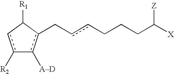
The cyclic lipid component of the microsphere may include one or more types of prostaglandin, prostaglandin analog, prostaglandin derivative, prostamide, prostamide analog, and a prostamide derivative, and any salts thereof, and mixtures thereof. In certain microspheres, the cyclic lipid component may comprise a compound having the following formula (VI)
Figure US07589057-20090915-C00001

wherein the dashed bonds represent a single or double bonds which can be in the cis or trans configuration, A is an alkyene or alkenylene radical having from two to six carbon atoms, which radical may be interrupted by one or more oxide radicals and substituted with one or more hydroxy, oxo, alkoxy or alkycarboxyl groups wherein said alkyl radical comprises from one to six carbon atoms; D is a branched or unbranched alkyl or heteroalkyl radical of from two to 10 carbon atoms, a cycloalkyl radical having from three to seven carbon atoms, or an aryl radical, selected from the group consisting of hydrocarbyl aryl and heteroaryl radicals having from four to ten carbon atoms wherein the heteroatom is selected from the group consisting of nitrogen, oxygen and sulfur atoms; X is a radical selected from the group consisting of hydrogen, a lower alkyl radical having from one to six carbon atoms, R5—C(═O)— or R5—O—C(═O)— wherein R5 is a lower alkyl radical having from one to six carbon atoms; Z is ═O or represents 2 hydrogen radicals; one of R1 and R2 is ═O, —OH or a —O—C(═O)—R6 group, and the other one is —OH or —O—C(═O)—R6, or R1 is ═O and R2 is H, wherein R6 is a saturated or unsaturated acyclic hydrocarbon group having from 1 to about 20 carbon atoms, or —(CH2)mR7 wherein m is 0-10, and R7 is cycloalkyl radical, having from three to seven carbon atoms, or a hydrocarbyl aryl or heteroaryl radical, as defined above, or a pharmaceutically acceptable salt thereof.
Pharmaceutically acceptable acid addition salts of certain of the cyclic lipids of the invention are those formed from acids which form non-toxic addition salts containing pharmaceutically acceptable anions, such as the hydrochloride, hydrobromide, hydroiodide, sulfate, or bisulfate, phosphate or acid phosphate, acetate, maleate, fumarate, oxalate, lactate, tartrate, citrate, gluconate, saccharate and p-toluene sulphonate salts.
In further embodiments, the cyclic lipid component comprises a compound having the following formula (VII)
Figure US07589057-20090915-C00002

wherein the radicals are as defined for Formula VI.
In another embodiment, the cyclic lipids of the present invention may comprise a compound having the following formula (VII)
Figure US07589057-20090915-C00003

wherein hatched lines indicate the α configuration and the solid triangles comprise the β configuration and the radicals are as defined for Formula VI.
In a further embodiment, the cyclic lipid component comprises a compound having the following formula (VIII)
Figure US07589057-20090915-C00004

wherein the radicals are as defined for Formula VI.
In a further embodiment, the cyclic lipid component comprises a compound having the following formula (IX)
Figure US07589057-20090915-C00005

wherein X is as defined for Formula VI.
In a further embodiment, the cyclic lipid component comprises a compound having the following formula (X)
Figure US07589057-20090915-C00006
In particular, the cyclic lipid component may comprise prostaglandin E1 or prostaglandin E2, or salts, esters or mixtures thereof. It will be understood that the cyclic lipids used in the present invention include, where appropriate, salts or esters of any compound disclosed herein.
In another embodiment, the cyclic lipid component of the present invention may comprise a compound having the formula (I)
Figure US07589057-20090915-C00007

wherein the dashed bonds represent a single or double bond which can be in the cis or trans configuration, A is an alkylene or alkenylene radical having from two to six carbon atoms, which radical may be interrupted by one or more oxide radicals and substituted with one or more hydroxy, oxo, alkyloxy or akylcarboxy groups wherein said alkyl radical comprises from one to six carbon atoms; B is a cycloalkyl radical having from three to seven carbon atoms, or an aryl radical, selected from the group consisting of hydrocarbyl aryl and heteroaryl radicals having from four to ten carbon atoms wherein the heteroatom is selected from the group consisting of nitrogen, oxygen and sulfur atoms; X is a radical selected from the group consisting of —OR4 and —N(R4)2 wherein R4 is selected from the group consisting of hydrogen, a lower alkyl radical having from one to six carbon atoms,
Figure US07589057-20090915-C00008
wherein R5 is a lower alkyl radical having from one to six carbon atoms; Z is ═O or represents 2 hydrogen radicals; one of R1 and R2 is ═O, —OH or a —O(CO)R6 group, and the other one is —OH or —O(CO)R6, or R1 is ═O and R2 is H, wherein R6 is a saturated or unsaturated acyclic hydrocarbon group having from 1 to about 20 carbon atoms, or —(CH2)mR7 wherein m is 0 or an integer of from 1 to 10, and R7 is cycloalkyl radical, having from three to seven carbon atoms, or a hydrocarbyl aryl or heteroaryl radical, as defined above, or a pharmaceutically-acceptable salt thereof, provided, however, that when B is not substituted with a pendant heteroatom-containing radical, and Z is ═O, then X is not —OR4.
Pharmaceutically acceptable acid addition salts of certain of the compounds of the invention are those formed from acids which form non-toxic addition salts containing pharmaceutically acceptable anions, such as the hydrochloride, hydrobromide, hydroiodide, sulfate, or bisulfate, phosphate or acid phosphate, acetate, maleate, fumarate, oxalate, lactate, tartrate, citrate, gluconate, saccharate and p-toluene sulphonate salts.
In more specific implants, the compound of the prostamide component has the following formula (II)
Figure US07589057-20090915-C00009
wherein y is 0 or 1, x is 0 or 1 and x+y are not both 1, Y is a radical selected from the group consisting of alkyl, halo, nitro, amino, thiol, hydroxy, alkyloxy, alkylcarboxy and halo substituted alkyl, wherein said alkyl radical comprises from one to six carbon atoms, n is 0 or an integer of from 1 to 3 and R3 is ═O, —OH or —O(CO)R6.
In additional implants, the compound of the prostamide component has the following formula (III)
Figure US07589057-20090915-C00010
wherein hatched lines indicate the α configuration and solid triangles indicate the β configuration.
In certain implants, the compound of the prostamide component has the following formula (IV)
Figure US07589057-20090915-C00011
wherein Y1 is Cl or trifluoromethyl, such as the compound having the following formula (V)
Figure US07589057-20090915-C00012
and the 9-and/or 11- and/or 15 esters thereof.
In another embodiment, the compounds used in conjunction with the resent invention include
  • a) cyclopentane heptenol-5-cis-2-(3α-hydroxy-5-phenyl-1-trans-pentenyl)-3,5-dihydroxy, [1α, 2β, 3α, 5α];
  • b) cyclopentane heptenamide-5-cis-2-(3α-hydroxy-5-phenyl-1-trans-pentenyl)-3,5-dihydroxy, [1α, 2β, 3α, 5α];
  • c) cyclopentane N,N-dimethylheptenamide-5-cis-2-(3α-hydroxy-5-phenyl-1-trans-penten-yl)-3,5-dihydroxy, [1α, 2β, 3α, 5α];
  • d) cyclopentane heptenyl methoxide-5-cis-2-(3α-hydroxy-5-phenyl-1-trans-pentenyl)-3-,5-dihydroxy, [1α, 2β, 3α, 5α];
  • e) cyclopentane heptenyl ethoxide-5-cis-2-(3α-hydroxy-4-meta-chloro-phenoxy-1-trans- -butenyl)-3,5-dihydroxy, [1α, 2β, 3α, 5α];
  • f) cyclopentane heptenylamide-5-cis-2-(3α-hydroxy-4-meta-chloro-phenox-y-1-trans-butenyl)-3,5-dihydroxy, [1α, 2β, 3α, 5α];
  • g) cyclopentane heptenylamide-5-cis-2-(3α-hydroxy-4-meta-tr-ifluoromethyl-phenoxy-1-trans-butenyl)-3,5-dihydroxy, [1α, 2β, 3α, 560 ];
  • h) cyclopentane N-isopropyl hepteneamide-5-cis-2-(3α-hydroxy-5-phenyl-1-trans-pentenyl)-3,5-dihydroxy, [1α, 2β, 3α, 5α];
  • i) cyclopentane N-ethyl heptenamide-5-cis-2-(3α-hydroxy-5-phenyl-1-trans-pentenyl)-3,5-dihydroxy, [1α, 2β, 3α, 5α];
  • j) cyclopentane N-methyl heptenamide-5-cis-2-(3α-hydroxy-5-phenyl-1-trans-pentenyl)-3,5-dihydroxy, [1α, 2β, 3α, 5α];
  • k) cyclopentane heptenol-5-cis-2-(3α-hydroxy-4-meta-chlorophenoxy-1-trans-butenyl)-3,5-dihydroxy, [1α, 2β, 3α, 5α];
  • l) cyclopentane heptenamide-5-cis-2-(3α-hydroxy-4-m-chlorophenoxy-1-trans-butenyl)-3,5-dihydroxy, [1α, 2β, 3α, 5α], and
  • m) cyclopentane heptenol-5-cis-2-(3α-hydroxy-5-phenylpentyl)3,5-dihydroxy, [1α, 2β, 3α, 5α].
The prostamide having a name cyclopentane N-ethyl heptenamide-5-cis2-cis-2-(3α-hydroxy-5-phenyl-1-trans-pentenyl)-3,5-dihydroxy, [1α,2β,3α,5α], and derivatives, analods, and/or esters thereof, is particularly preferred in this aspect of the invention. This compound is also known as bimatoprost and is publicly available in a topical ophthalmic solution under the tradename, Lumigan® (Allergan, Inc., CA).
Thus, the microparticles may comprise a therapeutic component which comprises, consists essentially of, or consists of bimatoprost, a salt thereof, or mixtures thereof.
The cyclic lipid component may be in a liquid, derivatized, particulate, or powder form and it may be entrapped by the biodegradable polymer matrix. Usually, cyclic lipid particles will have an effective average size less than about 3000 nanometers. In certain implants, the particles may have an effective average particle size about an order of magnitude smaller than 3000 nanometers. For example, the particles may have an effective average particle size of less than about 500 nanometers. In additional implants, the particles may have an-effective average particle size of less than about 400 nanometers, and in still further embodiments, a size less than about 200 nanometers.
The cyclic lipid component of the microspheres is preferably from about 10% to 90% by weight of the microspheres. More preferably, the cyclic lipid component is from about 20% to about 80% by weight of the microspheres. In a preferred embodiment, the cyclic lipid component comprises about 20% by weight of the microsphere (e.g., 15%-25%). In another embodiment, the cyclic lipid component comprises about 50% by weight of the microspheres.
Suitable polymeric materials or compositions for use in the microspheres include those materials which are compatible, that is biocompatible, with the eye so as to cause no substantial interference with the functioning or physiology of the eye. Such materials preferably are at least partially and more preferably substantially completely biodegradable or bioerodible.
Examples of useful polymeric materials include, without limitation, such materials derived from and/or including organic esters and organic ethers, which when degraded result in physiologically acceptable degradation products, including the monomers. Also, polymeric materials derived from and/or including, anhydrides, amides, orthoesters and the like, by themselves or in combination with other monomers, may also find use. The polymeric materials may be addition or condensation polymers, advantageously condensation polymers. The polymeric materials may be cross-linked or non-cross-linked, for example not more than lightly cross-linked, such as less than about 5%, or less than about 1% of the polymeric material being cross-linked. For the most part, besides carbon and hydrogen, the polymers will include at least one of oxygen and nitrogen, advantageously oxygen. The oxygen may be present as oxy, e.g. hydroxy or ether, carbonyl, e.g. non-oxo-carbonyl, such as carboxylic acid ester, and the like. The nitrogen may be present as amide, cyano and amino. The polymers set forth in Heller, Biodegradable Polymers in Controlled Drug Delivery, In: CRC Critical Reviews in Therapeutic Drug Carrier Systems, Vol. 1, CRC Press, Boca Raton, Fla. 1987, pp 39-90, which describes encapsulation for controlled drug delivery, may find use in the present microspheres.
Of additional interest are polymers of hydroxyaliphatic carboxylic acids, either homopolymers or copolymers, and polysaccharides. Polyesters of interest include polymers of D-lactic acid, L-lactic acid, racemic lactic acid, glycolic acid, polycaprolactone, and combinations thereof. Generally, by employing the L-lactate or D-lactate, a slowly eroding polymer or polymeric material is achieved, while erosion is substantially enhanced with the lactate racemate.
Among the useful polysaccharides are, without limitation, calcium alginate, and functionalized celluloses, particularly carboxymethylcellulose esters characterized by being water insoluble, a molecular weight of about 5 kD to 500 kD, for example.
Other polymers of interest include, without limitations polyvinyl alcohol, polyesters, polyethers and combinations thereof which are biocompatible and may be biodegradable and/or bioerodible.
Some preferred characteristics of the polymers or polymeric materials for use in the present invention may include biocompatibility, compatibility with the therapeutic component, ease of use of the polymer in making the drug delivery systems of the present invention, a half-life in the physiological environment of at least about 6 hours, preferably greater than about one day, and water insolubility.
The biodegradable polymeric materials which are included to form the matrix are desirably subject to enzymatic or hydrolytic instability. Water soluble polymers may be cross-linked with hydrolytic or biodegradable unstable cross-links to provide useful water insoluble polymers. The degree of stability can be varied widely, depending upon the choice of monomer, whether a homopolymer or copolymer is employed, employing mixtures of polymers, and whether the polymer includes terminal acid groups.
Equally important to controlling the biodegradation of the polymer and hence the extended release profile of the implant is the relative average molecular weight of the polymeric composition employed in the microspheres. Different molecular weights of the same or different polymeric compositions may be included in the microspheres to modulate the release profile. In certain implants, the relative average molecular weight of the polymer will range from about 9 to about 64 kD, usually from about 10 to about 54 kD, and more usually from about 12 to about 45 kD.
In some microspheres, copolymers of glycolic acid and lactic acid are used, where the rate of biodegradation is controlled by the ratio of glycolic acid to lactic acid. The most rapidly degraded copolymer has roughly equal amounts of glycolic acid and lactic acid. Homopolymers, or copolymers having ratios other than equal, are more resistant to degradation. The ratio of glycolic acid to lactic acid will also affect the brittleness of the microspheres. The percentage of polylactic acid in the polylactic acid polyglycolic acid (PLGA) copolymer can be from 0-100%, preferably about 15-85%, more preferably about 35-65%. In some implants, a 50/50 PLGA copolymer is used.
The biodegradable polymer matrix of the subconjunctival microspheres may comprise a mixture of two or more biodegradable polymers. For example, the microspheres may comprise a mixture of a first biodegradable polymer and a different second biodegradable polymer. One or more of the biodegradable polymers may have terminal acid groups.
Release of a drug from an erodible polymer is the consequence of several mechanisms or combinations of mechanisms. Some of these mechanisms include desorption from the microsphere's surface, dissolution, diffusion through porous channels of the hydrated polymer and erosion. Erosion can be bulk or surface or a combination of both. As discussed herein, the matrix of the microspheres may release drug at a rate effective to sustain release of an amount of the prostamide component for more than one week after implantation into an eye. In certain microspheres, therapeutic amounts of the cyclic lipid component are released for no more than about 30-35 days after administration to the subconjunctival space. For example, a microsphere may comprise bimatoprost, and the matrix of the microsphere degrades at a rate effective to sustain release of a therapeutically effective amount of bimatoprost for about one month after being placed under the conjunctiva. As another example, the microspheres may comprise bimatoprost, and the matrix releases drug at a rate effective to sustain release of a therapeutically effective amount of bimatoprost for more than forty days, such as for about six months.
One example of the biodegradable microsphere comprises an cyclic lipid component associated with a biodegradable polymer matrix, which comprises a mixture of different biodegradable polymers. At least one of the biodegradable polymers is a polylactide having a molecular weight of about 63.3 kD. A second biodegradable polymer is a polylactide having a molecular weight of about 14 kD. Such a mixture is effective in sustaining release of a therapeutically effective amount of the cyclic lipid component for a time period greater than about one month from the time the microspheres are placed administered under the conjuctiva.
Another example of a biodegradable microsphere comprises a cyclic lipid component associated with a biodegradable polymer matrix, which comprises a mixture of different biodegradable polymers, each biodegradable polymer having an inherent viscosity from about 0.16 dl/g to about 1.0 dl/g. For example, one of the biodegradable polymers may have an inherent viscosity of about 0.3 dl/g. A second biodegradable polymer may have an inherent viscosity of about 1.0 dl/g. Additional microspheres may comprise biodegradable polymers that have an inherent viscosity between about 0.2 dl/g and 0.5 dl/g. The inherent viscosities identified above may be determined in 0.1% chloroform at 25° C.
One particular microsphere formulation comprises bimatoprost associated with a combination of two different polylactide polymers. The bimatoprost is present in about 20% by weight of the microsphere. One polylactide polymer has a molecular weight of about 14 kD and an inherent viscosity of about 0.3 dl/g, and the other polylactide polymer has a molecular weight of about 63.3 kD and an inherent viscosity of about 1.0 dl/g. The two polylactide polymers are present in the microsphere in a 1:1 ratio. Such a microsphere may be effective in releasing the bimatoprost for more than two months.
The release of the cyclic lipid component from microspheres into the subconjuctiva may include an initial burst of release followed by a gradual increase in the amount of the cyclic lipid component released, or the release may include an initial delay in release of the prostamide component followed by an increase in release. When the microspheres are substantially completely degraded, the percent of the cyclic lipid component that has been released is about one hundred. The microsphere disclosed herein do not completely release, or release about 100% of the cyclic lipid component, until after about one week of being placed in an eye.
It may be desirable to provide a relatively constant rate of release of the cyclic lipid component from the microspheres over the life of the implant. For example, it may be desirable for the cyclic lipid component to be released in amounts from about 0.01 μg to about 2 μg per day for the life of the microspheres. However, the release rate may change to either increase or decrease depending on the formulation of the biodegradable polymer matrix. In addition, the release profile of the prostamide component may include one or more linear portions and/or one or more non-linear portions. Preferably, the release rate is greater than zero once the microspheres has begun to degrade or erode.
The microspheres may be monolithic, i.e. having the active agent or agents homogenously distributed through the polymeric matrix, or encapsulated, where a reservoir of active agent is encapsulated by the polymeric matrix. Due to ease of manufacture, monolithic implants are usually preferred over encapsulated forms. However, the greater control afforded by the encapsulated microspheres may be of benefit in some circumstances, where the therapeutic level of the drug falls within a narrow window. In addition, the therapeutic component, including the cyclic lipid component, may be distributed in a non-homogenous pattern in the matrix. For example, the microspheres may include a portion that has a greater concentration of the cyclic lipid component relative to a second portion of the microspheres.
The microspheres disclosed herein may have a size of between about 5 μm and about 1 mm, or between about 10 μm and about 0.8 mm for administration with a needle. For needle-injected microspheres, the microsphere may have any appropriate dimensions so long as the longest dimension of the microsphere permits the microsphere to move through a needle. This is generally not a problem in the administration of microspheres. The subconjunctival space in humans is able to accommodate relatively large volumes of microspheres, for example, about 100 μl, or about 150 μl, or about 50-200 μl or more.
The total weight of microsphere in a single dosage an optimal amount, depending on the volume of the subconjunctival space and the activity or solubility of the active agent. Most often, the dose is usually about 10 mg to about 500 mg of microspheres per dose. For example, a single subconjunctival injection may contain about 20 mg, or about 50 mg, or about 75 mg, or about 100 mg, or about 125 mg or about 150 mg, or about 175 mg, or about 200 mg of microspheres, including the incorporated therapeutic component. For non-human individuals, the dimensions and total weight of the microsphere(s) may be larger or smaller, depending on the type of individual.
The dosage of the therapeutic component in the microsphere is generally in the range from about 0.001% to about 100 mg per eye per dose, but also can vary from this depending upon the activity of the agent and its solubility.
Thus, microspheres can be prepared where the center may be of one material and the surface may have one or more layers of the same or a different composition, where the layers may be cross-linked, or of a different molecular weight, different density or porosity, or the like. For example, where it is desirable to quickly release an initial bolus of drug, the center of the microsphere may be a polylactate coated with a polylactate-polyglycolate copolymer, so as to enhance the rate of initial degradation. Alternatively, the center may be polyvinyl alcohol coated with polylactate, so that upon degradation of the polylactate exterior the center would dissolve and be rapidly washed out of the eye.
The microspheres may be of any particulate geometry including micro and nanospheres, micro and nanoparticles, spheres, powders, fragments and the like. The upper limit for the microsphere size will be determined by factors such as toleration for the implant, size limitations on insertion, desired rate of release, ease of handling, etc. Spheres may be in the range of about 0.5 μm to 4 mm in diameter, with comparable volumes for other shaped particles.
The size and form of the microspheres can also be used to control the rate of release, period of treatment, and drug concentration at the site of implantation. Larger microspheres will deliver a proportionately larger dose, but depending on the surface to mass ratio, may have a slower release rate. The particular size and geometry of the microspheres are chosen to suit the activity of the active agent and the location of its target tissue.
The proportions of the cyclic lipid component, polymer, and any other modifiers may be empirically determined by formulating several microsphere batches with varying average proportions. A USP approved method for dissolution or release test can be used to measure the rate of release (USP 23; NF 18 (1995) pp. 1790-1798). For example, using the infinite sink method, a weighed sample of the microspheres is added to a measured volume of a solution containing 0.9% NaCl in water, where the solution volume will be such that the drug concentration is after release is less than 5% of saturation. The mixture is maintained at 37° C. and stirred slowly to maintain the microspheres in suspension. The appearance of the dissolved drug as a function of time may be followed by various methods known in the art, such as spectrophotometrically, HPLC, mass spectroscopy, etc. until the absorbance becomes constant or until greater than 90% of the drug has been released.
In addition to the cyclic lipid component included in the microspheres disclosed herein, the microsphere may also include one or more additional ophthalmically acceptable therapeutic agents. For example, the microspheres may include one or more antihistamines, one or more antibiotics, one or more beta blockers, one or more steroids, one or more antineoplastic agents, one or more immunosuppressive agents, one or more antiviral agents, one or more antioxidant agents, and mixtures thereof. Alternatively, a single injection of microspheres may include two or more microsphere batches each containing a different therapeutic component or components. Such a mixture of different microspheres in included within the present invention so long as a therapeutic component comprises a cyclic lipid.
Additional pharmacologic or therapeutic agents which may find use in the present systems, include, without limitation, those disclosed in U.S. Pat. No. 4,474,451, columns 4-6 and U.S. Pat. No. 4,327,725, columns 7-8.
Examples of antihistamines include, and are not limited to, loradatine, hydroxyzine, diphenhydramine, chlorpheniramine, brompheniramine, cyproheptadine, terfenadine, clemastine, triprolidine, carbinoxamine, diphenylpyraline, phenindamine, azatadine, tripelennamine, dexchlorpheniramine, dexbrompheniramine, methdilazine, and trimprazine doxylamine, pheniramine, pyrilamine, chiorcyclizine, thonzylamine, and derivatives thereof.
Examples of antibiotics include without limitation, cefazolin, cephradine, cefaclor, cephapirin, ceftizoxime, cefoperazone, cefotetan, cefutoxime, cefotaxime, cefadroxil, ceftazidime, cephalexin, cephalothin, cefamandole, cefoxitin, cefonicid, ceforanide, ceftriaxone, cefadroxil, cephradine, cefuroxime, ampicillin, amoxicillin, cyclacillin, ampicillin, penicillin G, penicillin V potassium, piperacillin, oxacillin, bacampicillin, cloxacillin, ticarcillin, azlocillin, carbenicillin, methicillin, nafcillin, erythromycin, tetracycline, doxycycline, minocycline, aztreonam, chloramphenicol, ciprofloxacin hydrochloride, clindamycin, metronidazole, gentamicin, lincomycin, tobramycin, vancomycin, polymyxin B sulfate, colistimethate, colistin, azithromycin, augmentin, sulfamethoxazole, trimethoprim, and derivatives thereof.
Examples of beta blockers include acebutolol, atenolol, labetalol, metoprolol, propranolol, timolol, and derivatives thereof.
Examples of steroids include corticosteroids, such as cortisone, prednisolone, flurometholone, dexamethasone, medrysone, loteprednol, fluazacort, hydrocortisone, prednisone, betamethasone, prednisone, methylprednisolone, riamcinolone hexacatonide, paramethasone acetate, diflorasone, fluocinonide, fluocinolone, triamcinolone, derivatives thereof, and mixtures thereof.
Examples of antineoplastic agents include adriamycin, cyclophosphamide, actinomycin, bleomycin, duanorubicin, doxorubicin, epirubicin, mitomycin, methotrexate, fluorouracil, carboplatin, carmustine (BCNU), methyl-CCNU, cisplatin, etoposide, interferons, camptothecin and derivatives thereof, phenesterine, taxol and derivatives thereof, taxotere and derivatives thereof, vinblastine, vincristine, tamoxifen, etoposide, piposulfan, cyclophosphamide, and flutamide, and derivatives thereof.
Examples of immunosuppressive agents include cyclosporine, azathioprine, tacrolimus, and derivatives thereof.
Examples of antiviral agents include interferon gamma, zidovudine, amantadine hydrochloride, ribavirin, acyclovir, valciclovir, dideoxycytidine, phosphonoformic acid, ganciclovir, and derivatives thereof.
Examples of antioxidant agents include ascorbate, alpha-tocopherol, mannitol, reduced glutathione, various carotenoids, cysteine, uric acid, taurine, tyrosine, superoxide dismutase, lutein, zeaxanthin, cryotpxanthin, astazanthin, lycopene, N-acetyl-cysteine, carnosine, gamma-glutamylcysteine, quercitin, lactoferrin, dihydrolipoic acid, citrate, Ginkgo Biloba extract, tea catechins, bilberry extract, vitamins E or esters of vitamin E, retinyl palmitate, and derivatives thereof.
Other therapeutic agents include squalamine, carbonic anhydrase inhibitors, alpha-2 adrenergic receptor agonists, antiparasitics, antifungals, and derivatives thereof.
The amount of active agent or agents employed in the microspheres, individually or in combination, will vary widely depending on the effective dosage required and the desired rate of release from the microspheres. Usually the agent will be at least about 1, more usually at least about 10 weight percent of the microsphere, and usually not more than about 80, more usually not more than about 40 weight percent of the microspheres.
Some of the present implants may comprise a cyclic lipid component that comprises a combination of two or more different cyclic lipid derivatives. One microsphere or dosage of microspheres may comprise a combination of bimatoprost and latanoprost. Another microsphere or dosage of microspheres may comprise a combination of bimatoprost and travoprost.
As discussed herein, the present microspheres may comprise additional therapeutic agents. For example, one microsphere or dosage of microspheres may comprise a combination of bimatoprost and a beta-adrenergic receptor antagonist. More specifically, the microsphere or dosage of microspheres may comprise a combination of bimatoprost and Timolol®. Or, a microsphere or dosage of microspheres may comprise a combination of bimatoprost and a carbonic anyhdrase inhibitor. For example, the microsphere or dosage of microspheres may comprise a combination of bimatoprost and dorzolamide (Trusopt®).
In addition to the therapeutic component, the microspheres disclosed herein may include or may be provided in compositions that include effective amounts of buffering agents, preservatives and the like. Suitable water soluble buffering agents include, without limitation, alkali and alkaline earth carbonates, phosphates, bicarbonates, citrates, borates, acetates, succinates and the like, such as sodium phosphate, citrate, borate, acetate, bicarbonate, carbonate and the like. These agents advantageously present in amounts sufficient to maintain a pH of the system of between about 2 to about 9 and more preferably about 4 to about 8. As such the buffering agent may be as much as about 5% by weight of the total implant. Suitable water soluble preservatives include sodium bisulfite, sodium bisulfate, sodium thiosulfate, ascorbate, benzalkonium chloride, chlorobutanol, thimerosal, phenylmercuric acetate, phenylmercuric borate, phenylmercuric nitrate, parabens, methylparaben, polyvinyl alcohol, benzyl alcohol, phenylethanol and the like and mixtures thereof. These agents may be present in amounts of from about 0.001% to about 5% by weight and preferably about 0.01% to about 2% by weight. In at least one of the present microspheres, a benzylalkonium chloride preservative is provided in the implant, such as when the cyclic lipid component consists essentially of bimatoprost.
In some situations mixtures of microspheres may be utilized employing the same or different pharmacological agents. In this way, a cocktail of release profiles, giving a biphasic or triphasic release with a single administration is achieved, where the pattern of release may be greatly varied.
Additionally, release modulators such as those described in U.S. Pat. No. 5,869,079 may be included in the microspheres. The amount of release modulator employed will be dependent on the desired release profile, the activity of the modulator, and on the release profile of the cyclic lipid component in the absence of modulator. Electrolytes such as sodium chloride and potassium chloride may also be included in the microspheres. Where the buffering agent or enhancer is hydrophilic, it may also act as a release accelerator. Hydrophilic additives act to increase the release rates through faster dissolution of the material surrounding the drug in the microspheres, which increases the surface area of the drug exposed, thereby increasing the rate of drug bioerosion. Similarly, a hydrophobic buffering agent or enhancer dissolves more slowly, slowing the exposure of drug, and thereby slowing the rate of drug bioerosion.
In certain microspheres, the combination of bimatoprost and a biodegradable polymer matrix is released or delivered an amount of bimatoprost between about 0.1 mg to about 0.5 mg for about 3-6 months after implantation into the eye.
Various techniques may be employed to produce the microspheres described herein. Useful techniques include, but are not necessarily limited to, self-emulsification methods, super critical fluid methods, solvent evaporation methods, phase separation methods, spray drying methods, grinding methods, interfacial methods, molding methods, injection molding methods, combinations thereof and the like.
Compression methods may be used to make the implants, and typically yield implants with faster release rates than extrusion methods. Compression methods may use pressures of about 50-150 psi, more preferably about 70-80 psi, even more preferably about 76 psi, and use temperatures of about 0 degrees C. to about 115 degrees C., more preferably about 25 degrees C.
Another method comprises using an oil-in-oil emulsion process to produce the present microparticles.
In one embodiment, a method for producing therapeutic polymeric microparticles comprises encapsulating a cyclic lipid component with a polymeric component to form a population of cyclic lipid-encapsulated microparticles by an oil-in-oil emulsion process. Such microparticles are effective in treating one or more ocular conditions, as described herein, and are suitable for administration to a patient into the subconjunctival space. The therapeutic activity of the cyclic lipid component remains stable during storage of the microspheres which may be attributed to the particular encapsulated form of the microspheres.
In more detail, a method of forming microparticles comprises forming an oil-in-oil emulsion containing the cyclic lipid component and the polymeric component. For example, the method may include mixing or combining a plurality, such as two or more, non-aqueous liquid compositions to form an emulsion. In one embodiment, a first composition may comprise an organic solvent, and a second composition may comprise an oil. At least one of the compositions contains the cyclic lipid component, the polymeric component, or the cyclic lipid component and the polymeric component.
The method also comprises drying the emulsion to form a dried emulsion product. The drying can be achieved using one or more techniques to remove the liquid from the emulsified product. For example, the drying process can include increasing the temperature of or near the emulsified product to facilitate evaporation of the liquid, can include the use of a vacuum to facilitate removal of the liquid, can include centrifugation to separate solid from the liquid, and combinations thereof.
The dried emulsion product can then be contacted with a solvent to form a solvent containing composition. Adding a solvent to the dried emulsion product, or otherwise contacting the product with the solvent results in the product being suspended in a volume of the liquid solvent. The contacting can include a step of stirring or mixing the combination of the solvent and dried emulsion product to form a suspension of the dried emulsion product in the solvent. The suspension can include particles of the dried emulsion product, for example, microspheres of various sizes and shapes, including microparticles.
After forming the solvent containing composition, the method can comprise removing the solvent from the solvent-containing composition to form a population of microparticles that comprise the cyclic lipid component and the polymeric component. The removing can include one or more steps of centrifuging and/or rinsing intermediate compositions, and can include one or more steps of drying the resulting composition. The resulting products are encapsulated microparticles or microimplants that comprise a cyclic lipid component encapsulated by a polymeric component, such as a biodegradable polymer coating.
As discussed herein, the cyclic lipid component can comprises a single type of cyclic lipid derivative or derivatives. In certain embodiments, the cyclic lipid component comprises at least one prostamide derivative selected from the group consisting of bimatoprost, esters thereof, and mixtures thereof. In a further embodiment, the cyclic lipid component consists essentially of bimatoprost.
In additional embodiments, the cyclic lipid component can comprise combinations of two or more different cyclic lipid derivatives, such as a combination of bimatoprost and latanoprost, bimatoprost and travoprost, and the like.
The present methods are effective in producing encapsulated cyclic lipid component microparticles that maintain or preserve a substantial portion, if not all, of the therapeutic activity after a terminal sterilization procedure. It can be understood, that the present methods may also comprise a step of terminally sterilizing the microparticles. The microparticles can be sterilized before packaging or in their packaging. Sterilization of packages containing the present microparticles or implants is often preferred. The method may comprise exposing the present microparticles or implants to sterilizing amounts of gamma radiation, e-beam radiation, and other terminal sterilization products. In one embodiment, a method may comprise a step of exposing the present microparticles to gamma radiation at a dose of about 25 kGy.
As discussed herein, the polymeric component recited in the present method may comprise a biodegradable polymer or biodegradable copolymer. In at least one embodiment, the polymeric component comprises a poly (lactide-co-glycolide) PLGA copolymer. In a further embodiment, the PLGA copolymer has a lactide/glycolide ratio of 75/25. In a still further embodiment, the PLGA copolymer has at least one of a molecular weight of about 63 kilodaltons and an inherent viscosity of about 0.6 dL/g.
The present methods may also comprise a step of forming a first composition which comprises a cyclic lipid component, a polymeric component, and an organic solvent, and a step of forming a second oil-containing composition, and mixing the first composition and the second oil-containing composition.
The present methods may also comprise evaporating the oil-in-oil emulsion to form an evaporated product, as described herein.
Further, the methods may comprise a step of suspending the evaporated product in a solvent before removing the solvent from the solvent containing composition. Such a step can be understood to be a way of forming a suspension.
As one example, a method of forming encapsulated bimatoprost biodegradable microparticles comprises forming an oil-in-oil emulsion comprising the bimatoprbst and PLGA, evaporating the liquid from the emulsion to form an evaporated product, suspending and rinsing the evaporated product, and drying the evaporated product.
In accordance with the disclosure herein, an embodiment of the present invention is a population of microparticles that comprise a polymeric component encapsulating a prostamide component in the form of oil-in-oil emulsified microparticles. In one specific embodiment, the polymeric component comprises a PLGA copolymer and the cyclic lipid component comprises at least one prostamide derivative selected from the group consisting of bimatoprost, salts thereof, and mixtures thereof.
The resulting population may be a terminally sterilized population of microparticles. Terminally sterilized microparticles retain their therapeutic activity during storage and therefore can provide successful treatment to patients. In certain embodiments, a major portion of the prostamide component of the terminally sterilized microparticles remains stable. For example, in certain embodiments, at least 80% of the prostamide component remains stable after sterilization. In further embodiments, at least 90%, at least 95%, or at least 99% of the prostamide component remains stable.
In addition, the present population of microparticles may have a maximum particle diameter less than about 200 μm. In certain embodiments, the population of microparticles has an average or mean particle diameter less than about 50 μm. In further embodiments, the population of microparticles has a mean particle diameter from about 30 μm to about 50 μm.
The present microparticles are structured or configured to release the cyclic lipid component for extended periods of time at controlled rates. In some embodiments, the cyclic lipid component is released at a substantially linear rate (e.g., a single rate) over the life of the microparticles (e.g., until the microparticles fully degrade). Other embodiments are capable of releasing the cyclic lipid component at multiple rates or different rates over the life of the microparticles. The rate at which the microparticles degrade can vary, as discussed herein, and therefore, the present microparticles can release the cyclic lipid component for different periods of time depending on the particular configuration and materials of the microparticles. In at least one embodiment, a microparticle can release about 1% of the cyclic lipid component in the microparticles per day. In a further embodiment, the microparticles may have a release rate of about 0.7% per day when measured in vitro. Thus, over a period of about 40 days, about 30% of the cyclic lipid component may have been released.
As discussed herein, the amount of the cyclic lipid component present in the microparticles can vary. In certain embodiments, about 10% wt/wt of the microparticles is the cyclic lipid component. In further embodiments, the cyclic lipid component constitutes about 5% wt/wt of the microparticles.
The microspheres, including the population of microparticles, of the present invention may be inserted into the subconjunctival space of an eye by a variety of methods. The method of placement may influence the therapeutic component or drug release kinetics. A preferred means of administration of the microspheres of the present invention is by subconjunctival injection. The location of the site of injection of the microspheres may influence the concentration gradients of therapeutic component or drug surrounding the element, and thus influence the delivery rate to a given tissue of the eye. For example, an injection into the conjunctiva toward the posterior of the eye will direct drug more efficiently to the tissues of the posterior segment, while a site of injection closer to the anterior of the eye (but avoiding the cornea) may direct drug more efficiently to the anterior segment.
Microparticles may be administered to patients by administering an ophthalmically acceptable composition which comprises the microparticles to the patient. For example, microparticles may be provided in a liquid composition, a suspension, an emulsion, and the like, and administered by injection or implantation into the subconjunctival space of the eye.
The present implants or microparticles are configured to release an amount of cyclic lipid component effective to treat an ocular condition, such as by reducing at least one symptom of the ocular condition. More specifically, the microparticles may be used in a method to treat glaucoma, such as open angle glaucoma, ocular hypertension, chronic angle-closure glaucoma, with patent iridotomy, psuedoexfoliative glaucoma, and pigmentary glaucoma. By injecting the cyclic lipid component-containing microspheres into the subconjunctival space of an eye, it is believed that the cyclic lipid component is effective to enhance aqueous humor flow thereby reducing intraocular pressure. Additionally, the present inventors have shown that subconjunctival delivery of microspheres containing a cyclic lipid component is able to provide quite high concentrations of the therapeutic agent to the retina of the eye.
The microspheres disclosed herein may also be configured to release the cyclic lipid component with or without additional agents, as described above.
Ocular conditions which can be treated or prevented in accordance with the present invention include, without limitation, the following:
Maculopathies/retinal degeneration: macular degeneration, including age related macular degeneration (ARMD), such as non-exudative age related macular degeneration and exudative age related macular degeneration, choroidal neovascularization, retinopathy, including diabetic retinopathy, acute and chronic macular neuroretinopathy, central serous chorioretinopathy, and macular edema, including cystoid macular edema, and diabetic macular edema. Uveitis/retinitis/choroiditis: acute multifocal placoid pigment epitheliopathy, Behcet's disease, birdshot retinochoroidopathy, infectious (syphilis, lyme, tuberculosis, toxoplasmosis), uveitis, including intermediate uveitis (pars planitis) and anterior uveitis, multifocal choroiditis, multiple evanescent white dot syndrome (MEWDS), ocular sarcoidosis, posterior scleritis, serpignous choroiditis, subretinal fibrosis, uveitis syndrome, and Vogt-Koyanagi-Harada syndrome. Vascular diseases/exudative diseases: retinal arterial occlusive disease, central retinal vein occlusion, disseminated intravascular coagulopathy, branch retinal vein occlusion, hypertensive fundus changes, ocular ischemic syndrome, retinal arterial microaneurysms, Coat's disease, parafoveal telangiectasis, hemi-retinal vein occlusion, papillophlebitis, central retinal artery occlusion, branch retinal artery occlusion, carotid artery disease (CAD), frosted branch angitis, sickle cell retinopathy and other hemoglobinopathies, angioid streaks, familial exudative vitreoretinopathy, Eales disease. Traumatic/surgical: sympathetic ophthalmia, uveitic retinal disease, retinal detachment, trauma, laser, PDT, photocoagulation, hypoperfusion during surgery, radiation retinopathy, bone marrow transplant retinopathy. Proliferative disorders: proliferative vitreal retinopathy and epiretinal membranes, proliferative diabetic retinopathy. Infectious disorders: ocular histoplasmosis, ocular toxocariasis, presumed ocular histoplasmosis syndrome (POHS), endophthalmitis, toxoplasmosis, retinal diseases associated with HIV infection, choroidal disease associated with HIV infection, uveitic disease associated with HIV Infection, viral retinitis, acute retinal necrosis, progressive outer retinal necrosis, fungal retinal diseases, ocular syphilis, ocular tuberculosis, diffuse unilateral subacute neuroretinitis, and myiasis. Genetic disorders: retinitis pigmentosa, systemic disorders with associated retinal dystrophies, congenital stationary night blindness, cone dystrophies, Stargardt's disease and fundus flavimaculatus, Bests disease, pattern dystrophy of the retinal pigmented epithelium, X-linked retinoschisis, Sorsby's fundus dystrophy, benign concentric maculopathy, Bietti's crystalline dystrophy, pseudoxanthoma elasticum. Retinal tears/holes: retinal detachment, macular hole, giant retinal tear. Tumors: retinal disease associated with tumors, congenital hypertrophy of the RPE, posterior uveal melanoma, choroidal hemangioma, choroidal osteoma, choroidal metastasis, combined hamartoma of the retina and retinal pigmented epithelium, retinoblastoma, vasoproliferative tumors of the ocular fundus, retinal astrocytoma, intraocular lymphoid tumors. Miscellaneous: punctate inner choroidopathy, acute posterior multifocal placoid pigment epitheliopathy, myopic retinal degeneration, acute retinal pigment epithelitis and the like.
In one embodiment, an implant, such as the microspheres disclosed herein, is administered to a subconjunctival space of an eye.
In at least one embodiment, a method of reducing intraocular pressure in an eye of a patient comprises administering a microsphere containing a cyclic lipid component, as disclosed herein, to a patient by subconjuctival injection. A syringe apparatus including an appropriately sized needle, for example, a 22 gauge needle, a 27 gauge needle or a 30 gauge needle, can be effectively used to inject the composition with into the subconjunctival space of an eye of a human or animal. Frequent repeat injections are often not necessary due to the extended release of the cyclic lipid component from the microspheres.
In addition, for dual therapy approaches to treating an ocular condition, the method may include one or more additional steps of administering additional therapeutic agents to the eye, such as by topically administering compositions containing timolol, dorzolamide, and iatoprost, among others.
In another aspect of the invention, kits and packages for treating an ocular condition of the eye are provided, comprising: a) a container comprising an extended release microsphere formulation comprising a therapeutic component including a cyclic lipid component, such as bimatoprost (Lumigan®), and a drug release sustaining component; and b) instructions for use. Instructions may include steps of how to handle the microspheres, how to insert the microspheres into an ocular region, and what to expect from using the microspheres.
In certain implants, the microspheres preparation comprises a therapeutic component which consists essentially of bimatoprost, salts thereof, and mixtures thereof, and a biodegradable polymer matrix. The biodegradable polymer matrix may consist essentially of PLA, PLGA, or a combination thereof. When placed in the eye, the preparation releases about 40% to about 60%. of the bimatoprost to provide a loading dose of the bimatoprost within about one day after subconjunctival administration. Subsequently, the microspheres release about 1% to about 2% of the bimatoprost per day to provide a sustained therapeutic effect. Such microsphere preparations may be effective in reducing and maintaining a reduced intraocular pressure, such as below about 15 mm Hg for several months, and potentially for one or two years.
Other microspheres disclosed herein may be configured such that the amount of the cyclic lipid component that is released from the microspheres within two days of subconjunctival injection is less than about 95% of the total amount of the cyclic lipid component in the microsphere. In certain formulations, 95% of the cyclic lipid component is not released until after about one week of injection. In certain microsphere formulations, about 50% of the cyclic lipid component is released within about one day of placement in the eye, and about 2% is released for about 1 month after being placed in the eye. In other microspheres, about 50% of the cyclic lipid component is released within about one day of subconjunctival administration, and about 1% is released for about 2 months after such administration.
In an alternative embodiment, the present invention may comprise the installation of a hollow depot device, made from a biocompatible, preferably non-biodegradable, polymer in connection with and preferably penetrating the conjunctiva of an eye. Such a device is described, for example, in US Patent Publication 20050112175, hereby incorporated by reference herein. The depot is preferably refillable with microspheres comprising an ophthalmically active therapeutic component, such as an ophthalmically active cyclic lipid component.
EXAMPLES
Without intending to limit the scope of the invention, embodiments of the invention are set forth by the following Examples.
Example 1 Manufacture and Testing of Implants Containing Bimatoprost and a Biodegradable Polymer Matrix
Biodegradable implants were made by combining bimatoprost with a biodegradable polymer composition. 800 mg of polylactic acid (PLA) was combined with 200 mg of bimatoprost. The combination was dissolved in 25 milliliters of dichloromethane. The mixture was placed in a vacuum at 45° C. overnight to evaporate the dichloromethane. The resulting mixture was in the form of a cast sheet. The cast sheet was cut and ground in a high shear grinder with dry ice until the particles could pass through a sieve having a pore size of about 125 μm. The percent of bimatoprost present in the microparticles was analyzed using high pressure liquid chromatography (HPLC). The percent release of bimatoprost from the microparticles was profiled using dialysis. The percent of bimatoprost remaining in the recovered particles was analyzed by HPLC.
The release profile is described in Table 1.
Time Elapsed Time Percent Percent Per
Point (Days) Released Day
Start
0
1 1.03 47.51 47.51
2 2.03 47.92 0.41
3 3.03 49.99 2.07
4 4.03 50.09 0.10
5 7.04 50.90 0.82
The percent loading of bimatoprost was 14.93%. The percent of bimatoprost remaining in the recovered release particles was 4.94%.
Example 2 Extrusion Process and Compression Method for Manufacturing Bimatoprost-Containing Biodegradable Intraocular Implants
Bimatoprost is combined with a biodegradable polymer composition in a mortar. The combination is mixed with a shaker set at about 96 RPM for about 15 minutes. The powder blend is scraped off the wall of the mortar and is then remixed for an additional 15 minutes. The mixed powder blend is heated to a semi-molten state at specified temperature for a total of 30 minutes, forming a polymer/drug melt.
Rods are manufactured by pelletizing the polymer/drug melt using a 9 gauge polytetrafluoroethylene (PTFE) tubing, loading the pellet into the barrel and extruding the material at the specified core extrusion temperature into filaments. The filaments are then cut into about 1 mg size implants or drug delivery systems. The rods may have dimensions of about 2 mm long×0.72 mm diameter. The rod implants weigh between about 900 μg and 1100 μg.
Wafers are formed by flattening the polymer melt with a Carver press at a specified temperature and cutting the flattened material into wafers, each weighing about 1 mg. The wafers have a diameter of about 2.5 mm and a thickness of about 0.13 mm. The wafer implants weigh between about 900 μg and 1100 μg.
In-vitro release testing is performed by placing each implant into a 24 mL screw cap vial with 10 mL of Phosphate Buffered Saline solution at 37° C. 1 mL aliquots are removed and are replaced with equal volume of fresh medium on day 1, 4, 7, 14, 28, and every two weeks thereafter.
Drug assays are performed by HPLC, which consists of a Waters 2690 Separation Module (or 2696), and a Waters 2996 Photodiode Array Detector. An Ultrasphere, C-18 (2), 5 □m; 4.6×150 mm column heated at 30° C. is used for separation and the detector is set at about 264 nm. The mobile phase is (10:90) MeOH—buffered mobile phase with a flow rate of 1 mL/min and a total run time of 12 min per sample. The buffered mobile phase may comprise (68:0.75:0.25:31) 13 mM 1-Heptane Sulfonic Acid, sodium salt—glacial acetic acid—triethylamine—Methanol. The release rates are determined by calculating the amount of drug being released in a given volume of medium over time in □g/day.
Polymers which may be used in the implants can be obtained from Boehringer Ingelheim. Examples of polymer include: RG502, RG752, R202H, R203 and R206, and Purac PDLG (50/50). RG502 is (50:50) poly(D,L-lactide-co-glycolide), RG752 is (75:25) poly(D,L-lactide-co-glycolide), R202H is 100% poly(D, L-lactide) with acid end group or terminal acid groups, R203 and R206 are both 100% poly(D, L-lactide). Purac PDLG (50/50) is (50:50) poly(D,L-lactide-co-glycolide). The inherent viscosity of RG502, RG752, R202H, R203, R206 , and Purac PDLG are 0.2, 0.2, 0.2, 0.3, 1.0, and 0.2 dL/g, respectively. The average molecular weight of RG502, RG752, R202H, R203, R206, and Purac PDLG are, 11700, 11200, 6500, 14000, 63300, and 9700 daltons, respectively.
Example 3 Bimatoprost/PLA/PLGA Intraocular Implants to Treat Glaucoma
A 72 year old female suffering from glaucoma in both eyes receives an intraocular implant containing bimatoprost and a combination of a PLA and PLGA in each eye. The implants weigh about 1 mg, and contain about 500 mg of bimatoprost. One implant is placed in the vitreous of each eye using a syringe. In about two days, the patient reports a substantial relief in ocular comfort. Examination reveals that the intraocular pressure has decreased, the average intraocular pressure measured at 8:00 AM has decreased from 28 mm Hg to 14.3 mm Hg. The patient is monitored monthly for about 6 months. Intraocular pressure levels remain below 15 mm Hg for six months, and the patient reports reduced ocular discomfort.
Example 4 Bimatoprost/PLA Intraocular Implants for Reducing Ocular Hypertension
A 62 year old male presents with an intraocular pressure in his left eye of 33 mm Hg. An implant containing 400 mg of bimatoprost and 600 mg of PLA is inserted into the vitreous of the left eye using a trocar. The patient's intraocular pressure is monitored daily for one week, and then monthly thereafter. One day after implantation, the intraocular pressure is reduced to 18 mm Hg. By day 7 after implantation, the intraocular pressure is relatively stable at 14 mm Hg. The patient does not experience any further signs of elevated intraocular pressure for 2 years.
Example 5 Oil in Oil Method for Microencapsulation of a Prostamide Derivative
This example describes a process for producing microparticles that include a prostamide derivative encapsulated by a biodegradable polymer. In the specific example, bimatoprost was used as the prostamide derivative. The procedures outlined herein can be used to make encapsulated microparticles of other prostamide derivatives as well.
FIG. 1 is a flow chart illustrating the steps used in the method of this example.
As shown in FIG. 1, a first composition was formed by adding 100 mg of bimatoprost to 20 mL of acetonitrile (CH3CN) in an Erlenmeyer flask with a magnetic stirrer and stopper. The bimatoprost was solubilized in the acetonitirile. PLGA (900 mg) was added to the solubilized composition and stirred until the PLGA was solubilized. This first composition can be understood to be a discontinuous phase or an acetonitrile phase composition.
A second composition was formed by combining 800 mL of cottonseed oil and 12.8 mL of Span® 85 in a 1000 mL beaker. Span® 85 is a fatty acid composition which comprises oleic acid (C18:1) approx. 74%; linoleic acid (C18:2) approx. 7%; linolenic acid (C18:2) approx. 2%; palmitoleic acid (C16:1) approx. 7%; and palmitic acid (C16:0). Span is a registered trademark of ICI Americas, Inc., and the Span® 85 can be obtained from public sources, such as Sigma Aldrich. Other Span® products can also be used, such as Span® 80. This composition can be understood to be a continuous phase or oil-containing composition.
An emulsion was formed by adding the first composition to the second composition. The speed impeller was set at 350 rotations per minute (rpm) to stir the second composition. The first composition was added to the stirring second composition, and the mixture was allowed to stir for 90 minutes.
The emulsion was then evaporated under filter air flow for 45 hours at 250 rpm air flow.
Hexane (250 mL) was added to the evaporated product and stirred for 1 hour. Subsequently, the suspension was centrifuged at 7000 rpm for 10 minutes.
The pellet containing microparticles was rinsed twice by centrifuging with 100 mL of hexane. The microparticles were resuspended in 15 mL of hexane.
The microspheres were then dried under filter air flow overnight.
Microspheres were packaged under nitrogen and reduced temperatures to maintain the temperature at about 5 C. The cooled microspheres were sterilized using gamma radiation (25-35 kGy).
FIGS. 2A, 2B, 2C, 2D, 2E, and 2F illustrate the chemical structure of bimatoprost (AGN 192024, FIG. 2A), 15β bimatoprost (FIG. 2B); 5,6-trans bimatoprost isomer (FIG. 2C); C1 acid of bimatoprost (FIG. 2D); triphenyphosphine oxide (TPPO; FIG. 2E); and 15-Keto bimatoprost (FIG. 2F). Bimatoprost is the active ingredient and the other chemicals are impurities that may be present. Bimatoprost has a molecular weight of 415.6 g/mol, a solubility in water at room temperature of 3.2 mg/mL (which is slightly soluble), and a partition coefficient Log P of 2.4 plus or minus 0.1. Bimatoprost is a non-ionizable compound.
The effects of gamma sterilization on the stability of bimatoprost are presented in the following table (Table 2). The formula abbreviations correspond to compounds illustrated in FIGS. 2A-2F.
% wt/wt % wt/wt, % wt/wt % wt/wt
Bimatoprost bimatoprost 15-keto 15β 5,6-trans
none 95.3 1.0 0.2 0.1
sterilized
gamma 85.3 2.3 0.1 0.1
sterilized
Gamma sterilization appeared to decrease the amount of active bimatoprost by about 10%. Gamma sterilization may also change the color of the material (e.g., from white to a yellow/brown) and the consistency of the material (e.g., from a fluffy powder to a more compact powder).
Two separate batches of microparticles were produced using PLGA copolymers from different suppliers, as presented in Table 3, below
Lactide/Glycolide
ratio
Batch Random/Block Molecular Inherent
Supplier Number End-groups Weight Viscosity
APT
1 75/25   63,200 Da 0.65 dL/g
Random
Ester & Hydroxyl
BPI
2 75/25 ~97,100 Da 0.67 dL/g
Random
Ester & Hydroxyl
APT in Table 3 refers to Absorbable Polymer Technologies, and BPI refers to Birmingham Polymers, Inc. The PLGA obtained from APT appeared to provide better results than PLGA from BPI.
High performance liquid chromatography was used to quantify bimatoprost and impurities present in the microparticles. Analytes were eluted from a Waters Symmetry® C18 reverse-phase column using a mobile phase composed of 72/18/10 (water/acetonitrile/methanol v/v/v) containing 0.03% (w/v) trifluoroacetic acid. UV detection was performed at 210 nm. Chromatograms of a bimatoprost standard (FIG. 3A), a non-sterilized and sterilized placebo compositions (FIG. 3D and FIG. 3E), and non-sterilized and sterilized bimatoprost microparticles (FIG. 3B and FIG. 3C) revealed that the microparticles did not contain detectable amounts of impurities as evidenced by the single absorption peak in FIGS. 3A-C.
Particle size was determined by suspending an aliquot of the bimatoprost microspheres in 1 mL of deionized water.
To this, 100 μL of 10% Tween 80 was added. The composition was sonicated for 5 minutes and vortexed for 15 seconds. A Coulter LS230 apparatus or equivalent apparatus were used to measure particle size. Mean particle diameters of tested batches were between about 30 μm and about 50 μm. Some particles had diameters up to about 200 μm. These larger particles were observed in batches that were not sieved. FIGS. 4A-4D illustrate particle diameters (differential volume and number) profile superposition for sterile and non sterile conditions. Each graph illustrates the particle diameter distribution for a batch of sterile microspheres and non-sterile microspheres. The data reveal that sterilization did not significantly impact the particle size distribution as evidenced by the substantial overlap in the distribution curves. FIG. 4A illustrates volume % as a function of particle diameter for batch DL042 (top left panel), FIG. 4B illustrates number % as a function of particle diameter for batch DL042 (top right panel), FIG. 4C illustrates volume % as a function of particle diameter for batch DL043 (bottom left panel), and FIG. 4D illustrates number % as a function of particle diameter for batch DL043 (bottom right panel).
Microscopic aspect was determined using the sample prepared for particle size determination, described above. Observations were performed at magnification 5 and 20. FIGS. 5A-5F illustrate shapes of four different non-sterile and two different sterile batches of microspheres (FIG. 5A and FIG. 5B (top two panels) and FIGS. 5C and 5E illustrate non-sterile batches and FIG. 5D and FIG. 5F (bottom two right panels) illustrate sterile batches of microspheres).
The dissolution profile for a batch of microparticles was monitored for 28 and 42 days for sterile and non-sterile microspheres using dialysis bags and dissolution media. The dissolution media was phosphate buffer (pH 7.4) and ethanol in a ratio of 90/10. Samples were monitored at 37 C in a shaker water bath at approximately 110 rpm. At each time point, an aliquot of sample was collected and replaced by fresh media. The dissolution profile for a batch of sterile and non-sterile microspheres is shown in FIG. 6. The dissolution rate for the non-sterile microparticles was about 0.7%/day (e.g., 30% released over 42 days). The dissolution rate for the sterile batch initially appeared to be about 0.7%/day and increased after about two weeks. FIG. 6 also shows that bimatoprost was released faster from gamma sterilized microspheres than non-sterilized microspheres.
Table 4 below provides information regarding the batches of microspheres prepared in accordance with the present methods and as discussed above.
Batch No. PLGA type/SPAN type Sterilized (Yes/No)
DL040 PLGA APT 75/25/ No
iv 0.65/SPAN 80
DL041 APT/SPAN 85 No
DL042 APT/SPAN 80 No
DL042 APT/SPAN 80 Yes
DL043 APT/SPAN 80 No
DL043 APT/SPAN 80 Yes
In Table 4, APT refers to the PLGA supplier, as discussed herein, iv refers to inherent viscosity, and SPAN refers to the fatty acid composition described herein, and which is publicly available.
Table 5 below provides information regarding the amount of bimatoprost present in non-sterile microspheres. In these microspheres, bimatoprost was present in an amount of about 5% w/w.
Batch No. Targeted loading % w/w bimatoprost
DL040
10% 4.6
DL041 10% 5.1
DL042 10% 4.8
DL043 Placebo ND
Batches DL042 and DL043 were gamma sterilized (25-35 kGy) in order to evaluate the stability of the active ingredient when encapsulated within PLGA. The results are presented in Table 6 below.
Targeted % w/w
Batch No. Loading bimatoprost % w/w keto
DL042
10% 4.3 ND
DL042 sterile 10% 4.2 ND
DL043 Placebo ND ND
DL043 sterile Placebo ND ND
Example 6 Preparation of Bimatoprost Microspheres
A series of three bimatoprost microsphere (microparticle) formulations, designated Formulations 1, 2 and 3, were prepared as follows:
Formulations 1 and 2 were prepared using a spray-drying technique. Bimatoprost was dissolved in a solution of a polylactide(co-glycolide) (PLGA) biodegradable polymer and dichloromethane at a theoretical bimatoprost content of 3 wt %. Different PLGA polymers were used in each of Formulations 1 and 2. The solution was atomized using a Buchi Model 190 spray dryer with an inlet temperature set at 40° C. The bimatoprost/PLGA polymer microparticles thus formed were recovered for testing.
Formulation 3 was prepared using a solvent-extraction “continuous” microencapsulation process. The bimatoprost was suspended in a solution of a PLGA polymer and ethyl acetate at a theoretical bimatoprost content of 4.3 wt %. The solution was homogenized in the presence of a polyvinyl alcohol solution saturated with 4 mg/mL bimatoprost to produce an oil in water emulsion. The microparticles were formed upon extraction of the solvent in a continuous flow of purified water. The microparticles were isolated by centrifugation and lyophilized. The dried particles were then suspended and stirred in purified water at room temperature for approximately 2 hours. The particles were again isolated by centrifugation and lyophilized.
All of the microparticles of Formulations 1, 2 and 3 microparticles were irradiated with 2.5 Mrad gamma radiation prior to use in the rabbit in vivo study, described in Example 8 below.
Analytical characterization of the microparticles was performed, and the analytical data are summarized in Table 7.
TABLE 7
Summary of characterization data of Bimatoprost
Microparticle Formulations.
Particle Size
Formula- Bimatoprost Encapsula- Distribution, Residual
tion Content, tion microns Solvent,
Number wt % Efficiency d10 d90 Mean wt %
1 2.9 96.7 1.8 30.3 14.4 <0.1
2 2.9 96.7 1.4 17.6 8.8 <0.1
3 4.0 88.9 2.4 41.4 19.0 0.8
Example 7 In Vitro Testing of Bimatoprost Microspheres
Samples of each of the Example 6 Formulations 1, 2 and 3, both irradiated with gamma radiation (as set forth in Example 6) and non-irradiated, were tested in vitro for rate of release of bimatoprost. These in vitro tests were carried out as follows:
Each individual sample of microparticles was placed in a glass vial filled with receptor medium (9% NaCl in water). To allow for “infinite sink” conditions, the receptor medium volume was chosen so that the concentration of bimatoprost would never exceed 5% of saturation. To minimize secondary transport phenomena, e.g. concentration polarization in the stagnant boundary layer, each of the glass vials was placed into a shaking water bath at 37° C. Samples were taken for HPLC analysis from each vial at defined time points. The HPLC method was as described in USP 23 (1995) pp. 1791-1798. The concentration values were used to calculate the cumulative release profiles.
The release profiles are shown in FIG. 7.
As shown in FIG. 7, for each of the samples of Formulations 1 and 2, both irradiated and not irradiated, the release rate of bimatoprost is somewhat faster than the target release rate 7. The samples of Formulation 3, both irradiated and not irradiated, reasonably approximate the target release rate 7.
These data demonstrate that bimatoprost/polymer microparticles can be produced with different release rates, for example, for use in different applications and/or to treat different conditions/diseases. In short, these data demonstrate that bimatoprost/polymer microparticles can be produced with a range of bimatoprost release profiles as needed to treat different ocular conditions and/or to improve visual acuity.
Example 8 Subconjunctivally Injected Bimatoprost Microspheres
A study of subconjunctival injection in New Zealand white rabbit eyes of the Formulation 1, 2 and 3 microparticles of Example 6 was carried out.
The Formulation 1, 2 and 3 microparticles were provided in the form of dry particulates as six-seven doses per vial. A duplicate set of vials was supplied for each formulation, dose level, and timepoint (i.e., 20 vials for each lot number).
A 2 mg/mL bimatoprost solution (Formulation 4) prepared in saline containing 0.1 wt % Tween 80 was provided for dose administration to any animals still available following completion of microparticle dosing.
Preparation: The microparticles in each vial were reconstituted in 2.2 mL of injection vehicle. Microparticle/vehicle compositions were used immediately upon reconstitution or as soon as possible within 2 hours of reconstitution. Details of test composition preparation are outlined in Table 8 below.
TABLE 8
Vol. of
Micro- injec-
Bimato- No. of spheres tion Suspen-
Formula- prost vials of per vehicle sion Dose
tion content 6-7 vial added Conc. volume
Number (wt %) doses (mg) (mL) (wt %) (mL)
1 2.9 10 168 2.2 7.1 0.15
10 336 2.2 13.2 0.15
2 2.9 10 168 2.2 7.1 0.15
10 336 2.2 13.2 0.15
3 3.9 10 139 2.2 6.2 0.15
10 278 2.2 11.2 0.15
4 2 mg/mL 10 mL none 10 mL 2 mg/mL 0.15
The injection vehicle was a sterile, aqueous saline solution containing 0.1 wt % Tween 80 (polyoxyethylene-sorbitan monooleate).
To ensure that the appropriate dose of microparticles was injected, the following procedure was used to administer the microparticle doses.
An 18-gauge needle was inserted into a vial of sterile injection vehicle to release the vacuum. An empty syringe was attached to the needle the vial containing the vehicle was inverted, and the appropriate volume (2.2 mL) of vehicle was drawn out. The needle and the syringe was withdrawn from the vial containing the vehicle, and the needle was inserted into the vial containing the microparticles to be injected. The vial of microparticles was vented by inserting a separate needle through the septum. Then, leaving this needle in place, the vehicle was injected into the vial containing the microparticles, and both needles were withdrawn.
To suspend the microparticles in the vehicle, the vial was vigorously shaken for at least 30 seconds. After the microparticles were well dispersed, the appropriate amount of suspension (0.20 mL) was pulled into a 1-cc syringe using an 18-gauge needle. The needle was cleared of any suspension by pulling all of the contents into the syringe.
The 18-gauge needle was replaced with a 25-gauge×⅝-inch needle, and the needle was purged of air. The microparticles were injected quickly to prevent the microparticles from settling out of solution. Between each injection, the microparticles were resuspended by agitating the sample vial.
The entire procedure was repeated as necessary to complete dose administration. Each vial was used for a maximum of six to seven injections.
The microparticle doses were stored at −20° C. and then allowed to equilibrate to room temperature for approximately 1-2 hours prior to reconstitution. The injection vehicle was stored at room temperature.
The animals were provided with approximately 150 grams of Teklad Certified Hi-Fiber Rabbit Diet daily. The animals were provided tap water ad libitum.
Prior to placement on study, a physical examination was performed on each animal. Each animal also had a pre-treatment ophthalmic examination (slit lamp and indirect ophthalmoscopy), performed by a veterinary ophthalmologist.
Prior to placement on study, intraocular pressure (IOP) was determined for both eyes of each animal. Proparacaine hydrochloride 0.5% (1-2 drops) was delivered to each eye prior to measurements.
Neomycin/Polymyxin/Bacitracin (NPB) Ophthalmic Ointment was placed in both eyes of each animal once daily on the day prior to injection (Day −1) and two days after injection (Days 2 and 3).
Prior to dosing, the animals were weighed and randomly assigned to 33 study groups (Groups A-GG). If any alternate animals remained following completion of microsphere dosing for Groups A-GG, these animals were assigned to Groups HH, II, and/or JJ.
Animals were anesthetized with an intravenous injection of a ketamine/xylazine cocktail (87 mg/mL ketamine, 13 mg/mL xylazine) at a volume of 0.1-0.2 mL/kg. Animals instead were anesthetized with an intramuscular injection of ketamine 100 mg/mL at 35 mg/kg plus xylazine 100 mg/mL at 7 mg/kg.
Eyes were prepared for injection as follows: Approximately five minutes prior to injection, eyes were moistened with an ophthalmic Betadine solution. After five minutes, the Betadine was washed out of the eyes with sterile saline. Proparacaine hydrochloride 0.5% (1-2 drops) was delivered to each eye.
On Day 1, each rabbit in Groups A-GG received a 150-μL subconjunctival injection of the appropriate microparticle formulation into the left eye using a 25-gauge×⅝-inch needle. Each rabbit assigned to Groups HH-JJ received a 150-μL subconjunctival injection of bimatoprost solution into the left eye using a 30-gauge×⅝-inch needle. The bulbar conjunctiva in the dorsotemporal quadrant was elevated using forceps. Microparticle formulation or bimatoprost solution was injected into the subconjunctival space. The actual dose delivered was calculated as a differential of the syringe weight before and after dosing. Injection was performed by board-certified veterinary ophthalmic surgeons.
The animals were observed for mortality/morbidity twice daily.
If an animal was determined to be moribund or under severe distress, the animal was euthanized with an intravenous injection of commercial euthanasia solution. Both eyes were explanted, placed in 10% formalin, and stored for possible future evaluation.
The animals were weighed at random, on Day 1, and prior to necropsy.
Intraocular pressure (IOP) was determined for both eyes of each remaining animal on Days 2, 8±1, 15±1, 22±1, and 29±1. Proparacaine hydrochloride 0.5% (1-2 drops) was delivered to each eye prior to measurements. IOP was evaluated with a Medtronic Solan, Model 30 classic pneumatonometer on conditioned rabbits. A three-point diurnal curve was established: Intraocular pressure was recorded at 8 a.m., 12 p.m., and 4 p.m., with a ±1 hour range for each of these times.
Gross ocular observations were performed once weekly.
Ophthalmic observations (slit lamp and indirect ophthalmoscopy) were performed on both eyes of each remaining animal on Days 2, 8±1, 15±1, 22±1, and 29±1. Observations were performed by the same veterinary ophthalmologist for all timepoints.
Blood (at least 0.5 mL) was collected from each animal via ear vein or cardiac puncture prior to euthanasia. Animals were weighed and anesthetized with an intravenous injection of a ketamine/xylazine cocktail (87 mg/mL ketamine, 13 mg/mL xylazine) at a volume of 0.1 mL/kg. Animals were euthanized with an intravenous injection of commercial euthanasia solution following blood collection.
Each approximate 0.5-mL blood sample was collected into a single, pre-labeled K3 EDTA tube. Each tube was inverted 5-10 times to ensure adequate mixing of blood and anticoagulant. The tubes were then placed on ice. Using a glass pipette, duplicate 0.2-mL aliquots of whole blood were transferred into silanized round bottom 13×100 mm screw top glass centrifuge tubes with Teflon caps containing 2.5 mL of 50% acetonitrile/50% methanol (high.purity). Tubes were then vortexed. Blood samples were stored temporarily on wet ice until transferred to a freezer (at or below −20° C.). Samples were maintained at or below −20° C. until submitted for testing.
For eyes designated for pharmacokinetics analysis, aqueous humor samples were collected from all eyes before the eyes were enucleated, and the volume collected was measured and recorded. Both globes were enucleated and frozen in liquid nitrogen (at −20° C. or below).
Eyes were dissected as follows: The iris/ciliary body (ICB) was collected first, followed by collection of the vitreous humor (VH) and the retina.
Tissue weights were recorded for all tissues collected from all eyes. Careful attention was used to ensure that there is no cross contamination during dissection.
Aqueous humor (AH) and VH were placed in silanized amber screw-cap glass vials (3 mL) with Teflon or other inert washer. ICB and retina samples were placed in silanized round bottom 13×100 mm screw top glass centrifuge tubes with Teflon caps. Collected tissues were frozen on dry ice immediately after collection. All samples were stored at −20° C. or below until analysis.
The acceptable time ranges for sample collections (blood and ocular tissues) are summarized in the following Table 9:
TABLE 9
Scheduled Collection Acceptable
Time (Day) Time Range
2 ±0 days
8, 15, 22, 29 ±1 day
Ocular tissue samples collected for pharmacokinetics were analyzed using a validated LC-MS/MS method. Samples were quantified with AH, ICB, VH and retina assay range for bimatoprost and a bimatoprost metabolite of 0.1 ng or ng/mL. Assay range for blood was 0.125 and 0.25 ng/mL for bimatoprost and the bimatoprost metabolite, respectively.
Following subconjunctival injection of the microparticles, mainly the bimatoprost metabolite was detected in the AH, and bimatoprost was detected in blood. Both bimatoprost and the bimatoprost metabolite were detected in the ICB, VH and retina. Contralateral diffusion of bimatoprost and the bimatoprost metabolite to the untreated eyes was detected in the ICB and VH. In contrast, no drug levels were detected in all tissues at all timepoints (Day 1-14) following subconjunctival injection of 300 μg bimatoprost solution. Significantly, the subconjunctival bimatoprost microparticles caused little or no eye redness (hyperemia) in the rabbits. This is an unexpected advantage, for example, since topical administration of bimatoprost-containing eye drops can cause instances of hyperemia.
For all formulations dosed either at 300 μg or 600 μg dose, AH bimatoprost levels were detected at below the limit of quantification (BLQ) (<0.1 ng/mL) for all timepoints. Similar levels were detected for the bimatoprost metabolite, with the exception of a few treated eyes which had an AH level slightly above the BLQ level.
For all formulations dosed either at 300 μg or 600 μg dose, bimatoprost and bimatoprost metabolite were detected in the ICB at the earliest timepoint of Day 1. Only one formulation resulted in drug levels beyond Day 1 up to Day 21.
For all formulations dosed either at 300 μg or 600 μg dose, bimatoprost was detected in the VH at the earliest timepoint of about Day 1. Only one formulation, Formulation 3, resulted in drug levels beyond Day 1 up to Day 21. The bimatoprost VH maximum concentration for this formulation dosed at 300 μg and 600 μg was 1.73 ng/mL (Day 1) and 13.4 ng/mL (Day 1), respectively.
For all formulations dosed either at 300 μg or 600 μg dose, bimatoprost and bimatoprost metabolite were detected in the retina at the earliest timepoint of Day 1. Only one formulation resulted in bimatoprost levels beyond Day 1 up to Day 7, Formulation 3 (300 μg) and beyond Day 1 up to Day 21, Formulation 3 (600 μg). The a bimatoprost retinal maximum concentration for this formulation dosed at 300 μg and 600 μg bimatoprost was 101 ng/g (Day 7) and 289 ng/g (Day 1), respectively.
For all formulations administered at either 300 μg or 600 μg dose, blood drug levels were mainly at BLQ.
No mortality or drug-related effects on ophthalmic observations, body weight, or gross ocular observations were observed up to 1 month after administration.
This study shows that bimatoprost microparticles can be administered subconjunctively to produce concentrations, for example therapeutically effective concentrations, of bimatoprost/metabolite in various portions of the eye for periods of time of at least about 1 week or at least about 2 weeks or at least about 3 weeks. Thus, these subconjunctival injected bimatoprost microspheres can be used to effectively treat glaucoma. The fact that a relatively large amount of the bimatoprost administered subconjunctivally was passed to the posterior chamber, as bimatoprost or the bimatoprost metabolite is significant, for example, in treating conditions/diseases of the retina, for example, dry age-related macular degeneration, as well as other retinal conditions/diseases.
Example 9 Subconjunctivally Injected Bimatoprost Microspheres to Treat Glaucoma
A 56 year old male suffering from glaucoma in both eyes receives a subconjunctival injection of microspheres containing bimatoprost and a combination of a PLA and PLGA within the subconjunctival space of each eye. The microspheres contain 4% bimatoprost, and 40 mg of microspheres are used for each injection. In about two days, the patient reports a substantial relief in ocular comfort. Determination of intraocular pressure (IOP) reveals that the IOP has decreased in each eye, the average intraocular pressure measured at 8:00 AM has decreased from 28 mm Hg to 14.3 mm Hg. The patient is monitored every other day for about 1 month. Intraocular pressure levels remain below 15 mm Hg for this period of time, and the patient reports reduced ocular discomfort. Little or no ocular hyperemia is observed.
Example 10 Oil in Water Method for Making Bimatoprost Microspheres
This example describes an oil in water emulsion evaporation for making microparticles that include a prostamide derivative encapsulated by a biodegradable polymer. In the specific example, bimatoprost was used as the prostamide derivative. The procedures outlined herein can be used to make encapsulated microparticles of other prostamide derivatives as well.
Preparation of bimatoprost containing microspheres is difficult due to a lack of a significant solubility differential of bimatoprost in water as well as in many organic phases such as ethyl acetate and methylene chloride. When known oil in water emulsion evaporation processes are used most of the bimatoprost escapes into the aqueous phase. This Example sets forth an oil in water emulsion evaporation process which has been successfully used to make bimatoprost containing PLGA microspheres. An oil in water process is used in which an excess amount of the bimatoprost is added to the aqueous phase so as to saturate the aqueous phase to thereby prevent or reduce escape of the bimatoprost from the aqueous phase, with the result of higher drug loading (up to about 10 wt %) in the microspheres made.
The following process was use to make biodegradable bimatoprost microspheres using Poly(DL-lactide-co-glycolide) (PLGA). An emulsion/solvent evaporation technique was used. The continuous or aqueous non-solvent phase was saturated with bimatoprost to prevent loss of bimatoprost from the polymer phase and increase loading efficiency. As drug diffuses away from the polymer phase during microsphere hardening counter-diffusion of drug into the polymer phase from the saturated aqueous phase minimizes loss of the bimatoprost.
Bimatoprost loaded PLGA microspheres were prepared using the emulsion/evaporation procedure as follows: (1) 1 gram of PLGA (75:25) was combined with 1 gram of bimatoprost in 60 milliliters of dichloromethane (an organic solvent) (this formed the polymer phase; also referred to as the oil phase or as the organic phase); (2) 10 grams of polyvinyl alcohol (“PVA”) was combined with 1 gram of bimatoprost in 300 milliliters of water to thereby form a saturated aqueous phase (aqueous phase); (3) the polymer phase was emulsified with the aqueous phase by drop wise addition of the polymer phase into the aqueous phase under continuous mixing with a high shear impeller; (4) the resulting PLGA microspheres were hardened through the evaporation of the dichloromethane by stirring the emulsion in an open beaker for 24 hours; (5) the hardened microspheres were separated from the remaining PVA solution by centrifugation; (6) the microsphere pellets were suspended and washed 3 times with water; (7) the microspheres were dried under vacuum at 45° C. overnight; (8) the dried microspheres were sized to 38 microns through sieves.
The percent of bimatoprost loaded into the microspheres by this process was analyzed by HPLC to be 11.91%. The particle size distribution of the microspheres was determined to have a mean of 8.7 microns. The percent release of bimatoprost from the microspheres was assessed using dialysis.
In variants of this process the solvent used can be, for example, ethyl acetate or methylene chloride. The PLGA polymer used can be, for example, 50:50 DL-PLG acid, 65:35 DL-PLG, 65:35 COOH, 75:25 COOH or 75:25 laurl ester capped. DL-PLG acid 50:50 DL-PLG acid. The mean microparticle diameter can be from about 5 microns and about 20 microns.
In another variant of our an oil in water emulsion evaporation process for making microparticles that include a prostamide derivative encapsulated by a biodegradable polymer, a solid bimatoprost can be present in the emulsion during preparation. In such a variant of our process, the drug (bimatoprost) concentration exceeds its solubilities in both the aqueous and the organic phases and the drug is present as microfine particles in both phases as well. As the PLGA hardens when the organic phase evaporates, the drug can be captured as microfine particles as well as from precipitation.
Significantly, bimatoprost encapsulation within PLGA allows terminal sterilization with gamma radiation (i.e. by exposing the microparticles to gamma radiation at a dose of about 25-35 kGy) with low to moderate impact on the biological stability of the encapsulated bimatoprost.
Example 11 Oil in Water Method for Making Alpha-2 Agonist Microspheres
In this example an oil in water emulsion, solvent evaporation process was used to make polymeric microparticles (synonymously microspheres) comprising an alpha-2 adrenergic receptor agonist as the active agent.
An alpha-2 adrenergic receptor agonist is a compound that selectively activates alpha-2 adrenergic receptors relative to alpha-1 adrenergic receptors. An alpha-2 adrenergic receptor agonist can selectively activate a subtype of the alpha-2 adrenergic receptors. For example, the agonist may selectively activate one or more of the alpha-2a, the alpha-2b, or the alpha-2c receptors.
An alpha-2 adrenergic receptor agonist can be a quinoxaline derivative, such as a (2-imidozolin-2-ylamino) quinoxaline or a 5-bromo-6-(2-imidozolin-2-ylamino) quinoxaline, or salts, derivatives thereof and mixtures thereof. An alpha-2 adrenergic receptor agonist can be clonidine, apraclonidine, or a brimonidine, such as a brimonidine freebase or a brimonidine salt, such as a brimonidine tartrate.
This Example 11 sets forth a process for making brimonidine PLGA microspheres in a saturated non-solvent. Specifically, an emulsion/solvent evaporation process was used to make biodegradable microspheres incorporating brimonidine free base). The non-solvent (continuous aqueous phase) was saturated with brimonidine to prevent loss of the brimonidine from the polymer phase and to increase loading efficiency. Additionally, brimonidine saturated methanol was used to quench the emulsion. The methanol served as a sink to remove the dichloromethane quickly, hardening the microspheres before the brimonidine can diffuse out of them.
Thus, brimonidine free base loaded PLGA microspheres were prepared using the following emulsion/evaporation procedure:
  • (1) 1 gm of a polylactide-co-glycolide [PLGA] resomer (a polylactide [PLA] resomer can also be used) was combined with 175 mg of brimonidine free base and mixed. The mixture can optionally be heated to 100° C. while being mixed. Additionally, the brimonidine free base can be a quinoline derivative such as compound A below:
Figure US07589057-20090915-C00013
  • (2) The polymer brimonidine mixture was then added to 60 milliliters of dichloromethane (an organic solvent) at 38° C. This formed an oil phase, also referred to as a polymer phase.
  • (3) 9.6 grams of polyvinyl alcohol (PVA) as emulsifying agent was combined with 80 mg of brimonidine free base in 320 milliliters of water at pH 7.8 to form an aqueous phase saturated with the brimonidine free base (saturated aqueous phase)
  • (4) The oil phase was emulsified with the saturated aqueous phase by drop wise addition of the oil phase into the saturated aqueous phase under continuous mixing with a high shear impeller.
  • (5) 350 milliliters of methanol containing 900 mg of brimonidine free base was prepared to form a saturated, second oil phase.
  • (6) The saturated second oil phase was added to the emulsion and stirred for 4 hours to pull the dichloromethane and the methanol out and harden the microspheres.
  • (7) The resulting PLGA microspheres were further hardened by evaporation under vacuum for 1 hour.
  • (8) The hardened microspheres were separated from the remaining PVA solution by centrifugation.
  • (9) The microsphere pellets were suspended and washed 3 times with acetate buffer.
  • (10) The microspheres were then dried under vacuum at 45° C. overnight.
  • (11) The dried microspheres were sized to 38 microns through sieves.
The percent of brimonidine free base loaded into the microspheres was analyzed by UV. The percent of brimonidine free base loaded into (incorporated into) the polymeric microspheres when the aqueous phase is not saturated with brimonidine was less than 0.1 wt %. By using this Example 11 process in which the aqueous phase was saturated with brimonidine the loading of brimonidine in the PLGA microspheres was increased by a log unit to about 0.9 wt %. It was determined that the microparticle mean size was 11.8 μm. The in vitro percent release of brimonidine free base from the microspheres was assessed using dialysis and is shown by FIG. 8 which shows that all the brimonidine had been released from the microspheres after about 35 days.
Prior to in vivo use, sterilization of the brimonidine containing microspheres with gamma radiation (as a terminal or last step of sterilization) by exposing the microspheres to gamma radiation at a dose of about 25-35 kGy) can be carried out with a low to moderate impact on the biological stability (i.e. on the biological activity) of the encapsulated brimonidine.
All references, articles, publications and patents and patent applications cited herein are incorporated by reference in their entireties.
While this invention has been described with respect to various specific examples and embodiments, it is to be understood that the invention is not limited thereto and that it can be variously practiced within the scope of the following claims.

Claims (2)

1. A method for making biodegradable polymeric microspheres for treating an ocular condition, the method comprising the steps of: (a) combining a polylactic acid polyglycolic acid (PLGA) polymer with an alpha-2 adrenergic receptor agonist in an organic solvent to form a first oil phase; (b) adding an excess amount of the alpha-2 adrenergic receptor agonist to water to form a saturated aqueous phase; (c) emulsifying the first oil phase in the saturated aqueous phase by mixing the two phases together to form an oil in water emulsion; and; (d) removing the organic solvent from the oil phase of the oil in water emulsion using a second oil phase comprising a second organic solvent saturated with the alpha-2 adrenergic receptor agonist, thereby making biodegradable PLGA microspheres comprising the alpha-2 adrenergic receptor agonist, wherein the alpha-2 adrenergic agonist is brimonidine.
2. The method of claim 1, wherein the alpha-2 adrenergic receptor agonist of the microparticles so produced is encapsulated by the biodegradable polymer to preserve therapeutic activity of the alpha-2 adrenergic receptor agonist after a terminal sterilization procedure.
US11/395,019 2004-04-30 2006-03-31 Oil-in-water method for making alpha-2 agonist polymeric drug delivery systems Active 2025-06-01 US7589057B2 (en)

Priority Applications (1)

Application Number Priority Date Filing Date Title
US11/395,019 US7589057B2 (en) 2004-04-30 2006-03-31 Oil-in-water method for making alpha-2 agonist polymeric drug delivery systems

Applications Claiming Priority (7)

Application Number Priority Date Filing Date Title
US10/837,260 US7799336B2 (en) 2004-04-30 2004-04-30 Hypotensive lipid-containing biodegradable intraocular implants and related methods
US10/837,143 US8425929B2 (en) 2004-04-30 2004-04-30 Sustained release intraocular implants and methods for preventing retinal dysfunction
US11/118,519 US20050244506A1 (en) 2004-04-30 2005-04-29 Sustained release intraocular implants and methods for preventing retinal dysfunction
US11/303,462 US7993634B2 (en) 2004-04-30 2005-12-15 Oil-in-oil emulsified polymeric implants containing a hypotensive lipid and related methods
US11/368,845 US20060182781A1 (en) 2004-04-30 2006-03-06 Methods for treating ocular conditions with cyclic lipid contraining microparticles
US11/371,117 US20070212395A1 (en) 2006-03-08 2006-03-08 Ocular therapy using sirtuin-activating agents
US11/395,019 US7589057B2 (en) 2004-04-30 2006-03-31 Oil-in-water method for making alpha-2 agonist polymeric drug delivery systems

Related Parent Applications (1)

Application Number Title Priority Date Filing Date
US11/371,117 Continuation-In-Part US20070212395A1 (en) 2004-04-30 2006-03-08 Ocular therapy using sirtuin-activating agents

Publications (2)

Publication Number Publication Date
US20060173060A1 US20060173060A1 (en) 2006-08-03
US7589057B2 true US7589057B2 (en) 2009-09-15

Family

ID=36757439

Family Applications (1)

Application Number Title Priority Date Filing Date
US11/395,019 Active 2025-06-01 US7589057B2 (en) 2004-04-30 2006-03-31 Oil-in-water method for making alpha-2 agonist polymeric drug delivery systems

Country Status (1)

Country Link
US (1) US7589057B2 (en)

Cited By (41)

* Cited by examiner, † Cited by third party
Publication number Priority date Publication date Assignee Title
US20050244479A1 (en) * 2004-04-30 2005-11-03 Allergan, Inc. Sustained release intraocular implants and methods for preventing retinal dysfunction
US20060182781A1 (en) * 2004-04-30 2006-08-17 Allergan, Inc. Methods for treating ocular conditions with cyclic lipid contraining microparticles
US20060233860A1 (en) * 2004-04-30 2006-10-19 Allergan, Inc. Alpha-2 agonist polymeric drug delivery systems
US20060246145A1 (en) * 2004-04-30 2006-11-02 Allergan, Inc. Methods for treating ocular conditions with cyclic lipid containing microparticles
US20070224246A1 (en) * 2004-04-30 2007-09-27 Hughes Patrick M Oil-in-oil emulsified polymeric implants containing a hypotensive lipid and related methods
US20070243136A1 (en) * 2004-01-15 2007-10-18 Fisher Edward A Methods and Compositions for Imaging
US20090017097A1 (en) * 2007-07-09 2009-01-15 Sawhney Amarpreet S Hydrogel polymeric compositions and methods
US20110152271A1 (en) * 2009-12-17 2011-06-23 Gerald Horn Compositions and methods for ophthalmic delivery of nasal decongestants
US20110160214A1 (en) * 2009-12-17 2011-06-30 Gerald Horn Compositions and methods for eye whitening
US20110182966A1 (en) * 2010-01-22 2011-07-28 Allergan, Inc. Intracameral sustained release therapeutic agent implants
US8206736B2 (en) * 2004-04-30 2012-06-26 Allergan, Inc. Hypotensive lipid-containing biodegradable intraocular implants and related methods
US8299079B2 (en) 2009-05-22 2012-10-30 Kaufman Herbert E Preparations and methods for ameliorating or reducing presbyopia
WO2012169972A1 (en) * 2011-06-09 2012-12-13 Agency For Science, Technology And Research Core-shell microspheres
US8409606B2 (en) 2009-02-12 2013-04-02 Incept, Llc Drug delivery through hydrogel plugs
US8445526B2 (en) 2011-02-03 2013-05-21 Glaucoma & Nasal Therapies Llc Compositions and methods for treatment of glaucoma
US8465778B2 (en) 2004-04-30 2013-06-18 Allergan, Inc. Method of making tyrosine kinase microspheres
US8580292B2 (en) 2004-04-30 2013-11-12 Allergan, Inc. Sustained release intraocular implants and methods for treating ocular vasculopathies
US8663194B2 (en) 2008-05-12 2014-03-04 University Of Utah Research Foundation Intraocular drug delivery device and associated methods
US8673341B2 (en) 2004-04-30 2014-03-18 Allergan, Inc. Intraocular pressure reduction with intracameral bimatoprost implants
US8715713B2 (en) 2011-04-29 2014-05-06 Allergan, Inc. Solvent cast film sustained release latanoprost implant
US8771745B2 (en) 2008-10-27 2014-07-08 Allergan, Inc. Prostaglandin and prostamide drug delivery systems and intraocular therapeutic uses thereof
US8834884B2 (en) 2006-11-03 2014-09-16 Allergan, Inc. Sustained release drug delivery systems comprising a water soluble therapeutic agent and a release modifier
US8952011B2 (en) 2008-08-01 2015-02-10 Eye Therapies Llc Compositions and methods for the treatment of nasal conditions
US20150051372A1 (en) * 2012-02-10 2015-02-19 Hybio Pharmaceutical Co., Ltd. Method for purifying solid-phase synthetic crude liraglutide
US8961501B2 (en) 2010-09-17 2015-02-24 Incept, Llc Method for applying flowable hydrogels to a cornea
US8987270B2 (en) 2009-07-27 2015-03-24 Eye Therapies Llc Formulations of selective alpha-2 agonists and methods of use thereof
US8999938B2 (en) 2013-06-21 2015-04-07 Gnt Llc Ophthalmic lipophilic drug delivery vehicle formulations
US9095404B2 (en) 2008-05-12 2015-08-04 University Of Utah Research Foundation Intraocular drug delivery device and associated methods
US9138480B2 (en) 2009-11-09 2015-09-22 Allergan, Inc. Compositions and methods for stimulating hair growth
US9205150B2 (en) 2011-12-05 2015-12-08 Incept, Llc Medical organogel processes and compositions
US9504653B2 (en) 2013-04-01 2016-11-29 Allergan, Inc. Microsphere drug delivery system for sustained intraocular release
US9610246B2 (en) 2013-02-15 2017-04-04 Allergan, Inc. Sustained drug delivery implant
US9775846B2 (en) 2004-04-30 2017-10-03 Allergan, Inc. Hypotensive lipid-containing biodegradable intraocular implants and related implants
US9877973B2 (en) 2008-05-12 2018-01-30 University Of Utah Research Foundation Intraocular drug delivery device and associated methods
US10064819B2 (en) 2008-05-12 2018-09-04 University Of Utah Research Foundation Intraocular drug delivery device and associated methods
US10226417B2 (en) 2011-09-16 2019-03-12 Peter Jarrett Drug delivery systems and applications
US11246838B2 (en) 2013-03-05 2022-02-15 University of Pittsburgh—of the Commonwealth System of Higher Education Thermoresponsive hydrogel containing polymer microparticles for noninvasive ocular drug delivery
US11357756B2 (en) 2017-01-20 2022-06-14 Warsaw Orthopedic, Inc. Anesthetic compositions and methods comprising imidazoline compounds
US11395853B2 (en) 2016-06-23 2022-07-26 University Of Pittsburgh - Of The Commonwealth System Of Higher Education Biomimetic drug delivery of an immunomodulatory agent for the treatment of ocular conditions
US11596600B2 (en) 2008-08-01 2023-03-07 Eye Therapies, Llc Vasoconstriction compositions and methods of use
US11696593B2 (en) 2018-08-24 2023-07-11 Bioscience Formulators, LLC Astaxanthin nutritional compositions and uses

Families Citing this family (28)

* Cited by examiner, † Cited by third party
Publication number Priority date Publication date Assignee Title
US7172762B1 (en) * 1999-01-29 2007-02-06 Pfizer Inc. Erysipelothrix rhusiopathiae antigens and vaccine compositions
US20070224278A1 (en) 2003-11-12 2007-09-27 Lyons Robert T Low immunogenicity corticosteroid compositions
US20050101582A1 (en) * 2003-11-12 2005-05-12 Allergan, Inc. Compositions and methods for treating a posterior segment of an eye
US8668676B2 (en) * 2006-06-19 2014-03-11 Allergan, Inc. Apparatus and methods for implanting particulate ocular implants
US8580307B2 (en) * 2006-06-22 2013-11-12 Ethicon, Inc. High glass transition temperature absorbable microspheres
US20080097335A1 (en) 2006-08-04 2008-04-24 Allergan, Inc. Ocular implant delivery assemblies
EP2178502A2 (en) * 2007-06-21 2010-04-28 Yale University Sustained delivery of drugs from biodegradable polymeric microparticles
MX2010002616A (en) * 2007-09-07 2010-08-04 Qlt Plug Delivery Inc Drug cores for sustained release of therapeutic agents.
US8557273B2 (en) 2008-04-18 2013-10-15 Medtronic, Inc. Medical devices and methods including polymers having biologically active agents therein
US8889173B2 (en) * 2008-04-18 2014-11-18 Warsaw Orthopedic, Inc. Alpha adrenergic receptor agonists for treatment of pain and/or inflammation
US9072727B2 (en) * 2008-04-18 2015-07-07 Warsaw Orthopedic, Inc. Alpha adrenergic receptor agonists for treatment of degenerative disc disease
US8956641B2 (en) * 2008-04-18 2015-02-17 Warsaw Orthopedic, Inc. Alpha adrenergic receptor agonists for treatment of inflammatory diseases
USRE48948E1 (en) 2008-04-18 2022-03-01 Warsaw Orthopedic, Inc. Clonidine compounds in a biodegradable polymer
US8420114B2 (en) * 2008-04-18 2013-04-16 Warsaw Orthopedic, Inc. Alpha and beta adrenergic receptor agonists for treatment of pain and / or inflammation
US9610243B2 (en) 2008-04-18 2017-04-04 Warsaw Orthopedic, Inc. Clonidine compounds in a biodegradable polymer
US8946277B2 (en) 2008-04-18 2015-02-03 Warsaw Orthopedic, Inc. Clonidine formulations in a biodegradable polymer carrier
US20110206773A1 (en) * 2008-05-20 2011-08-25 Yale University Sustained delivery of drugs from biodegradable polymeric microparticles
WO2009155353A1 (en) * 2008-06-17 2009-12-23 The Regents Of The University Of California Process and system for reducing sizes of emulsion droplets and emulsions having reduced droplet sizes
US8821870B2 (en) * 2008-07-18 2014-09-02 Allergan, Inc. Method for treating atrophic age related macular degeneration
US20100098772A1 (en) * 2008-10-21 2010-04-22 Allergan, Inc. Drug delivery systems and methods for treating neovascularization
US8545554B2 (en) * 2009-01-16 2013-10-01 Allergan, Inc. Intraocular injector
US20100239632A1 (en) 2009-03-23 2010-09-23 Warsaw Orthopedic, Inc. Drug depots for treatment of pain and inflammation in sinus and nasal cavities or cardiac tissue
US8529492B2 (en) 2009-12-23 2013-09-10 Trascend Medical, Inc. Drug delivery devices and methods
US10350196B2 (en) 2013-09-30 2019-07-16 Zoetis Services Llc Long-acting spiro-isoxazoline formulations
BR112016009575B1 (en) 2013-10-31 2022-12-13 Allergan, Inc BIODEGRADABLE INTRAOCULAR IMPLANT, USE THEREOF, APPLIANCE FOR DELIVERING A BIODEGRADABLE INTRAOCULAR IMPLANT AND METHOD FOR PREPARING A BIODEGRADABLE INTRAOCULAR IMPLANT
EP3813793B1 (en) * 2018-05-25 2023-09-20 ADS Therapeutics LLC A composition for treating ocular hyperemia and a method for treating ocular hyperemia with the same
CN108888608A (en) * 2018-07-09 2018-11-27 华南理工大学 A kind of application of the preparation method and its fat reducing protect liver of the nano-carrier carrying resveratrol
CN108888609A (en) * 2018-07-11 2018-11-27 华南理工大学 A kind of controlled release nanometer carrier and preparation method thereof for oral load fat reducing drug resveratrol

Citations (26)

* Cited by examiner, † Cited by third party
Publication number Priority date Publication date Assignee Title
US4327725A (en) 1980-11-25 1982-05-04 Alza Corporation Osmotic device with hydrogel driving member
US4474451A (en) 1982-02-19 1984-10-02 Olympus Optical Co., Ltd. Diaphragm control circuit for TTL automatic electronic flash
US4494274A (en) 1982-05-28 1985-01-22 Thurlow Heida L Cookware with covers having metal handles
US4521210A (en) 1982-12-27 1985-06-04 Wong Vernon G Eye implant for relieving glaucoma, and device and method for use therewith
US4599353A (en) 1982-05-03 1986-07-08 The Trustees Of Columbia University In The City Of New York Use of eicosanoids and their derivatives for treatment of ocular hypertension and glaucoma
US4853224A (en) 1987-12-22 1989-08-01 Visionex Biodegradable ocular implants
US4997652A (en) 1987-12-22 1991-03-05 Visionex Biodegradable ocular implants
US5034413A (en) 1989-07-27 1991-07-23 Allergan, Inc. Intraocular pressure reducing 9,11-diacyl prostaglandins
US5164188A (en) 1989-11-22 1992-11-17 Visionex, Inc. Biodegradable ocular implants
US5443505A (en) 1993-11-15 1995-08-22 Oculex Pharmaceuticals, Inc. Biocompatible ocular implants
US5501856A (en) 1990-11-30 1996-03-26 Senju Pharmaceutical Co., Ltd. Controlled-release pharmaceutical preparation for intra-ocular implant
US5612364A (en) * 1995-11-08 1997-03-18 Alcon Laboratories, Inc. Substantially pure apraclonidine
US5688819A (en) 1992-09-21 1997-11-18 Allergan Cyclopentane heptanoic acid, 2-cycloalkyl or arylalkyl derivatives as therapeutic agents
US5869079A (en) 1995-06-02 1999-02-09 Oculex Pharmaceuticals, Inc. Formulation for controlled release of drugs by combining hydrophilic and hydrophobic agents
US6074661A (en) 1997-08-11 2000-06-13 Allergan Sales, Inc. Sterile bioerodible occular implant device with a retinoid for improved biocompatability
US6331313B1 (en) 1999-10-22 2001-12-18 Oculex Pharmaceticals, Inc. Controlled-release biocompatible ocular drug delivery implant devices and methods
US6369116B1 (en) 1995-06-02 2002-04-09 Oculex Pharmaceuticals, Inc. Composition and method for treating glaucoma
US6403649B1 (en) 1992-09-21 2002-06-11 Allergan Sales, Inc. Non-acidic cyclopentane heptanoic acid,2-cycloalkyl or arylalkyl derivatives as therapeutic agents
US20030185873A1 (en) * 1995-06-09 2003-10-02 Mark Chasin Formulations and methods for providing prolonged local anesthesia
US6699493B2 (en) 2000-11-29 2004-03-02 Oculex Pharmaceuticals, Inc. Method for reducing or preventing transplant rejection in the eye and intraocular implants for use therefor
US20050112175A1 (en) 1999-10-21 2005-05-26 Yoseph Yaacobi Drug delivery device
US20050113806A1 (en) 2001-08-29 2005-05-26 De Carvalho Ricardo A.P. Implantable and sealable medical device for unidirectional delivery of therapeutic agents to tissues
US20050244479A1 (en) 2004-04-30 2005-11-03 Allergan, Inc. Sustained release intraocular implants and methods for preventing retinal dysfunction
US20050244464A1 (en) 2004-04-30 2005-11-03 Allergan, Inc. Hypotensive lipid-containing biodegradable intraocular implants and related methods
US20060013859A1 (en) 2002-12-04 2006-01-19 Santen Pharmaceutical Co., Ltd. Drug delivery system using subconjunctival depot
US20060182781A1 (en) 2004-04-30 2006-08-17 Allergan, Inc. Methods for treating ocular conditions with cyclic lipid contraining microparticles

Patent Citations (29)

* Cited by examiner, † Cited by third party
Publication number Priority date Publication date Assignee Title
US4327725A (en) 1980-11-25 1982-05-04 Alza Corporation Osmotic device with hydrogel driving member
US4474451A (en) 1982-02-19 1984-10-02 Olympus Optical Co., Ltd. Diaphragm control circuit for TTL automatic electronic flash
US4599353A (en) 1982-05-03 1986-07-08 The Trustees Of Columbia University In The City Of New York Use of eicosanoids and their derivatives for treatment of ocular hypertension and glaucoma
US4494274A (en) 1982-05-28 1985-01-22 Thurlow Heida L Cookware with covers having metal handles
US4521210A (en) 1982-12-27 1985-06-04 Wong Vernon G Eye implant for relieving glaucoma, and device and method for use therewith
US4853224A (en) 1987-12-22 1989-08-01 Visionex Biodegradable ocular implants
US4997652A (en) 1987-12-22 1991-03-05 Visionex Biodegradable ocular implants
US5034413A (en) 1989-07-27 1991-07-23 Allergan, Inc. Intraocular pressure reducing 9,11-diacyl prostaglandins
US5164188A (en) 1989-11-22 1992-11-17 Visionex, Inc. Biodegradable ocular implants
US5501856A (en) 1990-11-30 1996-03-26 Senju Pharmaceutical Co., Ltd. Controlled-release pharmaceutical preparation for intra-ocular implant
US5688819A (en) 1992-09-21 1997-11-18 Allergan Cyclopentane heptanoic acid, 2-cycloalkyl or arylalkyl derivatives as therapeutic agents
US6403649B1 (en) 1992-09-21 2002-06-11 Allergan Sales, Inc. Non-acidic cyclopentane heptanoic acid,2-cycloalkyl or arylalkyl derivatives as therapeutic agents
US5824072A (en) 1993-11-15 1998-10-20 Oculex Pharmaceuticals, Inc. Biocompatible ocular implants
US5443505A (en) 1993-11-15 1995-08-22 Oculex Pharmaceuticals, Inc. Biocompatible ocular implants
US5766242A (en) 1993-11-15 1998-06-16 Oculex Pharmaceuticals, Inc. Biocompatible ocular implants
US5869079A (en) 1995-06-02 1999-02-09 Oculex Pharmaceuticals, Inc. Formulation for controlled release of drugs by combining hydrophilic and hydrophobic agents
US6369116B1 (en) 1995-06-02 2002-04-09 Oculex Pharmaceuticals, Inc. Composition and method for treating glaucoma
US20030185873A1 (en) * 1995-06-09 2003-10-02 Mark Chasin Formulations and methods for providing prolonged local anesthesia
US5612364A (en) * 1995-11-08 1997-03-18 Alcon Laboratories, Inc. Substantially pure apraclonidine
US6074661A (en) 1997-08-11 2000-06-13 Allergan Sales, Inc. Sterile bioerodible occular implant device with a retinoid for improved biocompatability
US20050112175A1 (en) 1999-10-21 2005-05-26 Yoseph Yaacobi Drug delivery device
US6331313B1 (en) 1999-10-22 2001-12-18 Oculex Pharmaceticals, Inc. Controlled-release biocompatible ocular drug delivery implant devices and methods
US6699493B2 (en) 2000-11-29 2004-03-02 Oculex Pharmaceuticals, Inc. Method for reducing or preventing transplant rejection in the eye and intraocular implants for use therefor
US20050113806A1 (en) 2001-08-29 2005-05-26 De Carvalho Ricardo A.P. Implantable and sealable medical device for unidirectional delivery of therapeutic agents to tissues
US20060013859A1 (en) 2002-12-04 2006-01-19 Santen Pharmaceutical Co., Ltd. Drug delivery system using subconjunctival depot
US20050244479A1 (en) 2004-04-30 2005-11-03 Allergan, Inc. Sustained release intraocular implants and methods for preventing retinal dysfunction
US20050244506A1 (en) 2004-04-30 2005-11-03 Allergan, Inc. Sustained release intraocular implants and methods for preventing retinal dysfunction
US20050244464A1 (en) 2004-04-30 2005-11-03 Allergan, Inc. Hypotensive lipid-containing biodegradable intraocular implants and related methods
US20060182781A1 (en) 2004-04-30 2006-08-17 Allergan, Inc. Methods for treating ocular conditions with cyclic lipid contraining microparticles

Non-Patent Citations (15)

* Cited by examiner, † Cited by third party
Title
Bito, L. Z., Arch. Ophthalmol. 105, 1036 (1987).
Brubaker, Mechanism of Action of Bimatoprost (Lumigan(TM)), Surv Ophthalmol 45 (Suppl 4):S347-S351 (2001).
Chen et al., Lumigan(R): A Novel Drug for Glaucoma Therapy, Optom In Pract, 3:95-102 (2002).
Coleman et al., A 3-Month Randomized Controlled Trial of Bimatoprost (Lumigan) versus Combined Timolol and Dorzolamide (Cosopt) in Patients with Glaucoma or Ocular Hypertension, Ophthalmology 110(12): 2362-8 (2003).
Heller, Biodegradable Polymers in Controlled Drug Delivery, In: CRC Critical Reviews in Therapeutic Drug Carrier Systems, vol. 1, CRC Press, Boca Raton, FL 1987, pp. 39-90.
Ladewig M.S. et al., Prostaglandins Leukot. Essent. Fatty Acids 72:251-6 (Apr. 2005).
Nilsson et al., Invest. Ophthalmol. Vis. Sci. 28(suppl), 284 (1987).
Siebold et al., Prodrug 5, 3 (1989).
U.S. Appl. No. 07/386,835, filed Jul. 27, 1989, Chan.
U.S. Appl. No. 11/303,462, filed Dec. 15, 2005, Hughes et al.
U.S. Appl. No. 11/371,117, filed Mar. 8, 2006, Donello et al.
USP 23; NF 18 (1995) pp. 1790-1798.
Watson et al., Ophthalmology 103:126 (1996).
Woodward et al., AGN 192024 (Lumigan(R)): A Synthetic Prostamide Analog that Lowers Primate Intraocular Pressure by Virtue of Its Inherent Pharmacological Activity, ARVO 2002; (CD-ROM):POS.
Woodward et al., The Pharmacology of Bimatoprost (Lumigan(TM)), Surv Ophthalmol 45 (Suppl 4) S337-S345 (2001).

Cited By (90)

* Cited by examiner, † Cited by third party
Publication number Priority date Publication date Assignee Title
US20070243136A1 (en) * 2004-01-15 2007-10-18 Fisher Edward A Methods and Compositions for Imaging
US9775846B2 (en) 2004-04-30 2017-10-03 Allergan, Inc. Hypotensive lipid-containing biodegradable intraocular implants and related implants
US20130236557A1 (en) * 2004-04-30 2013-09-12 Allergan, Inc. Oil-in-Oil emulsified polymeric implants containing a hypotensive lipid and related methods
US20060246145A1 (en) * 2004-04-30 2006-11-02 Allergan, Inc. Methods for treating ocular conditions with cyclic lipid containing microparticles
US20070224246A1 (en) * 2004-04-30 2007-09-27 Hughes Patrick M Oil-in-oil emulsified polymeric implants containing a hypotensive lipid and related methods
US20060182781A1 (en) * 2004-04-30 2006-08-17 Allergan, Inc. Methods for treating ocular conditions with cyclic lipid contraining microparticles
US10864218B2 (en) 2004-04-30 2020-12-15 Allergan, Inc. Hypotensive lipid-containing biodegradable intraocular implants and related methods
US10406168B2 (en) 2004-04-30 2019-09-10 Allergan, Inc. Oil-in-oil emulsified polymeric implants containing a hypotensive lipid and related methods
US10398707B2 (en) 2004-04-30 2019-09-03 Allergan, Inc. Hypotensive lipid-containing biodegradable intraocular implants and related implants
US10328086B2 (en) 2004-04-30 2019-06-25 Allergan, Inc. Hypotensive lipid-containing biodegradable intraocular implants and related methods
US10201641B2 (en) 2004-04-30 2019-02-12 Allergan, Inc. Sustained release intraocular implants and methods for treating ocular vasculopathies
US7993634B2 (en) * 2004-04-30 2011-08-09 Allergan, Inc. Oil-in-oil emulsified polymeric implants containing a hypotensive lipid and related methods
US8206736B2 (en) * 2004-04-30 2012-06-26 Allergan, Inc. Hypotensive lipid-containing biodegradable intraocular implants and related methods
US8206737B2 (en) 2004-04-30 2012-06-26 Allergan, Inc. Hypotensive lipid-containing biodegradable intraocular implants and related methods
US20120238633A1 (en) * 2004-04-30 2012-09-20 Allergan, Inc. Hypotensive lipid-containing biodegradable intraocular implants and related methods
US8293210B2 (en) 2004-04-30 2012-10-23 Allergan, Inc. Sustained release intraocular implants and methods for preventing retinal dysfunction
US10064872B2 (en) 2004-04-30 2018-09-04 Allergan, Inc. Oil-in-water method for making polymeric implants containing a hypotensive lipid
US20050244479A1 (en) * 2004-04-30 2005-11-03 Allergan, Inc. Sustained release intraocular implants and methods for preventing retinal dysfunction
US9750751B2 (en) 2004-04-30 2017-09-05 Allergan, Inc. Hypotensive lipid-containing biodegradable intraocular implants and related methods
US8425929B2 (en) 2004-04-30 2013-04-23 Allergan, Inc. Sustained release intraocular implants and methods for preventing retinal dysfunction
US9707238B2 (en) 2004-04-30 2017-07-18 Allergan, Inc. Oil-in-water method for making polymeric implants containing a hypotensive lipid
US8445027B2 (en) 2004-04-30 2013-05-21 Allergan, Inc. Oil-in-oil emulsified polymeric implants containing a hypotensive lipid and prostamide
US9669039B2 (en) 2004-04-30 2017-06-06 Allergan, Inc. Oil-in-oil emulsified polymeric implants containing a hypotensive lipid and related methods
US8465778B2 (en) 2004-04-30 2013-06-18 Allergan, Inc. Method of making tyrosine kinase microspheres
US8529927B2 (en) 2004-04-30 2013-09-10 Allergan, Inc. Alpha-2 agonist polymeric drug delivery systems
US8999397B2 (en) * 2004-04-30 2015-04-07 Allergan, Inc. Oil-in-oil emulsified polymeric implants containing a hypotensive lipid and related methods
US9393223B2 (en) 2004-04-30 2016-07-19 Allergan, Inc. Hypotensive lipid-containing biodegradable intraocular implants and related methods
US8580292B2 (en) 2004-04-30 2013-11-12 Allergan, Inc. Sustained release intraocular implants and methods for treating ocular vasculopathies
US8609144B2 (en) 2004-04-30 2013-12-17 Allergan, Inc. Sustained release intraocular implants and methods for preventing retinal dysfunction
US8637068B2 (en) * 2004-04-30 2014-01-28 Allergan, Inc. Hypotensive prostamide-containing biodegradable intraocular implants and related methods
US9326949B2 (en) 2004-04-30 2016-05-03 Allergan, Inc. Method of making oil-in-oil emulsified polymeric implants containing a hypotensive lipid
US9144543B2 (en) 2004-04-30 2015-09-29 Allergan, Inc. Sustained release intraocular implants and methods for preventing retinal dysfunction
US8673341B2 (en) 2004-04-30 2014-03-18 Allergan, Inc. Intraocular pressure reduction with intracameral bimatoprost implants
US9101583B2 (en) * 2004-04-30 2015-08-11 Allergan, Inc. Microparticles manufactured in an oil-in-water process comprising a prostamide
US8722097B2 (en) * 2004-04-30 2014-05-13 Allergan, Inc. Oil-in-water method for making polymeric implants containing a hypotensive lipid
US20150004243A1 (en) * 2004-04-30 2015-01-01 Allergan, Inc. Oil-in-water method for making polymeric implants containing a hypotensive lipid
US20060233860A1 (en) * 2004-04-30 2006-10-19 Allergan, Inc. Alpha-2 agonist polymeric drug delivery systems
US8911767B2 (en) 2004-04-30 2014-12-16 Allergan, Inc. Hypotensive lipid-containing biodegradable intraocular implants and related methods
US8900622B1 (en) 2004-04-30 2014-12-02 Allergan, Inc. Hypotensive lipid-containing biodegradable intraocular implants and related methods
US9056045B2 (en) 2004-04-30 2015-06-16 Allergan, Inc. Intraocular biodegradable microspheres
US8834884B2 (en) 2006-11-03 2014-09-16 Allergan, Inc. Sustained release drug delivery systems comprising a water soluble therapeutic agent and a release modifier
US9125807B2 (en) 2007-07-09 2015-09-08 Incept Llc Adhesive hydrogels for ophthalmic drug delivery
US11324828B2 (en) 2007-07-09 2022-05-10 Incept, Llc Hydrogel polymeric compositions and methods
US20090252781A1 (en) * 2007-07-09 2009-10-08 Incept Llc Hydrogel polymeric compositions and methods
US10251954B2 (en) 2007-07-09 2019-04-09 Incept, Llc Hydrogel polymeric compositions and methods
US9775906B2 (en) 2007-07-09 2017-10-03 Incept Llc Hydrogel polymeric compositions and methods
US9370485B2 (en) 2007-07-09 2016-06-21 Incept, Llc Hydrogel polymeric compositions and methods
US20090017097A1 (en) * 2007-07-09 2009-01-15 Sawhney Amarpreet S Hydrogel polymeric compositions and methods
US9095404B2 (en) 2008-05-12 2015-08-04 University Of Utah Research Foundation Intraocular drug delivery device and associated methods
US8663194B2 (en) 2008-05-12 2014-03-04 University Of Utah Research Foundation Intraocular drug delivery device and associated methods
US9877973B2 (en) 2008-05-12 2018-01-30 University Of Utah Research Foundation Intraocular drug delivery device and associated methods
US10064819B2 (en) 2008-05-12 2018-09-04 University Of Utah Research Foundation Intraocular drug delivery device and associated methods
US11596600B2 (en) 2008-08-01 2023-03-07 Eye Therapies, Llc Vasoconstriction compositions and methods of use
US8952011B2 (en) 2008-08-01 2015-02-10 Eye Therapies Llc Compositions and methods for the treatment of nasal conditions
US11833245B2 (en) 2008-08-01 2023-12-05 Eye Therapies Llc Vasoconstriction compositions and methods of use
US8771745B2 (en) 2008-10-27 2014-07-08 Allergan, Inc. Prostaglandin and prostamide drug delivery systems and intraocular therapeutic uses thereof
US10463772B2 (en) 2008-10-27 2019-11-05 Allergan, Inc. Prostaglandin and prostamide drug delivery systems and intraocular therapeutic uses thereof
US8409606B2 (en) 2009-02-12 2013-04-02 Incept, Llc Drug delivery through hydrogel plugs
US8563027B2 (en) 2009-02-12 2013-10-22 Incept, Llc Drug delivery through hydrogel plugs
US8299079B2 (en) 2009-05-22 2012-10-30 Kaufman Herbert E Preparations and methods for ameliorating or reducing presbyopia
US8455494B2 (en) 2009-05-22 2013-06-04 Hek Development, Llc Preparations and methods for ameliorating or reducing presbyopia
US8987270B2 (en) 2009-07-27 2015-03-24 Eye Therapies Llc Formulations of selective alpha-2 agonists and methods of use thereof
US9138480B2 (en) 2009-11-09 2015-09-22 Allergan, Inc. Compositions and methods for stimulating hair growth
US9259425B2 (en) 2009-12-17 2016-02-16 Eye Therapies Llc Compositions and methods for eye whitening
US20110152271A1 (en) * 2009-12-17 2011-06-23 Gerald Horn Compositions and methods for ophthalmic delivery of nasal decongestants
US8765758B2 (en) 2009-12-17 2014-07-01 Eye Therapies Llc Compositions and methods for eye whitening
US20110160214A1 (en) * 2009-12-17 2011-06-30 Gerald Horn Compositions and methods for eye whitening
US10278919B2 (en) 2010-01-22 2019-05-07 Allergan, Inc. Intracameral sustained release therapeutic agent implants
US9504696B2 (en) 2010-01-22 2016-11-29 Allergan, Inc. Intracameral sustained release therapeutic agent implants
US8647659B2 (en) 2010-01-22 2014-02-11 Allergan, Inc. Intracameral sustained release therapeutic agent implants
US20110182966A1 (en) * 2010-01-22 2011-07-28 Allergan, Inc. Intracameral sustained release therapeutic agent implants
US8961501B2 (en) 2010-09-17 2015-02-24 Incept, Llc Method for applying flowable hydrogels to a cornea
US8445526B2 (en) 2011-02-03 2013-05-21 Glaucoma & Nasal Therapies Llc Compositions and methods for treatment of glaucoma
US8715713B2 (en) 2011-04-29 2014-05-06 Allergan, Inc. Solvent cast film sustained release latanoprost implant
US9161929B2 (en) 2011-04-29 2015-10-20 Allergan, Inc. Solvent cast film sustained release latanoprost implant
WO2012169972A1 (en) * 2011-06-09 2012-12-13 Agency For Science, Technology And Research Core-shell microspheres
US9155707B2 (en) 2011-06-09 2015-10-13 Agency For Science, Technology And Research Core-shell microspheres
US10226417B2 (en) 2011-09-16 2019-03-12 Peter Jarrett Drug delivery systems and applications
US9205150B2 (en) 2011-12-05 2015-12-08 Incept, Llc Medical organogel processes and compositions
US10905765B2 (en) 2011-12-05 2021-02-02 Incept, Llc Medical organogel processes and compositions
US11890343B2 (en) 2011-12-05 2024-02-06 Incept, Llc Medical organogel processes and compositions
US20150051372A1 (en) * 2012-02-10 2015-02-19 Hybio Pharmaceutical Co., Ltd. Method for purifying solid-phase synthetic crude liraglutide
US10231926B2 (en) 2013-02-15 2019-03-19 Allergan, Inc. Sustained drug delivery implant
US9610246B2 (en) 2013-02-15 2017-04-04 Allergan, Inc. Sustained drug delivery implant
US11246838B2 (en) 2013-03-05 2022-02-15 University of Pittsburgh—of the Commonwealth System of Higher Education Thermoresponsive hydrogel containing polymer microparticles for noninvasive ocular drug delivery
US9504653B2 (en) 2013-04-01 2016-11-29 Allergan, Inc. Microsphere drug delivery system for sustained intraocular release
US8999938B2 (en) 2013-06-21 2015-04-07 Gnt Llc Ophthalmic lipophilic drug delivery vehicle formulations
US11395853B2 (en) 2016-06-23 2022-07-26 University Of Pittsburgh - Of The Commonwealth System Of Higher Education Biomimetic drug delivery of an immunomodulatory agent for the treatment of ocular conditions
US11357756B2 (en) 2017-01-20 2022-06-14 Warsaw Orthopedic, Inc. Anesthetic compositions and methods comprising imidazoline compounds
US11696593B2 (en) 2018-08-24 2023-07-11 Bioscience Formulators, LLC Astaxanthin nutritional compositions and uses

Also Published As

Publication number Publication date
US20060173060A1 (en) 2006-08-03

Similar Documents

Publication Publication Date Title
US20210361670A1 (en) Oil-in-water method for making polymeric implants containing a hypotensive lipid
US20210260075A1 (en) Oil-in-oil emulsified polmeric implants containing a hypotensive lipid and related methods
US7589057B2 (en) Oil-in-water method for making alpha-2 agonist polymeric drug delivery systems
US10864218B2 (en) Hypotensive lipid-containing biodegradable intraocular implants and related methods
US20060182781A1 (en) Methods for treating ocular conditions with cyclic lipid contraining microparticles
US10398707B2 (en) Hypotensive lipid-containing biodegradable intraocular implants and related implants

Legal Events

Date Code Title Description
AS Assignment

Owner name: ALLERGAN, INC., CALIFORNIA

Free format text: ASSIGNMENT OF ASSIGNORS INTEREST;ASSIGNORS:CHANG, JAMES;HUGHES, PATRICK;REEL/FRAME:017748/0418

Effective date: 20060330

STCF Information on status: patent grant

Free format text: PATENTED CASE

CC Certificate of correction
FPAY Fee payment

Year of fee payment: 4

FPAY Fee payment

Year of fee payment: 8

MAFP Maintenance fee payment

Free format text: PAYMENT OF MAINTENANCE FEE, 12TH YEAR, LARGE ENTITY (ORIGINAL EVENT CODE: M1553); ENTITY STATUS OF PATENT OWNER: LARGE ENTITY

Year of fee payment: 12