US7503916B2 - Integrated scanning and ocular tomography system and method - Google Patents
Integrated scanning and ocular tomography system and method Download PDFInfo
- Publication number
- US7503916B2 US7503916B2 US11/421,450 US42145006A US7503916B2 US 7503916 B2 US7503916 B2 US 7503916B2 US 42145006 A US42145006 A US 42145006A US 7503916 B2 US7503916 B2 US 7503916B2
- Authority
- US
- United States
- Prior art keywords
- tissue
- light
- measurement
- ablative
- wavelengths
- Prior art date
- Legal status (The legal status is an assumption and is not a legal conclusion. Google has not performed a legal analysis and makes no representation as to the accuracy of the status listed.)
- Expired - Fee Related, expires
Links
Images


Classifications
-
- A—HUMAN NECESSITIES
- A61—MEDICAL OR VETERINARY SCIENCE; HYGIENE
- A61F—FILTERS IMPLANTABLE INTO BLOOD VESSELS; PROSTHESES; DEVICES PROVIDING PATENCY TO, OR PREVENTING COLLAPSING OF, TUBULAR STRUCTURES OF THE BODY, e.g. STENTS; ORTHOPAEDIC, NURSING OR CONTRACEPTIVE DEVICES; FOMENTATION; TREATMENT OR PROTECTION OF EYES OR EARS; BANDAGES, DRESSINGS OR ABSORBENT PADS; FIRST-AID KITS
- A61F9/00—Methods or devices for treatment of the eyes; Devices for putting-in contact lenses; Devices to correct squinting; Apparatus to guide the blind; Protective devices for the eyes, carried on the body or in the hand
- A61F9/007—Methods or devices for eye surgery
- A61F9/008—Methods or devices for eye surgery using laser
-
- A—HUMAN NECESSITIES
- A61—MEDICAL OR VETERINARY SCIENCE; HYGIENE
- A61B—DIAGNOSIS; SURGERY; IDENTIFICATION
- A61B3/00—Apparatus for testing the eyes; Instruments for examining the eyes
- A61B3/10—Objective types, i.e. instruments for examining the eyes independent of the patients' perceptions or reactions
- A61B3/1005—Objective types, i.e. instruments for examining the eyes independent of the patients' perceptions or reactions for measuring distances inside the eye, e.g. thickness of the cornea
-
- A—HUMAN NECESSITIES
- A61—MEDICAL OR VETERINARY SCIENCE; HYGIENE
- A61F—FILTERS IMPLANTABLE INTO BLOOD VESSELS; PROSTHESES; DEVICES PROVIDING PATENCY TO, OR PREVENTING COLLAPSING OF, TUBULAR STRUCTURES OF THE BODY, e.g. STENTS; ORTHOPAEDIC, NURSING OR CONTRACEPTIVE DEVICES; FOMENTATION; TREATMENT OR PROTECTION OF EYES OR EARS; BANDAGES, DRESSINGS OR ABSORBENT PADS; FIRST-AID KITS
- A61F9/00—Methods or devices for treatment of the eyes; Devices for putting-in contact lenses; Devices to correct squinting; Apparatus to guide the blind; Protective devices for the eyes, carried on the body or in the hand
- A61F9/007—Methods or devices for eye surgery
- A61F9/008—Methods or devices for eye surgery using laser
- A61F9/00802—Methods or devices for eye surgery using laser for photoablation
- A61F9/00804—Refractive treatments
-
- G—PHYSICS
- G01—MEASURING; TESTING
- G01B—MEASURING LENGTH, THICKNESS OR SIMILAR LINEAR DIMENSIONS; MEASURING ANGLES; MEASURING AREAS; MEASURING IRREGULARITIES OF SURFACES OR CONTOURS
- G01B9/00—Measuring instruments characterised by the use of optical techniques
- G01B9/02—Interferometers
- G01B9/02001—Interferometers characterised by controlling or generating intrinsic radiation properties
- G01B9/02002—Interferometers characterised by controlling or generating intrinsic radiation properties using two or more frequencies
-
- G—PHYSICS
- G01—MEASURING; TESTING
- G01B—MEASURING LENGTH, THICKNESS OR SIMILAR LINEAR DIMENSIONS; MEASURING ANGLES; MEASURING AREAS; MEASURING IRREGULARITIES OF SURFACES OR CONTOURS
- G01B9/00—Measuring instruments characterised by the use of optical techniques
- G01B9/02—Interferometers
- G01B9/02001—Interferometers characterised by controlling or generating intrinsic radiation properties
- G01B9/02007—Two or more frequencies or sources used for interferometric measurement
-
- G—PHYSICS
- G01—MEASURING; TESTING
- G01B—MEASURING LENGTH, THICKNESS OR SIMILAR LINEAR DIMENSIONS; MEASURING ANGLES; MEASURING AREAS; MEASURING IRREGULARITIES OF SURFACES OR CONTOURS
- G01B9/00—Measuring instruments characterised by the use of optical techniques
- G01B9/02—Interferometers
- G01B9/02041—Interferometers characterised by particular imaging or detection techniques
- G01B9/02044—Imaging in the frequency domain, e.g. by using a spectrometer
-
- G—PHYSICS
- G01—MEASURING; TESTING
- G01B—MEASURING LENGTH, THICKNESS OR SIMILAR LINEAR DIMENSIONS; MEASURING ANGLES; MEASURING AREAS; MEASURING IRREGULARITIES OF SURFACES OR CONTOURS
- G01B9/00—Measuring instruments characterised by the use of optical techniques
- G01B9/02—Interferometers
- G01B9/0209—Low-coherence interferometers
- G01B9/02091—Tomographic interferometers, e.g. based on optical coherence
-
- A—HUMAN NECESSITIES
- A61—MEDICAL OR VETERINARY SCIENCE; HYGIENE
- A61B—DIAGNOSIS; SURGERY; IDENTIFICATION
- A61B90/00—Instruments, implements or accessories specially adapted for surgery or diagnosis and not covered by any of the groups A61B1/00 - A61B50/00, e.g. for luxation treatment or for protecting wound edges
- A61B90/06—Measuring instruments not otherwise provided for
- A61B2090/061—Measuring instruments not otherwise provided for for measuring dimensions, e.g. length
-
- A—HUMAN NECESSITIES
- A61—MEDICAL OR VETERINARY SCIENCE; HYGIENE
- A61F—FILTERS IMPLANTABLE INTO BLOOD VESSELS; PROSTHESES; DEVICES PROVIDING PATENCY TO, OR PREVENTING COLLAPSING OF, TUBULAR STRUCTURES OF THE BODY, e.g. STENTS; ORTHOPAEDIC, NURSING OR CONTRACEPTIVE DEVICES; FOMENTATION; TREATMENT OR PROTECTION OF EYES OR EARS; BANDAGES, DRESSINGS OR ABSORBENT PADS; FIRST-AID KITS
- A61F9/00—Methods or devices for treatment of the eyes; Devices for putting-in contact lenses; Devices to correct squinting; Apparatus to guide the blind; Protective devices for the eyes, carried on the body or in the hand
- A61F9/007—Methods or devices for eye surgery
- A61F9/008—Methods or devices for eye surgery using laser
- A61F2009/00842—Permanent Structural Change [PSC] in index of refraction; Limit between ablation and plasma ignition
-
- A—HUMAN NECESSITIES
- A61—MEDICAL OR VETERINARY SCIENCE; HYGIENE
- A61F—FILTERS IMPLANTABLE INTO BLOOD VESSELS; PROSTHESES; DEVICES PROVIDING PATENCY TO, OR PREVENTING COLLAPSING OF, TUBULAR STRUCTURES OF THE BODY, e.g. STENTS; ORTHOPAEDIC, NURSING OR CONTRACEPTIVE DEVICES; FOMENTATION; TREATMENT OR PROTECTION OF EYES OR EARS; BANDAGES, DRESSINGS OR ABSORBENT PADS; FIRST-AID KITS
- A61F9/00—Methods or devices for treatment of the eyes; Devices for putting-in contact lenses; Devices to correct squinting; Apparatus to guide the blind; Protective devices for the eyes, carried on the body or in the hand
- A61F9/007—Methods or devices for eye surgery
- A61F9/008—Methods or devices for eye surgery using laser
- A61F2009/00844—Feedback systems
- A61F2009/00851—Optical coherence topography [OCT]
-
- A—HUMAN NECESSITIES
- A61—MEDICAL OR VETERINARY SCIENCE; HYGIENE
- A61F—FILTERS IMPLANTABLE INTO BLOOD VESSELS; PROSTHESES; DEVICES PROVIDING PATENCY TO, OR PREVENTING COLLAPSING OF, TUBULAR STRUCTURES OF THE BODY, e.g. STENTS; ORTHOPAEDIC, NURSING OR CONTRACEPTIVE DEVICES; FOMENTATION; TREATMENT OR PROTECTION OF EYES OR EARS; BANDAGES, DRESSINGS OR ABSORBENT PADS; FIRST-AID KITS
- A61F9/00—Methods or devices for treatment of the eyes; Devices for putting-in contact lenses; Devices to correct squinting; Apparatus to guide the blind; Protective devices for the eyes, carried on the body or in the hand
- A61F9/007—Methods or devices for eye surgery
- A61F9/008—Methods or devices for eye surgery using laser
- A61F2009/00853—Laser thermal keratoplasty or radial keratotomy
-
- A—HUMAN NECESSITIES
- A61—MEDICAL OR VETERINARY SCIENCE; HYGIENE
- A61F—FILTERS IMPLANTABLE INTO BLOOD VESSELS; PROSTHESES; DEVICES PROVIDING PATENCY TO, OR PREVENTING COLLAPSING OF, TUBULAR STRUCTURES OF THE BODY, e.g. STENTS; ORTHOPAEDIC, NURSING OR CONTRACEPTIVE DEVICES; FOMENTATION; TREATMENT OR PROTECTION OF EYES OR EARS; BANDAGES, DRESSINGS OR ABSORBENT PADS; FIRST-AID KITS
- A61F9/00—Methods or devices for treatment of the eyes; Devices for putting-in contact lenses; Devices to correct squinting; Apparatus to guide the blind; Protective devices for the eyes, carried on the body or in the hand
- A61F9/007—Methods or devices for eye surgery
- A61F9/008—Methods or devices for eye surgery using laser
- A61F2009/00861—Methods or devices for eye surgery using laser adapted for treatment at a particular location
- A61F2009/00872—Cornea
-
- A—HUMAN NECESSITIES
- A61—MEDICAL OR VETERINARY SCIENCE; HYGIENE
- A61F—FILTERS IMPLANTABLE INTO BLOOD VESSELS; PROSTHESES; DEVICES PROVIDING PATENCY TO, OR PREVENTING COLLAPSING OF, TUBULAR STRUCTURES OF THE BODY, e.g. STENTS; ORTHOPAEDIC, NURSING OR CONTRACEPTIVE DEVICES; FOMENTATION; TREATMENT OR PROTECTION OF EYES OR EARS; BANDAGES, DRESSINGS OR ABSORBENT PADS; FIRST-AID KITS
- A61F9/00—Methods or devices for treatment of the eyes; Devices for putting-in contact lenses; Devices to correct squinting; Apparatus to guide the blind; Protective devices for the eyes, carried on the body or in the hand
- A61F9/007—Methods or devices for eye surgery
- A61F9/008—Methods or devices for eye surgery using laser
- A61F2009/00878—Planning
- A61F2009/0088—Planning based on wavefront
-
- A—HUMAN NECESSITIES
- A61—MEDICAL OR VETERINARY SCIENCE; HYGIENE
- A61F—FILTERS IMPLANTABLE INTO BLOOD VESSELS; PROSTHESES; DEVICES PROVIDING PATENCY TO, OR PREVENTING COLLAPSING OF, TUBULAR STRUCTURES OF THE BODY, e.g. STENTS; ORTHOPAEDIC, NURSING OR CONTRACEPTIVE DEVICES; FOMENTATION; TREATMENT OR PROTECTION OF EYES OR EARS; BANDAGES, DRESSINGS OR ABSORBENT PADS; FIRST-AID KITS
- A61F9/00—Methods or devices for treatment of the eyes; Devices for putting-in contact lenses; Devices to correct squinting; Apparatus to guide the blind; Protective devices for the eyes, carried on the body or in the hand
- A61F9/007—Methods or devices for eye surgery
- A61F9/008—Methods or devices for eye surgery using laser
- A61F2009/00878—Planning
- A61F2009/00882—Planning based on topography
-
- A—HUMAN NECESSITIES
- A61—MEDICAL OR VETERINARY SCIENCE; HYGIENE
- A61F—FILTERS IMPLANTABLE INTO BLOOD VESSELS; PROSTHESES; DEVICES PROVIDING PATENCY TO, OR PREVENTING COLLAPSING OF, TUBULAR STRUCTURES OF THE BODY, e.g. STENTS; ORTHOPAEDIC, NURSING OR CONTRACEPTIVE DEVICES; FOMENTATION; TREATMENT OR PROTECTION OF EYES OR EARS; BANDAGES, DRESSINGS OR ABSORBENT PADS; FIRST-AID KITS
- A61F9/00—Methods or devices for treatment of the eyes; Devices for putting-in contact lenses; Devices to correct squinting; Apparatus to guide the blind; Protective devices for the eyes, carried on the body or in the hand
- A61F9/007—Methods or devices for eye surgery
- A61F9/008—Methods or devices for eye surgery using laser
- A61F2009/00897—Scanning mechanisms or algorithms
-
- A—HUMAN NECESSITIES
- A61—MEDICAL OR VETERINARY SCIENCE; HYGIENE
- A61F—FILTERS IMPLANTABLE INTO BLOOD VESSELS; PROSTHESES; DEVICES PROVIDING PATENCY TO, OR PREVENTING COLLAPSING OF, TUBULAR STRUCTURES OF THE BODY, e.g. STENTS; ORTHOPAEDIC, NURSING OR CONTRACEPTIVE DEVICES; FOMENTATION; TREATMENT OR PROTECTION OF EYES OR EARS; BANDAGES, DRESSINGS OR ABSORBENT PADS; FIRST-AID KITS
- A61F9/00—Methods or devices for treatment of the eyes; Devices for putting-in contact lenses; Devices to correct squinting; Apparatus to guide the blind; Protective devices for the eyes, carried on the body or in the hand
- A61F9/007—Methods or devices for eye surgery
- A61F9/008—Methods or devices for eye surgery using laser
- A61F9/00802—Methods or devices for eye surgery using laser for photoablation
- A61F9/00812—Inlays; Onlays; Intraocular lenses [IOL]
-
- G—PHYSICS
- G01—MEASURING; TESTING
- G01B—MEASURING LENGTH, THICKNESS OR SIMILAR LINEAR DIMENSIONS; MEASURING ANGLES; MEASURING AREAS; MEASURING IRREGULARITIES OF SURFACES OR CONTOURS
- G01B2290/00—Aspects of interferometers not specifically covered by any group under G01B9/02
- G01B2290/25—Fabry-Perot in interferometer, e.g. etalon, cavity
-
- G—PHYSICS
- G01—MEASURING; TESTING
- G01B—MEASURING LENGTH, THICKNESS OR SIMILAR LINEAR DIMENSIONS; MEASURING ANGLES; MEASURING AREAS; MEASURING IRREGULARITIES OF SURFACES OR CONTOURS
- G01B2290/00—Aspects of interferometers not specifically covered by any group under G01B9/02
- G01B2290/70—Using polarization in the interferometer
Definitions
- the present invention relates generally to measurements of tissues, optical elements and other structures, and, more particularly, to methods and systems for integrated ocular tomography and scanning laser ablation.
- Corneal shape corrective surgeries are commonly used to treat myopia, hyperopia, astigmatism, and the like. Procedures employing an excimer laser include LASIK (Laser Assisted In-Situ Keratomileusis), PRK (Photo Refractive Keratectomy) and LASEK (Laser Subepithelial Keratomileusis).
- LASIK Laser Assisted In-Situ Keratomileusis
- PRK Photo Refractive Keratectomy
- LASEK Laser Subepithelial Keratomileusis
- a suction ring is typically placed over sclera tissue (the white part of the eye) to hold the eye firmly.
- a surgeon first uses a microkeratome with an oscillating steel blade to make a partial cut through a front surface of a cornea. A microkeratome automatically passing across a cornea creates a thin flap of clear tissue on a front central part of an eye.
- a suction ring is then removed, and a flap is lifted back to sufficiently expose tissue for ablation with a laser.
- a laser is programmed to correct a desired amount of visual effect, and directs a laser beam.
- a rapid, continuous emission of laser pulses removes very small precise amounts of corneal tissue. After irrigation with saline solution, a corneal flap is folded back to adhere to its original position.
- corneal thickness may benefit LASIK and other corneal shape corrective surgeries.
- An ability to monitor corneal thickness during surgery is desirable because it may facilitate improved control over ablation and may lead to more precise reshaping of a cornea.
- in situ surgical tomographic measurement of a cornea may prevent over and under correction with ablation and excessive thinning of a cornea having associated problems such as kera ectasia.
- the present invention is generally directed to systems and methods measuring at least one reflecting surface of an object disposed along an optical path.
- a measured optical interference signal for each of at least three wavelengths of reflected light is used to determine a frequency component of a Fourier series.
- Frequency components of a Fourier series may be transformed to spatial components.
- Spatial components describe intensities and positions of light reflected along an optical path.
- Systems and methods of the present invention permit rapid measuring and monitoring of corneal thickness during surgery. Specific embodiments of the invention provide simple and efficient ways of measuring tomography of a cornea during ablation. The invention may do so by integrating an ablation device and a measurement apparatus into a single system.
- the invention comprises a method of measuring a thickness of a tissue.
- the method includes reflecting at least three wavelengths of light from a tissue by directing a measurement light beam along an optical path toward a tissue.
- An interference signal for each of at least three wavelengths of reflected light is measured.
- a separation distance between positions of at least two reflecting tissue surfaces along an optical path is determined by combining measured interference signals.
- a measurement light beam may comprise at least three light wavelengths simultaneously directed along a path toward a tissue, and at least three interference signals may be measured simultaneously.
- Frequency components of a Fourier series may be determined from an interference signal for each of at least three wavelengths.
- Measured frequency components of a Fourier series may be transformed to spatial components.
- Spatial components describe positions and intensities of a light beam reflected from a tissue along an optical path.
- a tomography of a tissue may be determined by directing a measurement beam to several locations of a tissue. Locations may have at least two reflecting tissue surfaces along an optical path.
- a light beam may be scanned from a first location to a second location. A first location and a second location may be among locations used to determine a tomography of a tissue.
- the invention comprises a method of treating a tissue.
- a desired shape is formed in a tissue by directing an ablative light beam toward a tissue.
- a tissue reflects at least three wavelengths of light from a measurement light beam directed along an optical path.
- An interference signal for each of at least three wavelengths of reflected light is measured.
- Positions of at least two reflecting tissue surfaces along a optical path are determined by combining measured interference signals while an ablative light beam is directed toward a tissue.
- a measurement light beam may comprise at least three wavelengths simultaneously directed along a path toward a tissue and at least three interference signals may be measured simultaneously.
- Frequency components of a Fourier series may be determined from an interference signal for each of at least three wavelengths.
- Measured frequency components of a Fourier series may be transformed to spatial components. Spatial components may describe positions and intensities of a light beam reflected from a tissue along an optical path.
- the invention comprises a method of treating a tissue.
- An ablative beam for ablating a tissue is directed via a scanning device to a tissue.
- a measurement beam for measuring a profile of a tissue is directed via a scanning device to a tissue.
- a path of an ablative beam and a path of a measurement beam are substantially concentric as directed onto a tissue.
- a path of an ablative beam and a path of a measurement beam may be substantially coaxial as directed onto a tissue.
- a tissue may be measured intermittently at time intervals between instances of ablation.
- a measurement beam for measuring a thickness of a tissue may be directed to a tissue via a scanning device.
- the invention comprises a system for measuring a thickness of a tissue.
- a system comprises a light source emitting a measurement light beam directed along an optical path toward a tissue. At least three wavelengths of a measurement light beam reflect from a tissue.
- An interferometer generates an interference signal for each of at least three wavelengths of a measurement light beam reflected from a tissue.
- a processor determines a separation distance between positions of at least two reflecting tissue surfaces along an optical path by combining interference signals.
- a measurement light beam may comprise at least three light wavelengths simultaneously directed along a path toward a tissue, and at least three interference signals may be measured simultaneously.
- An interference signal of each of at least three light wavelengths may be used to determine frequency components of a Fourier series.
- a processor may transform frequency components of a Fourier series to spatial components. Spatial components may describe positions and intensities of a light beam reflected from a tissue along an optical path.
- An optical system may direct a measurement beam to several locations of a tissue so as to determine a tomography of a tissue at locations having at least two reflecting tissue surfaces along an optical path.
- An optical system may scan a light beam from a first location to a second location. A first location and a second location may be among locations used to determine a tomography of a tissue.
- the invention comprises a system for treating a tissue.
- a system comprises an ablative light source emitting an ablative light beam.
- a light source emits a measurement light beam directed along an optical path toward a tissue. At least three wavelengths of a measurement light beam reflect from a tissue.
- An interferometer generates an interference signal for each of at least three wavelengths of a measurement light beam reflected from a tissue.
- a processor controls an ablative light beam and determines positions of at least two reflecting tissue surfaces along an optical path by combining interference signals.
- a measurement light beam may comprise at least three wavelengths simultaneously directed along an optical path toward a tissue, and at least three interference signals may be measured simultaneously.
- An interference signal of each of at least three wavelengths may be used to determine a frequency component of a Fourier series.
- a processor may transform frequency components of a Fourier series to spatial components describing positions and intensities of a light beam reflected from a tissue along an optical path.
- the invention comprises an apparatus for treating tissue.
- An ablative light source produces an ablative light beam.
- a measurement light source produces a measurement light beam.
- a scanner receives an ablative light beam from an ablative light source and a measurement light beam from a measurement light source.
- a scanner includes optical elements directing an ablative beam and a measurement beam to locations across a tissue so as to ablate a tissue with an ablative beam and measure a profile of a tissue with a measurement beam.
- a path of an ablative beam and a path of a measurement beam are substantially concentric at a tissue.
- a path of an ablative beam and a path of a measurement beam may be substantially coaxial as directed onto a tissue.
- a processor may be electrically connected with an ablative light source and a measurement light source.
- a processor may control an ablative light beam and a measurement light beam.
- the invention comprises an apparatus for treating tissue.
- An ablative light source produces an ablative beam.
- a beam delivery device directs an ablative beam onto a tissue.
- a microscope has a viewing port.
- An optical pachymeter emits a measurement light beam directed along an optical path toward a tissue. At least three wavelengths of a light beam reflect from a tissue.
- An optical pachymeter comprises an interferometer generating an interference signal for each of at least three wavelengths of a measurement light beam reflected from a tissue.
- a pachymeter includes a processor determining a separation distance between positions of at least two reflecting tissue surfaces along an optical path by combining interference signals.
- a measurement light beam may comprise at least three wavelengths simultaneously directed along a path toward a tissue, and at least three interference signals may be measured simultaneously.
- An interference signal of each of at least three wavelengths may be used to determine frequency components of a Fourier series.
- a processor may transform frequency components of a Fourier series to spatial components. Spatial components may describe positions and intensities of a light beam reflected from a tissue along an optical path.
- the present invention comprises a method of measuring a separation distance between positions of at least two reflections along an optical path. At least three wavelengths of light are reflected at the positions by directing a measurement light beam along an optical path. An interference signal for each of the at least three wavelengths of reflected light is measured. A separation distance between positions of at least two reflections along an optical path is determined by combining interference signals.
- FIG. 1 illustrates a laser eye surgery system incorporating the present invention.
- FIGS. 2 and 3 schematically illustrate a laser beam delivery system for selectively directing a laser beam onto a corneal tissue for incorporation with the present invention.
- FIG. 4 is a schematic view of a laser delivery system incorporating a tomography system in accordance with an embodiment of the invention.
- FIG. 5 is a schematic view of an alternate laser delivery system for incorporating a tomography system in accordance with another embodiment of the present invention.
- FIG. 6 is a schematic illustration of a Fourier reconstruction tomography system for rapidly measuring a shape and thickness of a cornea in accordance with an embodiment of the present invention.
- FIG. 7 illustrates an reference spectrum obtained from a calibration measurement of a system as in FIG. 6 , in accordance with an embodiment of the present invention.
- FIG. 8 illustrates an interference spectrum obtained from a cornea measurement with a system as in FIGS. 6 and 7 .
- FIG. 9 illustrates components of an interference signal in accordance with an embodiment of the present invention.
- FIG. 10 illustrates measured reflected light intensities and positions along an optical path for a cornea after LASIK eye surgery, in accordance with an embodiment of the present invention.
- FIG. 10A illustrates phantom signals that may occur in an embodiment of the present invention.
- FIG. 11 is a schematic illustration of a tomographic system in accordance with an embodiment of the present invention.
- FIGS. 11A-11D illustrate dimensions, wavelengths and signals in accordance with an embodiment of the present invention.
- FIG. 12 is a schematic diagram illustrating integration a tomography system with an operating microscope in accordance with an embodiment of the invention.
- the present invention is particularly useful for enhancing accuracy and efficacy of laser eye surgical procedures, such as photorefractive keratectomy (PRK), phototherapeutic keratectomy (PTK), laser assisted in situ keratomileusis (LASIK), laser subepithelial keratomileusis (LASEK) and the like.
- PRK photorefractive keratectomy
- PTK phototherapeutic keratectomy
- LASIK laser assisted in situ keratomileusis
- LASEK laser subepithelial keratomileusis
- the present invention can provide enhanced optical accuracy of refractive procedures by improving a corneal ablation of a refractive treatment program.
- Systems and methods of the present invention permit rapid measurements of an object having reflecting and scattering surfaces, and are able to rapidly measure a thickness and a tomography of a cornea.
- Systems and methods of the present invention may be integrated into a surgical laser for sculpting a corneal surface. In embodiments using several wavelengths of light and spectral decomposition techniques corneal thickness may be obtained very rapidly.
- an “optical tissue surface” may encompass a theoretical tissue surface derived from an optical measurement of light refraction of an eye (exemplified by wavefront sensor data and manifest refraction data), an actual tissue surface, and/or a tissue surface formed for purposes of treatment (for example, by incising corneal tissues so as to allow a flap of the corneal epithelium to be displaced and expose the underlying stroma during a LASIK procedure).
- a laser ablating a surface of an eye is illustrated in FIG. 1 .
- a laser eye surgery system 10 includes a laser module 12 that produces a laser beam 14 .
- An eye 2 is illustrated in cross section as being ablated by a laser system 10 having a laser module 12 emiffing a beam 14 of an ablative light energy.
- An eye 2 has a cornea 4 .
- a cornea 4 has a front surface 6 and a back surface 7 .
- a flap 8 of tissue is often excised from a cornea 4 .
- a bed 10 of remaining tissue is exposed when a flap 8 is resected.
- a thickness between a front surface 6 and a back surface 7 may vary across a corneal tissue.
- Laser delivery optical system 16 is in a path of laser beam 14 .
- Delivery optical system 16 direct laser beam 14 to an eye 2 .
- An input device 20 is used to align laser system 10 in relation to an eye 2 .
- a microscope 21 is often used to image a cornea 4 of an eye 2 .
- a display 19 is viewable by a user of system 10 .
- a processor 22 of system 10 includes a tangible media 29 .
- a tomography measurement system 9 has a measurement beam 13 .
- Elements of delivery optical system 16 are common to both measurement beam 13 and laser beam 14 .
- a laser eye surgery system 10 includes at least some portions of a Star S 3 Active Trak® Excimer Laser System available from VISX, INCORPORATED of Santa Clara, CA.
- an input device 20 is here schematically illustrated as a joystick, a variety of input components may be used. Suitable input components may include trackballs, touch screens, or a wide variety of alternative pointing devices. Still further alternative input components include keypads, data transmission mechanisms such as an Ethernet, intranet, internet, a modem, or the like.
- a laser module 12 generally comprises an excimer laser and ideally comprises an argon-fluoride laser producing pulses of laser light having a wavelength of approximately 193 nm.
- a pulse of laser light typically has a fixed pulse duration having a full width half maximum (FWHM) of about 15 nano seconds during a treatment.
- Laser module 12 is preferably designed to provide a feedback-stabilized fluence at the patient's eye, delivered via delivery optical system 16 .
- the present invention may also be useful with alternative sources of ultraviolet or infrared radiation, particularly those adapted to controllably ablate a corneal tissue without causing significant damage to adjacent and/or underlying tissues of the eye.
- the laser system may include, but is not limited to, excimer lasers such as argon-fluoride excimer lasers (producing laser energy with a wavelength of about 193 nm), solid state lasers, including frequency multiplied solid state lasers such as flash-lamp and diode pumped solid state lasers.
- Exemplary solid state lasers include UV solid state lasers (approximately 193-215 nm) such as those described in U.S. Pat. Nos. 5,144,630 and 5,742,626, Borsuztky et al., “ Tunable UV Radiation at Short Wavelengths (188-240 nm ) Generated by Sum Frequency Mixing in Lithium Borate”, Appl. Phys.
- Laser energy may comprise a beam formed as a series of discreet laser pulses.
- a variety of alternative lasers might also be used.
- an excimer laser is the illustrative source of an ablative beam, other lasers may be used in the present invention.
- Laser module 12 and delivery optical system 16 will generally direct laser beam 14 to an eye 2 of patient under direction of a processor 22 .
- Processor 22 will often selectively adjust laser beam 14 to expose portions of a cornea to pulses of laser energy so as to effect a predetermined sculpting of a cornea and alter refractive characteristics of an eye.
- both laser 14 and a delivery optical system 16 will be under computer control of processor 22 to effect a desired laser sculpting process, with processor 22 effecting (and optionally modifying) a pattern of laser pulses.
- a pattern of pulses may be summarized in a treatment table listing of machine readable data of a tangible media 29 .
- a treatment table may be adjusted according to feedback input into processor 22 from an automated cornea shape analysis system (manually input into processor 22 by a system operator) in response to feedback data provided from an ablation monitoring system feedback system.
- Feedback is provided by a rapid tomographic measurement system 9 integrated with a laser treatment system 10 , and processor 22 may continue and/or terminate a sculpting treatment in response to feedback, and may optionally also modify a planned sculpting based at least in part on feedback.
- Information related to a surgical procedure is shown on a display 19 .
- Information on display 19 may include a profile in cross section 18 of a corneal tissue as feedback measured by a rapid tomographic measurement system 9 .
- Display 19 may also show a video image of a cornea 4 as seen through a microscope 21 .
- a CCD camera 17 mounted to a microscope 21 is in electrical communication with a display 19 .
- Laser beam 14 may be adjusted to produce a desired sculpting using a variety of alternative mechanisms.
- a laser beam 14 may be selectively limited using one or more variable apertures.
- An exemplary variable aperture system having a variable iris and a variable width slit is described in U.S. Pat. No. 5,713,892, the full disclosure of which is incorporated herein by reference.
- a laser beam may also be tailored by varying a size and offset of a laser spot from an axis of an eye, as described in U.S. Pat. No. 5,683,379, and as also described in co-pending U.S. patent application Ser. No. 08/968,380, filed Nov. 12, 1997; and Ser. No. 09/274,999 filed Mar. 22, 1999, the full disclosures of which are incorporated herein by reference.
- Still further alternatives are possible, including scanning a laser beam over a surface of an eye and controlling a number of pulses and/or dwell time at each location, as described, for example, by U.S. Pat. No. 4,665,913 (the full disclosure of which is incorporated herein by reference); using masks in an optical path of laser beam 14 which ablate to vary a profile of a beam incident on a cornea, as described in U.S. patent application Ser. No. 08/468,898, filed Jun. 6, 1995 (the full disclosure of which is incorporated herein by reference); hybrid profile-scanning systems in which a variable size beam (typically controlled by a variable width slit and/or variable diameter iris diaphragm) is scanned across the cornea as described in U.S. Pat. Nos. 6,319,247; 6,280,435; and 6,203,539, the full disclosures of which are incorporated herein by reference; or the like.
- the computer programs and control methodology for these laser pattern tailoring techniques are well described in the patent literature
- laser system 10 Additional components and subsystems may be included with laser system 10 , as should be understood by those of skill in the art.
- spatial and/or temporal integrators may be included to control a distribution of energy within a laser beam, as described in U.S. Pat. Nos. 5,646,791 and 5,912,779 the full disclosures of which are incorporated herein by reference.
- An ablation effluent evacuator/filter, and other ancillary components of a laser surgery system that are not necessary to an understanding of the invention, which may be optionally employed, need not be described in detail for an understanding of the present invention.
- Processor 22 may comprise (or interface with) a conventional PC system including standard user interface devices such as a keyboard, a display monitor, and the like.
- Processor 22 will typically include an input device such as a magnetic or optical disk drive, an internet connection, or the like. Such input devices will often be used to download a computer executable code from a tangible storage media 29 embodying any methods of the present invention.
- Tangible storage media 29 may comprise a floppy disk, an optical disk, a data tape, a volatile or non-volatile memory, or the like, and a processor 22 will include memory boards and other standard components of modern computer systems for storing and executing a computer program code.
- Tangible storage media 29 may optionally embody wavefront sensor data, wavefront gradients, a wavefront elevation map, a treatment map, a corneal topography map, a measurement of a refraction of an eye, and an ablation table.
- a laser beam delivery system 16 for directing a laser beam 14 at an eye 2 will often include a number of mirrors 30 , as well as one or more temporal integrators 32 which may adjust (or otherwise tailor) an energy distribution across a laser beam.
- Laser module 12 will often comprise an excimer laser as described above.
- a variable aperture 34 changes a diameter and/or slot width to profile laser beam 14 , ideally including both a variable diameter iris and a variable width slot.
- a prism 36 separates laser beam 14 into a plurality of beamlets, which may partially overlap on eye 2 to smooth edges of an ablation or “crater” formed from each pulse of a laser beam.
- an offset module 38 includes motors 40 which vary an angular offset of an offset lens 42 , and which also change a radial orientation of an offset. Hence, offset module 38 can selectively direct laser beam 14 at a desired lateral region of a cornea.
- a system and method for using a laser beam delivery system 16 and an offset module 38 are more fully described in U.S. Pat. Nos. 6,331,177; 6,203,539; 5,912,775; and 5,646,791 the full disclosures of which are incorporated herein by reference.
- FIG. 4 illustrates an embodiment of an integrated scanning laser and ocular tomography system 50 having separate scanning modules 52 A and 52 B.
- Scanning device 52 A having mirrors 56 E and 56 F scans ablative light beam 14 over a cornea 4 of an eye 2 .
- Scanning device 52 B scans a measurement light beam 13 from a measurement light source module 9 A over a cornea 4 of an eye 2 .
- paths of an ablative light beam 14 and a measurement light beam 13 may be combined after passing through scanning modules 52 A and 52 B.
- the scanning modules 52 A and 52 B may include any suitable arrangement of optical elements for delivering beams to the cornea 4 .
- Several known methods and systems for ablating tissue surfaces with laser beams can be combined with a measurement light source module and a scanning module as illustrated in FIG. 4 .
- FIG. 5 illustrates an integrated scanning and ocular tomography measurement system 9 including a scanning device 52 for delivering an ablative beam 14 of light energy from an ablative light energy source module 12 to a cornea 4 of an eye 2 .
- An ablative light energy source module 12 typically includes a laser as the light energy source as described above.
- a measurement beam 13 of light energy from a measurement light energy source module 9 A also passes through the scanning device 52 which delivers a measurement beam 13 to a cornea 4 .
- a measurement light energy source module 9 A will typically include a source of light energy and optical elements for generating measurement data.
- An ablative beam 14 and measurement beam 13 typically have different wavelengths.
- an ablative light energy beam 14 will typically have a wavelength of about 200 nm, preferably 193 nm as described above, and measurement beam 13 will typically have a wavelength between about 700 nm and 1700 nm.
- Paths of laser beam 14 and measurement beam 13 are combined with a mirror 60 before scanning device 52 .
- Mirror 60 preferably has a dielectric coating selectively reflecting ultraviolet light and passing visible light. Such mirrors are well known in the art.
- a scanning device 52 is shown to include a pair of mirrors 56 A, 56 B.
- a processor 22 is provided to control operation of the system 10 as described above, and is in electrical communication with scanning mirror pair 56 A, 56 B.
- an ablative beam 14 and measurement beam 13 are concentric so that ablation and measurement may take place at the same location.
- an ablative beam 14 and measurement beam 13 are desirably coaxial and concentric as directed toward a cornea 4 .
- a coaxial and concentric arrangement of measurement beam 13 and ablative beam 14 eliminates axial displacement errors that may be caused by angular variations between the beams.
- a terminus 72 of optical fiber 70 emits measurement light beam 13 .
- a scanning device 52 includes optical elements configured to focus ablative and measurement beams confocally and coaxially.
- lens 64 forms an image of an aperture in module 34 near a surface of cornea 4
- lens 64 also forms an image of a terminus 72 of optical fiber 70 near a surface of cornea 4
- Mirrors 62 and 66 reflect light from measurement beam 13 and ablative light beam 14 to direct both measurement light beam 13 and ablative light beam 14 to a cornea 4
- Use of the scanning device 52 to deliver both an ablative beam 14 and a measurement beam 13 may provide effective delivery of both beams confocally and coaxially, and avoids a need to introduce two separate sets of beam delivery apparatus.
- Processor 22 is in electrical communication with a measurement light source module 9 to control generation of a measurement beam 13 and to process measurement data.
- a measurement beam 13 may generate an interference pattern with reflection from a cornea 4 and reflection from a reference surface.
- Processor 22 processes measured interference patterns to extract tomography data of a cornea 4 , preferably at a sufficient processing rate to allow real time monitoring of ocular tomography during ablation by ablative beam 14 .
- a processing rate may be higher than a pulse repetition rate of a laser in an ablative light source module generating ablative beam 14 .
- the ocular tomography can be monitored by processor 22 via measurement light source module 9 A on a pulse-by-pulse basis. As a result, substantially simultaneous ablation and measurement may be accomplished.
- a target tomographic shape of a cornea 4 may be preset based on a diagnosis of the patient, and be stored in a memory of processor 22 .
- a processor 22 may compare measured tomography with a target tomography for a tissue shape in real time to determine an ablative depth needed to achieve a targeted tomography.
- a processor 22 may dynamically direct operation of an ablative light source module 12 and a scanning device 52 to adjust energy and/or positioning of ablative beam 14 and scan ablative beam 14 across a cornea 4 at desired locations with appropriate intensity levels to achieve a desired beam exposure and targeted correction in real time.
- a processor 22 can also provide temporal control of the delivery of the ablative beam 14 and measurement beam 13 . Although ablation and measurement can take place simultaneously, the ablative process may perturb the measurement beam 13 and lead to inaccurate measurements. Some embodiments provide temporal interleaving of ablation and measurement to ensure accuracy.
- a processor 22 may direct operation of an ablative light source module 12 and measurement light source module 9 A to produce an intermittent ablative beam 14 and intermittent measurements of beam 13 that alternate in time. Alternately, a measurement beam 13 may be measured continuously, while an ablative beam 14 is intermittent.
- a processor 22 may direct operation of a measurement light source module to take measurements in time intervals between pulses of an intermittent ablative beam 14 .
- a variety of methods of generating and using a measurement beam 13 for tomography measurement may be adapted to a laser system 10 .
- a measurement beam 13 may be generated by a variety of sources including, for instance, a white light source, a super-luminescent diode, and a tunable laser source providing laser light tuned to a specific wavelength.
- sources including, for instance, a white light source, a super-luminescent diode, and a tunable laser source providing laser light tuned to a specific wavelength.
- the following describes a Fourier reconstruction interferometer apparatus and an optical coherence tomography apparatus.
- Each apparatus includes a light source and optical elements that comprise a measurement light source module 9 A emitting a measurement beam 13 as described above.
- FIG. 6 illustrates an embodiment of a tomographic measurement system that may be integrated with a laser system 10 as described above.
- a Fourier reconstruction interferometer apparatus 100 measures a tomography of corneal tissue. Many elements of interferometer apparatus 100 are preferably comprised within light source module 9 A as described above.
- a processor 22 is electrically connected with a scanning module 52 as described above. In an alternate embodiment surface topography may be measured.
- a super luminescent diode 102 emits a beam of light energy and is electrically connected with a super luminescent diode driver 104 .
- a super luminescent diode driver 104 is electrically connected with processor 22 .
- any super luminescent diode emitting any wavelength of light can be used, a preferred diode emits infrared light at about 1500 nm.
- a commercially available super luminescent diode 102 and diode driver may be comprised within a single subsystem.
- a LightPAKTM LP-2000 intelligent optical source available from FIBERBYTE of Sydney, SA, Australia includes many super luminescent diodes and a diode driver.
- a super luminescent diode emits light at about 1500 nm and has a full width half maximum (FWHM) of about 40 nm.
- An InGaAs linear image sensor 108 is in electrical communication with a sensor module 106 having a driver amplifier circuit and control subsystem. Examples of a suitable linear image sensor and sensor module are a G 8161 - 512 S Linear Imaging Sensor and a C 7557 Sensor Module, both available from HAMAMATSU of Hamamatsu City, Japan.
- a lens 110 collimates light emitted by super luminescent diode 102 .
- An etalon 112 selectively passes frequencies of light emitted by super luminescent diode 102 .
- the frequencies of light passed by an etalon 112 are frequency components of a Fourier series.
- a lens 114 focuses light into a first end 115 of first optical fiber 116 .
- An optional mirror 113 between lens 114 and first end 115 reflects focused light.
- a 50/50 coupler 118 combines light from a first optical fiber 116 with light from a second optical fiber 120 . Light entering a first optical fiber 116 is enters a second optical fiber 120 at 50/50 coupler 118 and is emitted from a first end 124 of second optical fiber 120 .
- Lens 122 collimates light emitted by a first end 124 . Collimated light passes through scanning module 52 as described above. A lens 123 images and focuses emitted light from scanning module 52 . Imaged and focused light is incident on cornea 4 . Light reflected from cornea 4 travels through lenses 122 and 123 , enters a first end of a second optical fiber 120 , and is emitted by a second end 125 of second optical fiber 120 .
- a lens 128 collimates light emitted by a second end 126 of a first optical fiber 116 .
- a mirror 130 reflects light back toward lens 128 and a second end 126 .
- Light reflected from mirror 130 is combined with light reflected from cornea 4 at 50/50 coupler 118 .
- Combined light is emitted from a second end 125 of a second fiber 120 .
- An optional mirror 132 reflects light toward a lens 134 .
- a lens 134 collimates light emitted by a second end 125 .
- a grating 136 diffracts light.
- a lens 138 focuses light onto a linear image detector 108 as described above.
- a separation distance between lens 138 and grating 136 may typically be about a focal length of lens 138
- a separation distance between a lens 138 and a linear image sensor 108 may typically be about a focal length of a lens 138
- Alternate embodiments may use different optical components and separation distances to separate wavelengths of light at a detector.
- Cornea 4 is positioned so that an optical path length of light traveling to a reference mirror 130 from source 102 will be near an optical path length of light traveling to a cornea 4 from a source 102 .
- a boundary of a measured tissue for example an apex of a front surface of a cornea, is positioned with control and at a location having an optical path length near that of a reference mirror 130 .
- An etalon can be used to provide several light frequency components and wavelengths of a Fourier series.
- an illustrative optical path length of an etalon of 2 mm and an illustrative wavelength of light of 800 nm an integer number of 2500 oscillations of light will occur. Additional light wavelengths are spaced at spectral line widths of about 0.32 nm.
- a light source having a full width half maximum of 32 nm and a central wavelength of about 800 nm about 100 frequencies of light are available as frequency components of a Fourier series.
- An inverse Fourier transform is made with frequencies of light that are measured.
- an inverse Fourier transform will provide valuable information even with a band of wavelengths having a limited number of measured frequencies.
- An inverse Fourier transform of measured interference signals for wavelengths having spectral line widths of 0.32 nm will determine intensities of reflected light at 0.32 nm increments along an optical path.
- Frequencies and wavelengths of light can be generated at any wavelength.
- white light having a wavelength between about 400 and 800 nm may be used.
- Wavelengths at about 1500 nm are desirable as longer wavelengths scatter light less than shorter wavelengths and will penetrate into a tissue structure.
- Wavelengths at about 10 um may be used to a measure topography of a surface appearing rough and diffuse when illuminated with shorter wavelengths of light, for example visible wavelengths.
- An etalon of any length and finesse can be used to generate frequency components at any desired spectral line width. While at least 3 wavelengths of reflected light are used to generate frequency components, in general more frequency components produce more accurate measurements. In some embodiments 10 or more, wavelengths of light are reflected from a cornea to generate frequency components. As described above 100 or more wavelengths of light may be generated and reflected from a cornea. A linear image sensor may have at least 500 pixel elements, and 200 or more wavelengths may be generated and reflected from a tissue. Light from a broad spectral source, for example a white light source, may be passed though an etalon to generate light having thousands of frequency components of a Fourier series for reflecting from a cornea.
- a broad spectral source for example a white light source
- reference intensity 150 in arbitrary units is measured for a wavelength 152 of several wavelengths of light energy emitted by a super luminescent diode. Such a measurement may be taken with light reflected from a reference mirror 130 while a reflecting surface such as a cornea 4 is removed from an optical path of light emitted from a first end 124 of a second optical fiber 120 as described above.
- Reference intensity 150 generally matches a spectral distribution of light energy emitted by a super luminescent diode 102 as described above. While a value for any wavelength can be measured, wavelengths within a full width half maximum band of wavelengths emitted by a super luminescent diode are often measured.
- a cornea is positioned along an optical path of emitted light as described above. Light emitted from a super luminescent diode is reflected and combined as described above. As shown in FIG. 8 , an interference signal 160 for each of several wavelengths of light 156 is measured at linear image detector 108 with linear image sensor 108 and sensor module 106 , as described above. Values above a reference intensity value for a given wavelength result from constructive interference and values below a reference intensity value for a given wavelength result from destructive interference. Measured signals are normalized with respect to reference values as described above to provide a normalized signal intensity 154 for each of several wavelengths 156 .
- a phase plot of a reflected tissue component 162 of interference signal 160 at a wavelength of light is illustrated in FIG. 9 .
- An intensity of a reference signal as described above is modulated by light reflected from a tissue.
- a phase plot of a reflected tissue component 162 of an interference signal 160 has a real component 164 and an imaginary component 166 .
- a real component 164 is measurable by linear image sensor 108 as a modulation of a reference signal as described above.
- An imaginary component 166 of a reflected tissue component 162 may be determined from a real component 164 .
- an angle of an imaginary component 166 is determined with an arccosine of a real component 164 .
- a magnitude of a reflected tissue component 162 may be determined by selecting a maximum amplitude modulation from among several normalized signal intensities having similar reflective properties in tissue.
- Each interference signal has an associated wavelength of light.
- a spatial frequency of light is determined by a speed of light in tissue and its oscillation frequency.
- An index of refraction, n determines a speed of light in a tissue.
- a cornea has an index of refraction of about 1.377 and an aqueous humor of an eye has an index of refraction of about 1.33.
- a spatial frequency for a wavelength of light is calculated for an appropriate index of refraction.
- An apparatus 100 calculates tissue tomography by combining spatial frequency components to determine a position and intensity of light reflected along an optical path as illustrated in FIG. 10 .
- a band-limited inverse Fourier transform can be used to combine frequencies to determine an intensity and position of light reflected along an optical path.
- a plot of an intensity 168 of reflected light at a position 170 relative to a reference mirror 130 as described above is shown in FIG. 10 .
- peaks of three reflecting surfaces are illustrated.
- a first peak 172 is located at a position indicating first reflecting surface of a cornea.
- a second peak 174 is located at a position indicating an interface between a LASIK flap and stromal bead.
- a third peak 176 is located at a position indicating a posterior surface of a cornea.
- an inverse Fourier transform may result in phantom intensities of reflected light at phantom positions as illustrated in FIG. 10A .
- Phantom intensities having positions may arise from incomplete phase information of a measured interference signals.
- Phantom peaks 172 A, 174 A and 176 A illustrate phantom intensities symmetrically positioned in relation to peaks 172 , 174 and 176 along an optical path in relation to an etalon and a reference mirror. Phantom intensities and positions may be isolated so as not to interfere with actual tissue signals by controlling alignment of a tissue sample in relation to a reference mirror and a length of a tissue sample in relation to a length of an etalon.
- a tissue sample may have an optical path length less than about half of an optical path length of an etalon, and a boundary of a measured surface may be positioned at an optical path length position nearly matching an optical path length of reference mirror as described above.
- a plot of intensities at positions may have an optical path length matching an optical path length of an etalon.
- an etalon may have an optical path length of 2 mm as illustrated in FIG. 10A .
- a dimension along a cornea is determined by dividing an optical path length by an index of refraction of a cornea.
- phantom intensities at phantom positions may appear at locations removed from true corneal reflections and be excluded from tomographic measurements.
- phantom peaks 172 A, 174 A and 176 A occur at locations removed from peaks 172 , 174 and 176 .
- a position is in relation to a reference mirror.
- phantom peaks 172 A, 174 A and 176 A may be excluded from tomographic measurements.
- Scanning a light beam across a cornea 4 and measuring interference signals at several locations across a cornea can make a three-dimensional tomography model of optical properties of a cornea 4 .
- Several plots for each of several known locations are made as illustrated in FIG. 10 . These plots of intensity are combined to make three dimensional tomography maps of corneal tissue.
- a controlled laser source emitting light at selected wavelengths of a Fourier series may be used as a light source.
- Several measurements may be sequentially taken at controlled wavelengths to generate interference signals for each of several wavelengths of a Fourier series.
- An interferometer having an optical path with a large cross section of several mm for example a Twyman Green interferometer, may be used to generate two-dimensional interference signals on two dimensional area of a CCD array.
- Several optical fibers, each measuring a tomography along an optical path as described above, may be directed at a cornea.
- An interferometric topography apparatus measuring several points on a cornea with several optical fibers is described in U.S. Pat. No. 5,317,389, the full disclosure of which is incorporated herein by reference.
- FIG. 11 shows an optical coherence tomography (“OCT”) apparatus 200 using a short coherence length light source for range measurements based on principles similar to white light interferometry.
- OCT apparatus measures a pachymetry (thickness) of a cornea and may be scanned across a cornea to measure a tomography of a cornea.
- pachymetry thickness
- FIG. 11 illustrates an embodiment of a system that may be integrated with a laser system 10 as described above.
- Many elements of tomography apparatus 200 are preferably included within light source module 9 A as described above.
- An apparatus 200 includes a radiation source 203 with a short coherent length and a Michelson interferometer 205 for measuring a thickness d of an object 201 such as the cornea.
- a radiation source 203 may be a super-luminescent diode having a coherence length in a range of 10-15 ⁇ m.
- An interferometer 205 includes a measuring branch 211 , a reference branch 213 , an illumination branch 216 , and an observation branch 209 , which are connected to one another by a 50/50% coupler 217 .
- a radiation detector 207 and associated evaluation unit 210 are disposed within observation branch 209 .
- a measured object 201 is arranged within measuring branch 211 .
- Disposed just downstream of the coupler 217 is a polarization control unit 219 A in the measuring branch 211 and another polarization control unit 219 B in the reference branch 213 .
- a radiation conductor 220 A with detachable coupling 221 A is connected to the polarization control unit 219 a in the measuring branch 211 , which leads to a measuring unit 223 .
- a measuring unit 223 is also connected via a detachable coupling 224 a to another end of the radiation conductor 220 A.
- a measuring unit 223 has a lens 225 for collimating radiation passing through radiation conductor 220 A.
- a focusing lens 226 focuses emitted radiation and collects radiation reflected from surfaces 227 A, 227 B of an object 201 .
- a focusing lens 226 may be arranged so that radiation is focused at a back surface 227 B having a very minimal degree of reflection in order to measure radiation reflected from a back surface 227 B.
- a radiation conductor 220 B with detachable coupling 221 b is also connected to a polarization control unit 219 B of a reference branch 213 , which leads to a reference unit 229 and a wavelength variator element 215 , and a reflector 230 connected downstream thereof. Another end of a radiation conductor 220 B is also connected via a detachable coupling 224 b to a reference unit 229 .
- a reference unit 229 radiation that passed through a radiation conductor 220 B is collimated through a lens 231 and beamed into a variator 215 . Radiation is passed in the reference branch 213 and measuring branch 211 in such a way that the differences in dispersion in both branches 211 , 213 can be disregarded, thereby preventing a dispersal of the interference signal.
- a wavelength variator 215 has a refractive index n e , and periodically changes an optical path length and wavelength of a beam in the reference branch 213 through natural rotation around a rotational axis 237 .
- a cross-sectional area of a variator 215 on which a reference beam 241 A of a reference branch 213 impinges is at least quadrilateral so that a reference beam path in a variator 215 is reflected at least twice at its inner surfaces.
- a reference beam 241 E leaving a variator 215 can be reflected back, typically on itself, by a fixed reflector 230 .
- dimensions of side surfaces of the variator 215 , a point of incidence of radiation thereon, and a refractive index of a variator material may be selected so that a wavelength difference achievable with rotation of variator 215 is approximately linear over an angle of rotation.
- Linearity provides a narrow bandwidth Doppler frequency shift to light emitted from a reference unit 220 .
- a narrow bandwidth of a Doppler frequency shift permits good filtration, thereby producing a high signal-to-noise ratio measurement signal.
- a coupler 217 , polarization control units 219 A, 219 B, radiation source 203 , radiation detector 207 , reflector 230 , lens 231 , variator 215 , and evaluation unit 210 are housed in a single device.
- a measuring unit 223 is linked via a radiation conductor 220 A of an appropriate length to a remainder of apparatus 200 , and is coupled to a scanning device 52 as described above for delivering a measurement beam to a cornea.
- an etalon 112 is positioned along an optical path of an illumination branch 216 of an interferometer as described above for selectively transmitting light as frequency and wavelength components of a Fourier series. Transmitted light is reflected from a cornea and combined as described above. Rotation of a variator 215 produces a Doppler shifted interference signal for each of several transmitted and reflected wavelengths.
- a reference mirror may be translated along an optical path.
- reflector 230 may be movably mounted so as to translate along an optical path of reference beam 241 e . Interference signals for each of several wavelengths of reflected light are measured from a signal comprising several interference signals.
- a measured interference signal for each of several wavelengths of reflected light is determined by taking a Fourier transform of a measured signal comprising several signals from several wavelengths of reflected light.
- a measured interference signal for each of several wavelengths of reflected light may be determined by least squares fitting of a measured interference signal.
- transmitted and reflected wavelengths may be known based on properties of an etalon and a light source, least squares fitting to a measured signal may determine measured amplitudes and phases for each of several interference signals from a measured signal comprising several signals.
- An etalon has an optical path length of 2 mm as described above, and a source emits light having a wavelength 302 centered near 800 nm and a spectral band of wavelengths with a full width half maximum of 32 nm.
- a source emits light having a wavelength 302 centered near 800 nm and a spectral band of wavelengths with a full width half maximum of 32 nm.
- frequency oscillations 304 of light having a wavelength 306 of 800 nm (8 ⁇ 10 ⁇ 7 m) will occur.
- a moving reference mirror has an optical path length velocity 308 of 1 meter per second (twice a velocity of a reference mirror) and produces a central Doppler shifted interference signal at a temporal frequency 310 of 1.25 MHz (1.25 ⁇ 10 6 cycles/s) and a spatial frequency 312 of 1.25 ⁇ 10 6 cycles per meter for a central wavelength of 800 nm. Additional wavelengths of light emitted by a light source are transmitted by an etalon and reflected from a cornea as described above. A wavelength 306 of additional frequencies of transmitted and reflected light are determined by a number of oscillations 304 . A difference in wavelength 314 illustrates a spectral line width among transmitted wavelengths of light.
- a Doppler frequency (Hz) 315 illustrates a frequency at which each spectral component is measured at a detector 207 .
- a difference in Doppler frequency 316 illustrates sideband frequencies at 500 Hz intervals about a central frequency of 1.25 MHz.
- a spatial frequency 318 in cycles per mm illustrates spatial frequencies of each wavelength of transmitted and reflected light.
- a difference in spatial frequency 320 illustrates a difference in spatial frequency between adjacent wavelengths of light.
- Each interference signal may be measured with a composite signal from a single detector 207 with a measured energy detector signal data acquisition rate of at least 5 MHz and a spatial sampling density of at least about 5000 measured energy detector signal data samples per mm.
- a Fourier transform of such a measured composite signal may determine a measured interference signal for each of several wavelengths of reflected light.
- FIG. 11B illustrates a composite signal 330 of an intensity 332 at a position 334 of a moving reference mirror as described above.
- a mirror may not move an optical path length by an entire length of an etalon.
- a mirror may move 0.25 mm to vary an optical path by 0.5 mm for an Etalon having a length of 2 mm.
- a signal may be padded with any arbitrary value, for example 0, to complete a data set in preparation for Fourier spectral analysis.
- a measurement data sampling frequency may be an integer multiple of Fourier frequencies passed by an etalon.
- a data sampling frequency may be 5000 cycles per mm and four times a transmitted optical signal spatial frequency.
- a data set may be padded with a value of zero for 3750 values from 0.5 mm to 2.0 mm.
- least squares regression may be used to fit a data set of 1250 samples over a 0.5 mm length with known frequency components produced by an etalon with an optical path of 2.0 mm so as to determine an amplitude and a phase of reflected frequency components.
- FIG. 11C illustrates measured interference signals for each of several wavelengths as determined by a Fourier transform.
- An intensity 338 is shown at a spatial frequency 340 for each of several measured interference signals 342 of light reflected from a cornea.
- Each interference signal may be a complex number.
- a digital filter in the form of a window 344 may selectively pass measured frequencies matching frequencies of light emitted by a source and transmitted by an etalon.
- FIG. 11A illustrates a spatial frequency 318 for each wavelength to be near 1250 cycles per mm.
- FIG. 11D illustrates positions of reflecting surfaces determined by combining measured interference signals.
- An intensity 350 of reflected light in arbitrary units is shown at a position 334 for each of several positions.
- a first peak 352 and a second peak 354 are illustrated.
- a first peak 352 is located at a position of a reflecting anterior surface of a cornea
- a second peak 354 is located at a position of a reflecting posterior surface of a cornea.
- Positions of other reflecting surfaces and tissues of a cornea may be determined. For example, an entire optical path of an eye may be measured from a cornea to a retina. Also, optical properties such a tissue scattering may be measured.
- Optical properties of a tissue may be determined over a distance along an optical path by varying an optical path length of a reference mirror only a fraction a distance along an optical path. As illustrated with reference to FIGS. 11A-11D , optical properties may be determined along a 2 mm distance of an optical path by moving a mirror 1 ⁇ 2 mm. An entire optical path of an eye having a distance of about 30 mm may be measured by moving a mirror a distance of about 1 mm and less. A distance a mirror is moved is related to a bandwidth and spatial frequencies of reflected light components.
- An inverse Fourier transform of several measured interference signals combines several interference signals and determines an intensity of reflected light along an optical path.
- An inverse transform may be limited to have spatial frequencies within a window of a filter as described above.
- a Fourier transform of a limited band of frequencies may be referred to as a band limited Fourier transform.
- a band limited transform may exhibit oscillations around a peak in reflected intensity as illustrated near first peak 352 and second peak 354 of FIG. 11D .
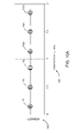
- FIG. 12 shows an embodiment integrating an optical pachymeter as described above with a microscope 402 of a system as described above.
- the microscope 402 has one viewing port coupled to a camera 404 , and another viewing port coupled to a pachymeter 400 .
- Microscope 402 may be a laser vision correction operating microscope commonly used in surgery, and provides a convenient coupling site for integrating a pachymeter 400 .
- a measurement beam 410 generated by a optical pachymeter 400 is directed to a cornea via a microscope 402 in a slightly off-axis manner.
- An off-axis measurement can be compensated for by a correction factor determined from geometric dimensions of the apparatus, and an off axis beam may be directed slightly away from an apex of a cornea to provide an increased amount of light reflected back into a microscope 402 .
- FIG. 12 illustrates an ablative beam 414 delivered by a beam delivery device such as a scanner 416 to a cornea 4 .
- a scanner 416 and a camera 402 are coupled together as indicated by electrical communication 420 to move synchronously.
- a system controller similar to a processor 22 of FIG. 1 may be in electrical communication with a scanner 416 , a microscope 402 , and a pachymeter 400 for controlling their operations.
Abstract
Description
Claims (7)
Priority Applications (1)
| Application Number | Priority Date | Filing Date | Title |
|---|---|---|---|
| US11/421,450 US7503916B2 (en) | 2002-06-27 | 2006-05-31 | Integrated scanning and ocular tomography system and method |
Applications Claiming Priority (3)
| Application Number | Priority Date | Filing Date | Title |
|---|---|---|---|
| US39233002P | 2002-06-27 | 2002-06-27 | |
| US10/601,119 US7133137B2 (en) | 2002-06-27 | 2003-06-19 | Integrated scanning and ocular tomography system and method |
| US11/421,450 US7503916B2 (en) | 2002-06-27 | 2006-05-31 | Integrated scanning and ocular tomography system and method |
Related Parent Applications (1)
| Application Number | Title | Priority Date | Filing Date |
|---|---|---|---|
| US10/601,119 Division US7133137B2 (en) | 2002-06-27 | 2003-06-19 | Integrated scanning and ocular tomography system and method |
Publications (2)
| Publication Number | Publication Date |
|---|---|
| US20060206102A1 US20060206102A1 (en) | 2006-09-14 |
| US7503916B2 true US7503916B2 (en) | 2009-03-17 |
Family
ID=30000847
Family Applications (2)
| Application Number | Title | Priority Date | Filing Date |
|---|---|---|---|
| US10/601,119 Expired - Fee Related US7133137B2 (en) | 2002-06-27 | 2003-06-19 | Integrated scanning and ocular tomography system and method |
| US11/421,450 Expired - Fee Related US7503916B2 (en) | 2002-06-27 | 2006-05-31 | Integrated scanning and ocular tomography system and method |
Family Applications Before (1)
| Application Number | Title | Priority Date | Filing Date |
|---|---|---|---|
| US10/601,119 Expired - Fee Related US7133137B2 (en) | 2002-06-27 | 2003-06-19 | Integrated scanning and ocular tomography system and method |
Country Status (2)
| Country | Link |
|---|---|
| US (2) | US7133137B2 (en) |
| WO (1) | WO2004003468A1 (en) |
Cited By (19)
| Publication number | Priority date | Publication date | Assignee | Title |
|---|---|---|---|---|
| US20090131921A1 (en) * | 2007-09-06 | 2009-05-21 | Lensx Lasers, Inc. | Precise Targeting of Surgical Photodisruption |
| US20110194743A1 (en) * | 2010-02-05 | 2011-08-11 | Ferenc Raksi | Gradient Search Integrated with Local Imaging in Laser Surgical Systems |
| US20110202044A1 (en) * | 2010-02-18 | 2011-08-18 | Ilya Goldshleger | Optical Coherence Tomographic System for Ophthalmic Surgery |
| US8398236B2 (en) | 2010-06-14 | 2013-03-19 | Alcon Lensx, Inc. | Image-guided docking for ophthalmic surgical systems |
| US8398238B1 (en) | 2011-08-26 | 2013-03-19 | Alcon Lensx, Inc. | Imaging-based guidance system for ophthalmic docking using a location-orientation analysis |
| US8459794B2 (en) | 2011-05-02 | 2013-06-11 | Alcon Lensx, Inc. | Image-processor-controlled misalignment-reduction for ophthalmic systems |
| US20130211387A1 (en) * | 2008-06-30 | 2013-08-15 | Wavelight Gmbh | Apparatus for ophthalmological, in particular refractive, laser surgery |
| US9023016B2 (en) | 2011-12-19 | 2015-05-05 | Alcon Lensx, Inc. | Image processor for intra-surgical optical coherence tomographic imaging of laser cataract procedures |
| US9066784B2 (en) | 2011-12-19 | 2015-06-30 | Alcon Lensx, Inc. | Intra-surgical optical coherence tomographic imaging of cataract procedures |
| US9492322B2 (en) | 2009-11-16 | 2016-11-15 | Alcon Lensx, Inc. | Imaging surgical target tissue by nonlinear scanning |
| US9532708B2 (en) | 2010-09-17 | 2017-01-03 | Alcon Lensx, Inc. | Electronically controlled fixation light for ophthalmic imaging systems |
| US9622913B2 (en) | 2011-05-18 | 2017-04-18 | Alcon Lensx, Inc. | Imaging-controlled laser surgical system |
| CN111063171A (en) * | 2019-12-13 | 2020-04-24 | 绍兴天宏激光科技有限公司 | Electronic product use monitoring system and method |
| US10758416B2 (en) | 2010-01-08 | 2020-09-01 | Amo Development, Llc | Method and system for modifying eye tissue and intraocular lenses |
| US10864114B2 (en) | 2010-01-08 | 2020-12-15 | Amo Development, Llc | Method and system for modifying eye tissue and intraocular lenses |
| US10973684B2 (en) * | 2004-03-23 | 2021-04-13 | Carl Zeiss Meditec Ag | Machining device and method |
| US11259963B2 (en) | 2010-04-30 | 2022-03-01 | Aleyegn Technologies Llc | Method and apparatus for treatment of ocular tissue using combined modalities |
| US11369516B2 (en) | 2013-03-15 | 2022-06-28 | Aleyegn Technologies Llc | Scleral translocation elasto-modulation methods and apparatus |
| US11857463B2 (en) | 2011-05-05 | 2024-01-02 | Michael S. Berlin | Methods for the treatment of glaucoma using visible and infrared ultrashort laser pulses |
Families Citing this family (85)
| Publication number | Priority date | Publication date | Assignee | Title |
|---|---|---|---|---|
| WO2001028477A1 (en) * | 1999-10-21 | 2001-04-26 | Technolas Gmbh Ophthalmologische Systeme | Multi-step laser correction of ophthalmic refractive errors |
| US8679089B2 (en) | 2001-05-21 | 2014-03-25 | Michael S. Berlin | Glaucoma surgery methods and systems |
| US9603741B2 (en) | 2000-05-19 | 2017-03-28 | Michael S. Berlin | Delivery system and method of use for the eye |
| CA2446143C (en) | 2000-05-19 | 2010-01-19 | Michael S. Berlin | Delivery system and method of use for the eye |
| JP2004153149A (en) * | 2002-10-31 | 2004-05-27 | Sumitomo Electric Ind Ltd | Light emitting module |
| DE60333389D1 (en) * | 2002-12-18 | 2010-08-26 | Koninkl Philips Electronics Nv | Method and arrangement for optical coherence tomography |
| DE10326527B8 (en) * | 2003-06-12 | 2015-08-06 | Carl Zeiss Meditec Ag | Method and device for determining a movement of a human eye |
| US20080269731A1 (en) * | 2003-11-19 | 2008-10-30 | Casimir Andrew Swinger | Method and apparatus applying patient-verified prescription of high order aberrations |
| US7481536B2 (en) * | 2004-02-19 | 2009-01-27 | Amo Manufacturing Usa, Llc | Methods and systems for differentiating left and right eye images |
| JP4381847B2 (en) * | 2004-02-26 | 2009-12-09 | 株式会社トプコン | Optical image measuring device |
| WO2006022342A1 (en) * | 2004-08-26 | 2006-03-02 | Nippon Telegraph And Telephone Corporation | Tissue measuring optical interference tomography-use light producing device and tissue measuring optical interference tomography device |
| CN100516772C (en) * | 2004-09-09 | 2009-07-22 | 鸿富锦精密工业(深圳)有限公司 | Optical signall processor and process thereof |
| US7450242B2 (en) * | 2004-12-10 | 2008-11-11 | Fujifilm Corporation | Optical tomography apparatus |
| TWI247096B (en) * | 2004-12-17 | 2006-01-11 | Hon Hai Prec Ind Co Ltd | Optical fiber interference device used for thickness measuring and the method of the same |
| US8394084B2 (en) | 2005-01-10 | 2013-03-12 | Optimedica Corporation | Apparatus for patterned plasma-mediated laser trephination of the lens capsule and three dimensional phaco-segmentation |
| US7365856B2 (en) * | 2005-01-21 | 2008-04-29 | Carl Zeiss Meditec, Inc. | Method of motion correction in optical coherence tomography imaging |
| ES2596365T3 (en) * | 2005-03-18 | 2017-01-09 | Wavelight Gmbh | Device for ophthalmic laser surgery |
| US7805009B2 (en) | 2005-04-06 | 2010-09-28 | Carl Zeiss Meditec, Inc. | Method and apparatus for measuring motion of a subject using a series of partial images from an imaging system |
| US7359057B2 (en) * | 2005-08-26 | 2008-04-15 | Ball Aerospace & Technologies Corp. | Method and apparatus for measuring small shifts in optical wavelengths |
| DE102005042436C5 (en) * | 2005-09-07 | 2010-05-27 | Carl Zeiss Surgical Gmbh | Ophthalmic surgical microscope with measuring device |
| JP2007085931A (en) * | 2005-09-22 | 2007-04-05 | Fujinon Corp | Optical tomographic imaging system |
| DE102005053297A1 (en) * | 2005-11-08 | 2007-05-10 | Bausch & Lomb Inc. | System and method for correcting ophthalmic refractive errors |
| US20070106285A1 (en) | 2005-11-09 | 2007-05-10 | Ferenc Raksi | Laser scanner |
| US20070173791A1 (en) * | 2006-01-20 | 2007-07-26 | Intralase Corp. | System for ophthalmic laser surgery |
| US20070219541A1 (en) * | 2006-03-14 | 2007-09-20 | Intralase Corp. | System and method for ophthalmic laser surgery on a cornea |
| US7416300B2 (en) * | 2006-05-25 | 2008-08-26 | Coopervision International Holding Company, Lp | Measurement of lenses and lens molds using optical coherence tomography |
| US20080009844A1 (en) * | 2006-06-26 | 2008-01-10 | Ingeborg Rolle | Device for Laser Surgery |
| DE102006036086A1 (en) * | 2006-08-02 | 2008-02-07 | Bausch & Lomb Incorporated | Method and apparatus for calculating a laser shot file for use in a refractive excimer laser |
| DE102006036085A1 (en) * | 2006-08-02 | 2008-02-07 | Bausch & Lomb Incorporated | Method and apparatus for calculating a laser shot file for use in an excimer laser |
| US8088124B2 (en) * | 2007-01-19 | 2012-01-03 | Technolas Perfect Vision Gmbh | System and method for precise beam positioning in ocular surgery |
| US20110319875A1 (en) * | 2007-01-19 | 2011-12-29 | Frieder Loesel | Apparatus and Method for Morphing a Three-Dimensional Target Surface into a Two-Dimensional Image for Use in Guiding a Laser Beam in Ocular Surgery |
| CN101631522B (en) | 2007-03-13 | 2014-11-05 | 眼科医疗公司 | Apparatus for creating ocular surgical and relaxing incisions |
| AU2015200832B2 (en) * | 2007-03-13 | 2018-02-22 | Amo Development, Llc | Apparatus for creating ocular surgical and relaxing incisions |
| US8042944B2 (en) * | 2007-03-20 | 2011-10-25 | Topcon Europe Medical B.V. | Apparatus and method for observing an eye, as well as OCT-module |
| US20090043211A1 (en) * | 2007-03-28 | 2009-02-12 | Albert Ting | Mems based optical coherence tomography probe |
| US7988290B2 (en) | 2007-06-27 | 2011-08-02 | AMO Wavefront Sciences LLC. | Systems and methods for measuring the shape and location of an object |
| US20170360609A9 (en) | 2007-09-24 | 2017-12-21 | Ivantis, Inc. | Methods and devices for increasing aqueous humor outflow |
| KR100900263B1 (en) * | 2007-11-08 | 2009-06-01 | 르호봇테크(주) | Guidance device for focus of iris and guidance method for the same |
| US8230866B2 (en) * | 2007-12-13 | 2012-07-31 | Carl Zeiss Meditec Ag | Systems and methods for treating glaucoma and systems and methods for imaging a portion of an eye |
| PL2230990T3 (en) * | 2007-12-21 | 2017-08-31 | Bausch & Lomb Incorporated | Ophthalmic instrument alignment apparatus and method of using same |
| DE102008008732A1 (en) | 2008-02-11 | 2009-08-13 | Carl Zeiss Meditec Ag | Method and device for determining corneal radii |
| AU2009221859B2 (en) | 2008-03-05 | 2013-04-18 | Alcon Inc. | Methods and apparatus for treating glaucoma |
| EP2108328B2 (en) * | 2008-04-09 | 2020-08-26 | Brainlab AG | Image-based control method for medicinal devices |
| US8418168B2 (en) | 2008-05-29 | 2013-04-09 | Research In Motion Limited | Method and system for performing a software upgrade on an electronic device connected to a computer |
| US8041346B2 (en) | 2008-05-29 | 2011-10-18 | Research In Motion Limited | Method and system for establishing a service relationship between a mobile communication device and a mobile data server for connecting to a wireless network |
| DE102008028509A1 (en) * | 2008-06-16 | 2009-12-24 | Technolas Gmbh Ophthalmologische Systeme | Treatment pattern monitoring device |
| DE102008035995A1 (en) * | 2008-08-01 | 2010-02-04 | Technolas Perfect Vision Gmbh | Combination of excimer laser ablation and femtosecond laser technique |
| CN102137617A (en) * | 2008-08-28 | 2011-07-27 | 泰克诺拉斯完美视觉股份有限公司 | Eye measurement and modeling techniques |
| DE102008053827A1 (en) | 2008-10-30 | 2010-05-12 | Technolas Perfect Vision Gmbh | Apparatus and method for providing a laser shot file |
| CN102481404B (en) | 2009-07-09 | 2014-03-05 | 伊万提斯公司 | Ocular implants |
| AU2010271274B2 (en) | 2009-07-09 | 2015-05-21 | Alcon Inc. | Single operator device for delivering an ocular implant |
| EP2547298B1 (en) * | 2010-03-19 | 2019-05-08 | Avedro, Inc. | Systems for applying and monitoring eye therapy |
| CN102869299A (en) * | 2010-05-04 | 2013-01-09 | 爱克透镜国际公司 | Corneal topographer |
| DE102010016862B3 (en) * | 2010-05-10 | 2011-09-22 | Precitec Optronik Gmbh | Material processing device with in-situ measurement of the machining distance |
| US9427493B2 (en) | 2011-03-07 | 2016-08-30 | The Regents Of The University Of Colorado | Shape memory polymer intraocular lenses |
| WO2012123947A2 (en) * | 2011-03-14 | 2012-09-20 | G & D Innovative Analysis, Ltd. | Method and apparatus for morphological analysis |
| US9033510B2 (en) | 2011-03-30 | 2015-05-19 | Carl Zeiss Meditec, Inc. | Systems and methods for efficiently obtaining measurements of the human eye using tracking |
| GB2489722B (en) | 2011-04-06 | 2017-01-18 | Precitec Optronik Gmbh | Apparatus and method for determining a depth of a region having a high aspect ratio that protrudes into a surface of a semiconductor wafer |
| US8657776B2 (en) | 2011-06-14 | 2014-02-25 | Ivantis, Inc. | Ocular implants for delivery into the eye |
| DE102011051146B3 (en) | 2011-06-17 | 2012-10-04 | Precitec Optronik Gmbh | Test method for testing a bonding layer between wafer-shaped samples |
| US8857988B2 (en) | 2011-07-07 | 2014-10-14 | Carl Zeiss Meditec, Inc. | Data acquisition methods for reduced motion artifacts and applications in OCT angiography |
| US8663150B2 (en) | 2011-12-19 | 2014-03-04 | Ivantis, Inc. | Delivering ocular implants into the eye |
| US9101294B2 (en) | 2012-01-19 | 2015-08-11 | Carl Zeiss Meditec, Inc. | Systems and methods for enhanced accuracy in OCT imaging of the cornea |
| US8807752B2 (en) | 2012-03-08 | 2014-08-19 | Technolas Perfect Vision Gmbh | System and method with refractive corrections for controlled placement of a laser beam's focal point |
| US9358156B2 (en) | 2012-04-18 | 2016-06-07 | Invantis, Inc. | Ocular implants for delivery into an anterior chamber of the eye |
| US20130338649A1 (en) * | 2012-06-02 | 2013-12-19 | Nidek Co., Ltd. | Ophthalmic laser surgical apparatus |
| US9437041B2 (en) * | 2012-10-30 | 2016-09-06 | The Penn State Research Foundation | 3D laser ablation tomography |
| DE102012111008B4 (en) | 2012-11-15 | 2014-05-22 | Precitec Optronik Gmbh | Optical measuring method and optical measuring device for detecting a surface topography |
| US10617558B2 (en) | 2012-11-28 | 2020-04-14 | Ivantis, Inc. | Apparatus for delivering ocular implants into an anterior chamber of the eye |
| US9398979B2 (en) | 2013-03-11 | 2016-07-26 | Technolas Perfect Vision Gmbh | Dimensional compensator for use with a patient interface |
| CN105324629B (en) | 2013-06-17 | 2018-08-24 | 普雷茨特激光技术有限公司 | Optical measuring device and measuring method for obtaining range difference |
| EP3646774A1 (en) * | 2013-07-25 | 2020-05-06 | Optimedica Corporation | In situ determination of refractive index of materials |
| JP6488540B2 (en) * | 2013-11-29 | 2019-03-27 | 株式会社ニデック | Ophthalmic measuring device |
| WO2016011056A1 (en) | 2014-07-14 | 2016-01-21 | Ivantis, Inc. | Ocular implant delivery system and method |
| US10029330B2 (en) * | 2015-06-17 | 2018-07-24 | The Boeing Company | Hybrid laser machining of multi-material stack-ups |
| CN108135470B (en) | 2015-08-14 | 2021-03-09 | 伊万提斯公司 | Ocular implant with pressure sensor and delivery system |
| WO2017106517A1 (en) | 2015-12-15 | 2017-06-22 | Ivantis, Inc. | Ocular implant and delivery system |
| US10234265B2 (en) | 2016-12-12 | 2019-03-19 | Precitec Optronik Gmbh | Distance measuring device and method for measuring distances |
| AU2018317499A1 (en) | 2017-08-18 | 2020-02-27 | Ellex Medical Pty Ltd | Multi-spot ophthalmic laser |
| DE102017126310A1 (en) | 2017-11-09 | 2019-05-09 | Precitec Optronik Gmbh | Distance measuring device |
| CN108606774B (en) * | 2018-05-16 | 2023-12-26 | 视微如著科技(上海)有限公司 | Automatic safety protection device and method suitable for ophthalmic optical scanning imaging device |
| DE102018130901A1 (en) | 2018-12-04 | 2020-06-04 | Precitec Optronik Gmbh | Optical measuring device |
| US11484361B2 (en) * | 2019-08-27 | 2022-11-01 | Nikolai Tankovich | Tip for multiple beam tissue therapy |
| FI129606B (en) * | 2020-08-11 | 2022-05-31 | Photono Oy | A pachymeter device and a method for measuring thickness of a cornea |
| EP4274529A1 (en) | 2021-01-11 | 2023-11-15 | Alcon Inc. | Systems and methods for viscoelastic delivery |
Citations (37)
| Publication number | Priority date | Publication date | Assignee | Title |
|---|---|---|---|---|
| US3735036A (en) | 1970-10-26 | 1973-05-22 | Express I M Co | Real time interferometry contour mapping system |
| US4665913A (en) | 1983-11-17 | 1987-05-19 | Lri L.P. | Method for ophthalmological surgery |
| US4884697A (en) | 1988-06-21 | 1989-12-05 | Takacs Peter Z | Surface profiling interferometer |
| US4984883A (en) | 1989-07-21 | 1991-01-15 | Joseph Winocur | Translation insensitive keratometer using moire deflectometry |
| US5062702A (en) | 1990-03-16 | 1991-11-05 | Intelligent Surgical Lasers, Inc. | Device for mapping corneal topography |
| US5116115A (en) | 1990-05-09 | 1992-05-26 | Wyko Corporation | Method and apparatus for measuring corneal topography |
| US5144630A (en) | 1991-07-29 | 1992-09-01 | Jtt International, Inc. | Multiwavelength solid state laser using frequency conversion techniques |
| US5260761A (en) | 1990-12-07 | 1993-11-09 | Ometron Limited | Apparatus for the measurement of surface shape |
| US5317389A (en) | 1989-06-12 | 1994-05-31 | California Institute Of Technology | Method and apparatus for white-light dispersed-fringe interferometric measurement of corneal topography |
| US5321501A (en) | 1991-04-29 | 1994-06-14 | Massachusetts Institute Of Technology | Method and apparatus for optical imaging with means for controlling the longitudinal range of the sample |
| US5349440A (en) | 1991-02-08 | 1994-09-20 | Hughes Aircraft Company | Interferometric laser profilometer including a multimode laser diode emitting a range of stable wavelengths |
| US5406342A (en) | 1992-01-15 | 1995-04-11 | Euclid Medical Instruments | System for determining the topography of a curved surface |
| US5521657A (en) | 1992-05-01 | 1996-05-28 | Summit Technology Inc. | Method and system for topographic measurement by measuring the distance between a rigid reference member and a surface of an eye |
| US5610716A (en) | 1995-08-28 | 1997-03-11 | Hewlett-Packard Company | Method and apparatus for measuring film thickness utilizing the slope of the phase of the Fourier transform of an autocorrelator signal |
| US5646791A (en) | 1995-01-04 | 1997-07-08 | Visx Incorporated | Method and apparatus for temporal and spatial beam integration |
| US5683379A (en) | 1992-10-01 | 1997-11-04 | Chiron Technolas Gmbh Ophthalmologische Systeme | Apparatus for modifying the surface of the eye through large beam laser polishing and method of controlling the apparatus |
| US5713892A (en) | 1991-08-16 | 1998-02-03 | Visx, Inc. | Method and apparatus for combined cylindrical and spherical eye corrections |
| US5742626A (en) | 1996-08-14 | 1998-04-21 | Aculight Corporation | Ultraviolet solid state laser, method of using same and laser surgery apparatus |
| US5782822A (en) * | 1995-10-27 | 1998-07-21 | Ir Vision, Inc. | Method and apparatus for removing corneal tissue with infrared laser radiation |
| WO1999001716A1 (en) | 1997-07-01 | 1999-01-14 | David Macpherson | Ablation profiler |
| US5909270A (en) | 1996-05-10 | 1999-06-01 | California Institute Of Technology | Conoscopic system for real-time corneal topography |
| US5912779A (en) | 1992-12-01 | 1999-06-15 | Nat Semiconductor Corp | Method of reading and writing data on a magnetic medium |
| US6004313A (en) * | 1998-06-26 | 1999-12-21 | Visx, Inc. | Patient fixation system and method for laser eye surgery |
| US6203539B1 (en) | 1993-05-07 | 2001-03-20 | Visx, Incorporated | Method and system for laser treatment of refractive errors using offset imaging |
| US6280435B1 (en) | 1998-03-04 | 2001-08-28 | Visx, Incorporated | Method and systems for laser treatment of presbyopia using offset imaging |
| US6319247B1 (en) | 1993-05-07 | 2001-11-20 | Visx, Incorporated | Systems and methods for corneal surface ablation to correct hyperopia |
| US6331177B1 (en) | 1998-04-17 | 2001-12-18 | Visx, Incorporated | Multiple beam laser sculpting system and method |
| US6448094B2 (en) | 2000-01-28 | 2002-09-10 | Tokyo Electron Limited | Method of detecting etching depth |
| US6451009B1 (en) | 2000-09-12 | 2002-09-17 | The Regents Of The University Of California | OCDR guided laser ablation device |
| US6454761B1 (en) | 1995-01-30 | 2002-09-24 | Philip D. Freedman | Laser surgery device and method |
| US6501551B1 (en) | 1991-04-29 | 2002-12-31 | Massachusetts Institute Of Technology | Fiber optic imaging endoscope interferometer with at least one faraday rotator |
| US20030025913A1 (en) | 2001-01-29 | 2003-02-06 | Izatt Joseph A. | Frequency-encoded parallel OCT and associated systems and methods |
| US6815228B2 (en) | 2000-06-20 | 2004-11-09 | Hitachi, Ltd. | Film thickness measuring method of member to be processed using emission spectroscopy and processing method of the member using the measuring method |
| US6873419B2 (en) | 2001-11-16 | 2005-03-29 | National Research Council Of Canada | Method and apparatus for three-dimensional compositional mapping of heterogeneous materials |
| US6882431B2 (en) | 2002-11-26 | 2005-04-19 | Malvin C. Teich | Quantum optical coherence tomography data collection apparatus and method for processing therefor |
| US7160288B2 (en) * | 2002-02-27 | 2007-01-09 | Nidek Co., Ltd. | Ophthalmic apparatus |
| US7282060B2 (en) * | 2003-12-23 | 2007-10-16 | Reliant Technologies, Inc. | Method and apparatus for monitoring and controlling laser-induced tissue treatment |
Family Cites Families (3)
| Publication number | Priority date | Publication date | Assignee | Title |
|---|---|---|---|---|
| US5118115A (en) * | 1989-05-04 | 1992-06-02 | Codinha J Albert | Economic and military conflict board game |
| US5408342A (en) * | 1993-02-25 | 1995-04-18 | Canon Kabushiki Kaisha | Color image processing apparatus, method, and printer driver |
| JPH08304741A (en) * | 1995-05-09 | 1996-11-22 | Olympus Optical Co Ltd | Optical system including diffraction optical element |
-
2003
- 2003-06-19 US US10/601,119 patent/US7133137B2/en not_active Expired - Fee Related
- 2003-06-26 WO PCT/US2003/020338 patent/WO2004003468A1/en not_active Application Discontinuation
-
2006
- 2006-05-31 US US11/421,450 patent/US7503916B2/en not_active Expired - Fee Related
Patent Citations (39)
| Publication number | Priority date | Publication date | Assignee | Title |
|---|---|---|---|---|
| US3735036A (en) | 1970-10-26 | 1973-05-22 | Express I M Co | Real time interferometry contour mapping system |
| US4665913A (en) | 1983-11-17 | 1987-05-19 | Lri L.P. | Method for ophthalmological surgery |
| US4884697A (en) | 1988-06-21 | 1989-12-05 | Takacs Peter Z | Surface profiling interferometer |
| US5317389A (en) | 1989-06-12 | 1994-05-31 | California Institute Of Technology | Method and apparatus for white-light dispersed-fringe interferometric measurement of corneal topography |
| US4984883A (en) | 1989-07-21 | 1991-01-15 | Joseph Winocur | Translation insensitive keratometer using moire deflectometry |
| US5062702A (en) | 1990-03-16 | 1991-11-05 | Intelligent Surgical Lasers, Inc. | Device for mapping corneal topography |
| US5116115A (en) | 1990-05-09 | 1992-05-26 | Wyko Corporation | Method and apparatus for measuring corneal topography |
| US5260761A (en) | 1990-12-07 | 1993-11-09 | Ometron Limited | Apparatus for the measurement of surface shape |
| US5349440A (en) | 1991-02-08 | 1994-09-20 | Hughes Aircraft Company | Interferometric laser profilometer including a multimode laser diode emitting a range of stable wavelengths |
| US5321501A (en) | 1991-04-29 | 1994-06-14 | Massachusetts Institute Of Technology | Method and apparatus for optical imaging with means for controlling the longitudinal range of the sample |
| US5459570A (en) | 1991-04-29 | 1995-10-17 | Massachusetts Institute Of Technology | Method and apparatus for performing optical measurements |
| US6501551B1 (en) | 1991-04-29 | 2002-12-31 | Massachusetts Institute Of Technology | Fiber optic imaging endoscope interferometer with at least one faraday rotator |
| US5144630A (en) | 1991-07-29 | 1992-09-01 | Jtt International, Inc. | Multiwavelength solid state laser using frequency conversion techniques |
| US5713892A (en) | 1991-08-16 | 1998-02-03 | Visx, Inc. | Method and apparatus for combined cylindrical and spherical eye corrections |
| US5406342A (en) | 1992-01-15 | 1995-04-11 | Euclid Medical Instruments | System for determining the topography of a curved surface |
| US5521657A (en) | 1992-05-01 | 1996-05-28 | Summit Technology Inc. | Method and system for topographic measurement by measuring the distance between a rigid reference member and a surface of an eye |
| US5683379A (en) | 1992-10-01 | 1997-11-04 | Chiron Technolas Gmbh Ophthalmologische Systeme | Apparatus for modifying the surface of the eye through large beam laser polishing and method of controlling the apparatus |
| US5912779A (en) | 1992-12-01 | 1999-06-15 | Nat Semiconductor Corp | Method of reading and writing data on a magnetic medium |
| US6319247B1 (en) | 1993-05-07 | 2001-11-20 | Visx, Incorporated | Systems and methods for corneal surface ablation to correct hyperopia |
| US6203539B1 (en) | 1993-05-07 | 2001-03-20 | Visx, Incorporated | Method and system for laser treatment of refractive errors using offset imaging |
| US5912775A (en) | 1995-01-04 | 1999-06-15 | Visx, Incorporated | Method and apparatus for temporal and spatial beam integration |
| US5646791A (en) | 1995-01-04 | 1997-07-08 | Visx Incorporated | Method and apparatus for temporal and spatial beam integration |
| US6454761B1 (en) | 1995-01-30 | 2002-09-24 | Philip D. Freedman | Laser surgery device and method |
| US5610716A (en) | 1995-08-28 | 1997-03-11 | Hewlett-Packard Company | Method and apparatus for measuring film thickness utilizing the slope of the phase of the Fourier transform of an autocorrelator signal |
| US5782822A (en) * | 1995-10-27 | 1998-07-21 | Ir Vision, Inc. | Method and apparatus for removing corneal tissue with infrared laser radiation |
| US5909270A (en) | 1996-05-10 | 1999-06-01 | California Institute Of Technology | Conoscopic system for real-time corneal topography |
| US5742626A (en) | 1996-08-14 | 1998-04-21 | Aculight Corporation | Ultraviolet solid state laser, method of using same and laser surgery apparatus |
| WO1999001716A1 (en) | 1997-07-01 | 1999-01-14 | David Macpherson | Ablation profiler |
| US6280435B1 (en) | 1998-03-04 | 2001-08-28 | Visx, Incorporated | Method and systems for laser treatment of presbyopia using offset imaging |
| US6331177B1 (en) | 1998-04-17 | 2001-12-18 | Visx, Incorporated | Multiple beam laser sculpting system and method |
| US6004313A (en) * | 1998-06-26 | 1999-12-21 | Visx, Inc. | Patient fixation system and method for laser eye surgery |
| US6448094B2 (en) | 2000-01-28 | 2002-09-10 | Tokyo Electron Limited | Method of detecting etching depth |
| US6815228B2 (en) | 2000-06-20 | 2004-11-09 | Hitachi, Ltd. | Film thickness measuring method of member to be processed using emission spectroscopy and processing method of the member using the measuring method |
| US6451009B1 (en) | 2000-09-12 | 2002-09-17 | The Regents Of The University Of California | OCDR guided laser ablation device |
| US20030025913A1 (en) | 2001-01-29 | 2003-02-06 | Izatt Joseph A. | Frequency-encoded parallel OCT and associated systems and methods |
| US6873419B2 (en) | 2001-11-16 | 2005-03-29 | National Research Council Of Canada | Method and apparatus for three-dimensional compositional mapping of heterogeneous materials |
| US7160288B2 (en) * | 2002-02-27 | 2007-01-09 | Nidek Co., Ltd. | Ophthalmic apparatus |
| US6882431B2 (en) | 2002-11-26 | 2005-04-19 | Malvin C. Teich | Quantum optical coherence tomography data collection apparatus and method for processing therefor |
| US7282060B2 (en) * | 2003-12-23 | 2007-10-16 | Reliant Technologies, Inc. | Method and apparatus for monitoring and controlling laser-induced tissue treatment |
Non-Patent Citations (2)
| Title |
|---|
| Bohnke et al., "Real-Time pachymetry during photorefractive keratectomy using low-coherence reflectometry" Journal of Biomedical Optics 6(4) (Oct. 2001), pp. 412-417. |
| Borsuztky et al., "Tunable UV Radiation at Short Wavelengths (188-240nm) Generated by Sum Frequency Mixing in Lithium Borate" Appl. Phys., B52:380-384 (1991). |
Cited By (27)
| Publication number | Priority date | Publication date | Assignee | Title |
|---|---|---|---|---|
| US10973684B2 (en) * | 2004-03-23 | 2021-04-13 | Carl Zeiss Meditec Ag | Machining device and method |
| US20090131921A1 (en) * | 2007-09-06 | 2009-05-21 | Lensx Lasers, Inc. | Precise Targeting of Surgical Photodisruption |
| US9408749B2 (en) | 2007-09-06 | 2016-08-09 | Alcon Lensx, Inc. | Precise targeting of surgical photodisruption |
| US9044303B2 (en) | 2007-09-06 | 2015-06-02 | Alcon Lensx, Inc. | Precise targeting of surgical photodisruption |
| US8764737B2 (en) | 2007-09-06 | 2014-07-01 | Alcon Lensx, Inc. | Precise targeting of surgical photodisruption |
| US20130211387A1 (en) * | 2008-06-30 | 2013-08-15 | Wavelight Gmbh | Apparatus for ophthalmological, in particular refractive, laser surgery |
| US9492322B2 (en) | 2009-11-16 | 2016-11-15 | Alcon Lensx, Inc. | Imaging surgical target tissue by nonlinear scanning |
| US11058583B2 (en) | 2010-01-08 | 2021-07-13 | Amo Development, Llc | Method and system for modifying eye tissue and intraocular lenses |
| US10758416B2 (en) | 2010-01-08 | 2020-09-01 | Amo Development, Llc | Method and system for modifying eye tissue and intraocular lenses |
| US10864114B2 (en) | 2010-01-08 | 2020-12-15 | Amo Development, Llc | Method and system for modifying eye tissue and intraocular lenses |
| US8265364B2 (en) | 2010-02-05 | 2012-09-11 | Alcon Lensx, Inc. | Gradient search integrated with local imaging in laser surgical systems |
| US20110194743A1 (en) * | 2010-02-05 | 2011-08-11 | Ferenc Raksi | Gradient Search Integrated with Local Imaging in Laser Surgical Systems |
| US8414564B2 (en) | 2010-02-18 | 2013-04-09 | Alcon Lensx, Inc. | Optical coherence tomographic system for ophthalmic surgery |
| US20110202044A1 (en) * | 2010-02-18 | 2011-08-18 | Ilya Goldshleger | Optical Coherence Tomographic System for Ophthalmic Surgery |
| US11259963B2 (en) | 2010-04-30 | 2022-03-01 | Aleyegn Technologies Llc | Method and apparatus for treatment of ocular tissue using combined modalities |
| US8398236B2 (en) | 2010-06-14 | 2013-03-19 | Alcon Lensx, Inc. | Image-guided docking for ophthalmic surgical systems |
| US9532708B2 (en) | 2010-09-17 | 2017-01-03 | Alcon Lensx, Inc. | Electronically controlled fixation light for ophthalmic imaging systems |
| US8459794B2 (en) | 2011-05-02 | 2013-06-11 | Alcon Lensx, Inc. | Image-processor-controlled misalignment-reduction for ophthalmic systems |
| US11857463B2 (en) | 2011-05-05 | 2024-01-02 | Michael S. Berlin | Methods for the treatment of glaucoma using visible and infrared ultrashort laser pulses |
| US9622913B2 (en) | 2011-05-18 | 2017-04-18 | Alcon Lensx, Inc. | Imaging-controlled laser surgical system |
| US8398238B1 (en) | 2011-08-26 | 2013-03-19 | Alcon Lensx, Inc. | Imaging-based guidance system for ophthalmic docking using a location-orientation analysis |
| US9456926B2 (en) | 2011-12-19 | 2016-10-04 | Alcon Lensx, Inc. | Intra-surgical optical coherence tomographic imaging of cataract procedures |
| US9456927B2 (en) | 2011-12-19 | 2016-10-04 | Alcon Lensx, Inc. | Image processor for intra-surgical optical coherence tomographic imaging of laser cataract procedures |
| US9066784B2 (en) | 2011-12-19 | 2015-06-30 | Alcon Lensx, Inc. | Intra-surgical optical coherence tomographic imaging of cataract procedures |
| US9023016B2 (en) | 2011-12-19 | 2015-05-05 | Alcon Lensx, Inc. | Image processor for intra-surgical optical coherence tomographic imaging of laser cataract procedures |
| US11369516B2 (en) | 2013-03-15 | 2022-06-28 | Aleyegn Technologies Llc | Scleral translocation elasto-modulation methods and apparatus |
| CN111063171A (en) * | 2019-12-13 | 2020-04-24 | 绍兴天宏激光科技有限公司 | Electronic product use monitoring system and method |
Also Published As
| Publication number | Publication date |
|---|---|
| US20040021874A1 (en) | 2004-02-05 |
| WO2004003468A1 (en) | 2004-01-08 |
| US7133137B2 (en) | 2006-11-07 |
| US20060206102A1 (en) | 2006-09-14 |
Similar Documents
| Publication | Publication Date | Title |
|---|---|---|
| US7503916B2 (en) | Integrated scanning and ocular tomography system and method | |
| US20210101020A1 (en) | Systems and methods for applying and monitoring eye therapy | |
| US10702146B2 (en) | Angular multiplexed optical coherence tomography systems and methods | |
| US9408749B2 (en) | Precise targeting of surgical photodisruption | |
| JP4651253B2 (en) | Hydration and topographic measurements for laser ablation | |
| JP6040578B2 (en) | Ophthalmic laser surgery device | |
| JP5763327B2 (en) | Imaging of surgical target tissue by nonlinear scanning | |
| US20090149841A1 (en) | Effective Laser Photodisruptive Surgery in a Gravity Field | |
| US20100106143A1 (en) | Apparatus, method and control program for ophthalmological, in particular, refractive laser surgery | |
| JP2010538703A (en) | Apparatus, system and technique for providing an interface to the eye in laser surgery | |
| WO1999065431A1 (en) | Z axis tracker | |
| RU2183108C1 (en) | Method and device for adjusting cornea refraction properties in performing in situ monitoring by applying optical coherent tomography method | |
| US10617564B1 (en) | Area scanning photomedicine device and method | |
| AU750778B2 (en) | Z axis tracker |
Legal Events
| Date | Code | Title | Description |
|---|---|---|---|
| AS | Assignment |
Owner name: BANK OF AMERICA, N.A., AS ADMINISTRATIVE AGENT,NOR Free format text: INTELLECTUAL PROPERTY SECURITY AGREEMENT;ASSIGNOR:VISX, INCORPORATED;REEL/FRAME:019501/0142 Effective date: 20070402 Owner name: BANK OF AMERICA, N.A., AS ADMINISTRATIVE AGENT, NO Free format text: INTELLECTUAL PROPERTY SECURITY AGREEMENT;ASSIGNOR:VISX, INCORPORATED;REEL/FRAME:019501/0142 Effective date: 20070402 |
|
| AS | Assignment |
Owner name: AMO MANUFACTURING USA, LLC, CALIFORNIA Free format text: CHANGE OF NAME;ASSIGNOR:VISX, INCORPORATED;REEL/FRAME:020308/0064 Effective date: 20071231 Owner name: AMO MANUFACTURING USA, LLC,CALIFORNIA Free format text: CHANGE OF NAME;ASSIGNOR:VISX, INCORPORATED;REEL/FRAME:020308/0064 Effective date: 20071231 |
|
| STCF | Information on status: patent grant |
Free format text: PATENTED CASE |
|
| AS | Assignment |
Owner name: AMO MANUFACTURING USA, LLC; FORMERLY VISX, INCORPO Free format text: RELEASE BY SECURED PARTY;ASSIGNOR:BANK OF AMERICA, N.A. AS ADMINISTRATIVE AGENT;REEL/FRAME:022331/0698 Effective date: 20090225 |
|
| CC | Certificate of correction | ||
| FPAY | Fee payment |
Year of fee payment: 4 |
|
| FPAY | Fee payment |
Year of fee payment: 8 |
|
| FEPP | Fee payment procedure |
Free format text: MAINTENANCE FEE REMINDER MAILED (ORIGINAL EVENT CODE: REM.); ENTITY STATUS OF PATENT OWNER: LARGE ENTITY |
|
| LAPS | Lapse for failure to pay maintenance fees |
Free format text: PATENT EXPIRED FOR FAILURE TO PAY MAINTENANCE FEES (ORIGINAL EVENT CODE: EXP.); ENTITY STATUS OF PATENT OWNER: LARGE ENTITY |
|
| STCH | Information on status: patent discontinuation |
Free format text: PATENT EXPIRED DUE TO NONPAYMENT OF MAINTENANCE FEES UNDER 37 CFR 1.362 |
|
| FP | Lapsed due to failure to pay maintenance fee |
Effective date: 20210317 |