EP1493395A2 - Control apparatus for an automated surgical biopsy device - Google Patents
Control apparatus for an automated surgical biopsy device Download PDFInfo
- Publication number
- EP1493395A2 EP1493395A2 EP04077757A EP04077757A EP1493395A2 EP 1493395 A2 EP1493395 A2 EP 1493395A2 EP 04077757 A EP04077757 A EP 04077757A EP 04077757 A EP04077757 A EP 04077757A EP 1493395 A2 EP1493395 A2 EP 1493395A2
- Authority
- EP
- European Patent Office
- Prior art keywords
- cutter
- translation
- motor
- tissue
- speed
- Prior art date
- Legal status (The legal status is an assumption and is not a legal conclusion. Google has not performed a legal analysis and makes no representation as to the accuracy of the status listed.)
- Granted
Links
Images


Classifications
-
- A—HUMAN NECESSITIES
- A61—MEDICAL OR VETERINARY SCIENCE; HYGIENE
- A61B—DIAGNOSIS; SURGERY; IDENTIFICATION
- A61B10/00—Other methods or instruments for diagnosis, e.g. instruments for taking a cell sample, for biopsy, for vaccination diagnosis; Sex determination; Ovulation-period determination; Throat striking implements
- A61B10/02—Instruments for taking cell samples or for biopsy
- A61B10/0233—Pointed or sharp biopsy instruments
- A61B10/0266—Pointed or sharp biopsy instruments means for severing sample
- A61B10/0275—Pointed or sharp biopsy instruments means for severing sample with sample notch, e.g. on the side of inner stylet
-
- A—HUMAN NECESSITIES
- A61—MEDICAL OR VETERINARY SCIENCE; HYGIENE
- A61B—DIAGNOSIS; SURGERY; IDENTIFICATION
- A61B17/00—Surgical instruments, devices or methods, e.g. tourniquets
- A61B17/32—Surgical cutting instruments
- A61B17/320016—Endoscopic cutting instruments, e.g. arthroscopes, resectoscopes
- A61B17/32002—Endoscopic cutting instruments, e.g. arthroscopes, resectoscopes with continuously rotating, oscillating or reciprocating cutting instruments
-
- A—HUMAN NECESSITIES
- A61—MEDICAL OR VETERINARY SCIENCE; HYGIENE
- A61B—DIAGNOSIS; SURGERY; IDENTIFICATION
- A61B10/00—Other methods or instruments for diagnosis, e.g. instruments for taking a cell sample, for biopsy, for vaccination diagnosis; Sex determination; Ovulation-period determination; Throat striking implements
- A61B10/02—Instruments for taking cell samples or for biopsy
- A61B2010/0208—Biopsy devices with actuators, e.g. with triggered spring mechanisms
Definitions
- the present invention relates, in general, to a method of controlling the excision of tissue in biopsy instruments and, more particularly, to a method of controlling the rotation and translation of a cutter in a biopsy instrument.
- Non-invasive methods for examining tissue include palpation, X-ray, magnetic resonance imaging (MRI), computed tomography (CT), and ultrasound imaging.
- MRI magnetic resonance imaging
- CT computed tomography
- a biopsy may be done using either an open procedure or a percutaneous procedure.
- a scalpel is used to create a large incision in the tissue to provide direct viewing and access to the tissue mass of interest. The entire mass (excisional biopsy) or a part of the mass (incisional biopsy) may then be removed.
- a needle-like instrument is inserted through a very small incision to access the tissue mass of interest and obtain a tissue sample for later examination and analysis.
- Aspiration and core sampling are two percutaneous methods for obtaining a portion of tissue from within the body.
- tissue is fragmented into pieces and drawn through a fine needle in a fluid medium.
- the method is less intrusive than most other sampling techniques, however, it has limited application since the structure of tissue excised by aspiration is destroyed leaving only individual cells for analysis (cytology) and not the tissue structure for analysis (pathology).
- cytology individual cells for analysis
- pathology tissue structure for analysis
- core biopsy a core or fragment of tissue is obtained in a manner, which preserves both the cells and the structure for histological examination.
- the type of biopsy used depends mainly on various factors, and no single procedure is ideal for all cases. Core biopsy, however, is very useful in a number of conditions and is widely used by physicians.
- core sampling biopsy instruments are described in U.S. Patents 5,562,822 and 5,769,086 (both issued to Ritchart, et al), and in U.S. Patent Application Serial No. 09/107,845 filed on June 30, 1998.
- Another example of a core sampling biopsy instrument is the biopsy instrument now marketed by Ethicon Endo-Surgery, Inc., Cincinnati, Ohio, under the trade name MAMMOTOME.
- Each of these instruments is a type of image-guided, percutaneous, coring, breast biopsy instrument, which uses a vacuum for retrieving tissue samples. A physician uses these instruments to capture "actively" (using the vacuum) tissue prior to severing it from the body.
- tissue is drawn into a port at the distal end of a piercing element, hereinafter referred to as a piercer.
- a cutting element hereinafter referred to as a cutter
- the cutter is rotated and advanced through a lumen of the piercer past the port.
- the cutter severs the tissue drawn into the port from the surrounding tissue.
- the cutter is generally rotated using some type of motor, it may be advanced either manually or automatically.
- the surgeon manually moves the cutter back and forth by lateral movement of a knob mounted on the outside of the instrument.
- the translation of the cutter may be automated to facilitate the procedure.
- the surgeon loses the benefit of the tactile feedback, which results when the cutter is advanced manually. It is generally desirable to ensure that the rotational speed of the cutter does not drop below a predetermined speed in order to sever cleanly the tissue sample from surrounding tissue and to avoid damage to the tissue sample. Automating translation of the cutter will, to some extent, eliminate the tactile feedback that the surgeon gets from moving the cutter manually. The advantageous method of automatically measuring and controlling the rotation and translation of the cutter is not described for either of the devices in the '822 or '086 patents.
- This automatic control method could be used, for example, to prevent the cutter from advancing when the opening is blocked by something other than tissue. Such an automatic control method could also be used to ensure that the cutter rotates at an optimal speed to ensure proper cutting of the tissue and to prevent the cutter rotational speed from dropping below a predetermined limit.
- a core sampling biopsy device being used with an automatic control method is that the operator would be able to perform the surgical procedure in less time than with a motorized device having no automatic control method. Since core sampling biopsy devices extract tissue samples from deep within the body of the surgical patient, the penetrating element, or piercer, for accessing the tissue, is necessarily long.
- the driven element, or cutter must translate from the proximal end of the piercer to the distal end in order to collect the tissue sample. Then the cutter transports the tissue sample from the distal end of the piercer to the proximal end, which is outside the body of the patient. As the cutter is actually cutting through tissue and collecting the tissue sample, the translational speed of the cutter should be maintained in an optimal range.
- the translational speed of the cutter may be relatively high without detrimental effects.
- the time required for obtaining each tissue sample might be reduced. Since many tissue samples may be extracted from the patient during a typical surgical procedure, the accumulated time saved could be significant, providing obvious benefit for both the surgeon and the patient.
- a core sampling, surgical instrument with a needle guiding stage of a stereotactic mammography biopsy system is disclosed in U.S. Patent 5,830,219 by Bird, et al.
- the driven element hereinafter referred to as a cutter
- This instrument couples feedback from an optical encoder with a microprocessor to calculate cutting resistance so that if the cutter encounters, for example, a dense tissue mass causing the cutter rotation to decrease, additional electrical current is automatically provided to the cutter motor to resume the desired cutter rotational speed.
- the encoder also provides information to the microprocessor to control automatically the angular stroke of the cutter as it oscillates between the clockwise and counterclockwise directions.
- This closed loop cutter control method is described in '219 as being provided only for when the cutter is in the cutting portion of its axial travel.
- An alternate control method to deal with an increase in cutting resistance (due to encountering dense tissue or obstructions) is to slow incrementally the translational speed of the cutter until a desired cutter rotational speed is resumed. If the cutter cannot penetrate the obstruction and the desired rotational speed cannot be resumed despite reducing translational speed, then the translation of the cutter could be completely halted and an error message, for example, could be transmitted to the operator.
- This alternate control method would have an advantage of preventing damage to the biopsy instrument because the tissue sample is obtained less aggressively due to the slowed advance of the cutter through the tissue, or else the cutter translation is completely halted if the obstruction is impenetrable.
- Another advantage for reducing the cutter translational speed rather than increasing the cutter rotational speed in response to an increase in rotational resistance on the cutter is that smaller, less powerful, and less expensive motors could be used to drive the cutter. Both the translation motor for advancing the cutter and the rotation motor for rotating the cutter may be smaller because the overall rate at which work would be done on the tissue by the cutter could be reduced. Using smaller, lighter weight motors would also facilitate their incorporation into the handheld portion of the biopsy instrument. As described in pending U.S. Patent Application Serial Number 09/178,075, the motors can be remotely located in a separate control unit and operationally connected to the handheld portion of the biopsy instrument by at least one rotatable shaft.
- the biopsy instrument could be more hand manipulatable by the operator during the surgical biopsy procedure.
- a single control method that combines a method for decreasing cutter translational speed and a method for increasing cutter rotational speed in response to decreasing cutter rotation may be used. For example, if the cutter encounters an obstruction in tissue and the cutting resistance rises sharply, the electrical current to the cutter rotational motor may automatically be increased by a predetermined amount. If the cutter rotational speed is measured and compared to a desired, predetermined cutter rotation speed, and it is determined that the cutter rotational speed is still not high enough, than the cutter translational speed may be decreased by a predetermined amount. These steps could be repeated automatically until the tissue sample is obtained, or until certain operational thresholds (minimal translation speed, maximum current to rotation motor, for example) are reached. By using such a combined method in response to increasing rotational resistance on the cutter, the cutter rotation translational motors may be smaller than when using a method to modif rotation alone.
- the operational configuration of the rotatable shaft may affect the efficiency of the mechanical energy transmitted to the handpiece. For example, if it is necessary for the operator to hold the handheld instrument during the surgical procedure so that the flexible, rotatable shaft is sharply curved, the resistance to rotation of the rotatable shaft is higher than if the rotatable shaft was in a straight configuration. Also, when the operator manipulates the probe of the instrument to penetrate into the tissue mass of interest, a bending moment may be unavoidably applied to the piercer of the probe, increasing the rotational resistance of the cutter which is constructed coaxially in close alignment with the piercer.
- each motor may be operationally connected with the cutter of the handpiece of the biopsy device by a flexible, rotatable shaft.
- a tradeoff associated with using a flexible, rotatable shaft is that the shaft may twist along its length, or "wind" under loading.
- a measurement on the proximal end of the rotatable shaft of the angular position of the shaft may vary from that on the distal end. Since the precise number of shaft rotations may be used to calculate axial position of the cutter, it is desirable to incorporate a high-resolution means within the biopsy device to measure and compensate for the twisting of the flexible, rotatable shaft operationally connected to the remotely located translation motor.
- a core sampling biopsy device having a control method and apparatus that allows the cutter translational speed to be automatically responsive to cutting resistance caused by obstructions or dense tissue encountered by the cutter.
- a core sampling biopsy device having a control method and apparatus that allows the cutter rotational speed to be automatically responsive to total rotational resistance on the cutter before and during the cutting of tissue.
- a core sampling biopsy device having a control method and apparatus that allows the cutter translational speed to be automatically responsive to cutter translational position so that surgical procedure time may be reduced.
- a high-resolution means within the biopsy device to measure and compensate for the twisting of the flexible, rotatable shaft operationally connected to the remotely located translation motor.
- the present invention is a control apparatus for removing at least one tissue sample from a surgical patient using a surgical biopsy device.
- the biopsy device comprises an elongated piercer having a piercer lumen extending therethrough, and a cutter rotatably and axially positionable relative to said piercer.
- the piercer has a port for receiving and transferring the tissue sample into the piercer lumen.
- the biopsy device further comprises a rotation motor for rotating the cutter and a translation motor for translating the cutter in the axial direction.
- the control apparatus comprises a computing device for the coordinated control of the rotation and translation motors.
- the control apparatus further comprises a first sensor operationally connected to the rotation motor for supplying a rotation signal to the computing device for measuring the rotational speed of the cutter.
- the control apparatus further comprises a second sensor operationally connected to the translation motor for supplying a translation signal to the computing device for measuring the translational position and translational speed of the cutter.
- the control apparatus further comprises a first driver for driving the translation motor in response to a translation command from the computing device, and a second driver for driving the rotation motor in response to a rotation command from said computing device.
- the control apparatus automatically modifies the rotational speed and the translational speed of the cutter in response to the translational position and rotational resistance acting on the cutter.
- Figure 1 shows a core sampling biopsy instrument comprising a probe assembly 40, a holster 140, a fluid collection system 22, a control unit 342, and a power transmission source 24.
- Probe assembly 40 is detachably connected to holster 140. Together they constitute a lightweight, ergonomically shaped, hand manipulatable portion referred to as a handpiece 20.
- Probe assembly 40 includes a piercer 70 extending distally from a hollow handle 43.
- Probe assembly 40 is fluidly connected to fluid collection system 22 by a first vacuum tube 94 and a second vacuum tube 136.
- First and second vacuum tubes are detachably connected to fluid collection system 22 by a first connector 27 and a second connector 25, respectively.
- First connector 27 has a male portion 32 and a female portion 28 attached to first vacuum tube 94.
- Second connector 25 has a female portion 30 and a male portion 26 attached to second vacuum tube 136. Connector portions, 26, 28, 30, and 32 are attached in this manner to prevent the accidental switching of first and second tubes, 136 and 94, to fluid collection system 22.
- Holster 140 includes a first rotatable shaft 34, a second rotatable shaft 36, and a control cord 38. First and second rotatable shafts, 34 and 36, are preferably flexible so that the operator may easily manipulate handpiece 20 with one hand.
- Control cord 38 operationally connects the handpiece 20 to power transmission source 24 and control unit 342.
- handpiece 20 Since handpiece 20 is manipulated by the operator's hand rather than by an electro-mechanical arm, the operator may steer the tip of handpiece 20 with great freedom towards the tissue mass of interest. The surgeon has tactile feedback while doing so and can thus ascertain, to a significant degree, the density and hardness of the tissue being encountered.
- handpiece 20 may be held approximately parallel to the chest wall of the patient for obtaining tissue portions closer to the chest wall then may be obtained when using a instrument mounted to an electro-mechanical arm.
- a mount or "nest” could be provided to hold handpiece 20 securely to the movable arm of an X-ray stereotactic table. This would provide the operator with the option to use handpiece 20 to access the tissue mass within the surgical patient in much the same manner as was described earlier for using the MAMMOTOME instrument. This versatility may be advantageous to the operator, for example, in a situation where the handheld imaging device was temporarily not available for use, and it would be necessary to use the X-ray stereotactic table.
- Figure 2 shows holster 140 and probe assembly 40 separated.
- a pair of tabs 144 project laterally from each side of a holster upper shell 142, and insert into right and left undercut ledges, 138 and 139 respectively, of hollow handle 43 of probe assembly 40.
- a plurality of indentations 66 is provided on handle 43 to improve the operator's grip on the instrument.
- a tube slot 162 in lower shell 156 of holster 140 provides clearance for first and second vacuum tubes, 94 and 136.
- a cutter forward switch 146 for moving a cutter 96 (see Figure 3) in the distal direction, a cutter reverse switch 148 for moving cutter 96 in the proximal direction, and a vacuum switch 150, are mounted in the distal portion of holster 140 so that the operator can use handpiece 20 with a single hand.
- One-handed operation allows the other hand to be free, for example, to hold an ultrasonic imaging device.
- a ridge 152 on the distal end of holster 140 is provided to assist the operator in grasping handpiece 20 and in operating switches 146, 148, and 150.
- probe assembly 40 includes a window 58 so that a portion of first vacuum tube 94 may be viewed.
- First and second vacuum tubes, 94 and 136 are made from a flexible, transparent or translucent material, such as silicone tubing. This enables visualization of the material flowing through the tubes, 94 and 136.
- window 58 in probe assembly 40 the operator can see the flow in first vacuum tube 94 without needing to look away from the tissue into which piercer 70 is inserted.
- a transverse opening 68 is provided in the distal end of hollow handle 43 which allows access from either side to a tissue sampling surface 64. The tissue extracted from the surgical patient is retrieved by the operator or by an assistant from tissue sampling surface 64.
- Figure 3 is an exploded isometric view of probe assembly 40.
- Handle 43 is formed from a right handle shell 42 and a left handle shell 44, each injection molded from a rigid, biocompatible plastic such as polycarbonate.
- left and right handle shells, 42 and 44 are joined together by ultrasonic welding along a joining edge 62, or joined by any of several other methods well known in the art.
- Probe assembly 40 comprises piercer 70 having an elongated, metallic piercer tube 74 and a piercer lumen 80. On the side of the distal end of piercer tube 74 is a port 78 for receiving the tissue to be extracted from the surgical patient.
- piercer tube 74 Joined alongside piercer tube 74 is an elongated, tubular, metallic vacuum chamber tube 76 having a vacuum lumen 82.
- Piercer lumen 80 is in fluid communication with vacuum lumen 82 via a plurality of vacuum holes 77 (see Figure 6B) located in the bottom of the "bowl" defined by port 78. These vacuum holes 77 are small enough to remove the fluids but not large enough to allow excised tissue portions to be removed through first vacuum tube 94 (see Figure 2) which is fluidly connected to vacuum chamber 76.
- a metallic, sharpened distal end 72 is attached to the distal end of piercer 70. It is designed to penetrate soft tissue such as the breast of a female surgical patient. In this embodiment, sharpened distal end 72 is a three-sided, pyramidal-shaped point, although the tip configuration may also have other shapes.
- proximal end of piercer 70 is attached to a union sleeve 90 having a longitudinal bore 84 through it, a widened center portion 86, and a transverse opening 88 through widened center portion 86.
- Union sleeve 90 is mounted between left and right handle shells, 44 and 42 respectively, on a pair of union sleeve ribs 50 (only the rib in the right handle shell is visible) projecting from each handle shell.
- An elongated, metallic, tubular cutter 96 is axially aligned within longitudinal bore 84 of union sleeve 90 and piercer lumen 80 of piercer 70 so that cutter 96 may slide easily in both the distal and proximal directions.
- a pair of cutter guides 46 are integrally molded into each of handle halves, 42 and 44, to slidably retain cutter 96 in an co-axially aligned position with the proximal end of piercer tube 74.
- Cutter 96 has a cutter lumen 95 through the entire length of cutter 96.
- the distal end of cutter 96 is sharpened to form a cutter blade 97 for cutting tissue held against cutter blade 97 as cutter 96 is rotated.
- the proximal end of cutter 96 is attached to the inside of a cutter gear bore 102 of a cutter gear 98.
- Cutter gear 98 may be metallic or polymeric, and has a plurality of cutter gear teeth 100, each tooth having a typical spur gear tooth configuration as is well known in the art.
- cutter gear 98 is driven by an elongated drive gear 104 having a plurality of drive gear teeth 106 designed to mesh with cutter gear teeth 100.
- the function of drive gear 104 is to rotate cutter gear 98 and cutter 96 as they translate in both longitudinal directions.
- Drive gear 104 is preferably made from a metal such as stainless steel.
- a distal drive axle 108 projects from the distal end of drive gear 104 and mounts into an axle support rib (not visible) molded on the inside of left handle shell 44.
- a gear shaft 110 projects from the proximal end of drive gear 104 and is supported by a gear shaft support rib (not visible) also molded on the inside of left handle shell 44.
- a left cross pin 112 is attached to the proximal end of gear shaft 110 as a means for rotationally engaging drive gear 104.
- a carriage 124 is provided to hold cutter gear 98 and to carry cutter gear 98 as it is rotated in the distal and proximal directions.
- Carriage 124 is preferably molded from a rigid polymer and is cylindrically shaped with a threaded bore 126 through it and with a carriage foot 130 extending from its side. Foot 130 has a recess 128 formed into it for rotatably holding cutter gear 98 in the proper orientation for cutter gear teeth 100 to mesh properly with drive gear teeth 106.
- Carriage 124 is attached via threaded bore 126 to an elongated screw 114, which is parallel to drive gear 104.
- Screw 114 has a plurality of conventional lead screw threads 116 and is preferably made from a stainless steel.
- screw 114 The rotation of screw 114 in one direction causes carriage 124 to move distally, while the reverse rotation of screw 114 causes carriage 124 to move proximally.
- cutter gear 98 moves distally and proximally according to the direction of the screw rotation, and cutter 96 is advanced or retracted.
- screw 114 is shown with a right hand thread so that clockwise rotation (looking from the proximal to distal direction) causes carriage 124 to translate in the proximal direction. It is also possible to use a left-hand thread for screw 114 as long as provisions are made to do so in control unit 342.
- a distal screw axle 118 and a proximal screw shaft 120 project from the distal and proximal ends, respectively, of screw 114.
- Distal screw axle mounts rotatably in a distal screw support 48 of right handle shell 42 while proximal screw shaft 120 mounts rotatably in a proximal screw support 54, also in right handle shell 42.
- a right cross pin 122 is attached to the proximal end of screw shaft 120 as a rotational engagement means.
- cutter 96 translates in either direction between a fully retracted position just proximal to tissue sampling surface 64 and a fully deployed position just distal to port 78 (see Figure 4).
- important adjustments to either the cutter rotational speed (sometimes referred to simply as rotation speed) or the cutter translational speed (sometimes referred to simply as translation speed), or both, are made automatically.
- rotation speed sometimes referred to simply as rotation speed
- translation speed sometimes referred to simply as translation speed
- cutter positions are to be understood as actually the positions of cutter blade 97 on the distal end of cutter 96.
- These four cutter positions are the following: a first position where cutter 96 is just proximal to the tissue sampling surface 64 (see Figure 6B); a second position where cutter 96 is just distal to the tissue sampling surface 64 (in Figure 6B, the cutter blade 97 would be located to the left of tissue sampling surface 64 instead of to the right); a third position where cutter 96 is just proximal to port 78 (see Figure 7B); and a fourth position where cutter 96 is just distal to port 78 (see Figure 8B).
- cutter positions are given by way of example although numerous other cutter positions may be used in the present invention for automatically signaling adjustments to cutter rotational speed and/or translational speed. These four positions are sometimes referred to as a position one, a position two, a position three, and a position four. They are also referred to as a position 1, a position 2, a position 3, and a position 4.
- a fifth position of the cutter 96 may be at a location about 2mm proximal to the port 78. The rotation of the cutter 96 may then be accelerated to the appropriate speed (1450 rpm, for example) slightly before the cutter 96 encounters tissue prolapsed into port 78.
- a sixth position of the cutter 96 may be at a location about 2mm distal to port 78 so that the cutter 96 is decelerated after it has traversed the entire length of the port 78.
- first vacuum tube 94 is attached to a polymeric vacuum fitting 92 which inserts tightly into transverse opening 88 of the union sleeve 90. This allows the communication of fluids in piercer lumen 80 to fluid collection system 22.
- First vacuum tube 94 is contained within the hollow handle 43 in an open space above screw 114 and drive gear 104, and exits the distal end of hollow handle 143 through an opening 57.
- Second vacuum tube 136 is fluidly attached to the proximal end of an elongated, metallic, tubular tissue remover 132. Second vacuum tube 136 exits the hollow handle 43 alongside first vacuum tube 94 out the opening 57.
- a strainer 134 is attached to the distal end of tissue remover 132 to prevent the passage of fragmented tissue portions through it and into fluid collection system 22.
- Tissue remover 132 inserts slidably into tubular cutter 96.
- tissue remover 132 is always stationary and is mounted between a pair of proximal supports 52 on the inside of the right and left handle shells, 42 and 44 respectively.
- cutter 96 is fully retracted to the first position, the distal end of tissue remover 132 is approximately even with the distal end of cutter 96.
- the distal end of cutter 96 when at its first, fully retracted position, is slightly distal to a vertical wall 69 which is proximal and perpendicular to tissue sampling surface 64.
- a right access hole 56 is shown in the proximal end of right handle shell 43.
- Right access hole 56 provides access to the proximal end of the screw 114 for operational engagement to power transmission source 24.
- a left access hole (not shown) is provided in left handle shell 44 to provide access to the proximal end of drive gear 104 for operational engagement with power transmission source 24.
- Tissue remover 132 has two functions. First, it helps to evacuate fluids contained in piercer lumen 80. This is accomplished by the attachment of second vacuum tube 136 to the proximal end of tissue remover 132. Since the distal end of tissue remover 132 is inserted into piercer lumen 80, piercer lumen 80 is fluidly connected to fluid collection system 22. Second, tissue remover 132 removes tissue from cutter 96 as follows. When a tissue sample is taken, cutter 96 advances to the fourth position just distal to port 78, and a severed tissue sample 200 (see Figure 9) is captured within cutter lumen 95 in the distal end of cutter 96. Then cutter 96 translates to the first position so that cutter blade 97 is just distal to tissue sampling surface 64.
- tissue remover 132 (which is always stationary) is approximately even with the distal end of cutter 96. Therefore, any tissue portion of significant size contained within cutter lumen 95 is pushed out of cutter lumen 95 and onto tissue sampling surface 64, as is shown in Figure 9. The operator or an assistant may then retrieve tissue sample 200.
- FIG 4 an isometric view of probe assembly 40 with left handle shell 44 removed reveals the placement of the components described for Figure 3. Part of first vacuum tube 94 has also been removed for clarity. Carriage 124 is shown in the fully retracted position so that cutter 96 is also at the fully retracted or first position. Cutter blade 97 is slightly distal to vertical wall 69 on handle 43. Foot 130 of carriage 124 is adapted to slide along a carriage guide surface 60 on the inside bottom of hollow handle 43.
- a cutter translational transmission 121 includes carriage 124, screw 114, and screw shaft 120.
- a cutter rotational transmission 109 includes drive gear 104, cutter gear 98, and gear shaft 110.
- FIG. 5 is an exploded isometric view of holster 140.
- a holster upper shell 142 and a holster lower shell 156 are each injection molded from a rigid, biocompatible plastic such as polycarbonate. Upon final assembly, the shells are joined together by screws (not shown) or other types of fasteners well known in the art, into a plurality of alignment holes 164.
- a gear drive shaft 180 and a screw drive shaft 182 are contained within the proximal, enclosed portion of holster 140. These shafts extend from a grommet 176 which has a groove 172 for retainably mounting onto shell edge 170 of both holster upper and lower shells, 142 and 156, respectively.
- Grommet 176 rotatably attaches first rotatable shaft 34 to screw drive shaft 182 and second rotatable shaft 36 to gear drive shaft 180.
- First rotatable shaft 34 rotatably inserts into a left bore 172 of grommet 176.
- Second rotatable shaft 36 rotatably inserts into a right bore 178.
- Grommet 176 also provides a strain-relieved attachment of control cord 38 to holster 140.
- gear drive shaft 180 is supported rotatably upon a pair of gear drive mounts 160 formed into a first wall 166 and a second wall 168 of the inside of holster shells, 142 and 156.
- Screw drive shaft 182 is likewise supported rotatably on screw drive mounts 158.
- a left coupler 184 is attached to the distal end of drive gear shaft 180 and has a left coupler mouth 192 for rotational engagement with left cross pin 112 attached to gear shaft 110.
- gear shaft 110 becomes rotatably engaged to gear drive shaft 180. This may be seen more clearly in Figure 6A.
- screw drive shaft 182 has a right coupler 186 with a mouth 194, which rotatably engages with cross pin 122 of screw shaft 120.
- Each of the left and right couplers, 184 and 186 have a coupler flange, 188 and 190, which rotatably insert into thrust slots 159 formed into the corresponding portions of drive mounts 158 and 160. Coupler flanges, 188 and 190, bear the translational loading of drive shafts, 180 and 182.
- holster 140 further includes an non-encased, rotation sensor 198 for providing an electronic signal to control unit 342 to be described later.
- a suitable example of an non-encased rotation sensor 198 is an optical encoder, Part Number HEDR-81002P, available from the Hewlett-Packard Corporation.
- non-encased rotation sensor 198 is mounted within the inside of holster upper shell 142 and in a position directly above screw drive shaft 182.
- a fluted wheel 199 is attached to screw drive shaft 182 and extends in front of a light emitting diode contained within non-encased rotation sensor 198.
- Non-encased rotation sensor leads 196 pass through grommet 176 and are part of the bundle of conductors within control cord 38.
- Holster 140 shown in Figure 5 has forward, reverse, and vacuum switches, 146, 148, and 150 respectively, mounted on the inside of holster upper shell 142.
- Switches 146, 148, and 150 are electronically connected to a plurality of conductors 193 contained in control cord 38.
- Vacuum switch 150 operates fluid communication with fluid collection system 22 and also sets control unit 342 to respond to various commands as described later.
- Reverse switch 148 operates the movement of cutter 96 in the proximal direction and sets control unit 342 to respond to various commands.
- Forward switch 150 operates the movement of cutter 96 in the distal direction and sets control unit 342 to respond to various commands.
- the physical locations of switches, 146, 148, and 150 on handpiece 20 are not restricted to the locations depicted in Figure 2.
- switches 146, 148, and 150 may be located elsewhere.
- switches 146, 148, and 150 may be of varying shapes and colors, or have varying surface treatments, so as to distinguish from one another, and to assist the operator in differentiating each one from the others either by tactile or visual identification.
- Figures 6A through 8A depict three of the four positions of the cutter 96 during the operation of the present invention as embodied in the prior Figures 1-5.
- the three positions are most easily distinguished by observing the relative positions of the carriage 124 (which moves together with cutter 96) and cutter blade 97 on the distal end of cutter 96.
- cutter 96 is at the first position.
- Carriage 124 begins its translation on the proximal ends of drive gear 104 and screw 114.
- Cutter blade 97 is shown to be immediately proximal to tissue sampling surface 64.
- tissue sample 200 may be retrieved from tissue-sampling surface 64 (see Figure 9).
- cutter 96 is at the third position.
- Carriage 124 is shown to have translated to the intermediate position that is a short distance from the distal ends of screw 114 and drive gear 104.
- Cutter blade 97 is shown by hidden lines to be located just proximal to port 78.
- Vacuum holes 77 are open to port 78 so that soft tissue adjacent to port 78 can be pulled into port 78 when first vacuum tube 94 is fluidly connected to the vacuum of fluid collection system 22.
- FIGS 8A and 8B show cutter 96 at the fourth position.
- Carriage 124 is located near the distal ends of screw 114 and drive gear 104.
- Cutter blade 97 is shown now (by hidden lines) to be distal to port 78 and to be covering vacuum holes 77.
- the tissue pulled into port 78 will have been severed by the rotating, advancing cutter blade 97 and stored inside cutter lumen 95 of the distal end of cutter 96.
- tissue sample 200 may be retrieved as shown in Figure 9.
- FIG 10 shows a further embodiment of the present invention, including an integrally motorized holster 221.
- integrally motorized holster 221 contains a first brushless, electric motor 234 and a second, brushless electric motor 236.
- a suitable example for first and second brushless, electric motors, 234 and 236, is Part Number B0508-050, available from Harowe Servo Controllers, Incorporated.
- rotatable shafts 34 and 36 have been eliminated so that only a control/electrical power cord 232 is required to electrically connect integrally motorized holster 221 to power transmission source 24 and control unit 342 (see Figure 1).
- a holster lower shell 222 has a first wall 242 and a second wall 244, which are spaced apart and adapted to support the pair of brushless, electric motors, 234 and 236, in a side-by-side arrangement.
- the use of brushless, electric motors, 234 and 236, eliminates the need for a separate rotation sensor to be mounted in the drive train of one or both of a screw 206 and a drive gear 204 as was described for holster 140 shown in Figure 5.
- a right coupler 238 rotationally engages a right cross pin 214 of a screw shaft 210.
- a left coupler 240 rotationally engages a left cross pin 216 of a gear shaft 212.
- An attachment slot 233 in the holster shell 222 retains a grommet 230 having a grommet groove 231.
- Fastener holes 228 are provided to fasten holster lower shell 222 to a holster upper shell (not shown) using screws or other types of fasteners well known in the art.
- probe assembly 202 comprises a lower shell 208 and an upper shell (not shown).
- Hollow handle 43 of holster 140 shown in Figure 5 is divided vertically into left and right shells, 44 and 42 respectively. This arrangement facilitates the mounting of brushless motors, 234 and 236, and additional features described next.
- Figure 11 shows an isometric view of probe lower shell 208 and holster lower shell 222 of integrally motorized holster 221 illustrated in Figure 10.
- the view in Figure 11 is upside-down with respect to the view in Figure 10 in order to show a probe latch 220 molded into probe lower shell 208.
- Probe latch 220 is a cantilever beam and can be deflected downwards by a force applied to a latch ramp surface 223.
- Probe latch 220 further comprises a latch projection 219 for insertion into a holster slot 224 as probe assembly 202 is inserted into integrally motorized holster 221.
- Ramp surface 220 is deflected downwards by interaction with an inside surface 225 of holster shell 222 and retainably snaps into a slot key 226 when probe assembly 202 is fully inserted into integrally motorized holster 221.
- the left and right couplers, 240 and 238, rotationally engage to drive shaft 212 and gear shaft 210, respectively, as shown in Figure 10.
- the operator presses on projection 219 while pulling them apart.
- Figure 12 shows a longitudinal section through the center axis of probe lower shell 208 and holster lower shell 222 of Figure 11 for when they are fully attached together.
- FIG 13 is an exploded isometric view of a further embodiment of the present invention that includes a switchboard 274 integrally mounted inside of a switch board-modified holster 251.
- Switch board-modified holster 251 may be used with probe assembly 40 shown in Figures 1-4.
- a first rotatable shaft 264 and a second rotatable shaft 266 are each attached by a grommet 262 to a drive shaft 258 and a screw shaft 260, respectively.
- Rotatable shafts, 264 and 266, are preferably flexible too, in order for switch board-modified holster 251, together with probe assembly 40 (see Figure 2), to be easily manipulatable with one hand.
- An encased rotation sensor 268 (also referred to as a third sensor) is shown mounted on a screw shaft 260.
- a suitable example for encased rotation sensor 268 is a miniature optical encoder, which is commercially available as Model Number SEH17 from CUI Stack, Incorporated. It is electrically connected to a switchboard 274 which mounts to the inside of the holster upper shell 252. Switchboard 274 also has a ribbon cable 270 containing a plurality of conductors for conveying electronic information to and from control unit 342.
- Switch board 274 has mounted on its distal end, three switches, 276, 278, and 280, for operation of the present invention in the same manner as described for holster 140 of Figure 5: a vacuum switch 280 for fluidic connection to the vacuum of fluid collection system 22; a forward switch 276 for the forward movement of cutter 96; and a reverse switch 278 for the reverse movement of cutter 96. Switches 276, 278 and 280 project through three switch openings 254 of holster upper shell 252. A holster lower shell 256 attaches to upper shell 252 as in the other embodiments to enclose the components of the proximal portion of holster 251. It is well known in the art that controls for a surgical instrument such as described in the embodiments herein may be incorporated into a foot operable mechanism in order to free the hands of the operator.
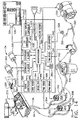
- Figure 14 is a schematic diagram which illustrates the interconnection of the electro-mechanical components of the biopsy device to control unit 342.
- Figure 14 illustrates the biopsy device illustrated in Figure 1 and comprises control unit 342, fluid collection system 22, power transmission source 24, and handpiece 20 (see Figure 1).
- a more detailed schematic diagram illustrating the elements of control unit 342 is shown in Figures 16A and 16B and will be described later. All of the components of Figure 14 may be packaged into a portable, wheeled unit, and moved from room to room such as in a physician's office.
- Handpiece 20 (see Figure 1), as described earlier, may be mounted to a stereotactic table already in the room, or handheld and used in combination with a handheld imaging device such as a handheld ultrasonic imager. Each time the biopsy device is used for a new patient, a new sterile probe assembly 40 may be used in handpiece 20.
- Figure 14 illustrates the interconnection of switchboard modified holster 251 with control unit 342, and the connection of power transmission source 24 to control unit 342.
- power transmission source 24 comprises a rotation motor 338 and a translation motor 340.
- Rotation motor 338 and translation motor 340 transmit rotational power to switchboard-modified holster 251 via first and second rotatable shafts, 264 and 266, respectively.
- An example of a motor which is suitable for either rotation motor 338 or translation motor 340 is available from Micro Motors Electronics, Incorporated, as DC Micro Motors Series 3863, with integral, miniature optical encoder, Part Number SHE 17.
- Rotation motor 338 has an integral rotation sensor also referred to as a first sensor.
- Translation motor 340 has an integral rotation sensor also referred to as a second sensor.
- control unit 342 uses the signals from the integral rotation sensor (also referred to as the first sensor) of translation motor 340 and rotation sensor 268 to calculate accurately the axial position of cutter 96.
- Second rotatable shaft 264 runs parallel to first rotatable shaft 266 between control unit 342 and holster 251.
- the mechanical efficiency of either shaft in transmitting rotation from the respective motor to holster 251 varies to some degree with the orientation of the rotatable shaft. If for example, it is necessary during the surgical procedure for the operator to drape first and second rotatable shafts, 266 and 264, so that they are bent significantly, then there will be more frictional energy losses than if the shafts were straight.
- the initial current supplied to rotation motor 338 is not sufficient to attain a predetermined cutter rotational speed, the current to rotation motor 338 increases until a desired rotational speed is reached.
- the rotation sensor integrated into rotation motor 338 provides feedback signals to control unit 342, so that the compensating current can be supplied to rotation motor 338. Once the desired rotational speed is reached, the current to rotation motor 338 is "locked” until the cutter 96 reaches position four at the end of its translation. This electrical compensation occurs for each time cutter 96 translates between the second and third positions, before cutter 96 begins to cut tissue. This allows for variations in the way rotatable shafts, 264 and 266, are oriented for each time the operator positions the biopsy instrument for collecting a tissue sample.
- fluid collection system 22 comprises a first valve 314, a second pinch valve 316, a fluid collection canister 318, a regulator valve 322, a pressure sensor 328, and a vacuum pump 330. These components are interconnected to each other, control unit 342, and probe assembly 40 ( Figure 1) as follows.
- First vacuum tube 94 comes from probe assembly 40 ( Figure 1), and is attached to a first vacuum Y-connector 302 which is fluidly connected to a first upper line 306 and a first lower line 308.
- the two lines, 306 and 308, pass through first pinch valve 314.
- An example of a suitable, commercially available, three-way pinch valve for this application is Model Number 373 12-7 15, available from Angar Scientific Company, Incorporated.
- Pinch valve 314 closes either the upper line 306 or the lower line 308, but never both lines simultaneously.
- Lower line 308 provides a vent to atmospheric pressure.
- Upper line 306 attaches to fluid collection canister 318.
- second vacuum line 136 from probe assembly 40 attaches to a second Y-connector 304 which is fluidly connected to a second upper line 310 and a second lower line 312.
- the first and second vacuum Y-connectors, 302 and 304 may be molded from a rigid polymer such as polycarbonate.
- Second upper line 310 passes through a second pinch valve 316, which is identical to the first, and to the canister 318.
- Second lower line 312 passes through second pinch valve 316 and vents to the atmosphere. Again, only one or the other of the two lines, 310 and 312, may be pinched closed at any time.
- a main vacuum line 320 attaches the canister 318 to electrically powered vacuum pump 330.
- An example of a suitable vacuum pump for this application is available as WOB-L PISTON Series 2639 from Thomas Compressors and Vacuum Pumps, Incorporated.
- Main vacuum line 320 passes through regulator valve 322 to adjust electronically the vacuum pressure supplied to canister 318.
- An example of a commercially available regulator valve for this application is model number VSONC6S11VHQ8 from Parker Hannifin Corporation, Pneutronics Division.
- Pressure sensor 328 is fluidly attached to main vacuum line 320 at a sensor connection 324. The signal from pressure sensor 328 is sent to an A/D converter 396 of control unit 342.
- An example of a commercially available, compensated pressure sensor for this application is model number SDX15 from SenSym, Incorporated.
- control unit 342 is shown to include the elements inside the drawn box, a liquid crystal display (LCD) 334, and a touchscreen 336.
- Figures 16A and 16B together form a detailed schematic of the elements of control unit 342.
- Figures 14, 16A, and 16B may be referred to concurrently for the description of the elements of control unit 342.
- a microprocessor 408 is 40 MHz, 32-bit microprocessor, available from Motorola, Incorporated as Part Number XCF5206EFT40.
- Microprocessor 408 is designed to perform logic operations that may be translated into simple electromechanical actions.
- LCD 334 prompts and informs the operator during the operation of the biopsy device.
- LCD 334 is 640 x 480 color TFT-LCD display available from Sharp Electronics Corporation as part number LQ64D343.
- a resistive touch screen 336 covers LCD 334 for the user interface.
- An example of a suitable touch screen 336 is available from Dynapro Thin Film Products, Incorporated as Part Number 95638.
- LCD 334 is electronically connected to a touch screen controller 402 in control unit 342.
- Oscillator 540 is available, for example, as Part Number ASV-40.000000-PCSA (40 megahertz) from Abracon Corporation.
- EPROM 542 is Part Number AT27BV4096-15JC available from Atmel Corporation.
- voltage supervisor 541 for a 2.93-volt supply is available as Part Number TLC773ID from Texas Instruments, Incorporated.
- Touch screen controller 402 allows control unit 342 to respond to the user's touch by interpreting touch inputs. Other more conventional devices, such as mechanical switches, may be used instead of touch screen controller 402 for controlling control unit 342. Touch screen controller 402, however, is easy to keep clean and is intuitive for the operator to use.
- Touch screen controller 402 comprises a microcontroller 511, an A-D converter 512, a multiplexer-demultiplexer 513, and an EEPROM 514.
- a suitable example for microcontroller 511 is 8-bit micro-controller Part Number 95705 from Microchip Technology, Incorporated.
- a suitable example for A-D converter 512 is 10-bit serial A-D converter Part Number TLV1543CDW from Texas Instruments, Incorporated.
- a suitable example for multiplexer-demultiplexer 513 is dual 4-to-1 line analog multiplexer-demultiplexer Part Number MC74HC4052D from Motorola, Incorporated.
- a suitable example for EEPROM 514 is 1K-bit serial EEPROM Part Number 93AA46SN from Microchip Technology, Incorporated.
- a LCD controller 404 is provided to interface between microprocessor 408 and LCD 334.
- LCD controller 404 reduces the burden of microprocessor 408 by efficiently controlling display parameters such as color, shading, screen update rates, and it typically accesses the memory chips of microprocessor 408 directly.
- LCD controller 404 comprises a 25-megahertz oscillator 539 that is available, for example, as part number ASV-25.000000-PCSA from Abracon Corporation.
- LCD controller 404 also comprises an LCD/CRT controller 508 that is available, for example, as part number SED1354FOA from Seiko Epson Corporation, and a 1-meg x 16-bit, 60 nanosecond, EDO DRAM 507 that is available, for example, as part number MT4LC1M16E5TG-6 from Micron Technology, Incorporated.
- LCD controller 404 further comprises a pair of 16-bit drivers, 509 and 510, of the non-inverting, buffer-line type, that are available, for example, as part number 74ACTQ16244SSCX from National Semiconductor Corporation.
- a miniature annunciator 332 is provided with control unit 342 in order to provide the operator with audible feedback "beeps" upon each activation of an icon control on the LCD 334.
- An example of a suitable annunciator for this application is model number BAS-45P104S from Matshusita Electric Corporation of America (Panasonic Division).
- Annunciator 332 interfaces with microprocessor 408 by an oscillator 400 which converts the digital input signal from microprocessor 408 to an analog, periodic output signal, thus controlling the audio frequency of the connector 332.
- the volume of the sound coming from annunciator 332 is controllable, as will be described later.
- oscillator 400 comprises a 62dB audio attenuator 517 that is available, for example, as Part Number LM1971M from National Semiconductor Corporation.
- Oscillator 400 further comprises an operational amplifier 516 that may be identical, for example, to operational amplifier 530 already described.
- Oscillator 515 further comprises a power audio amplifier 515 that is available, for example, as part number LM486M from National Semiconductor Corporation.
- a first motor controller and driver 390 interfaces with translation motor 340 and with microprocessor 408.
- Translation motor 340 is operationally connected to second rotatable shaft 266.
- Controller and driver 390 converts digital input signals from microprocessor 408 into analog motor input signals for controlling motor rotational direction and speed. Closed loop digital speed control of translation motor 340 is also achieved within controller and driver 390 using feedback signals from encased rotation sensor 268 in holster 251 and rotation sensor integrated within translation motor 340.
- First motor controller and driver 390 comprises a first H-bridge motor driver 552 (also referred to as a first driver) and a first motor controller 523.
- a suitable example of a first H-bridge motor driver is available as Part Number LMD18200T from National Semiconductor Corporation.
- a suitable example of a motor controller is available as Part Number LM629M-8 from National Semiconductor Corporation.
- rotation motor 338 drives first rotatable shaft 264.
- Rotation motor 338 interfaces with microprocessor 408 through second controller and driver 406 which comprises a second H-bridge motor driver 551 (also referred to as a second driver) and a second motor controller 522.
- Second H-bridge motor driver 551 may be identical to first H-bridge motor driver 552, already described.
- Second motor controller 522 may be identical to first motor controller 523, already described.
- Microprocessor 408 via second controller and driver 406 continually calculates and updates the rotational positions of cutter 96, as well as the rotational speed and acceleration, using feedback signals from the rotation sensor integrated within rotation motor 338.
- a serial controller 380 is electronically connected to switchboard 274 by ribbon cable 270 and control cord 265. Ribbon cable 270 is contained within holster 251. Control cord 265 runs along, and may be attached to, first rotatable shaft 264 and second rotatable shaft 266. Serial controller 380 coordinates information exchange across the serial communication link between switchboard 274 and microprocessor 408.
- An optional card reader 382 may be provided in control unit 342 for reading data from memory card in order to facilitate future software upgrades and servicing.
- a serial port 384 is provided for the bi-directional data exchange in a serial transmission mode, again to facilitate future software upgrades and servicing.
- Serial controller 380 includes a quad differential line receiver 524 that is available, for example, as Part Number DS90C032TM from National Semiconductor Corporation. Serial controller 380 further includes an ESD (electrostatic discharge) over-voltage protection array 525 that is available, for example, as Part Number SP723AB from Harris Semiconductor Products.
- ESD electrostatic discharge
- a first PWM (pulse width modulation) driver 386 interfaces first pinch valve 314 with microprocessor 408.
- First PWM driver 386 converts a digital input signal from microprocessor 408 to an analog output signal having a wave of fixed frequency and amplitude, but varying duty cycle.
- PWM driver 386 is used when the duty cycle is high to initially move the solenoid. Once pinch valve 314 is actuated, the duty cycle is reduced to a level, which maintains valve position, thus minimizing power requirements.
- a second PWM driver 388 similarly interfaces a second pinch valve 316 with microprocessor 408.
- a suitable example for both first PWM driver 386 and second PWM driver 388 is FET (60 volt, 3.5 amp, 0.10 ohm, N-channel dual) Part Number NDS9945 available from Fairchild Semiconductor Corporation.
- a first EPLD (Erasable Programmable Logic Device) 521 interfaces with LCD controller 404, PWM driver 388, PWM driver 386, an FET 554, oscillator 400, a first 8 MHz. oscillator 538, serial controller 380, and microprocessor 408 (via the path represented by the encircled "A").
- a suitable example for first EPLD 521 is available as Part Number EPM7256ATC144-7 from Altera Corporation.
- FET 554 may be identical, for example, to FET 556 of second PWM driver 388.
- First oscillator 538 is available, for example, as Part Number ASL-8.000000-PCSA from Abracon Corporation.
- a second EPLD 520 interfaces microprocessor 408 with serial port 384, first controller and driver 390, second controller and driver 406, touch screen controller 402, RAM 392, flash memory 398, and oscillator 540.
- EPLD 520 is capable of operating at 166.7 megahertz and is available, for example, as Part Number EPM7256ATC144-7 from Altera Corporation.
- a third PWM driver 394 interfaces with regulator valve 322 and A/D converter 396.
- PWM driver 394 comprises a voltage reference device 526 comprising a first operational amplifier and a voltage reference.
- PWM driver 394 further comprises a second operational amplifier 527, a third operational amplifier 528, a fourth operational amplifier 529, a fifth operational amplifier 530, a sixth operational amplifier 531, and a seventh operational amplifier 532.
- the operational amplifier in voltage reference device 526, and operational amplifiers 527, 528, 529, 530, 531, and 532 are more descriptively referred to as "Quad Rail-to-Rail Operational Amplifiers".
- a suitable example for each is available as Part Number LMC6484IM from the National Semiconductor Corporation.
- PWM driver 394 further comprises a first FET (Field Effect Transistor) 553.
- FET 553 is available as Part Number NDS9945 (60 volt, 3.5 amp, 0.10 ohm, N-channel dual) from Fairchild Semiconductor Corporation.
- a RAM (Random Access Memory) memory device 392 (also referred to as a temporary memory device) is provided with microprocessor 408, and inherently loses stored data when power is removed.
- a flash memory device 398 (also referred to as a non-volatile memory device), on the other hand, is provided with microprocessor 408 to store data even without power, but it has slower access time than RAM memory device 392.
- RAM memory device 392 comprises four EDO DRAM devices, 547, 548, 549, and 550. These devices may be identical and a suitable example of each is available as Part Number MT4LC1M16F5TG-6 from Micron Technology, Incorporated.
- Flash memory device 398 comprises four RAM devices which may be identical and a suitable example of each is available as Part Number AM29LV800BT-70REC from Advanced Micro Devices, Incorporated.
- the combination of the RAM memory device (temporary memory device) 392, the flash memory device (non-volatile memory device) 398, and the microprocessor 408 is sometimes referred to simply as a computing device.
- the computing device may also include the first controller 523 and the second controller 522 in an alternate embodiment.
- Serial port 384 comprises a dual, universal, asynchronous receiver/transmitter 533 available, for example, as part number ST16C2552CJ44 from Exar Corporation.
- Serial port 384 further comprises a first driver-receiver 534 and a second driver-receiver 535, each more descriptively called a "TIA/EIA-232, 3x5 driver-receiver" and available, for example, as Part Number DS14C335MSA from National Semiconductor Corporation.
- Serial port 384 further includes a first transient suppressor 536 and a second transient suppressor 537, each a bi-directional, 24 volt, 300 watt unit available, for example, as Part Number SMDA24C-8 from General Semiconductor, Incorporated.
- Card reader 382 may be used in future embodiments of the biopsy device to program control unit 342 with alternate values, for example, of the desired translation and rotation speeds of the cutter 96.
- An A/D converter 396 converts voltage signals from pressure sensor 328 into digital signals which are transmitted to microprocessor 408, and used by microprocessor 408 to maintain a desired vacuum pressure in fluid collection system 22.
- a suitable example of A/D converter 396 is ADC-DAC, 8-bit, 12C bus interface available as Part Number PCF8591AT from Philips Electronics N.V.
- the biopsy device is provided with a conventional, 48-volt DC power supply used in combination with standard DC-to-DC converters and electrical voltage regulators in order to supply reduced voltages to the components of control unit 342.
- the microprocessor 408 may be used to monitor the output value of second controller and driver 406 PID filter such that if the output of it exceeds a predefined maximum value, the translational speed of cutter 96 is reduced a set amount by sending an updated speed command to first controller and driver 390.
- This closed-loop system insures that the desired cutter rotational speed is maintained by decreasing the translational speed of cutter 96. This automatic adjustment to cutter translational speed occurs when cutter rotational resistance becomes abnormally high.
- Cutter rotational resistance is the combination of cutting resistance (when the cutter 96 encounters obstructions, very dense tissue, or calcified lesions, for example) and mechanical resistance (when the operator, for example, manipulates piercer 70 into tissue with enough force to place a significant bending moment on piercer 70 so that cutter 96 binds inside piercer lumen 80.)
- the cutter translational speed is decreased in order to reduce the cutter rotational resistance. In one embodiment of the present invention, this is accomplished in the following manner.
- microprocessor 408 While in the sampling mode and with cutter 96 advancing toward the third position (proximal to port 78), when cutter 96 reaches a predetermined translational position, microprocessor 408 sends a signal to second controller and driver 406 to initiate cutter rotation.
- the rotational speed of cutter 96 follows a predefined speed profile which insures that the cutter rotational speed is at a predetermined Q (also referred to as predetermined rotational speed) revolutions per minute (rpm) when cutter 96 reaches the third position.
- Q also referred to as predetermined rotational speed
- rpm revolutions per minute
- microprocessor 408 sends a signal to first controller and driver 390 to advance cutter 96 at a predetermined translation speed T (also referred to as a third, predetermined translation speed) inches per second (in/sec).
- Cutter 96 then progresses through port 78 at predetermined translation speed T in/sec while rotating at velocity Q rpm. While advancing through port 78, cutter 96 rotational speed is monitored by second controller and driver 406, using signals from the rotation sensor integrated within rotation motor 338. If the rotational speed is greater than Q rpm, electrical current to translation motor 340 is increased. If the cutter rotational speed is less than Q rpm, electrical current to translation motor 340 is decreased.
- one way to do so is to generate an error signal based on the difference between the desired speed (translation or rotation, depending on which motor is controlled) and the actual speed.
- the error signal is then input into a proportional, integral, and derivative (PID) digital filter, which is part of the respective controller and driver, either first controller and driver 390, or second controller and driver 406.
- PID proportional, integral, and derivative
- a PWM signal is input to first controller and driver 390 to generate an analog output signal to drive translation motor 340.
- a PWM signal is input to the second controller and driver 406 to generate an analog output signal to drive rotation motor 338.
- FIG 15 is an enlarged view of LCD 334 having a display area 344, and touch screen 336, both of which are part of control unit 342 of Figure 14.
- twelve separate operating modes are available to a user.
- a control switch for each operating mode is displayed graphically on LCD 334 in the form of icons, 346, 348, 350, 352, 354, 356, 358, 360, 362, 364, 366, and 368.
- the user may initiate a particular operation by pressing touch screen 336 in the region of the appropriate icon at the appropriate time during the surgical procedure to electronically control the operation of the biopsy device.
- Each mode of operation is utilized for a particular portion of the general biopsy procedure.
- the "Prime” mode of operation is selected when the operator is preparing the instrument for use.
- display area 344 indicates the status as being "Prime Mode”.
- Cutter 96 then translates to the third position just proximal to port 78. Once cutter 96 is in the third position, display area 344 instructs the operator to apply saline to port 78 and to depress vacuum switch 150 as needed to draw saline into piercer 70 and through probe assembly 40. The operator may observe the flow of saline through window 58.
- first pinch valve 314 and second pinch valve 316 are each set to respond to vacuum switch 150.
- the "Insert” mode of operation is next selected when the operator is preparing to insert the piercer into tissue.
- display area 344 indicates the status as being "Insert Mode”.
- Cutter 96 then translates to the fourth position, just distal to port 78. Once cutter 96 translates to the fourth position, the display indicates that the instrument is ready to insert.
- the "Verify” mode of operation is selected when the operator wants to verify whether or not cutter 96 is at the fourth position.
- display area 344 indicates the status as being "Verify Mode”. If cutter 96 is not at the fourth position, translation motor 340 is set to respond to forward switch 146 on handpiece 20. Then display area 344 instructs the operator to close port 78 by pressing forward switch 146 on handpiece 20. When the operator presses forward switch 146, cutter 96 translates to the fourth position. Translation motor 340 is then set to respond to reverse switch 148 on handpiece 20.
- second motor 340 is set to respond to reverse switch 148. Then display area 344 instructs the operator to open port 78 by pressing reverse switch 148 on handpiece 20. When the operator presses reverse switch 148, cutter 96 translates to the third position just proximal to port 78. Then translation motor 340 is set to respond to forward switch 146.
- the "Sample” mode of operation is selected when the operator desires to extract a portion of tissue from the surgical patient.
- display area 344 indicates the status as being ""Sample Mode”.
- Cutter 96 then translates to the third position, which is just proximal to port 78.
- translation motor 340 is set to respond to forward switch 146.
- display area 344 instructs the operator to take a tissue sample by pressing forward switch 146 on handpiece 20.
- forward switch 146 is pressed, first pinch valve 314 and second pinch valve 316 are opened, and rotation motor 338 is activated to rotate cutter 96 at the appropriate speed.
- cutter 96 translates to the fourth position, severing the tissue portion prolapsed into port 78 as cutter 96 moves distally.
- the operator may abort the sampling stroke, prior to cutter 96 reaching the midpoint of port 78, by pressing any one of the forward, reverse, or vacuum switches, 146, 147, or 150 respectively, on handpiece 20.
- cutter 96 translation is halted and display area 344 instructs the operator to either continue sampling by pressing forward switch 146 or reverse switch 148 to return to the first, fully retracted position.
- rotation motor 338 is deactivated and cutter 96 stops rotating.
- first pinch valve 314 is activated to close first upper line 306.
- Next display area 344 instructs an operator to retrieve a tissue sample by pressing reverse switch 148 on handpiece 20.
- Translation motor 340 is set to respond to reverse button 148 on handpiece 20.
- cutter 96 translates to the first, fully retracted position, just distal to sampling surface 64.
- second pinch valve 316 is activated to close the vacuum for tissue remover 132.
- a "smart-vacuum” is also activated and a plurality of vacuum pulses (0.5 seconds on and 0.5 seconds off) are supplied to second vacuum tube 136.
- a detailed description of the "smart vacuum” is provided in U.S. Patent Application Serial Number 08/878,468 filed on June 18, 1997 and hereby incorporated herein by reference.
- Display area 344 then instructs the operator to remove the tissue sample 200. If there was no sample extracted, that is, the severed tissue sample remained at the distal end of piercer 70 and was not deposited onto tissue sample surface 64, the operator is instructed to select "Dry Tap". The operator is also instructed to select "Remove Air/Blood” if required to remove excessive fluids in the patient and in probe assembly 40. The operator is finally instructed to press forward switch 146 on handpiece 20 to extract the next sample.
- translation motor 340 is set to respond to forward switch 146 on handpiece 20. When forward switch 146 is pressed by the operator, the "smart-vacuum” is stopped and first and second pinch valves, 314 and 316, are activated to open. Rotation motor 338 is activated to rotate cutter 96, which then translates to the fourth, fully distal position. Then cutter 96 rotation is stopped and first pinch valve 314 is closed to stop the vacuum to vacuum pressure chamber tube 76 supplied by first vacuum tube 94.
- the "Mark” mode of operation is selected when the operator desires to implant a metallic marker within the surgical patient at the location from which the tissue sample 200 was extracted.
- display area 344 indicates the status as being “Marker Mode” and also prompts the operator to select “Dry Tap” if required. Then the operator is instructed to press vacuum switch 150 on handpiece 20 to activate the "Mark” mode.
- a marking instrument which may be used in combination with the present invention for marking tissue is commercially available under the trade name MICROMARK from Ethicon Endo-Surgery, Inc., Cincinnati, Ohio. A complete description of the MICROMARK applier and clip, and the method of its use, is included in U.S.
- Next display area 344 instructs the operator to reposition the MICROMARK instrument if marker deployment was not complete, to press vacuum switch 150 on the handpiece 20 when ready to deploy the marker, to deploy the marker, and if the marker deployment is complete, to remove the MICROMARK instrument.
- the "Remove” mode of operation is selected when the operator is ready to remove piercer 70 from within the tissue of the surgical patient.
- display area 344 indicates the status as being "Remove Mode”.
- Cutter 96 translates to the fourth, fully distal position and closes port 78.
- Display area 344 instructs the operator that the instrument is ready to remove.
- the "Remove Air/Blood" mode of operation is selected when the operator desires to remove any fluids present near the distal end of piercer 78 and within probe assembly 40.
- Display area 344 indicates the status as being "Remove Air/Blood Mode”.
- Cutter 96 then translates to the third position just proximal to the port 78.
- First pinch valve 314 and second pinch valve 316 are each set to respond to vacuum switch 150 on handpiece 20.
- Display area 344 then instructs the operator to remove the air/blood by pressing vacuum switch 150 on handpiece 20.
- first pinch valve 314 and second pinch valve 316 are activated to open for five seconds. When they are closed, cutter 96 then translates to the first, fully retracted position just proximal to tissue sampling surface 64. Then the "Remove Air/Blood" mode is automatically exited and the previous mode selected is automatically reset.
- the "Dry Tap” mode of operation is selected for when the operator had attempted to extract a tissue portion from the surgical patient using the "Sample” mode of operation, but a tissue sample 200 was not deposited onto tissue sample surface 64. This may occur when the tissue sample 200 is properly severed from the surgical patient, but remained in the distal end of piercer 78.
- display area 344 indicates the status as being "Dry Tap Mode”.
- Cutter 96 then translates to the third position just proximal to port 78.
- second pinch valve 316 is activated to open for 0.5 seconds and to close for 0.5 seconds three times in order to pulse the vacuum supplied to tissue remover 132 through second vacuum tube 136.
- Cutter 96 then translates to the first, fully retracted position just distal to tissue sampling surface 64.
- the "Dry Tap" mode of operation is then exited and the previously selected mode of operation is automatically selected.
- the "Flush” mode of operation is selected when the operator desires to clear any obstructions (tissue fragments, etc.) on the distal end of tissue remover 132 to enable the passage of fluids through it.
- Display area 344 indicates the status as being "Flush Mode”.
- Cutter 96 then translates to the first, fully retracted position, thus exposing the distal end of tissue remover 132.
- control unit 342 is set to respond to vacuum switch 150, which when pressed by the operator, causes the "Flush” mode of operation to be exited and the previously selected mode of operation to be automatically reset. Before pressing vacuum switch 150, however, the operator may temporarily disconnect second connector 304, inject fluid such as saline into second vacuum tube 136 using a syringe, and reconnect second connector 304.
- the "Inject" mode of operation is selected when the operator desires to inject a fluid, such as a local anesthetic, into the tissue surrounding the distal end of piercer 78.
- a fluid such as a local anesthetic
- Display area 344 indicates the status as being "Inject Mode”.
- Cutter 96 then translates to the second position just proximal to port 78.
- control unit 342 is set to respond to vacuum switch 150 on the handpiece 20.
- LCD display instructs the operator to inject the fluid into second vacuum tube 136, and to press vacuum switch 150 again once the injection is complete.
- cutter 96 When the operator has completed the injection into second vacuum tube 136, reconnected it to fluid collection system 22, and pressed vacuum switch 150, cutter 96 translates to the first, fully retracted position. At that point, the "Inject" mode of operation is exited, and the previously selected mode of operation is automatically reset.
- the "Manual" mode of operation is selected when the operator desires to perform cutter positioning and/or vacuum functions manually.
- the operator activates the "Manual" mode of operation by, for example, touching LCD 334 in the region of icon 366, Display area 344 indicates the status as being “Manual Mode.”
- translation motor 340 is set to respond to forward switch 146 and reverse switch 148 for the duration of switch depression. The operator at any point between the first, fully retracted position, and the fourth, fully distal position may halt translation of cutter 96.
- first pinch valve 314 and second pinch valve 316 are each set to respond to vacuum switch 150.
- a mode indicator display 370 includes a representation of the probe assembly 40 showing the instantaneous position of cutter 96, referred to as a cutter position indicator 373. Mode indicator display 370 also shows activation of a front vacuum indicator 372 (corresponding with first vacuum tube 94), and activation of a rear vacuum indicator 371 (corresponding with the second vacuum tube 136).
- Figures 17A, 17B, 17C, and 17D illustrate a flow diagram of a first control method embodiment according to the present invention, wherein the cutter 96 has four distinct positions.
- Figures 17A, 17B, 17C, and 17E illustrate a flow diagram of a second control method embodiment according to the present invention, wherein the cutter 96 also has four distinct positions.
- the steps of the control method are represented in the flow chart. Even though each box may represent more than one step, or may only be part of a step, each box is referred to simply as a step. Progression of the steps occurs generally in the direction of the arrows connecting the boxes.
- the first and second control method embodiments may be used with any of the biopsy instrument embodiments shown in Figures 5, 10, and 13. In the following description of the first and second control method embodiments, however, the biopsy instrument embodiment shown in Figure 13, also shown in Figure 14, will be referred to.
- step 410 represents the beginning of the control method.
- the serial communication loop is completed through microprocessor 408 and EPLD 520 to receive the switch interface data for the initiation of controller 523 to enable H-bridge motor driver 552 to send the appropriate current to translation motor 340.
- Cutter 96 begins to move distally from the first position, but does not rotate.
- cutter 96 continues to translate from position one to position two.
- a signal is read from the encoder (also referred to as the first sensor) of translation motor 340 and compared with a first, predetermined translation speed R programmed in controller 523.
- the value of R may vary for different embodiments, but a preferred value for R is approximately in the range of 0.67 inches/second.
- a difference between R and the actual translation speed of the cutter 96 is calculated and in step 416, the difference is compared to a programmed value called RD (also referred to as a first predetermined differential translation speed), which represents an allowable differential for the value R.
- RD also referred to as a first predetermined differential translation speed
- step 4128 If the calculated difference between R and the actual speed is greater than RD, then current to translation motor 340 is stopped (step 418) and the error is reported on the display area 344. If the translation speed difference is less than RD, then the current to translation motor 340 is adjusted to reduce the translation speed difference, as represented by step 420.
- the cutter 96 continuously adjusts translation speed in this way until cutter 96 reaches position two, as shown in step 422.
- the number of translation motor 340 rotations are counted by rotation sensor 268, so that the number of screw rotations of screw 114 (see Figure 9) can be determined, thus providing the position of carriage 124 (see Figure 9), and finally the position of cutter 96. Correction for twisting of rotatable shaft 266 is calculated at this point of the control method by using the signals from both rotation sensor 268 and the encoder integral to translation motor 340.
- step 424 of Figure 17B The control method next progresses to step 424 of Figure 17B for continuing the translation of the cutter 96 from position two to position three.
- step 426 if cutter 96 has reached a predetermined intermediate position between two and three, then the control method progresses to step 428.
- the location of the predetermined intermediate position is based on the actual translation speed and predetermined cutter rotation speed Q. This is to allow a sufficient amount of time for cutter 96 to accelerate from zero to the predetermined rotation speed Q before cutter 96 has reached position 3, where cutting of tissue begins.
- the actual translation speed is compared to a second, predetermined translation speed S, which is programmed into the controller 523.
- the difference is calculated in step 428 and compared in step 430 to a value SD, the allowable differential, (also referred to as a second, differential translation speed) which is programmed in controller 523.
- SD is approximately in the range of 0.02 inches/second.
- Value S may vary for different embodiments, but a preferred value of predetermined translation speed S is approximately in the range of 1.45 inches/second.
- the second predetermined translation speed S is much higher than first predetermined translation speed R because the distance traversed by the cutter 96 is much greater between positions 2 and 3 than between positions 1 and 2. Reducing the time to operate the device during the sample mode is important in reducing the overall duration of the surgical procedure because several samples may be taken from the tissue of the patient.
- step 430 if the calculated difference between S and actual speed is greater than SD, than controller 523 signals H-bridge motor driver 552 to stop the current to translation motor 340 in step 432, thus stopping the advancement of cutter 96. If the difference between S and the actual speed is not greater than SD, then the current is adjusted to reduce the speed difference, as indicated in step 434.
- the control method next progresses to step 424 to continue the translation of cutter 96 from position two to three. For step 434, if cutter 96 has not reached position three, the adjustments to translation motor speed continue as already described. If cutter 96 has reached position three, then the control method progresses to step 436 of Figure 17C.
- cutter 96 is still translating from position two to position three.
- microprocessor 408 and EPLD 520 initiate second controller 522 to enable second H-bridge driver 551 to supply the appropriate current to rotation motor 338 to rotate at a predetermined rotation speed of Q.
- the value of Q may vary for different embodiments, but a preferred value for Q is approximately 1350 revolutions per minute (rpm). It has been found through experimentation that at this speed, the tissue is normally cut cleanly and without tearing, resulting in a good core tissue sample.
- the difference between predetermined rotation speed Q and the actual rotation speed is calculated in controller 522 using the signal from the integral encoder of rotation motor 338.
- step 440 the current to rotation motor 338 is adjusted to reduce the rotation speed difference.
- the actual translation speed is compared to predetermined translation speed S, which is programmed into controller 523.
- the difference is calculated in step 444 and compared in step 446 to a value SD, the allowable differential translation speed, also referred to as a second, predetermined differential translation speed, which is programmed in controller 523.
- controller 523 signals H-bridge motor driver 552 to stop the current to translation motor 340 and rotation motor 338, thus stopping the advancement and rotation of cutter 96.
- step 450 if cutter 96 has not reached position three, then control goes back to step 438. If cutter 96 has reached position three, then the control method proceeds to step 454, where first controller 523 initiates first H-bridge driver 552 to modify the current to translation motor 340 to change the cutter translation speed to a new commanded translation speed.
- a preferred initial value for the commanded translation speed is approximately in the range of 0.28 inches/second. It was found through experimentation that this value for initial commanded translation speed provides a clean core tissue sample.
- the initial commanded translation speed is slower than the speeds during the other segments of the cutter 96 journey because a slower speed is desired for severing tissue between positions 3 and 4 along the length of the port 78.
- the subsequently commanded translation speeds may be less due to increased cutter rotational resistance, as is described next.
- the control method may continue either as an embodiment identified as "First Control Method” (encircled letter “C") or as a further embodiment identified as "Second Control Method” (encircled letter “D").
- the control method of the present invention is not limited to these two embodiments; they are provided as examples of the method according to the present invention.
- step 456 cutter 96 continues to translate distally from position three towards position four.
- the actual rotation speed of cutter 96 is compared to a lowest allowable rotation speed QL (also referred to as a predetermined minimal rotation speed).
- QL also referred to as a predetermined minimal rotation speed
- a preferred value for QL is about 1200 rpm, although this value may vary.
- the integral encoder of rotation motor 338 sends a signal to be compared with the programmed value for QL in second controller 522. If the actual rotation speed is less than QL, then both rotation motor 338 and translation motor 340 are stopped (step 460) and the error is reported on display area 344.
- step 461 If rotation speed is greater than QL, than the control method proceeds to step 461 to calculate W, where W equals predetermined rotation speed minus actual rotation speed.
- step 462 if the predetermined rotation speed minus the actual rotation speed is less than QD, a predetermined differential rotation speed, then the control method proceeds to step 470.
- a preferred value for QD is approximately in the range of 200 rpm.
- W is not less than QD, the commanded translational speed is decreased by a predefined amount as specified in step 464. An approximate value for the predefined amount is in the range of .06 in/sec. Then the control method progresses to step 466. If commanded translation speed is less than a predetermined minimal translation speed, TL, then the control method proceeds to step 468.
- step 470 An approximate value for TL is in the range of .06 in/sec. If not, then the control method proceeds to step 470 where, as in previous steps, the commanded translation speed is compared to the actual translation speed as measured by the integral encoder on translation motor 340. If the calculated difference in step 472 is greater than an allowable TD (also referred to as a third, predetermined differential translation speed) in step 450, then current to translation motor 340 and rotation motor 338 is stopped and the error is reported in step 468. If the difference is not greater than TD, then the current to translation motor 340 is adjusted in step 474 to reduce the speed difference in the same manner as before.
- TD allowsable TD
- step 476 if cutter 96 has reached position four (the most distal position of the cutter) then rotation and translation motors, 338 and 340 respectively, are stopped (step 468), and cutter 96 stops immediately, regardless of the translational position. If cutter 96 has not reached position four, then the control method goes back to step 456 and the adjustments to translation and rotation speeds continue as before.
- cutter 96 translates from position three to four.
- step 456 cutter 96 continues to translate from position three to four.
- step 458 actual rotation speed is compared to QL.
- the difference between the predetermined rotation speed Q and the actual rotation speed is calculated in controller 522 using the signal from the integral encoder of rotation motor 338. If the actual rotation speed is less than QL which is programmed into controller 522, then current to rotation motor 338 and translation motor 340 is stopped. Otherwise, in step 459 the difference is calculated between Q and the actual rotation speed.
- step 463 current is adjusted to rotation motor 338 to reduce the difference between Q and the actual rotation speed.
- step 465 if the current to rotation motor 338 is greater than a value X, the control method proceeds to step 464 where the commanded translation speed is decreased by a predefined amount.
- the value for X depends on the specifications of the particular motor used. For the example provided in the description of Figure 14, a preferred value of X is approximately in the range of 3.5 amps.
- the control method continues through the same steps as described for the first control method embodiment of Figure 17D. If in step 465 the current to rotation motor 338 is not greater than X, then the second control method embodiment proceeds to step 470 and continues through the same steps as described for the first control method of Figure 17D.
- cutter 96 When the operator activates cutter reverse switch 148 (see Figure 2), cutter 96 translates proximally back to position one at a fourth predetermined translation speed.
- a value of a fourth predetermined translational speed is approximately in the range of 1.45 inches/second in this embodiment, although this speed may vary for other embodiments.
- the severed tissue portion is deposited on the tissue sampling surface 64 and may be retrieved by the operator as described earlier. The above control method is repeated for each time a tissue sample is extracted using the "manual" or “sample” modes.
Abstract
Description
- The present invention relates, in general, to a method of controlling the excision of tissue in biopsy instruments and, more particularly, to a method of controlling the rotation and translation of a cutter in a biopsy instrument.
- The diagnosis and treatment of patients with cancerous tumors, pre-malignant conditions, and other disorders has long been an area of intense investigation. Non-invasive methods for examining tissue include palpation, X-ray, magnetic resonance imaging (MRI), computed tomography (CT), and ultrasound imaging. When a physician suspects that tissue may contain cancerous cells, a biopsy may be done using either an open procedure or a percutaneous procedure. For an open procedure, a scalpel is used to create a large incision in the tissue to provide direct viewing and access to the tissue mass of interest. The entire mass (excisional biopsy) or a part of the mass (incisional biopsy) may then be removed. In most percutaneous biopsy procedures, a needle-like instrument is inserted through a very small incision to access the tissue mass of interest and obtain a tissue sample for later examination and analysis.
- Aspiration and core sampling are two percutaneous methods for obtaining a portion of tissue from within the body. In an aspiration procedure, tissue is fragmented into pieces and drawn through a fine needle in a fluid medium. The method is less intrusive than most other sampling techniques, however, it has limited application since the structure of tissue excised by aspiration is destroyed leaving only individual cells for analysis (cytology) and not the tissue structure for analysis (pathology). In core biopsy, a core or fragment of tissue is obtained in a manner, which preserves both the cells and the structure for histological examination. The type of biopsy used depends mainly on various factors, and no single procedure is ideal for all cases. Core biopsy, however, is very useful in a number of conditions and is widely used by physicians.
- Examples of core sampling biopsy instruments are described in U.S. Patents 5,562,822 and 5,769,086 (both issued to Ritchart, et al), and in U.S. Patent Application Serial No. 09/107,845 filed on June 30, 1998. Another example of a core sampling biopsy instrument is the biopsy instrument now marketed by Ethicon Endo-Surgery, Inc., Cincinnati, Ohio, under the trade name MAMMOTOME. Each of these instruments is a type of image-guided, percutaneous, coring, breast biopsy instrument, which uses a vacuum for retrieving tissue samples. A physician uses these instruments to capture "actively" (using the vacuum) tissue prior to severing it from the body. In particular, in these biopsy instruments, tissue is drawn into a port at the distal end of a piercing element, hereinafter referred to as a piercer. A cutting element, hereinafter referred to as a cutter, is rotated and advanced through a lumen of the piercer past the port. As the cutter advances through the port, it severs the tissue drawn into the port from the surrounding tissue. While the cutter is generally rotated using some type of motor, it may be advanced either manually or automatically. In the MAMMOTOME instrument, the surgeon manually moves the cutter back and forth by lateral movement of a knob mounted on the outside of the instrument. Once the cutter is in place, proximal to the tissue port, further lateral movement of the knob is prevented and the cutter is advanced through the tissue port to sever tissue by twisting the knob. This arrangement is advantageous because the surgeon is able, through tactile and/or audible feedback, to determine whether the cutter is effectively cutting tissue or if there is a problem, such as binding or stalling. The surgeon may then adjust the speed at which the cutter is moved through the tissue, stop the cutter or back the cutter away from the tissue.
- As described in U.S. Patents 5,562,822 and 5,769,086, the translation of the cutter may be automated to facilitate the procedure. However, if the procedure is automated as described in those references, the surgeon loses the benefit of the tactile feedback, which results when the cutter is advanced manually. It is generally desirable to ensure that the rotational speed of the cutter does not drop below a predetermined speed in order to sever cleanly the tissue sample from surrounding tissue and to avoid damage to the tissue sample. Automating translation of the cutter will, to some extent, eliminate the tactile feedback that the surgeon gets from moving the cutter manually. The advantageous method of automatically measuring and controlling the rotation and translation of the cutter is not described for either of the devices in the '822 or '086 patents. This automatic control method could be used, for example, to prevent the cutter from advancing when the opening is blocked by something other than tissue. Such an automatic control method could also be used to ensure that the cutter rotates at an optimal speed to ensure proper cutting of the tissue and to prevent the cutter rotational speed from dropping below a predetermined limit.
- Another advantage of a core sampling biopsy device being used with an automatic control method is that the operator would be able to perform the surgical procedure in less time than with a motorized device having no automatic control method. Since core sampling biopsy devices extract tissue samples from deep within the body of the surgical patient, the penetrating element, or piercer, for accessing the tissue, is necessarily long. The driven element, or cutter, must translate from the proximal end of the piercer to the distal end in order to collect the tissue sample. Then the cutter transports the tissue sample from the distal end of the piercer to the proximal end, which is outside the body of the patient. As the cutter is actually cutting through tissue and collecting the tissue sample, the translational speed of the cutter should be maintained in an optimal range. But for all other portions of cutter translation, the translational speed of the cutter may be relatively high without detrimental effects. Thus the time required for obtaining each tissue sample might be reduced. Since many tissue samples may be extracted from the patient during a typical surgical procedure, the accumulated time saved could be significant, providing obvious benefit for both the surgeon and the patient.
- A core sampling, surgical instrument with a needle guiding stage of a stereotactic mammography biopsy system is disclosed in U.S. Patent 5,830,219 by Bird, et al. In this instrument, the driven element (hereinafter referred to as a cutter) translates to cut tissue captured in the distal end of the instrument. This instrument couples feedback from an optical encoder with a microprocessor to calculate cutting resistance so that if the cutter encounters, for example, a dense tissue mass causing the cutter rotation to decrease, additional electrical current is automatically provided to the cutter motor to resume the desired cutter rotational speed. The encoder also provides information to the microprocessor to control automatically the angular stroke of the cutter as it oscillates between the clockwise and counterclockwise directions. This closed loop cutter control method is described in '219 as being provided only for when the cutter is in the cutting portion of its axial travel.
- An alternate control method, however, to deal with an increase in cutting resistance (due to encountering dense tissue or obstructions) is to slow incrementally the translational speed of the cutter until a desired cutter rotational speed is resumed. If the cutter cannot penetrate the obstruction and the desired rotational speed cannot be resumed despite reducing translational speed, then the translation of the cutter could be completely halted and an error message, for example, could be transmitted to the operator. This alternate control method would have an advantage of preventing damage to the biopsy instrument because the tissue sample is obtained less aggressively due to the slowed advance of the cutter through the tissue, or else the cutter translation is completely halted if the obstruction is impenetrable.
- Another advantage for reducing the cutter translational speed rather than increasing the cutter rotational speed in response to an increase in rotational resistance on the cutter is that smaller, less powerful, and less expensive motors could be used to drive the cutter. Both the translation motor for advancing the cutter and the rotation motor for rotating the cutter may be smaller because the overall rate at which work would be done on the tissue by the cutter could be reduced. Using smaller, lighter weight motors would also facilitate their incorporation into the handheld portion of the biopsy instrument. As described in pending U.S. Patent Application Serial Number 09/178,075, the motors can be remotely located in a separate control unit and operationally connected to the handheld portion of the biopsy instrument by at least one rotatable shaft. In such a biopsy instrument, using small motors would be advantageous in allowing the use of small diameter, lightweight, rotatable shafts. In addition to the cost savings realized in the manufacture of the device, the biopsy instrument could be more hand manipulatable by the operator during the surgical biopsy procedure.
- It is also advantageous to use both types of responses to increased rotational resistance on the cutter. That is, a single control method that combines a method for decreasing cutter translational speed and a method for increasing cutter rotational speed in response to decreasing cutter rotation may be used. For example, if the cutter encounters an obstruction in tissue and the cutting resistance rises sharply, the electrical current to the cutter rotational motor may automatically be increased by a predetermined amount. If the cutter rotational speed is measured and compared to a desired, predetermined cutter rotation speed, and it is determined that the cutter rotational speed is still not high enough, than the cutter translational speed may be decreased by a predetermined amount. These steps could be repeated automatically until the tissue sample is obtained, or until certain operational thresholds (minimal translation speed, maximum current to rotation motor, for example) are reached. By using such a combined method in response to increasing rotational resistance on the cutter, the cutter rotation translational motors may be smaller than when using a method to modif rotation alone.
- When an operator uses a handheld instrument operationally connected to a remotely located motor by a flexible, rotatable shaft, the operational configuration of the rotatable shaft may affect the efficiency of the mechanical energy transmitted to the handpiece. For example, if it is necessary for the operator to hold the handheld instrument during the surgical procedure so that the flexible, rotatable shaft is sharply curved, the resistance to rotation of the rotatable shaft is higher than if the rotatable shaft was in a straight configuration. Also, when the operator manipulates the probe of the instrument to penetrate into the tissue mass of interest, a bending moment may be unavoidably applied to the piercer of the probe, increasing the rotational resistance of the cutter which is constructed coaxially in close alignment with the piercer. There also may be additional mechanical losses, for example, due to wear or misalignment of power transmission components. Therefore, it would be advantageous to be able to measure the total, rotational resistance before the cutter encounters tissue, so that the cutter rotation may be increased to the desired, predetermined rotational speed for cutting tissue.
- For an automated, handheld biopsy device, the cutter rotation and translation motors may be mounted in a remote unit in order to minimize the overall size and weight of the handpiece. As described in U.S. Patent Application Serial Number 09/178075, each motor may be operationally connected with the cutter of the handpiece of the biopsy device by a flexible, rotatable shaft. A tradeoff associated with using a flexible, rotatable shaft is that the shaft may twist along its length, or "wind" under loading. A measurement on the proximal end of the rotatable shaft of the angular position of the shaft may vary from that on the distal end. Since the precise number of shaft rotations may be used to calculate axial position of the cutter, it is desirable to incorporate a high-resolution means within the biopsy device to measure and compensate for the twisting of the flexible, rotatable shaft operationally connected to the remotely located translation motor.
- What is needed is a core sampling biopsy device having a control method and apparatus that allows the cutter translational speed to be automatically responsive to cutting resistance caused by obstructions or dense tissue encountered by the cutter. What is further needed is a core sampling biopsy device having a control method and apparatus that allows the cutter rotational speed to be automatically responsive to total rotational resistance on the cutter before and during the cutting of tissue. What is further needed is a core sampling biopsy device having a control method and apparatus that allows the cutter translational speed to be automatically responsive to cutter translational position so that surgical procedure time may be reduced. What is finally needed is a high-resolution means within the biopsy device to measure and compensate for the twisting of the flexible, rotatable shaft operationally connected to the remotely located translation motor.
- The present invention is a control apparatus for removing at least one tissue sample from a surgical patient using a surgical biopsy device. The biopsy device comprises an elongated piercer having a piercer lumen extending therethrough, and a cutter rotatably and axially positionable relative to said piercer. The piercer has a port for receiving and transferring the tissue sample into the piercer lumen. The biopsy device further comprises a rotation motor for rotating the cutter and a translation motor for translating the cutter in the axial direction.
- In one embodiment of the present invention, the control apparatus comprises a computing device for the coordinated control of the rotation and translation motors. The control apparatus further comprises a first sensor operationally connected to the rotation motor for supplying a rotation signal to the computing device for measuring the rotational speed of the cutter. The control apparatus further comprises a second sensor operationally connected to the translation motor for supplying a translation signal to the computing device for measuring the translational position and translational speed of the cutter. The control apparatus further comprises a first driver for driving the translation motor in response to a translation command from the computing device, and a second driver for driving the rotation motor in response to a rotation command from said computing device. The control apparatus automatically modifies the rotational speed and the translational speed of the cutter in response to the translational position and rotational resistance acting on the cutter.
- The novel features of the invention are set forth with particularity in the appended claims. The invention itself, however, both as to organization and methods of operation, together with further objects and advantages thereof, may best be understood by reference to the following description, taken in conjunction with the accompanying drawings in which:
- Figure 1 is an isometric view of the present invention, a biopsy instrument, which includes a handpiece for the collection of soft tissue;
- Figure 2 is an isometric view of the handpiece showing a probe assembly prior to attachment to a holster;
- Figure 3 is an exploded isometric view of the probe assembly illustrated in Figure 2;
- Figure 4 is an isometric view of the probe assembly of Figure 2 with the left handle shell removed to reveal the internal components;
- Figure 5 is an exploded isometric view of the holster illustrating a non-encased rotation sensor mounted on a screw drive shaft;
- Figure 6A is a top view in section of the probe assembly and a distal portion of the holster, revealing a cutter in a first, fully retracted position;
- Figure 6B is a top view in partial section of the distal end of the probe assembly illustrating the cutter in the first, fully retracted position wherein the port on the distal end of the piercer is open;
- Figure 7A is a top view in section of the probe assembly and a distal portion of the holster, revealing the cutter in the third position wherein the distal end of the cutter is immediately proximal to the port;
- Figure 7B is a top view in partial section of the distal end of the probe assembly with the port on the distal end of the piercer open and the distal end of the cutter in the third position immediately proximal to the port;
- Figure 8A is a top view in section of the probe assembly and a distal portion of the holster illustrating the cutter in the fourth, fully deployed position;
- Figure 8B is a top view in partial section of the distal end of the probe assembly illustrating the distal end of the cutter in the fourth position distal to the port at the distal end of the piercer;
- Figure 9 is an isometric view of the probe assembly with the left handle shell removed, showing the cutter in the first position, with a tissue sample shown deposited onto a tissue sampling surface;
- Figure 10 is a partial top view of a further embodiment of the present invention wherein a first and a second motor are contained within a handheld holster rather than in a remotely located control unit as for the embodiment of Figure 5, and wherein the holster upper shell and the probe assembly upper shell have been removed to reveal the internal components;
- Figure 11 is an isometric view of the holster and probe assembly lower shells shown in Figure 10, wherein the holster lower shell includes a slot for the removable attachment to a latch on the probe assembly lower shell;
- Figure 12 is a longitudinal section of the holster and probe assembly lower shells of Figure 11, illustrating their removable attachment to each other;
- Figure 13 is an exploded isometric view of a further embodiment of the holster illustrated in Figure 5, wherein the further embodiment includes the three switches being mounted on a switch board electrically connected by a ribbon cable to the control cord (instead of the three switches being electrically connected to the control cord by discrete switch conductors as illustrated in Figure 5), and wherein the further embodiment includes an encased rotation sensor rather than the non-encased rotation sensor of the embodiment illustrated in Figure 5;
- Figure 14 is a schematic diagram of a control unit according to the present invention;
- Figure 15 is an enlarged diagram of an LCD display illustrated in Figure 14;
- Figure 16A is the first of two portions of a divided schematic diagram of the control unit components illustrated in Figure 14;
- Figure 16B is the second of two portions of the divided schematic diagram of the control unit components illustrated in Figure 14;
- Figure 17A is a first portion of a flow chart pertaining to a first embodiment and a second embodiment of a control method for the operation of the cutter, showing the control unit logic for when the cutter translates from the first position to the second position;
- Figure 17B is a second portion of a flow chart pertaining to the first embodiment and the second embodiment of the control method for the operation of the cutter, showing the control unit logic for when the cutter translates from the second position to the third position;
- Figure 17C is a third portion of a flow chart pertaining to the first embodiment and the second embodiment of the control method for the operation of the cutter, showing additional control unit logic for when the cutter translates from the second position to the third position;
- Figure 17D is a fourth portion of a flow chart pertaining specifically to the first control method embodiment for the operation of the cutter, showing the control unit logic for when the cutter translates from the third position to the fourth position; and
- Figure 17E is a fourth portion of a flow chart pertaining specifically to the second control method embodiment for the operation of the cutter, showing the control unit logic for when the cutter translates from the third position to the fourth position.
-
- Figure 1 shows a core sampling biopsy instrument comprising a
probe assembly 40, aholster 140, afluid collection system 22, acontrol unit 342, and apower transmission source 24. Probeassembly 40 is detachably connected to holster 140. Together they constitute a lightweight, ergonomically shaped, hand manipulatable portion referred to as ahandpiece 20. Probeassembly 40 includes apiercer 70 extending distally from ahollow handle 43. Probeassembly 40 is fluidly connected tofluid collection system 22 by afirst vacuum tube 94 and asecond vacuum tube 136. First and second vacuum tubes are detachably connected tofluid collection system 22 by afirst connector 27 and asecond connector 25, respectively.First connector 27 has amale portion 32 and afemale portion 28 attached tofirst vacuum tube 94.Second connector 25 has afemale portion 30 and amale portion 26 attached tosecond vacuum tube 136. Connector portions, 26, 28, 30, and 32 are attached in this manner to prevent the accidental switching of first and second tubes, 136 and 94, tofluid collection system 22.Holster 140 includes a firstrotatable shaft 34, a secondrotatable shaft 36, and acontrol cord 38. First and second rotatable shafts, 34 and 36, are preferably flexible so that the operator may easily manipulatehandpiece 20 with one hand.Control cord 38 operationally connects thehandpiece 20 topower transmission source 24 andcontrol unit 342. - Since
handpiece 20 is manipulated by the operator's hand rather than by an electro-mechanical arm, the operator may steer the tip ofhandpiece 20 with great freedom towards the tissue mass of interest. The surgeon has tactile feedback while doing so and can thus ascertain, to a significant degree, the density and hardness of the tissue being encountered. In addition,handpiece 20 may be held approximately parallel to the chest wall of the patient for obtaining tissue portions closer to the chest wall then may be obtained when using a instrument mounted to an electro-mechanical arm. - Those skilled in the art may appreciate that a mount or "nest" could be provided to hold
handpiece 20 securely to the movable arm of an X-ray stereotactic table. This would provide the operator with the option to usehandpiece 20 to access the tissue mass within the surgical patient in much the same manner as was described earlier for using the MAMMOTOME instrument. This versatility may be advantageous to the operator, for example, in a situation where the handheld imaging device was temporarily not available for use, and it would be necessary to use the X-ray stereotactic table. - Figure 2 shows
holster 140 and probeassembly 40 separated. A pair of tabs 144 project laterally from each side of a holsterupper shell 142, and insert into right and left undercut ledges, 138 and 139 respectively, ofhollow handle 43 ofprobe assembly 40. A plurality ofindentations 66 is provided onhandle 43 to improve the operator's grip on the instrument. Atube slot 162 inlower shell 156 ofholster 140 provides clearance for first and second vacuum tubes, 94 and 136. A cutter forward switch 146 for moving a cutter 96 (see Figure 3) in the distal direction, a cutterreverse switch 148 for movingcutter 96 in the proximal direction, and avacuum switch 150, are mounted in the distal portion ofholster 140 so that the operator can usehandpiece 20 with a single hand. One-handed operation allows the other hand to be free, for example, to hold an ultrasonic imaging device. Aridge 152 on the distal end ofholster 140 is provided to assist the operator in graspinghandpiece 20 and in operatingswitches - Still in Figure 2,
probe assembly 40 includes awindow 58 so that a portion offirst vacuum tube 94 may be viewed. First and second vacuum tubes, 94 and 136, are made from a flexible, transparent or translucent material, such as silicone tubing. This enables visualization of the material flowing through the tubes, 94 and 136. By havingwindow 58 inprobe assembly 40, the operator can see the flow infirst vacuum tube 94 without needing to look away from the tissue into whichpiercer 70 is inserted. Atransverse opening 68 is provided in the distal end ofhollow handle 43 which allows access from either side to atissue sampling surface 64. The tissue extracted from the surgical patient is retrieved by the operator or by an assistant fromtissue sampling surface 64. - Figure 3 is an exploded isometric view of
probe assembly 40.Handle 43 is formed from aright handle shell 42 and aleft handle shell 44, each injection molded from a rigid, biocompatible plastic such as polycarbonate. Upon final assembly ofprobe assembly 40, left and right handle shells, 42 and 44, are joined together by ultrasonic welding along a joiningedge 62, or joined by any of several other methods well known in the art. Probeassembly 40 comprisespiercer 70 having an elongated,metallic piercer tube 74 and apiercer lumen 80. On the side of the distal end ofpiercer tube 74 is aport 78 for receiving the tissue to be extracted from the surgical patient. Joined alongsidepiercer tube 74 is an elongated, tubular, metallicvacuum chamber tube 76 having avacuum lumen 82.Piercer lumen 80 is in fluid communication withvacuum lumen 82 via a plurality of vacuum holes 77 (see Figure 6B) located in the bottom of the "bowl" defined byport 78. These vacuum holes 77 are small enough to remove the fluids but not large enough to allow excised tissue portions to be removed through first vacuum tube 94 (see Figure 2) which is fluidly connected to vacuumchamber 76. A metallic, sharpeneddistal end 72 is attached to the distal end ofpiercer 70. It is designed to penetrate soft tissue such as the breast of a female surgical patient. In this embodiment, sharpeneddistal end 72 is a three-sided, pyramidal-shaped point, although the tip configuration may also have other shapes. - Still referring to Figure 3, the proximal end of
piercer 70 is attached to aunion sleeve 90 having alongitudinal bore 84 through it, a widenedcenter portion 86, and atransverse opening 88 through widenedcenter portion 86.Union sleeve 90 is mounted between left and right handle shells, 44 and 42 respectively, on a pair of union sleeve ribs 50 (only the rib in the right handle shell is visible) projecting from each handle shell. An elongated, metallic,tubular cutter 96 is axially aligned withinlongitudinal bore 84 ofunion sleeve 90 andpiercer lumen 80 ofpiercer 70 so thatcutter 96 may slide easily in both the distal and proximal directions. A pair of cutter guides 46 are integrally molded into each of handle halves, 42 and 44, to slidably retaincutter 96 in an co-axially aligned position with the proximal end ofpiercer tube 74.Cutter 96 has a cutter lumen 95 through the entire length ofcutter 96. The distal end ofcutter 96 is sharpened to form acutter blade 97 for cutting tissue held againstcutter blade 97 ascutter 96 is rotated. The proximal end ofcutter 96 is attached to the inside of a cutter gear bore 102 of acutter gear 98.Cutter gear 98 may be metallic or polymeric, and has a plurality ofcutter gear teeth 100, each tooth having a typical spur gear tooth configuration as is well known in the art. - Still in Figure 3,
cutter gear 98 is driven by anelongated drive gear 104 having a plurality ofdrive gear teeth 106 designed to mesh withcutter gear teeth 100. The function ofdrive gear 104 is to rotatecutter gear 98 andcutter 96 as they translate in both longitudinal directions.Drive gear 104 is preferably made from a metal such as stainless steel. Adistal drive axle 108 projects from the distal end ofdrive gear 104 and mounts into an axle support rib (not visible) molded on the inside ofleft handle shell 44. Agear shaft 110 projects from the proximal end ofdrive gear 104 and is supported by a gear shaft support rib (not visible) also molded on the inside ofleft handle shell 44. Aleft cross pin 112 is attached to the proximal end ofgear shaft 110 as a means for rotationally engagingdrive gear 104. - Still referring to Figure 3, a
carriage 124 is provided to holdcutter gear 98 and to carrycutter gear 98 as it is rotated in the distal and proximal directions.Carriage 124 is preferably molded from a rigid polymer and is cylindrically shaped with a threadedbore 126 through it and with acarriage foot 130 extending from its side.Foot 130 has arecess 128 formed into it for rotatably holdingcutter gear 98 in the proper orientation forcutter gear teeth 100 to mesh properly withdrive gear teeth 106.Carriage 124 is attached via threaded bore 126 to anelongated screw 114, which is parallel to drivegear 104.Screw 114 has a plurality of conventionallead screw threads 116 and is preferably made from a stainless steel. The rotation ofscrew 114 in one direction causescarriage 124 to move distally, while the reverse rotation ofscrew 114 causescarriage 124 to move proximally. Inturn cutter gear 98 moves distally and proximally according to the direction of the screw rotation, andcutter 96 is advanced or retracted. In this embodiment,screw 114 is shown with a right hand thread so that clockwise rotation (looking from the proximal to distal direction) causescarriage 124 to translate in the proximal direction. It is also possible to use a left-hand thread forscrew 114 as long as provisions are made to do so incontrol unit 342. Adistal screw axle 118 and aproximal screw shaft 120 project from the distal and proximal ends, respectively, ofscrew 114. Distal screw axle mounts rotatably in adistal screw support 48 ofright handle shell 42 whileproximal screw shaft 120 mounts rotatably in aproximal screw support 54, also inright handle shell 42. Aright cross pin 122 is attached to the proximal end ofscrew shaft 120 as a rotational engagement means. - At this point in the detailed description, it is important to point out that during operation of the present invention,
cutter 96 translates in either direction between a fully retracted position just proximal totissue sampling surface 64 and a fully deployed position just distal to port 78 (see Figure 4). There are key intermediate positions along the length (about six inches for this particular embodiment) of the cutter translation. When the distal end ofcutter 96 reaches each of these positions, important adjustments to either the cutter rotational speed (sometimes referred to simply as rotation speed) or the cutter translational speed (sometimes referred to simply as translation speed), or both, are made automatically. For the embodiment of the biopsy device described herein, there are four positions along the length of the cutter translation. At these positions, signals to controlunit 342 are sent in order to make appropriate adjustments to cutter rotational speed and/or cutter translational speed. To facilitate description of the cutter positions, they are to be understood as actually the positions ofcutter blade 97 on the distal end ofcutter 96. These four cutter positions are the following: a first position wherecutter 96 is just proximal to the tissue sampling surface 64 (see Figure 6B); a second position wherecutter 96 is just distal to the tissue sampling surface 64 (in Figure 6B, thecutter blade 97 would be located to the left oftissue sampling surface 64 instead of to the right); a third position wherecutter 96 is just proximal to port 78 (see Figure 7B); and a fourth position wherecutter 96 is just distal to port 78 (see Figure 8B). These four cutter positions are given by way of example although numerous other cutter positions may be used in the present invention for automatically signaling adjustments to cutter rotational speed and/or translational speed. These four positions are sometimes referred to as a position one, a position two, a position three, and a position four. They are also referred to as aposition 1, aposition 2, aposition 3, and aposition 4. - It is possible to have more or less than the four cutter positions identified, depending on what is programmed into
control unit 342. For example, a fifth position of thecutter 96 may be at a location about 2mm proximal to theport 78. The rotation of thecutter 96 may then be accelerated to the appropriate speed (1450 rpm, for example) slightly before thecutter 96 encounters tissue prolapsed intoport 78. Likewise, a sixth position of thecutter 96 may be at a location about 2mm distal to port 78 so that thecutter 96 is decelerated after it has traversed the entire length of theport 78. - Now referring again to Figure 3, the distal end of
first vacuum tube 94 is attached to a polymeric vacuum fitting 92 which inserts tightly intotransverse opening 88 of theunion sleeve 90. This allows the communication of fluids inpiercer lumen 80 tofluid collection system 22.First vacuum tube 94 is contained within thehollow handle 43 in an open space abovescrew 114 and drivegear 104, and exits the distal end of hollow handle 143 through anopening 57.Second vacuum tube 136 is fluidly attached to the proximal end of an elongated, metallic,tubular tissue remover 132.Second vacuum tube 136 exits thehollow handle 43 alongsidefirst vacuum tube 94 out theopening 57. Astrainer 134 is attached to the distal end oftissue remover 132 to prevent the passage of fragmented tissue portions through it and intofluid collection system 22.Tissue remover 132 inserts slidably intotubular cutter 96. During operation of the biopsy instrument,tissue remover 132 is always stationary and is mounted between a pair ofproximal supports 52 on the inside of the right and left handle shells, 42 and 44 respectively. Whencutter 96 is fully retracted to the first position, the distal end oftissue remover 132 is approximately even with the distal end ofcutter 96. The distal end ofcutter 96 when at its first, fully retracted position, is slightly distal to avertical wall 69 which is proximal and perpendicular totissue sampling surface 64. - In Figure 3, a
right access hole 56 is shown in the proximal end ofright handle shell 43.Right access hole 56 provides access to the proximal end of thescrew 114 for operational engagement topower transmission source 24. Similarly, a left access hole (not shown) is provided inleft handle shell 44 to provide access to the proximal end ofdrive gear 104 for operational engagement withpower transmission source 24. -
Tissue remover 132 has two functions. First, it helps to evacuate fluids contained inpiercer lumen 80. This is accomplished by the attachment ofsecond vacuum tube 136 to the proximal end oftissue remover 132. Since the distal end oftissue remover 132 is inserted intopiercer lumen 80,piercer lumen 80 is fluidly connected tofluid collection system 22. Second,tissue remover 132 removes tissue fromcutter 96 as follows. When a tissue sample is taken,cutter 96 advances to the fourth position just distal toport 78, and a severed tissue sample 200 (see Figure 9) is captured within cutter lumen 95 in the distal end ofcutter 96. Thencutter 96 translates to the first position so thatcutter blade 97 is just distal totissue sampling surface 64. At this position ofcutter 96, the distal end of tissue remover 132 (which is always stationary) is approximately even with the distal end ofcutter 96. Therefore, any tissue portion of significant size contained within cutter lumen 95 is pushed out of cutter lumen 95 and ontotissue sampling surface 64, as is shown in Figure 9. The operator or an assistant may then retrievetissue sample 200. - Now turning to Figure 4, an isometric view of
probe assembly 40 withleft handle shell 44 removed reveals the placement of the components described for Figure 3. Part offirst vacuum tube 94 has also been removed for clarity.Carriage 124 is shown in the fully retracted position so thatcutter 96 is also at the fully retracted or first position.Cutter blade 97 is slightly distal tovertical wall 69 onhandle 43.Foot 130 ofcarriage 124 is adapted to slide along acarriage guide surface 60 on the inside bottom ofhollow handle 43. - As shown in Figure 4, a cutter
translational transmission 121 includescarriage 124,screw 114, and screwshaft 120. A cutterrotational transmission 109 includesdrive gear 104,cutter gear 98, andgear shaft 110. - Figure 5 is an exploded isometric view of
holster 140. A holsterupper shell 142 and a holsterlower shell 156 are each injection molded from a rigid, biocompatible plastic such as polycarbonate. Upon final assembly, the shells are joined together by screws (not shown) or other types of fasteners well known in the art, into a plurality of alignment holes 164. Agear drive shaft 180 and ascrew drive shaft 182 are contained within the proximal, enclosed portion ofholster 140. These shafts extend from agrommet 176 which has agroove 172 for retainably mounting ontoshell edge 170 of both holster upper and lower shells, 142 and 156, respectively.Grommet 176 rotatably attaches firstrotatable shaft 34 to screwdrive shaft 182 and secondrotatable shaft 36 to geardrive shaft 180. Firstrotatable shaft 34 rotatably inserts into aleft bore 172 ofgrommet 176. Secondrotatable shaft 36 rotatably inserts into aright bore 178.Grommet 176 also provides a strain-relieved attachment ofcontrol cord 38 to holster 140. - Still referring to Figure 5,
gear drive shaft 180 is supported rotatably upon a pair of gear drive mounts 160 formed into afirst wall 166 and asecond wall 168 of the inside of holster shells, 142 and 156.Screw drive shaft 182 is likewise supported rotatably on screw drive mounts 158. Aleft coupler 184 is attached to the distal end ofdrive gear shaft 180 and has aleft coupler mouth 192 for rotational engagement withleft cross pin 112 attached to gearshaft 110. Whenprobe assembly 40 shown in Figure 4 is attached to holster 140,gear shaft 110 becomes rotatably engaged togear drive shaft 180. This may be seen more clearly in Figure 6A. Similarly, screwdrive shaft 182 has aright coupler 186 with amouth 194, which rotatably engages withcross pin 122 ofscrew shaft 120. Each of the left and right couplers, 184 and 186, have a coupler flange, 188 and 190, which rotatably insert intothrust slots 159 formed into the corresponding portions of drive mounts 158 and 160. Coupler flanges, 188 and 190, bear the translational loading of drive shafts, 180 and 182. - Still referring to Figure 5,
holster 140 further includes an non-encased,rotation sensor 198 for providing an electronic signal to controlunit 342 to be described later. A suitable example of annon-encased rotation sensor 198 is an optical encoder, Part Number HEDR-81002P, available from the Hewlett-Packard Corporation. In this first embodiment,non-encased rotation sensor 198 is mounted within the inside of holsterupper shell 142 and in a position directly abovescrew drive shaft 182. Afluted wheel 199 is attached to screwdrive shaft 182 and extends in front of a light emitting diode contained withinnon-encased rotation sensor 198. Asfluted wheel 192 rotates, the interrupted light beams are electronically detected and transmitted back tocontrol unit 342 to provide information about the rotational speed ofscrew drive shaft 182. By counting the number of screw rotations from the beginning of operation, the instantaneous axial translation position and speed in either direction of thecutter 96 may be calculated bycontrol unit 342. Non-encased rotation sensor leads 196 pass throughgrommet 176 and are part of the bundle of conductors withincontrol cord 38. -
Holster 140 shown in Figure 5 has forward, reverse, and vacuum switches, 146, 148, and 150 respectively, mounted on the inside of holsterupper shell 142.Switches conductors 193 contained incontrol cord 38.Vacuum switch 150 operates fluid communication withfluid collection system 22 and also setscontrol unit 342 to respond to various commands as described later.Reverse switch 148 operates the movement ofcutter 96 in the proximal direction and setscontrol unit 342 to respond to various commands.Forward switch 150 operates the movement ofcutter 96 in the distal direction and setscontrol unit 342 to respond to various commands. The physical locations of switches, 146, 148, and 150 onhandpiece 20 are not restricted to the locations depicted in Figure 2. Other embodiments ofhandpiece 20 of the present invention may incorporate certain ergonomic or other considerations, and switches 146, 148, and 150 may be located elsewhere. In addition, switches 146, 148, and 150 may be of varying shapes and colors, or have varying surface treatments, so as to distinguish from one another, and to assist the operator in differentiating each one from the others either by tactile or visual identification. - As already described, Figures 6A through 8A depict three of the four positions of the
cutter 96 during the operation of the present invention as embodied in the prior Figures 1-5. The three positions are most easily distinguished by observing the relative positions of the carriage 124 (which moves together with cutter 96) andcutter blade 97 on the distal end ofcutter 96. - In Figures 6A and 6B,
cutter 96 is at the first position.Carriage 124 begins its translation on the proximal ends ofdrive gear 104 andscrew 114.Cutter blade 97 is shown to be immediately proximal totissue sampling surface 64. In the first position,tissue sample 200 may be retrieved from tissue-sampling surface 64 (see Figure 9). - In Figures 7A and 7B,
cutter 96 is at the third position.Carriage 124 is shown to have translated to the intermediate position that is a short distance from the distal ends ofscrew 114 and drivegear 104.Cutter blade 97 is shown by hidden lines to be located just proximal toport 78. Vacuum holes 77 are open to port 78 so that soft tissue adjacent to port 78 can be pulled intoport 78 whenfirst vacuum tube 94 is fluidly connected to the vacuum offluid collection system 22. - Figures 8A and
8B show cutter 96 at the fourth position.Carriage 124 is located near the distal ends ofscrew 114 and drivegear 104.Cutter blade 97 is shown now (by hidden lines) to be distal toport 78 and to be covering vacuum holes 77. The tissue pulled intoport 78 will have been severed by the rotating, advancingcutter blade 97 and stored inside cutter lumen 95 of the distal end ofcutter 96. Whencutter 96 retracts back to the first position as shown in Figures 6A and 6B,tissue sample 200 may be retrieved as shown in Figure 9. - Figure 10 shows a further embodiment of the present invention, including an integrally
motorized holster 221. The main difference from the embodiment ofholster 140 shown in Figure 5 is that integrallymotorized holster 221 contains a first brushless,electric motor 234 and a second, brushlesselectric motor 236. A suitable example for first and second brushless, electric motors, 234 and 236, is Part Number B0508-050, available from Harowe Servo Controllers, Incorporated. In the embodiment of Figure 10,rotatable shafts electrical power cord 232 is required to electrically connect integrallymotorized holster 221 topower transmission source 24 and control unit 342 (see Figure 1). A holsterlower shell 222 has afirst wall 242 and asecond wall 244, which are spaced apart and adapted to support the pair of brushless, electric motors, 234 and 236, in a side-by-side arrangement. The use of brushless, electric motors, 234 and 236, eliminates the need for a separate rotation sensor to be mounted in the drive train of one or both of ascrew 206 and adrive gear 204 as was described forholster 140 shown in Figure 5. As forholster 140 of Figure 5, when aprobe assembly 202 is attached to integrallymotorized holster 221, aright coupler 238 rotationally engages a right cross pin 214 of ascrew shaft 210. A left coupler 240 rotationally engages aleft cross pin 216 of agear shaft 212. Anattachment slot 233 in theholster shell 222 retains agrommet 230 having agrommet groove 231. Fastener holes 228 are provided to fasten holsterlower shell 222 to a holster upper shell (not shown) using screws or other types of fasteners well known in the art. - Another difference of integrally
motorized holster 221 shown in Figure 10 fromholster 140 shown in Figure 5 is thatprobe assembly 202 comprises alower shell 208 and an upper shell (not shown). Hollow handle 43 ofholster 140 shown in Figure 5, however, is divided vertically into left and right shells, 44 and 42 respectively. This arrangement facilitates the mounting of brushless motors, 234 and 236, and additional features described next. - Figure 11 shows an isometric view of probe
lower shell 208 and holsterlower shell 222 of integrallymotorized holster 221 illustrated in Figure 10. The view in Figure 11 is upside-down with respect to the view in Figure 10 in order to show aprobe latch 220 molded into probelower shell 208.Probe latch 220 is a cantilever beam and can be deflected downwards by a force applied to alatch ramp surface 223.Probe latch 220 further comprises alatch projection 219 for insertion into aholster slot 224 asprobe assembly 202 is inserted into integrallymotorized holster 221.Ramp surface 220 is deflected downwards by interaction with aninside surface 225 ofholster shell 222 and retainably snaps into aslot key 226 whenprobe assembly 202 is fully inserted into integrallymotorized holster 221. By engagingprobe latch 220 in this way, the left and right couplers, 240 and 238, rotationally engage to driveshaft 212 andgear shaft 210, respectively, as shown in Figure 10. To removeprobe assembly 202 from integrallymotorized holster 221, the operator presses onprojection 219 while pulling them apart. Figure 12 shows a longitudinal section through the center axis of probelower shell 208 and holsterlower shell 222 of Figure 11 for when they are fully attached together. - Figure 13 is an exploded isometric view of a further embodiment of the present invention that includes a
switchboard 274 integrally mounted inside of a switch board-modifiedholster 251. Switch board-modifiedholster 251 may be used withprobe assembly 40 shown in Figures 1-4. A firstrotatable shaft 264 and a secondrotatable shaft 266 are each attached by agrommet 262 to adrive shaft 258 and a screw shaft 260, respectively. Rotatable shafts, 264 and 266, are preferably flexible too, in order for switch board-modifiedholster 251, together with probe assembly 40 (see Figure 2), to be easily manipulatable with one hand. An encased rotation sensor 268 (also referred to as a third sensor) is shown mounted on a screw shaft 260. A suitable example for encasedrotation sensor 268 is a miniature optical encoder, which is commercially available as Model Number SEH17 from CUI Stack, Incorporated. It is electrically connected to aswitchboard 274 which mounts to the inside of the holsterupper shell 252.Switchboard 274 also has aribbon cable 270 containing a plurality of conductors for conveying electronic information to and fromcontrol unit 342.Switch board 274 has mounted on its distal end, three switches, 276, 278, and 280, for operation of the present invention in the same manner as described forholster 140 of Figure 5: avacuum switch 280 for fluidic connection to the vacuum offluid collection system 22; aforward switch 276 for the forward movement ofcutter 96; and areverse switch 278 for the reverse movement ofcutter 96.Switches switch openings 254 of holsterupper shell 252. A holsterlower shell 256 attaches toupper shell 252 as in the other embodiments to enclose the components of the proximal portion ofholster 251. It is well known in the art that controls for a surgical instrument such as described in the embodiments herein may be incorporated into a foot operable mechanism in order to free the hands of the operator. - Figure 14 is a schematic diagram which illustrates the interconnection of the electro-mechanical components of the biopsy device to control
unit 342. Figure 14 illustrates the biopsy device illustrated in Figure 1 and comprisescontrol unit 342,fluid collection system 22,power transmission source 24, and handpiece 20 (see Figure 1). A more detailed schematic diagram illustrating the elements ofcontrol unit 342 is shown in Figures 16A and 16B and will be described later. All of the components of Figure 14 may be packaged into a portable, wheeled unit, and moved from room to room such as in a physician's office. Handpiece 20 (see Figure 1), as described earlier, may be mounted to a stereotactic table already in the room, or handheld and used in combination with a handheld imaging device such as a handheld ultrasonic imager. Each time the biopsy device is used for a new patient, a newsterile probe assembly 40 may be used inhandpiece 20. - In particular, Figure 14 illustrates the interconnection of switchboard modified
holster 251 withcontrol unit 342, and the connection ofpower transmission source 24 to controlunit 342. In the embodiment of the invention illustrated in Figure 14,power transmission source 24 comprises arotation motor 338 and atranslation motor 340.Rotation motor 338 andtranslation motor 340 transmit rotational power to switchboard-modifiedholster 251 via first and second rotatable shafts, 264 and 266, respectively. An example of a motor which is suitable for eitherrotation motor 338 ortranslation motor 340 is available from Micro Motors Electronics, Incorporated, as DC Micro Motors Series 3863, with integral, miniature optical encoder, Part Number SHE 17.Rotation motor 338 has an integral rotation sensor also referred to as a first sensor.Translation motor 340 has an integral rotation sensor also referred to as a second sensor. - By having encased
rotation sensor 268, as shown in Figure 14, mounted in switchboard modifiedholster 251, it is possible forcontrol unit 342 to calculate the amount of twisting along the length of firstrotatable shaft 266 by comparing the output of the encoder ofrotation motor 338 to the output of encasedrotation sensor 268. Since the number of revolutions ofrotatable shaft 266 is used to determine wherecutter 96 is located axially, this twisting could cause significant error, especially ifrotatable shaft 266 is very long. This error could result, for example, incutter 96 not stopping immediately whentranslation motor 340 is turned off, because firstrotatable shaft 266 is continuing to "unwind". As a result,control unit 342 uses the signals from the integral rotation sensor (also referred to as the first sensor) oftranslation motor 340 androtation sensor 268 to calculate accurately the axial position ofcutter 96. - Second
rotatable shaft 264 runs parallel to firstrotatable shaft 266 betweencontrol unit 342 andholster 251. The mechanical efficiency of either shaft in transmitting rotation from the respective motor to holster 251 varies to some degree with the orientation of the rotatable shaft. If for example, it is necessary during the surgical procedure for the operator to drape first and second rotatable shafts, 266 and 264, so that they are bent significantly, then there will be more frictional energy losses than if the shafts were straight. In one embodiment of the present invention, if the initial current supplied torotation motor 338 is not sufficient to attain a predetermined cutter rotational speed, the current torotation motor 338 increases until a desired rotational speed is reached. The rotation sensor integrated intorotation motor 338 provides feedback signals to controlunit 342, so that the compensating current can be supplied torotation motor 338. Once the desired rotational speed is reached, the current torotation motor 338 is "locked" until thecutter 96 reaches position four at the end of its translation. This electrical compensation occurs for eachtime cutter 96 translates between the second and third positions, beforecutter 96 begins to cut tissue. This allows for variations in the way rotatable shafts, 264 and 266, are oriented for each time the operator positions the biopsy instrument for collecting a tissue sample. - Referring now to
fluid collection system 22 shown in Figure 14,fluid collection system 22 comprises afirst valve 314, asecond pinch valve 316, afluid collection canister 318, aregulator valve 322, apressure sensor 328, and avacuum pump 330. These components are interconnected to each other,control unit 342, and probe assembly 40 (Figure 1) as follows.First vacuum tube 94 comes from probe assembly 40 (Figure 1), and is attached to a first vacuum Y-connector 302 which is fluidly connected to a firstupper line 306 and a firstlower line 308. The two lines, 306 and 308, pass throughfirst pinch valve 314. An example of a suitable, commercially available, three-way pinch valve for this application isModel Number 373 12-7 15, available from Angar Scientific Company, Incorporated. Pinchvalve 314 closes either theupper line 306 or thelower line 308, but never both lines simultaneously.Lower line 308 provides a vent to atmospheric pressure.Upper line 306 attaches tofluid collection canister 318. Similarly,second vacuum line 136 fromprobe assembly 40 attaches to a second Y-connector 304 which is fluidly connected to a secondupper line 310 and a secondlower line 312. The first and second vacuum Y-connectors, 302 and 304, may be molded from a rigid polymer such as polycarbonate. Secondupper line 310 passes through asecond pinch valve 316, which is identical to the first, and to thecanister 318. Secondlower line 312 passes throughsecond pinch valve 316 and vents to the atmosphere. Again, only one or the other of the two lines, 310 and 312, may be pinched closed at any time. - Still referring to
fluid collection system 22 of Figure 14, amain vacuum line 320 attaches thecanister 318 to electricallypowered vacuum pump 330. An example of a suitable vacuum pump for this application is available as WOB-L PISTON Series 2639 from Thomas Compressors and Vacuum Pumps, Incorporated.Main vacuum line 320 passes throughregulator valve 322 to adjust electronically the vacuum pressure supplied tocanister 318. An example of a commercially available regulator valve for this application is model number VSONC6S11VHQ8 from Parker Hannifin Corporation, Pneutronics Division.Pressure sensor 328 is fluidly attached tomain vacuum line 320 at asensor connection 324. The signal frompressure sensor 328 is sent to an A/D converter 396 ofcontrol unit 342. An example of a commercially available, compensated pressure sensor for this application is model number SDX15 from SenSym, Incorporated. - In Figure 14
control unit 342 is shown to include the elements inside the drawn box, a liquid crystal display (LCD) 334, and atouchscreen 336. Figures 16A and 16B together form a detailed schematic of the elements ofcontrol unit 342. Figures 14, 16A, and 16B may be referred to concurrently for the description of the elements ofcontrol unit 342. At the heart ofcontrol unit 342 is amicroprocessor 408. An example of asuitable microprocessor 408 is 40 MHz, 32-bit microprocessor, available from Motorola, Incorporated as Part Number XCF5206EFT40.Microprocessor 408 is designed to perform logic operations that may be translated into simple electromechanical actions.LCD 334 prompts and informs the operator during the operation of the biopsy device. An suitable example forLCD 334 is 640 x 480 color TFT-LCD display available from Sharp Electronics Corporation as part number LQ64D343. Aresistive touch screen 336 coversLCD 334 for the user interface. An example of asuitable touch screen 336 is available from Dynapro Thin Film Products, Incorporated as Part Number 95638.LCD 334 is electronically connected to atouch screen controller 402 incontrol unit 342. - Interfacing with
microprocessor 408 is anoscillator 540, anEPROM 542, and avoltage supervisor 541.Oscillator 540 is available, for example, as Part Number ASV-40.000000-PCSA (40 megahertz) from Abracon Corporation. A suitable example forEPROM 542 is Part Number AT27BV4096-15JC available from Atmel Corporation. A suitable example for voltage supervisor 541 (for a 2.93-volt supply) is available as Part Number TLC773ID from Texas Instruments, Incorporated. -
Touch screen controller 402 allowscontrol unit 342 to respond to the user's touch by interpreting touch inputs. Other more conventional devices, such as mechanical switches, may be used instead oftouch screen controller 402 for controllingcontrol unit 342.Touch screen controller 402, however, is easy to keep clean and is intuitive for the operator to use.Touch screen controller 402 comprises amicrocontroller 511, anA-D converter 512, a multiplexer-demultiplexer 513, and anEEPROM 514. A suitable example formicrocontroller 511 is 8-bit micro-controller Part Number 95705 from Microchip Technology, Incorporated. A suitable example forA-D converter 512 is 10-bit serial A-D converter Part Number TLV1543CDW from Texas Instruments, Incorporated. A suitable example for multiplexer-demultiplexer 513 is dual 4-to-1 line analog multiplexer-demultiplexer Part Number MC74HC4052D from Motorola, Incorporated. A suitable example forEEPROM 514 is 1K-bit serial EEPROM Part Number 93AA46SN from Microchip Technology, Incorporated. - A
LCD controller 404 is provided to interface betweenmicroprocessor 408 andLCD 334.LCD controller 404 reduces the burden ofmicroprocessor 408 by efficiently controlling display parameters such as color, shading, screen update rates, and it typically accesses the memory chips ofmicroprocessor 408 directly.LCD controller 404 comprises a 25-megahertz oscillator 539 that is available, for example, as part number ASV-25.000000-PCSA from Abracon Corporation.LCD controller 404 also comprises an LCD/CRT controller 508 that is available, for example, as part number SED1354FOA from Seiko Epson Corporation, and a 1-meg x 16-bit, 60 nanosecond,EDO DRAM 507 that is available, for example, as part number MT4LC1M16E5TG-6 from Micron Technology, Incorporated.LCD controller 404 further comprises a pair of 16-bit drivers, 509 and 510, of the non-inverting, buffer-line type, that are available, for example, as part number 74ACTQ16244SSCX from National Semiconductor Corporation. - A
miniature annunciator 332 is provided withcontrol unit 342 in order to provide the operator with audible feedback "beeps" upon each activation of an icon control on theLCD 334. An example of a suitable annunciator for this application is model number BAS-45P104S from Matshusita Electric Corporation of America (Panasonic Division).Annunciator 332 interfaces withmicroprocessor 408 by anoscillator 400 which converts the digital input signal frommicroprocessor 408 to an analog, periodic output signal, thus controlling the audio frequency of theconnector 332. The volume of the sound coming fromannunciator 332 is controllable, as will be described later. Referring to Figure 16B,oscillator 400 comprises a 62dB audio attenuator 517 that is available, for example, as Part Number LM1971M from National Semiconductor Corporation.Oscillator 400 further comprises an operational amplifier 516 that may be identical, for example, tooperational amplifier 530 already described.Oscillator 515 further comprises apower audio amplifier 515 that is available, for example, as part number LM486M from National Semiconductor Corporation. - Still referring to control
unit 342 shown in Figures 14, 16A and 16B, a first motor controller anddriver 390 interfaces withtranslation motor 340 and withmicroprocessor 408.Translation motor 340 is operationally connected to secondrotatable shaft 266. Controller anddriver 390 converts digital input signals frommicroprocessor 408 into analog motor input signals for controlling motor rotational direction and speed. Closed loop digital speed control oftranslation motor 340 is also achieved within controller anddriver 390 using feedback signals from encasedrotation sensor 268 inholster 251 and rotation sensor integrated withintranslation motor 340. First motor controller anddriver 390 comprises a first H-bridge motor driver 552 (also referred to as a first driver) and afirst motor controller 523. A suitable example of a first H-bridge motor driver is available as Part Number LMD18200T from National Semiconductor Corporation. A suitable example of a motor controller is available as Part Number LM629M-8 from National Semiconductor Corporation. - Still referring to Figures 14, 16A, and 16B,
rotation motor 338 drives firstrotatable shaft 264.Rotation motor 338 interfaces withmicroprocessor 408 through second controller anddriver 406 which comprises a second H-bridge motor driver 551 (also referred to as a second driver) and asecond motor controller 522. Second H-bridge motor driver 551 may be identical to first H-bridge motor driver 552, already described.Second motor controller 522 may be identical tofirst motor controller 523, already described.Microprocessor 408 via second controller anddriver 406 continually calculates and updates the rotational positions ofcutter 96, as well as the rotational speed and acceleration, using feedback signals from the rotation sensor integrated withinrotation motor 338. - Still referring to control
unit 342 shown in Figures 14, 16A, and 16B, aserial controller 380 is electronically connected toswitchboard 274 byribbon cable 270 andcontrol cord 265.Ribbon cable 270 is contained withinholster 251.Control cord 265 runs along, and may be attached to, firstrotatable shaft 264 and secondrotatable shaft 266.Serial controller 380 coordinates information exchange across the serial communication link betweenswitchboard 274 andmicroprocessor 408. Anoptional card reader 382 may be provided incontrol unit 342 for reading data from memory card in order to facilitate future software upgrades and servicing. Aserial port 384 is provided for the bi-directional data exchange in a serial transmission mode, again to facilitate future software upgrades and servicing.Serial controller 380 includes a quaddifferential line receiver 524 that is available, for example, as Part Number DS90C032TM from National Semiconductor Corporation.Serial controller 380 further includes an ESD (electrostatic discharge)over-voltage protection array 525 that is available, for example, as Part Number SP723AB from Harris Semiconductor Products. - A first PWM (pulse width modulation)
driver 386 interfacesfirst pinch valve 314 withmicroprocessor 408.First PWM driver 386 converts a digital input signal frommicroprocessor 408 to an analog output signal having a wave of fixed frequency and amplitude, but varying duty cycle. To drive the solenoid inpinch valve 314,PWM driver 386 is used when the duty cycle is high to initially move the solenoid. Oncepinch valve 314 is actuated, the duty cycle is reduced to a level, which maintains valve position, thus minimizing power requirements. Asecond PWM driver 388 similarly interfaces asecond pinch valve 316 withmicroprocessor 408. A suitable example for bothfirst PWM driver 386 andsecond PWM driver 388 is FET (60 volt, 3.5 amp, 0.10 ohm, N-channel dual) Part Number NDS9945 available from Fairchild Semiconductor Corporation. - Referring to Figure 16B, a first EPLD (Erasable Programmable Logic Device) 521 interfaces with
LCD controller 404,PWM driver 388,PWM driver 386, anFET 554,oscillator 400, a first 8 MHz.oscillator 538,serial controller 380, and microprocessor 408 (via the path represented by the encircled "A"). A suitable example forfirst EPLD 521 is available as Part Number EPM7256ATC144-7 from Altera Corporation.FET 554 may be identical, for example, to FET 556 ofsecond PWM driver 388.First oscillator 538 is available, for example, as Part Number ASL-8.000000-PCSA from Abracon Corporation. - A
second EPLD 520interfaces microprocessor 408 withserial port 384, first controller anddriver 390, second controller anddriver 406,touch screen controller 402,RAM 392,flash memory 398, andoscillator 540.EPLD 520 is capable of operating at 166.7 megahertz and is available, for example, as Part Number EPM7256ATC144-7 from Altera Corporation. - A
third PWM driver 394 interfaces withregulator valve 322 and A/D converter 396.PWM driver 394 comprises avoltage reference device 526 comprising a first operational amplifier and a voltage reference.PWM driver 394 further comprises a secondoperational amplifier 527, a thirdoperational amplifier 528, a fourthoperational amplifier 529, a fifthoperational amplifier 530, a sixthoperational amplifier 531, and a seventh operational amplifier 532. The operational amplifier involtage reference device 526, andoperational amplifiers PWM driver 394 further comprises a first FET (Field Effect Transistor) 553. A suitable example ofFET 553 is available as Part Number NDS9945 (60 volt, 3.5 amp, 0.10 ohm, N-channel dual) from Fairchild Semiconductor Corporation. - A RAM (Random Access Memory) memory device 392 (also referred to as a temporary memory device) is provided with
microprocessor 408, and inherently loses stored data when power is removed. A flash memory device 398 (also referred to as a non-volatile memory device), on the other hand, is provided withmicroprocessor 408 to store data even without power, but it has slower access time thanRAM memory device 392.RAM memory device 392 comprises four EDO DRAM devices, 547, 548, 549, and 550. These devices may be identical and a suitable example of each is available as Part Number MT4LC1M16F5TG-6 from Micron Technology, Incorporated.Flash memory device 398 comprises four RAM devices which may be identical and a suitable example of each is available as Part Number AM29LV800BT-70REC from Advanced Micro Devices, Incorporated. The combination of the RAM memory device (temporary memory device) 392, the flash memory device (non-volatile memory device) 398, and themicroprocessor 408 is sometimes referred to simply as a computing device. The computing device may also include thefirst controller 523 and thesecond controller 522 in an alternate embodiment. -
Serial port 384 comprises a dual, universal, asynchronous receiver/transmitter 533 available, for example, as part number ST16C2552CJ44 from Exar Corporation.Serial port 384 further comprises a first driver-receiver 534 and a second driver-receiver 535, each more descriptively called a "TIA/EIA-232, 3x5 driver-receiver" and available, for example, as Part Number DS14C335MSA from National Semiconductor Corporation.Serial port 384 further includes a firsttransient suppressor 536 and a secondtransient suppressor 537, each a bi-directional, 24 volt, 300 watt unit available, for example, as Part Number SMDA24C-8 from General Semiconductor, Incorporated. - Location for an
optional card reader 382 interfacing withmicroprocessor 408 is also shown in Figure 16A.Card reader 382 may be used in future embodiments of the biopsy device toprogram control unit 342 with alternate values, for example, of the desired translation and rotation speeds of thecutter 96. - An A/
D converter 396 converts voltage signals frompressure sensor 328 into digital signals which are transmitted tomicroprocessor 408, and used bymicroprocessor 408 to maintain a desired vacuum pressure influid collection system 22. A suitable example of A/D converter 396 is ADC-DAC, 8-bit, 12C bus interface available as Part Number PCF8591AT from Philips Electronics N.V. - The biopsy device is provided with a conventional, 48-volt DC power supply used in combination with standard DC-to-DC converters and electrical voltage regulators in order to supply reduced voltages to the components of
control unit 342. - The
microprocessor 408 may be used to monitor the output value of second controller anddriver 406 PID filter such that if the output of it exceeds a predefined maximum value, the translational speed ofcutter 96 is reduced a set amount by sending an updated speed command to first controller anddriver 390. This closed-loop system insures that the desired cutter rotational speed is maintained by decreasing the translational speed ofcutter 96. This automatic adjustment to cutter translational speed occurs when cutter rotational resistance becomes abnormally high. Cutter rotational resistance is the combination of cutting resistance (when thecutter 96 encounters obstructions, very dense tissue, or calcified lesions, for example) and mechanical resistance (when the operator, for example, manipulatespiercer 70 into tissue with enough force to place a significant bending moment onpiercer 70 so thatcutter 96 binds insidepiercer lumen 80.) Rather than attempting to maintain cutter translational speed by ramping up cutter rotational speed, the cutter translational speed is decreased in order to reduce the cutter rotational resistance. In one embodiment of the present invention, this is accomplished in the following manner. While in the sampling mode and withcutter 96 advancing toward the third position (proximal to port 78), whencutter 96 reaches a predetermined translational position,microprocessor 408 sends a signal to second controller anddriver 406 to initiate cutter rotation. The rotational speed ofcutter 96 follows a predefined speed profile which insures that the cutter rotational speed is at a predetermined Q (also referred to as predetermined rotational speed) revolutions per minute (rpm) whencutter 96 reaches the third position. Whencutter 96 reaches the third position,microprocessor 408 sends a signal to first controller anddriver 390 to advancecutter 96 at a predetermined translation speed T (also referred to as a third, predetermined translation speed) inches per second (in/sec).Cutter 96 then progresses throughport 78 at predetermined translation speed T in/sec while rotating at velocity Q rpm. While advancing throughport 78,cutter 96 rotational speed is monitored by second controller anddriver 406, using signals from the rotation sensor integrated withinrotation motor 338. If the rotational speed is greater than Q rpm, electrical current totranslation motor 340 is increased. If the cutter rotational speed is less than Q rpm, electrical current totranslation motor 340 is decreased. - If it is desired to control the speed of either
translation motor 340 orrotation motor 338 in response to increased cutter rotation resistance, such as in a further embodiment of the present invention, one way to do so is to generate an error signal based on the difference between the desired speed (translation or rotation, depending on which motor is controlled) and the actual speed. The error signal is then input into a proportional, integral, and derivative (PID) digital filter, which is part of the respective controller and driver, either first controller anddriver 390, or second controller anddriver 406. The sum of these three terms is used to generate the pulse width modulation (PWM) signal. First and second controller and driver, 390 and 406, each generate the error signal and the PWM signal. A PWM signal is input to first controller anddriver 390 to generate an analog output signal to drivetranslation motor 340. Similarly, a PWM signal is input to the second controller anddriver 406 to generate an analog output signal to driverotation motor 338. - Next is described the operator interface for the biopsy device according to the present invention. Figure 15 is an enlarged view of
LCD 334 having adisplay area 344, andtouch screen 336, both of which are part ofcontrol unit 342 of Figure 14. In one embodiment of the present invention, twelve separate operating modes are available to a user. A control switch for each operating mode is displayed graphically onLCD 334 in the form of icons, 346, 348, 350, 352, 354, 356, 358, 360, 362, 364, 366, and 368. The user may initiate a particular operation by pressingtouch screen 336 in the region of the appropriate icon at the appropriate time during the surgical procedure to electronically control the operation of the biopsy device. - Each mode of operation is utilized for a particular portion of the general biopsy procedure. The "Prime" mode of operation is selected when the operator is preparing the instrument for use. When an operator activates the "Prime" mode of operation by, for example, touching the
LCD 334 in the region oficon 346,display area 344 indicates the status as being "Prime Mode".Cutter 96 then translates to the third position just proximal toport 78. Oncecutter 96 is in the third position,display area 344 instructs the operator to apply saline to port 78 and to depressvacuum switch 150 as needed to draw saline intopiercer 70 and throughprobe assembly 40. The operator may observe the flow of saline throughwindow 58. Finally,first pinch valve 314 andsecond pinch valve 316 are each set to respond tovacuum switch 150. - The "Insert" mode of operation is next selected when the operator is preparing to insert the piercer into tissue. When an operator activates the "Insert" mode of operation by, for example, touching
LCD 334 in the region ofIcon 348,display area 344 indicates the status as being "Insert Mode".Cutter 96 then translates to the fourth position, just distal toport 78. Oncecutter 96 translates to the fourth position, the display indicates that the instrument is ready to insert. - The "Verify" mode of operation is selected when the operator wants to verify whether or not
cutter 96 is at the fourth position. When an operator activates the "Verify" mode of operation by, for example, touchingLCD 334 in the region ofIcon 350,display area 344 indicates the status as being "Verify Mode". Ifcutter 96 is not at the fourth position,translation motor 340 is set to respond toforward switch 146 onhandpiece 20. Then displayarea 344 instructs the operator to closeport 78 by pressing forward switch 146 onhandpiece 20. When the operator pressesforward switch 146,cutter 96 translates to the fourth position.Translation motor 340 is then set to respond to reverseswitch 148 onhandpiece 20. Ifcutter 96 is already at the fourth position when the "Verify" mode is selected, then second motor 340 is set to respond to reverseswitch 148. Then displayarea 344 instructs the operator to openport 78 by pressingreverse switch 148 onhandpiece 20. When the operator pressesreverse switch 148,cutter 96 translates to the third position just proximal toport 78. Thentranslation motor 340 is set to respond toforward switch 146. - The "Sample" mode of operation is selected when the operator desires to extract a portion of tissue from the surgical patient. When the operator activates the "Sample" mode of operation by, for example, touching
LCD 334 in the region oficon 352,display area 344 indicates the status as being ""Sample Mode".Cutter 96 then translates to the third position, which is just proximal toport 78. Thentranslation motor 340 is set to respond toforward switch 146. Oncecutter 96 is in the third position,display area 344 instructs the operator to take a tissue sample by pressing forward switch 146 onhandpiece 20. When forward switch 146 is pressed,first pinch valve 314 andsecond pinch valve 316 are opened, androtation motor 338 is activated to rotatecutter 96 at the appropriate speed. Thencutter 96 translates to the fourth position, severing the tissue portion prolapsed intoport 78 ascutter 96 moves distally. During translation to the fourth position, the operator may abort the sampling stroke, prior tocutter 96 reaching the midpoint ofport 78, by pressing any one of the forward, reverse, or vacuum switches, 146, 147, or 150 respectively, onhandpiece 20. At thispoint cutter 96 translation is halted anddisplay area 344 instructs the operator to either continue sampling by pressing forward switch 146 orreverse switch 148 to return to the first, fully retracted position. Oncecutter 96 reaches the fourth position,rotation motor 338 is deactivated andcutter 96 stops rotating. Thenfirst pinch valve 314 is activated to close firstupper line 306.Next display area 344 instructs an operator to retrieve a tissue sample by pressingreverse switch 148 onhandpiece 20.Translation motor 340 is set to respond to reversebutton 148 onhandpiece 20. When the operator pressesreverse switch 148,cutter 96 translates to the first, fully retracted position, just distal to samplingsurface 64. Thensecond pinch valve 316 is activated to close the vacuum fortissue remover 132. A "smart-vacuum" is also activated and a plurality of vacuum pulses (0.5 seconds on and 0.5 seconds off) are supplied tosecond vacuum tube 136. A detailed description of the "smart vacuum" is provided in U.S. Patent Application Serial Number 08/878,468 filed on June 18, 1997 and hereby incorporated herein by reference.Display area 344 then instructs the operator to remove thetissue sample 200. If there was no sample extracted, that is, the severed tissue sample remained at the distal end ofpiercer 70 and was not deposited ontotissue sample surface 64, the operator is instructed to select "Dry Tap". The operator is also instructed to select "Remove Air/Blood" if required to remove excessive fluids in the patient and inprobe assembly 40. The operator is finally instructed to press forward switch 146 onhandpiece 20 to extract the next sample. Next,translation motor 340 is set to respond toforward switch 146 onhandpiece 20. When forward switch 146 is pressed by the operator, the "smart-vacuum" is stopped and first and second pinch valves, 314 and 316, are activated to open.Rotation motor 338 is activated to rotatecutter 96, which then translates to the fourth, fully distal position. Thencutter 96 rotation is stopped andfirst pinch valve 314 is closed to stop the vacuum to vacuumpressure chamber tube 76 supplied byfirst vacuum tube 94. - The "Mark" mode of operation is selected when the operator desires to implant a metallic marker within the surgical patient at the location from which the
tissue sample 200 was extracted. When the operator activates the "Mark" mode of operation by, for example, touchingLCD 334 in the region oficon 354,display area 344 indicates the status as being "Marker Mode" and also prompts the operator to select "Dry Tap" if required. Then the operator is instructed to pressvacuum switch 150 onhandpiece 20 to activate the "Mark" mode. A marking instrument which may be used in combination with the present invention for marking tissue is commercially available under the trade name MICROMARK from Ethicon Endo-Surgery, Inc., Cincinnati, Ohio. A complete description of the MICROMARK applier and clip, and the method of its use, is included in U.S. Patent applications Serial Number 09/105,757 and Serial Number 09/105,570, both filed on June 26, 1998, and which are hereby incorporated herein by reference. When the operator pressesvacuum switch 150,cutter 96 translates to the first position just proximal totissue sampling area 64.Display area 344 then instructs the operator to insert the MICROMARK instrument, to pressvacuum switch 150 onhandpiece 20 when ready to deploy, and to deploy the marker. Then whenvacuum switch 150 is pressed,first pinch valve 314 is activated to the open position for five seconds to supply vacuum to theport 78 throughvacuum chamber 76.Next display area 344 instructs the operator to reposition the MICROMARK instrument if marker deployment was not complete, to pressvacuum switch 150 on thehandpiece 20 when ready to deploy the marker, to deploy the marker, and if the marker deployment is complete, to remove the MICROMARK instrument. - The "Remove" mode of operation is selected when the operator is ready to remove
piercer 70 from within the tissue of the surgical patient. When the operator activates the "Remove" mode of operation by, for example, touchingLCD 334 in the region oficon 356,display area 344 indicates the status as being "Remove Mode".Cutter 96 translates to the fourth, fully distal position and closesport 78.Display area 344 instructs the operator that the instrument is ready to remove. - The "Remove Air/Blood" mode of operation is selected when the operator desires to remove any fluids present near the distal end of
piercer 78 and withinprobe assembly 40. When the operator activates the "Remove Air/Blood" mode of operation by, for example, pressingLCD 334 in the region oficon 360,Display area 344 indicates the status as being "Remove Air/Blood Mode".Cutter 96 then translates to the third position just proximal to theport 78.First pinch valve 314 andsecond pinch valve 316 are each set to respond tovacuum switch 150 onhandpiece 20.Display area 344 then instructs the operator to remove the air/blood by pressingvacuum switch 150 onhandpiece 20. Whenvacuum switch 150 is pressed,first pinch valve 314 andsecond pinch valve 316 are activated to open for five seconds. When they are closed,cutter 96 then translates to the first, fully retracted position just proximal totissue sampling surface 64. Then the "Remove Air/Blood" mode is automatically exited and the previous mode selected is automatically reset. - The "Dry Tap" mode of operation is selected for when the operator had attempted to extract a tissue portion from the surgical patient using the "Sample" mode of operation, but a
tissue sample 200 was not deposited ontotissue sample surface 64. This may occur when thetissue sample 200 is properly severed from the surgical patient, but remained in the distal end ofpiercer 78. When the operator activates the "Dry Tap" mode of operation by, for example, touchingLCD 334 in the region oficon 358,display area 344 indicates the status as being "Dry Tap Mode".Cutter 96 then translates to the third position just proximal toport 78. Thensecond pinch valve 316 is activated to open for 0.5 seconds and to close for 0.5 seconds three times in order to pulse the vacuum supplied totissue remover 132 throughsecond vacuum tube 136.Cutter 96 then translates to the first, fully retracted position just distal totissue sampling surface 64. The "Dry Tap" mode of operation is then exited and the previously selected mode of operation is automatically selected. - The "Flush" mode of operation is selected when the operator desires to clear any obstructions (tissue fragments, etc.) on the distal end of
tissue remover 132 to enable the passage of fluids through it. When an operator activates the "Flush" mode of operation by, for example, touchingLCD 334 in the region oficon 362,Display area 344 indicates the status as being "Flush Mode".Cutter 96 then translates to the first, fully retracted position, thus exposing the distal end oftissue remover 132. Then controlunit 342 is set to respond tovacuum switch 150, which when pressed by the operator, causes the "Flush" mode of operation to be exited and the previously selected mode of operation to be automatically reset. Before pressingvacuum switch 150, however, the operator may temporarily disconnect second connector 304, inject fluid such as saline intosecond vacuum tube 136 using a syringe, and reconnect second connector 304. - The "Inject" mode of operation is selected when the operator desires to inject a fluid, such as a local anesthetic, into the tissue surrounding the distal end of
piercer 78. When the operator activates the "Inject" mode of operation by, for example, touchingLCD 334 in the region oficon 364,Display area 344 indicates the status as being "Inject Mode".Cutter 96 then translates to the second position just proximal toport 78. Then controlunit 342 is set to respond tovacuum switch 150 on thehandpiece 20. Next LCD display instructs the operator to inject the fluid intosecond vacuum tube 136, and to pressvacuum switch 150 again once the injection is complete. When the operator has completed the injection intosecond vacuum tube 136, reconnected it tofluid collection system 22, and pressedvacuum switch 150,cutter 96 translates to the first, fully retracted position. At that point, the "Inject" mode of operation is exited, and the previously selected mode of operation is automatically reset. - The "Manual" mode of operation is selected when the operator desires to perform cutter positioning and/or vacuum functions manually. When the operator activates the "Manual" mode of operation by, for example, touching
LCD 334 in the region oficon 366,Display area 344 indicates the status as being "Manual Mode." In this mode,translation motor 340 is set to respond toforward switch 146 andreverse switch 148 for the duration of switch depression. The operator at any point between the first, fully retracted position, and the fourth, fully distal position may halt translation ofcutter 96. Additionally,first pinch valve 314 andsecond pinch valve 316 are each set to respond tovacuum switch 150. - An alternative way of selecting the operating mode is available to the operator. By a rapid double clicking of
vacuum switch 150 onhandpiece 20, the unit is placed in a "Scroll" mode of operation.Display area 344 instructs the operator to press forward switch 146 orreverse switch 148 to move to the desired operational mode. Upon reaching the selected mode, it is actuated by pressingvacuum switch 150. This way of selecting the operating mode is especially useful to an operator who does not have an assistant (with clean hands) to usetouch screen 336 while the operator is manipulating the imaging device andhandpiece 20. - Each time one of the available operating modes is selected,
Display area 344 provides written and graphic information to prompt the user as to the correct usage of the instrument and the next operational steps. Amode indicator display 370 includes a representation of theprobe assembly 40 showing the instantaneous position ofcutter 96, referred to as acutter position indicator 373.Mode indicator display 370 also shows activation of a front vacuum indicator 372 (corresponding with first vacuum tube 94), and activation of a rear vacuum indicator 371 (corresponding with the second vacuum tube 136). - Figures 17A, 17B, 17C, and 17D illustrate a flow diagram of a first control method embodiment according to the present invention, wherein the
cutter 96 has four distinct positions. Figures 17A, 17B, 17C, and 17E illustrate a flow diagram of a second control method embodiment according to the present invention, wherein thecutter 96 also has four distinct positions. The steps of the control method are represented in the flow chart. Even though each box may represent more than one step, or may only be part of a step, each box is referred to simply as a step. Progression of the steps occurs generally in the direction of the arrows connecting the boxes. The first and second control method embodiments may be used with any of the biopsy instrument embodiments shown in Figures 5, 10, and 13. In the following description of the first and second control method embodiments, however, the biopsy instrument embodiment shown in Figure 13, also shown in Figure 14, will be referred to. - The steps for the first and second control method embodiments are identical for Figures 17A, 17B, and 17C. Please see Figure 16 for references to elements of
control unit 342. Referring first to Figure 17A,step 410 represents the beginning of the control method. When either the "manual" or "sample" modes of operation are activated, the serial communication loop is completed throughmicroprocessor 408 andEPLD 520 to receive the switch interface data for the initiation ofcontroller 523 to enable H-bridge motor driver 552 to send the appropriate current totranslation motor 340.Cutter 96 begins to move distally from the first position, but does not rotate. Atstep 412,cutter 96 continues to translate from position one to position two. Atstep 414, a signal is read from the encoder (also referred to as the first sensor) oftranslation motor 340 and compared with a first, predetermined translation speed R programmed incontroller 523. The value of R may vary for different embodiments, but a preferred value for R is approximately in the range of 0.67 inches/second. A difference between R and the actual translation speed of thecutter 96 is calculated and instep 416, the difference is compared to a programmed value called RD (also referred to as a first predetermined differential translation speed), which represents an allowable differential for the value R. A preferred value for RD is approximately in the range of 0.02 inches/second. If the calculated difference between R and the actual speed is greater than RD, then current totranslation motor 340 is stopped (step 418) and the error is reported on thedisplay area 344. If the translation speed difference is less than RD, then the current totranslation motor 340 is adjusted to reduce the translation speed difference, as represented bystep 420. Thecutter 96 continuously adjusts translation speed in this way untilcutter 96 reaches position two, as shown instep 422. The number oftranslation motor 340 rotations are counted byrotation sensor 268, so that the number of screw rotations of screw 114 (see Figure 9) can be determined, thus providing the position of carriage 124 (see Figure 9), and finally the position ofcutter 96. Correction for twisting ofrotatable shaft 266 is calculated at this point of the control method by using the signals from bothrotation sensor 268 and the encoder integral totranslation motor 340. - The control method next progresses to step 424 of Figure 17B for continuing the translation of the
cutter 96 from position two to position three. Next instep 426 ifcutter 96 has reached a predetermined intermediate position between two and three, then the control method progresses to step 428. The location of the predetermined intermediate position is based on the actual translation speed and predetermined cutter rotation speed Q. This is to allow a sufficient amount of time forcutter 96 to accelerate from zero to the predetermined rotation speed Q beforecutter 96 has reachedposition 3, where cutting of tissue begins. The actual translation speed is compared to a second, predetermined translation speed S, which is programmed into thecontroller 523. The difference is calculated instep 428 and compared instep 430 to a value SD, the allowable differential, (also referred to as a second, differential translation speed) which is programmed incontroller 523. A preferred value for SD is approximately in the range of 0.02 inches/second. Value S may vary for different embodiments, but a preferred value of predetermined translation speed S is approximately in the range of 1.45 inches/second. The second predetermined translation speed S is much higher than first predetermined translation speed R because the distance traversed by thecutter 96 is much greater betweenpositions positions piercer 70. Sharpdistal end 72, however, is exposed between positions one and two, making it advantageous to move slower between positions one and two. Instep 430, if the calculated difference between S and actual speed is greater than SD, thancontroller 523 signals H-bridge motor driver 552 to stop the current totranslation motor 340 instep 432, thus stopping the advancement ofcutter 96. If the difference between S and the actual speed is not greater than SD, then the current is adjusted to reduce the speed difference, as indicated instep 434. The control method next progresses to step 424 to continue the translation ofcutter 96 from position two to three. Forstep 434, ifcutter 96 has not reached position three, the adjustments to translation motor speed continue as already described. Ifcutter 96 has reached position three, then the control method progresses to step 436 of Figure 17C. - Now referring to Figure 17C,
cutter 96 is still translating from position two to position three. Atstep 436microprocessor 408 andEPLD 520 initiatesecond controller 522 to enable second H-bridge driver 551 to supply the appropriate current torotation motor 338 to rotate at a predetermined rotation speed of Q. The value of Q may vary for different embodiments, but a preferred value for Q is approximately 1350 revolutions per minute (rpm). It has been found through experimentation that at this speed, the tissue is normally cut cleanly and without tearing, resulting in a good core tissue sample. Instep 438 the difference between predetermined rotation speed Q and the actual rotation speed is calculated incontroller 522 using the signal from the integral encoder ofrotation motor 338. Then instep 440 the current torotation motor 338 is adjusted to reduce the rotation speed difference. As before, the actual translation speed is compared to predetermined translation speed S, which is programmed intocontroller 523. The difference is calculated instep 444 and compared instep 446 to a value SD, the allowable differential translation speed, also referred to as a second, predetermined differential translation speed, which is programmed incontroller 523. Instep 452, if the calculated difference between S and actual speed is greater than SD, thancontroller 523 signals H-bridge motor driver 552 to stop the current totranslation motor 340 androtation motor 338, thus stopping the advancement and rotation ofcutter 96. If the difference between S and the actual speed is not greater than SD, then the current totranslation motor 340 is adjusted to reduce the speed difference, as indicated instep 448. Instep 450, ifcutter 96 has not reached position three, then control goes back tostep 438. Ifcutter 96 has reached position three, then the control method proceeds to step 454, wherefirst controller 523 initiates first H-bridge driver 552 to modify the current totranslation motor 340 to change the cutter translation speed to a new commanded translation speed. A preferred initial value for the commanded translation speed is approximately in the range of 0.28 inches/second. It was found through experimentation that this value for initial commanded translation speed provides a clean core tissue sample. The initial commanded translation speed is slower than the speeds during the other segments of thecutter 96 journey because a slower speed is desired for severing tissue betweenpositions port 78. The subsequently commanded translation speeds may be less due to increased cutter rotational resistance, as is described next. After reachingstep 454, the control method may continue either as an embodiment identified as "First Control Method" (encircled letter "C") or as a further embodiment identified as "Second Control Method" (encircled letter "D"). The control method of the present invention is not limited to these two embodiments; they are provided as examples of the method according to the present invention. - Now referring to Figure 17D which describes the last portion of the first control method embodiment, in
step 456cutter 96 continues to translate distally from position three towards position four. Instep 458, the actual rotation speed ofcutter 96 is compared to a lowest allowable rotation speed QL (also referred to as a predetermined minimal rotation speed). A preferred value for QL is about 1200 rpm, although this value may vary. The integral encoder ofrotation motor 338 sends a signal to be compared with the programmed value for QL insecond controller 522. If the actual rotation speed is less than QL, then bothrotation motor 338 andtranslation motor 340 are stopped (step 460) and the error is reported ondisplay area 344. If rotation speed is greater than QL, than the control method proceeds to step 461 to calculate W, where W equals predetermined rotation speed minus actual rotation speed. Instep 462, if the predetermined rotation speed minus the actual rotation speed is less than QD, a predetermined differential rotation speed, then the control method proceeds to step 470. A preferred value for QD is approximately in the range of 200 rpm. If W is not less than QD, the commanded translational speed is decreased by a predefined amount as specified instep 464. An approximate value for the predefined amount is in the range of .06 in/sec. Then the control method progresses to step 466. If commanded translation speed is less than a predetermined minimal translation speed, TL, then the control method proceeds to step 468. An approximate value for TL is in the range of .06 in/sec. If not, then the control method proceeds to step 470 where, as in previous steps, the commanded translation speed is compared to the actual translation speed as measured by the integral encoder ontranslation motor 340. If the calculated difference instep 472 is greater than an allowable TD (also referred to as a third, predetermined differential translation speed) instep 450, then current totranslation motor 340 androtation motor 338 is stopped and the error is reported instep 468. If the difference is not greater than TD, then the current totranslation motor 340 is adjusted instep 474 to reduce the speed difference in the same manner as before. A preferred value of TD is approximately in the range of 0.01 inches/second although this value may vary in other embodiments. Instep 476, ifcutter 96 has reached position four (the most distal position of the cutter) then rotation and translation motors, 338 and 340 respectively, are stopped (step 468), andcutter 96 stops immediately, regardless of the translational position. Ifcutter 96 has not reached position four, then the control method goes back to step 456 and the adjustments to translation and rotation speeds continue as before. - Now referring to Figure 17E which illustrates the last portion of the second control method embodiment,
cutter 96 translates from position three to four. Instep 456,cutter 96 continues to translate from position three to four. Instep 458, actual rotation speed is compared to QL. The difference between the predetermined rotation speed Q and the actual rotation speed is calculated incontroller 522 using the signal from the integral encoder ofrotation motor 338. If the actual rotation speed is less than QL which is programmed intocontroller 522, then current torotation motor 338 andtranslation motor 340 is stopped. Otherwise, instep 459 the difference is calculated between Q and the actual rotation speed. Instep 463, current is adjusted torotation motor 338 to reduce the difference between Q and the actual rotation speed. Then instep 465, if the current torotation motor 338 is greater than a value X, the control method proceeds to step 464 where the commanded translation speed is decreased by a predefined amount. The value for X depends on the specifications of the particular motor used. For the example provided in the description of Figure 14, a preferred value of X is approximately in the range of 3.5 amps. Then the control method continues through the same steps as described for the first control method embodiment of Figure 17D. If instep 465 the current torotation motor 338 is not greater than X, then the second control method embodiment proceeds to step 470 and continues through the same steps as described for the first control method of Figure 17D. - When the operator activates cutter reverse switch 148 (see Figure 2),
cutter 96 translates proximally back to position one at a fourth predetermined translation speed. A value of a fourth predetermined translational speed is approximately in the range of 1.45 inches/second in this embodiment, although this speed may vary for other embodiments. The severed tissue portion is deposited on thetissue sampling surface 64 and may be retrieved by the operator as described earlier. The above control method is repeated for each time a tissue sample is extracted using the "manual" or "sample" modes. - While preferred embodiments of the present invention have been shown and described herein, it will be obvious to those skilled in the art that such embodiments are provided by way of example only. Numerous variations, changes, and substitutions will now occur to those skilled in the art without departing from the spirit and scope of the appended claims.
- Further statements of invention:
- 1. A control apparatus for removing at least one tissue sample from a surgical
patient using a surgical biopsy device comprising an elongated piercer having a
piercer lumen extending therethrough, a cutter rotatably and axially positionable
relative to said piercer, said piercer having a port for receiving and transferring
said tissue sample into said piercer lumen, said biopsy device further comprising a
rotation motor for rotating said cutter and a translation motor for translating said
cutter in the axial direction, wherein said control apparatus comprises:
- a) a computing device for the coordinated control of said rotation and translation motors;
- b) a first sensor operationally connected to said rotation motor for supplying a rotation signal to said computing device for measuring the rotational speed of said cutter;
- c) a second sensor operationally connected to said translation motor for supplying a translation signal to said computing device for measuring the translational position and translational speed of said cutter;
- d) a first driver for driving said translation motor in response to a translation command from said computing device; and
- e) a second driver for driving said rotation motor in response to a rotation command from said computing device; wherein said control apparatus automatically modifies the rotational speed and the translational speed of said cutter in response to the translational position and rotational resistance acting on said cutter.
- 2. The control apparatus of
statement 1, wherein said computing device comprises a microprocessor, a non-volatile memory device, and a temporary memory device. - 3. The control apparatus of
statement 2, wherein said computing device further comprises a first controller for controlling said translation motor, and a second controller for controlling said rotation motor. - 4. The control apparatus of any of
statements 1 to 3, wherein: - said cutter is operationally connected to said translational motor by a flexible, rotatable
shaft having a proximal and a distal end:
- said second sensor is operationally connected near the proximal end of said flexible rotatable shaft; and
- said apparatus further comprises a third sensor operationally connected to said translation motor and near the distal end of said first flexible, rotatable shaft for supplying a second translation signal to said computing device, wherein said second translation signal is compared to said first translation signal to determine the translational position of said cutter.
- 5. The control apparatus of
statement 4, wherein said third sensor is an optical encoder. -
Claims (10)
- A biopsy device comprising:a handpiece adapted to be held and operated with a single hand, the handpiece comprising an elongated piercer having a piercer lumen extending therethrough, a cutter rotatably and axially positionable relative to said piercer; and at least one motor for providing translation and/or rotation of the cutter; andcontrol apparatus operatively associated with the handpiece, the control apparatus adapted to vary at least one of the rotational speed and the translational speed of the cutter.
- The biopsy device of Claim 1 wherein the handpiece comprises a first motor for providing translation of the cutter and a second motor for providing rotation of the cutter.
- The biopsy device of any preceding Claim wherein the handpiece comprises an electric motor.
- The biopsy device of any preceding Claim wherein the control apparatus is adapted to vary both the rotational speed and the translational speed of the cutter.
- The biopsy device of any preceding Claim wherein the control apparatus is adapted to vary the translational speed of the cutter in response to a translational position of the cutter.
- The biopsy device of any preceding Claim wherein the control apparatus is adapted to vary the translational speed of the cutter in response to a sensed rotational resistance acting on the cutter.
- The biopsy device of any preceding Claim comprising a sensor for sensing the rotational speed of the cutter.
- The biopsy device of any preceding Claim comprising a sensor for sensing the translational position of the cutter.
- The biopsy device of any preceding Claim wherein said control apparatus automatically modifies the rotational speed and the translational speed of said cutter in response to the translational position and rotational resistance acting on said cutter.
- The biopsy device of any preceding Claim further comprising a computing device comprising a microprocessor, a non-volatile memory device, and a temporary memory device.
Priority Applications (1)
| Application Number | Priority Date | Filing Date | Title |
|---|---|---|---|
| EP08075472A EP1952774B1 (en) | 1999-03-31 | 2000-03-30 | Method of controlling the translation speed of a cutter in a biopsy device |
Applications Claiming Priority (3)
| Application Number | Priority Date | Filing Date | Title |
|---|---|---|---|
| US282142 | 1999-03-31 | ||
| US09/282,142 US6086544A (en) | 1999-03-31 | 1999-03-31 | Control apparatus for an automated surgical biopsy device |
| EP00302654A EP1044651B1 (en) | 1999-03-31 | 2000-03-30 | Control apparatus for an automated surgical biopsy device |
Related Parent Applications (1)
| Application Number | Title | Priority Date | Filing Date |
|---|---|---|---|
| EP00302654A Division EP1044651B1 (en) | 1999-03-31 | 2000-03-30 | Control apparatus for an automated surgical biopsy device |
Related Child Applications (1)
| Application Number | Title | Priority Date | Filing Date |
|---|---|---|---|
| EP08075472A Division EP1952774B1 (en) | 1999-03-31 | 2000-03-30 | Method of controlling the translation speed of a cutter in a biopsy device |
Publications (3)
| Publication Number | Publication Date |
|---|---|
| EP1493395A2 true EP1493395A2 (en) | 2005-01-05 |
| EP1493395A3 EP1493395A3 (en) | 2005-03-16 |
| EP1493395B1 EP1493395B1 (en) | 2008-09-03 |
Family
ID=23080292
Family Applications (3)
| Application Number | Title | Priority Date | Filing Date |
|---|---|---|---|
| EP04077757A Expired - Lifetime EP1493395B1 (en) | 1999-03-31 | 2000-03-30 | Automated surgical biopsy device comprising a control apparatus |
| EP08075472A Expired - Lifetime EP1952774B1 (en) | 1999-03-31 | 2000-03-30 | Method of controlling the translation speed of a cutter in a biopsy device |
| EP00302654A Expired - Lifetime EP1044651B1 (en) | 1999-03-31 | 2000-03-30 | Control apparatus for an automated surgical biopsy device |
Family Applications After (2)
| Application Number | Title | Priority Date | Filing Date |
|---|---|---|---|
| EP08075472A Expired - Lifetime EP1952774B1 (en) | 1999-03-31 | 2000-03-30 | Method of controlling the translation speed of a cutter in a biopsy device |
| EP00302654A Expired - Lifetime EP1044651B1 (en) | 1999-03-31 | 2000-03-30 | Control apparatus for an automated surgical biopsy device |
Country Status (7)
| Country | Link |
|---|---|
| US (1) | US6086544A (en) |
| EP (3) | EP1493395B1 (en) |
| JP (1) | JP4489239B2 (en) |
| AU (1) | AU769342B2 (en) |
| CA (1) | CA2303015C (en) |
| DE (2) | DE60016303T8 (en) |
| ES (2) | ES2233285T3 (en) |
Cited By (1)
| Publication number | Priority date | Publication date | Assignee | Title |
|---|---|---|---|---|
| EP2988676A4 (en) * | 2013-04-26 | 2017-01-11 | Iogyn, Inc | Tissue resecting systems and methods |
Families Citing this family (936)
| Publication number | Priority date | Publication date | Assignee | Title |
|---|---|---|---|---|
| US5649547A (en) | 1994-03-24 | 1997-07-22 | Biopsys Medical, Inc. | Methods and devices for automated biopsy and collection of soft tissue |
| US6391005B1 (en) | 1998-03-30 | 2002-05-21 | Agilent Technologies, Inc. | Apparatus and method for penetration with shaft having a sensor for sensing penetration depth |
| US6358260B1 (en) | 1998-04-20 | 2002-03-19 | Med-Logics, Inc. | Automatic corneal shaper with two separate drive mechanisms |
| US20010047183A1 (en) * | 2000-04-05 | 2001-11-29 | Salvatore Privitera | Surgical device for the collection of soft tissue |
| US20080146965A1 (en) | 2003-08-11 | 2008-06-19 | Salvatore Privitera | Surgical Device for The Collection of Soft Tissue |
| CA2287087C (en) * | 1998-10-23 | 2007-12-04 | Ethicon Endo-Surgery, Inc. | Surgical device for the collection of soft tissue |
| US6402701B1 (en) * | 1999-03-23 | 2002-06-11 | Fna Concepts, Llc | Biopsy needle instrument |
| US6345436B1 (en) * | 1999-06-22 | 2002-02-12 | Ernest Richardson Codrington | Combination torque tool and method of adjusting valves and injectors |
| US6702832B2 (en) | 1999-07-08 | 2004-03-09 | Med Logics, Inc. | Medical device for cutting a cornea that has a vacuum ring with a slitted vacuum opening |
| US6699285B2 (en) | 1999-09-24 | 2004-03-02 | Scieran Technologies, Inc. | Eye endoplant for the reattachment of a retina |
| US6432065B1 (en) * | 1999-12-17 | 2002-08-13 | Ethicon Endo-Surgery, Inc. | Method for using a surgical biopsy system with remote control for selecting and operational mode |
| US6428487B1 (en) * | 1999-12-17 | 2002-08-06 | Ethicon Endo-Surgery, Inc. | Surgical biopsy system with remote control for selecting an operational mode |
| US6428508B1 (en) | 2000-02-01 | 2002-08-06 | Enlighten Technologies, Inc. | Pulsed vacuum cataract removal system |
| KR20010104111A (en) * | 2000-05-12 | 2001-11-24 | 김용성 | Operating method for reducing large pore |
| US6663644B1 (en) | 2000-06-02 | 2003-12-16 | Med-Logics, Inc. | Cutting blade assembly for a microkeratome |
| US6355275B1 (en) | 2000-06-23 | 2002-03-12 | Carbon Medical Technologies, Inc. | Embolization using carbon coated microparticles |
| US6585664B2 (en) | 2000-08-02 | 2003-07-01 | Ethicon Endo-Surgery, Inc. | Calibration method for an automated surgical biopsy device |
| US6394965B1 (en) | 2000-08-15 | 2002-05-28 | Carbon Medical Technologies, Inc. | Tissue marking using biocompatible microparticles |
| US7494494B2 (en) * | 2000-08-30 | 2009-02-24 | Johns Hopkins University | Controllable motorized device for percutaneous needle placement in soft tissue target and methods and systems related thereto |
| US6712773B1 (en) | 2000-09-11 | 2004-03-30 | Tyco Healthcare Group Lp | Biopsy system |
| US6610020B2 (en) | 2000-10-13 | 2003-08-26 | Ethicon Endo-Surgery, Inc. | Fork assembly for a surgical biopsy device |
| US6656133B2 (en) * | 2000-10-13 | 2003-12-02 | Ethicon Endo-Surgery, Inc. | Transmission assembly for a surgical biopsy device |
| US6730044B2 (en) | 2000-10-13 | 2004-05-04 | Ethicon Endo-Surgery, Inc. | Firing mechanism for use in a surgical biopsy device |
| AU778338B2 (en) * | 2000-10-13 | 2004-12-02 | Devicor Medical Products, Inc. | Firing mechanism for a surgical biopsy device |
| US6602203B2 (en) * | 2000-10-13 | 2003-08-05 | Ethicon Endo-Surgery, Inc. | Remote thumbwheel for a surgical biopsy device |
| US6712774B2 (en) * | 2000-10-13 | 2004-03-30 | James W. Voegele | Lockout for a surgical biopsy device |
| US6758824B1 (en) * | 2000-11-06 | 2004-07-06 | Suros Surgical Systems, Inc. | Biopsy apparatus |
| US7458940B2 (en) * | 2000-11-06 | 2008-12-02 | Suros Surgical Systems, Inc. | Biopsy apparatus |
| GB2376633B (en) | 2000-11-06 | 2004-11-10 | Suros Surgical Systems Inc | Biopsy apparatus |
| US8641644B2 (en) | 2000-11-21 | 2014-02-04 | Sanofi-Aventis Deutschland Gmbh | Blood testing apparatus having a rotatable cartridge with multiple lancing elements and testing means |
| EP1339326B1 (en) * | 2000-11-27 | 2013-03-06 | Covidien LP | Tissue sampling and removal apparatus |
| US6425905B1 (en) | 2000-11-29 | 2002-07-30 | Med-Logics, Inc. | Method and apparatus for facilitating removal of a corneal graft |
| US6432064B1 (en) | 2001-04-09 | 2002-08-13 | Ethicon Endo-Surgery, Inc. | Biopsy instrument with tissue marking element |
| US6620111B2 (en) | 2001-04-20 | 2003-09-16 | Ethicon Endo-Surgery, Inc. | Surgical biopsy device having automatic rotation of the probe for taking multiple samples |
| JP2002345745A (en) * | 2001-05-22 | 2002-12-03 | Olympus Optical Co Ltd | Endoscopic system |
| CA2448902C (en) | 2001-06-12 | 2010-09-07 | Pelikan Technologies, Inc. | Self optimizing lancing device with adaptation means to temporal variations in cutaneous properties |
| US7749174B2 (en) | 2001-06-12 | 2010-07-06 | Pelikan Technologies, Inc. | Method and apparatus for lancet launching device intergrated onto a blood-sampling cartridge |
| US9795747B2 (en) | 2010-06-02 | 2017-10-24 | Sanofi-Aventis Deutschland Gmbh | Methods and apparatus for lancet actuation |
| US7033371B2 (en) | 2001-06-12 | 2006-04-25 | Pelikan Technologies, Inc. | Electric lancet actuator |
| US9427532B2 (en) | 2001-06-12 | 2016-08-30 | Sanofi-Aventis Deutschland Gmbh | Tissue penetration device |
| US9226699B2 (en) | 2002-04-19 | 2016-01-05 | Sanofi-Aventis Deutschland Gmbh | Body fluid sampling module with a continuous compression tissue interface surface |
| US7981056B2 (en) | 2002-04-19 | 2011-07-19 | Pelikan Technologies, Inc. | Methods and apparatus for lancet actuation |
| US7041068B2 (en) | 2001-06-12 | 2006-05-09 | Pelikan Technologies, Inc. | Sampling module device and method |
| US8337419B2 (en) | 2002-04-19 | 2012-12-25 | Sanofi-Aventis Deutschland Gmbh | Tissue penetration device |
| US7344507B2 (en) | 2002-04-19 | 2008-03-18 | Pelikan Technologies, Inc. | Method and apparatus for lancet actuation |
| US7510534B2 (en) * | 2001-07-20 | 2009-03-31 | Ethicon Endo-Surgery, Inc. | Method for operating biopsy device |
| US6626849B2 (en) | 2001-11-01 | 2003-09-30 | Ethicon Endo-Surgery, Inc. | MRI compatible surgical biopsy device |
| US7192404B2 (en) * | 2001-12-12 | 2007-03-20 | Ethicon Endo-Surgery, Inc. | MRI compatible surgical biopsy device having a tip which leaves an artifact |
| US7004928B2 (en) | 2002-02-08 | 2006-02-28 | Rosedale Medical, Inc. | Autonomous, ambulatory analyte monitor or drug delivery device |
| EP1524940B1 (en) * | 2002-03-19 | 2011-08-24 | Bard Dublin ITC Limited | Biopsy device and biopsy needle module that can be inserted into the biopsy device |
| JP4260024B2 (en) | 2002-03-19 | 2009-04-30 | バード ダブリン アイティーシー リミティッド | Vacuum biopsy device |
| JP3954888B2 (en) * | 2002-04-11 | 2007-08-08 | テルモ株式会社 | Ultrasound catheter |
| US9314194B2 (en) | 2002-04-19 | 2016-04-19 | Sanofi-Aventis Deutschland Gmbh | Tissue penetration device |
| US8579831B2 (en) | 2002-04-19 | 2013-11-12 | Sanofi-Aventis Deutschland Gmbh | Method and apparatus for penetrating tissue |
| US8267870B2 (en) | 2002-04-19 | 2012-09-18 | Sanofi-Aventis Deutschland Gmbh | Method and apparatus for body fluid sampling with hybrid actuation |
| US7491178B2 (en) | 2002-04-19 | 2009-02-17 | Pelikan Technologies, Inc. | Method and apparatus for penetrating tissue |
| US7229458B2 (en) | 2002-04-19 | 2007-06-12 | Pelikan Technologies, Inc. | Method and apparatus for penetrating tissue |
| US7547287B2 (en) | 2002-04-19 | 2009-06-16 | Pelikan Technologies, Inc. | Method and apparatus for penetrating tissue |
| US8360992B2 (en) | 2002-04-19 | 2013-01-29 | Sanofi-Aventis Deutschland Gmbh | Method and apparatus for penetrating tissue |
| US9248267B2 (en) | 2002-04-19 | 2016-02-02 | Sanofi-Aventis Deustchland Gmbh | Tissue penetration device |
| US7976476B2 (en) | 2002-04-19 | 2011-07-12 | Pelikan Technologies, Inc. | Device and method for variable speed lancet |
| US8784335B2 (en) | 2002-04-19 | 2014-07-22 | Sanofi-Aventis Deutschland Gmbh | Body fluid sampling device with a capacitive sensor |
| US7232451B2 (en) | 2002-04-19 | 2007-06-19 | Pelikan Technologies, Inc. | Method and apparatus for penetrating tissue |
| US9795334B2 (en) | 2002-04-19 | 2017-10-24 | Sanofi-Aventis Deutschland Gmbh | Method and apparatus for penetrating tissue |
| US8221334B2 (en) | 2002-04-19 | 2012-07-17 | Sanofi-Aventis Deutschland Gmbh | Method and apparatus for penetrating tissue |
| US7892183B2 (en) | 2002-04-19 | 2011-02-22 | Pelikan Technologies, Inc. | Method and apparatus for body fluid sampling and analyte sensing |
| US8702624B2 (en) | 2006-09-29 | 2014-04-22 | Sanofi-Aventis Deutschland Gmbh | Analyte measurement device with a single shot actuator |
| US7909778B2 (en) | 2002-04-19 | 2011-03-22 | Pelikan Technologies, Inc. | Method and apparatus for penetrating tissue |
| US8372016B2 (en) | 2002-04-19 | 2013-02-12 | Sanofi-Aventis Deutschland Gmbh | Method and apparatus for body fluid sampling and analyte sensing |
| US7297122B2 (en) | 2002-04-19 | 2007-11-20 | Pelikan Technologies, Inc. | Method and apparatus for penetrating tissue |
| US7674232B2 (en) | 2002-04-19 | 2010-03-09 | Pelikan Technologies, Inc. | Method and apparatus for penetrating tissue |
| US7331931B2 (en) | 2002-04-19 | 2008-02-19 | Pelikan Technologies, Inc. | Method and apparatus for penetrating tissue |
| US7226461B2 (en) | 2002-04-19 | 2007-06-05 | Pelikan Technologies, Inc. | Method and apparatus for a multi-use body fluid sampling device with sterility barrier release |
| US7901362B2 (en) | 2002-04-19 | 2011-03-08 | Pelikan Technologies, Inc. | Method and apparatus for penetrating tissue |
| EP1820453B1 (en) | 2002-04-23 | 2013-11-06 | Devicor Medical Products, Inc. | An MRI compatible biopsy device with detachable probe |
| US20030199753A1 (en) * | 2002-04-23 | 2003-10-23 | Ethicon Endo-Surgery | MRI compatible biopsy device with detachable probe |
| US7769426B2 (en) | 2002-04-23 | 2010-08-03 | Ethicon Endo-Surgery, Inc. | Method for using an MRI compatible biopsy device with detachable probe |
| US9314228B2 (en) | 2002-05-31 | 2016-04-19 | Vidacare LLC | Apparatus and method for accessing the bone marrow |
| WO2008033872A2 (en) | 2006-09-12 | 2008-03-20 | Vidacare Corporation | Biopsy devices and related methods |
| WO2008033873A2 (en) | 2006-09-12 | 2008-03-20 | Vidacare Corporation | Medical procedures trays and related methods |
| US9072543B2 (en) | 2002-05-31 | 2015-07-07 | Vidacare LLC | Vascular access kits and methods |
| US8641715B2 (en) | 2002-05-31 | 2014-02-04 | Vidacare Corporation | Manual intraosseous device |
| WO2008033871A2 (en) | 2006-09-12 | 2008-03-20 | Vidacare Corporation | Apparatus and methods for biopsy and aspiration of bone marrow |
| US10973545B2 (en) | 2002-05-31 | 2021-04-13 | Teleflex Life Sciences Limited | Powered drivers, intraosseous devices and methods to access bone marrow |
| US8668698B2 (en) | 2002-05-31 | 2014-03-11 | Vidacare Corporation | Assembly for coupling powered driver with intraosseous device |
| US7951089B2 (en) | 2002-05-31 | 2011-05-31 | Vidacare Corporation | Apparatus and methods to harvest bone and bone marrow |
| US8142365B2 (en) | 2002-05-31 | 2012-03-27 | Vidacare Corporation | Apparatus and method for accessing the bone marrow of the sternum |
| US11337728B2 (en) | 2002-05-31 | 2022-05-24 | Teleflex Life Sciences Limited | Powered drivers, intraosseous devices and methods to access bone marrow |
| US8690791B2 (en) | 2002-05-31 | 2014-04-08 | Vidacare Corporation | Apparatus and method to access the bone marrow |
| US20070049945A1 (en) | 2002-05-31 | 2007-03-01 | Miller Larry J | Apparatus and methods to install, support and/or monitor performance of intraosseous devices |
| US11298202B2 (en) | 2002-05-31 | 2022-04-12 | Teleflex Life Sciences Limited | Biopsy devices and related methods |
| DE60336939D1 (en) | 2002-05-31 | 2011-06-09 | Vidacare Corp | Device for access to bone marrow |
| US10973532B2 (en) | 2002-05-31 | 2021-04-13 | Teleflex Life Sciences Limited | Powered drivers, intraosseous devices and methods to access bone marrow |
| US7811260B2 (en) | 2002-05-31 | 2010-10-12 | Vidacare Corporation | Apparatus and method to inject fluids into bone marrow and other target sites |
| US6981949B2 (en) | 2002-06-06 | 2006-01-03 | Ethicon Endo-Surgery, Inc. | Perimeter cut biopsy probe |
| US7066893B2 (en) * | 2002-06-06 | 2006-06-27 | Ethicon Endo-Surgery, Inc. | Biopsy method |
| US6855140B2 (en) * | 2002-06-06 | 2005-02-15 | Thomas E. Albrecht | Method of tissue lesion removal |
| CA2493556C (en) | 2002-07-25 | 2012-04-03 | Thomas L. Ii Buchman | Electrosurgical pencil with drag sensing capability |
| US7438692B2 (en) * | 2002-10-18 | 2008-10-21 | Mark Tsonton | Localization mechanism for an MRI compatible biopsy device |
| US7244257B2 (en) | 2002-11-05 | 2007-07-17 | Sherwood Services Ag | Electrosurgical pencil having a single button variable control |
| US7351210B2 (en) * | 2002-12-11 | 2008-04-01 | Ethicon-Endo-Surgery, Inc. | Biopsy device with piston advance |
| US7740597B2 (en) * | 2002-12-11 | 2010-06-22 | Ethicon Endo-Surgery, Inc. | Biopsy device with sample tube |
| US8574895B2 (en) | 2002-12-30 | 2013-11-05 | Sanofi-Aventis Deutschland Gmbh | Method and apparatus using optical techniques to measure analyte levels |
| US7235072B2 (en) | 2003-02-20 | 2007-06-26 | Sherwood Services Ag | Motion detector for controlling electrosurgical output |
| US7025732B2 (en) * | 2003-02-25 | 2006-04-11 | Ethicon Endo-Surgery, Inc. | Biopsy device with variable speed cutter advance |
| CN100453052C (en) * | 2003-02-25 | 2009-01-21 | 伊西康内外科公司 | Biopsy device with variable speed cutter advance |
| US7052652B2 (en) | 2003-03-24 | 2006-05-30 | Rosedale Medical, Inc. | Analyte concentration detection devices and methods |
| DE10314240A1 (en) | 2003-03-29 | 2004-10-07 | Bard Dublin Itc Ltd., Crawley | Pressure generating unit |
| DE20305093U1 (en) | 2003-03-29 | 2003-09-11 | Heske Norbert F | Coaxial cannula with sealing element |
| US20050070819A1 (en) * | 2003-03-31 | 2005-03-31 | Rosedale Medical, Inc. | Body fluid sampling constructions and techniques |
| US7311673B2 (en) * | 2003-04-24 | 2007-12-25 | Acueity, Inc. | Biopsy device |
| US20070084897A1 (en) | 2003-05-20 | 2007-04-19 | Shelton Frederick E Iv | Articulating surgical stapling instrument incorporating a two-piece e-beam firing mechanism |
| US9060770B2 (en) | 2003-05-20 | 2015-06-23 | Ethicon Endo-Surgery, Inc. | Robotically-driven surgical instrument with E-beam driver |
| ES2347248T3 (en) | 2003-05-30 | 2010-10-27 | Pelikan Technologies Inc. | PROCEDURE AND APPLIANCE FOR FLUID INJECTION. |
| US9504477B2 (en) | 2003-05-30 | 2016-11-29 | Vidacare LLC | Powered driver |
| WO2004107964A2 (en) | 2003-06-06 | 2004-12-16 | Pelikan Technologies, Inc. | Blood harvesting device with electronic control |
| WO2006001797A1 (en) | 2004-06-14 | 2006-01-05 | Pelikan Technologies, Inc. | Low pain penetrating |
| WO2005033659A2 (en) | 2003-09-29 | 2005-04-14 | Pelikan Technologies, Inc. | Method and apparatus for an improved sample capture device |
| US7988642B2 (en) | 2003-10-14 | 2011-08-02 | Suros Surgical Systems, Inc. | Vacuum assisted biopsy device |
| US8048003B2 (en) | 2003-10-14 | 2011-11-01 | Suros Surgical Systems, Inc. | Vacuum assisted biopsy device |
| JP4500315B2 (en) | 2003-10-14 | 2010-07-14 | シュロス・サージカル・システムズ・インコーポレーテッド | Vacuum assisted biopsy needle set |
| US9351680B2 (en) | 2003-10-14 | 2016-05-31 | Sanofi-Aventis Deutschland Gmbh | Method and apparatus for a variable user interface |
| US7879033B2 (en) | 2003-11-20 | 2011-02-01 | Covidien Ag | Electrosurgical pencil with advanced ES controls |
| US7156842B2 (en) | 2003-11-20 | 2007-01-02 | Sherwood Services Ag | Electrosurgical pencil with improved controls |
| US7503917B2 (en) | 2003-11-20 | 2009-03-17 | Covidien Ag | Electrosurgical pencil with improved controls |
| US7822454B1 (en) | 2005-01-03 | 2010-10-26 | Pelikan Technologies, Inc. | Fluid sampling device with improved analyte detecting member configuration |
| EP1706026B1 (en) | 2003-12-31 | 2017-03-01 | Sanofi-Aventis Deutschland GmbH | Method and apparatus for improving fluidic flow and sample capture |
| US7815642B2 (en) | 2004-01-26 | 2010-10-19 | Vidacare Corporation | Impact-driven intraosseous needle |
| EP2098181B1 (en) | 2004-01-26 | 2016-10-19 | Vidacare LLC | Manual interosseous device |
| US20050182432A1 (en) * | 2004-02-18 | 2005-08-18 | Fanton Gary S. | Apparatus and methods for clearing obstructions from surgical cutting instruments |
| US8182501B2 (en) | 2004-02-27 | 2012-05-22 | Ethicon Endo-Surgery, Inc. | Ultrasonic surgical shears and method for sealing a blood vessel using same |
| US20100198066A1 (en) | 2004-03-12 | 2010-08-05 | Voegele James W | Method and Device for Obtaining Tissue Samples |
| US20050203441A1 (en) * | 2004-03-12 | 2005-09-15 | Voegele James W. | Electrode sleeve for biopsy device |
| US9345456B2 (en) * | 2004-03-24 | 2016-05-24 | Devicor Medical Products, Inc. | Biopsy device |
| US7445739B2 (en) * | 2004-03-24 | 2008-11-04 | Ethicon Endo-Surgery, Inc. | Method of forming a biopsy device |
| US7465279B2 (en) * | 2004-03-31 | 2008-12-16 | Ethicon Endo-Surgery, Inc. | Marker device and method of deploying a cavity marker using a surgical biopsy device |
| US8828203B2 (en) | 2004-05-20 | 2014-09-09 | Sanofi-Aventis Deutschland Gmbh | Printable hydrogels for biosensors |
| US8932233B2 (en) | 2004-05-21 | 2015-01-13 | Devicor Medical Products, Inc. | MRI biopsy device |
| US7708751B2 (en) | 2004-05-21 | 2010-05-04 | Ethicon Endo-Surgery, Inc. | MRI biopsy device |
| US9638770B2 (en) * | 2004-05-21 | 2017-05-02 | Devicor Medical Products, Inc. | MRI biopsy apparatus incorporating an imageable penetrating portion |
| US9775553B2 (en) | 2004-06-03 | 2017-10-03 | Sanofi-Aventis Deutschland Gmbh | Method and apparatus for a fluid sampling device |
| US9820684B2 (en) | 2004-06-03 | 2017-11-21 | Sanofi-Aventis Deutschland Gmbh | Method and apparatus for a fluid sampling device |
| DK1768572T3 (en) | 2004-07-09 | 2008-07-28 | Bard Peripheral Vascular Inc | Length detection system for biopsy device |
| US8215531B2 (en) | 2004-07-28 | 2012-07-10 | Ethicon Endo-Surgery, Inc. | Surgical stapling instrument having a medical substance dispenser |
| US11896225B2 (en) | 2004-07-28 | 2024-02-13 | Cilag Gmbh International | Staple cartridge comprising a pan |
| DE102004038415A1 (en) * | 2004-07-30 | 2006-03-23 | Aesculap Ag & Co. Kg | Surgical machine and method for controlling and / or regulating a surgical machine |
| DE102004038414A1 (en) * | 2004-07-30 | 2006-03-23 | Aesculap Ag & Co. Kg | Surgical machine and method for operating a surgical machine |
| DE602004005043T2 (en) * | 2004-09-27 | 2007-11-22 | Vibratech Ab | Cell collection device |
| US7740596B2 (en) | 2004-09-29 | 2010-06-22 | Ethicon Endo-Surgery, Inc. | Biopsy device with sample storage |
| US20060074345A1 (en) * | 2004-09-29 | 2006-04-06 | Hibner John A | Biopsy apparatus and method |
| US20060074344A1 (en) | 2004-09-29 | 2006-04-06 | Hibner John A | Fluid control for biopsy device |
| US8062230B1 (en) * | 2004-10-14 | 2011-11-22 | Suros Surgical Systems, Inc. | Surgical site marker delivery system |
| US8998848B2 (en) | 2004-11-12 | 2015-04-07 | Vidacare LLC | Intraosseous device and methods for accessing bone marrow in the sternum and other target areas |
| US7611474B2 (en) * | 2004-12-29 | 2009-11-03 | Ethicon Endo-Surgery, Inc. | Core sampling biopsy device with short coupled MRI-compatible driver |
| US20060144548A1 (en) * | 2004-12-30 | 2006-07-06 | Beckman Andrew T | Method of manufacturing a needle assembly for use with a biopsy device |
| US8652831B2 (en) | 2004-12-30 | 2014-02-18 | Sanofi-Aventis Deutschland Gmbh | Method and apparatus for analyte measurement test time |
| US7470237B2 (en) * | 2005-01-10 | 2008-12-30 | Ethicon Endo-Surgery, Inc. | Biopsy instrument with improved needle penetration |
| US7517321B2 (en) * | 2005-01-31 | 2009-04-14 | C. R. Bard, Inc. | Quick cycle biopsy system |
| US7517322B2 (en) | 2005-03-04 | 2009-04-14 | Ethicon Endo-Surgery, Inc. | Biopsy device with variable side aperture |
| US20060200041A1 (en) * | 2005-03-04 | 2006-09-07 | Ethicon Endo-Surgery, Inc. | Biopsy device incorporating an adjustable probe sleeve |
| US20060241385A1 (en) * | 2005-04-12 | 2006-10-26 | Ethicon Endo-Surgery, Inc. | Guided disposable fiducial for breast biopsy localization fixture |
| WO2006111173A1 (en) * | 2005-04-16 | 2006-10-26 | Aesculap Ag & Co. Kg | Surgical machine and method for controlling and/or regulating the same |
| US7556622B2 (en) * | 2005-05-18 | 2009-07-07 | Suros Surgical Systems, Inc. | Selectively openable tissue filter |
| US20060281187A1 (en) | 2005-06-13 | 2006-12-14 | Rosedale Medical, Inc. | Analyte detection devices and methods with hematocrit/volume correction and feedback control |
| US20060286004A1 (en) * | 2005-06-15 | 2006-12-21 | Jacobs Merrit N | Containers for reducing or eliminating foaming |
| US7500974B2 (en) | 2005-06-28 | 2009-03-10 | Covidien Ag | Electrode with rotatably deployable sheath |
| US7896817B2 (en) | 2005-08-05 | 2011-03-01 | Devicor Medical Products, Inc. | Biopsy device with manually rotated sample barrel |
| US7854707B2 (en) | 2005-08-05 | 2010-12-21 | Devicor Medical Products, Inc. | Tissue sample revolver drum biopsy device |
| US7867173B2 (en) * | 2005-08-05 | 2011-01-11 | Devicor Medical Products, Inc. | Biopsy device with replaceable probe and incorporating vibration insertion assist and static vacuum source sample stacking retrieval |
| US20080004545A1 (en) | 2005-08-05 | 2008-01-03 | Garrison William A | Trigger Fired Radial Plate Specimen Retrieval Biopsy Instrument |
| US7828748B2 (en) * | 2005-08-05 | 2010-11-09 | Devicor Medical Products, Inc. | Vacuum syringe assisted biopsy device |
| USRE46135E1 (en) | 2005-08-05 | 2016-09-06 | Devicor Medical Products, Inc. | Vacuum syringe assisted biopsy device |
| US7662109B2 (en) * | 2006-02-01 | 2010-02-16 | Ethicon Endo-Surgery, Inc. | Biopsy device with replaceable probe incorporating static vacuum source dual valve sample stacking retrieval and saline flush |
| JP5102207B2 (en) | 2005-08-10 | 2012-12-19 | シー・アール・バード・インコーポレーテッド | Single-insertion, multiple-sampling biopsy device that can be used with various transport systems and integrated markers |
| ES2403126T3 (en) | 2005-08-10 | 2013-05-14 | C.R.Bard, Inc. | Multi-sample biopsy device with single insertion |
| JP4955681B2 (en) * | 2005-08-10 | 2012-06-20 | シー・アール・バード・インコーポレーテッド | Single insertion multiple sampling biopsy device with linear drive |
| US7828794B2 (en) | 2005-08-25 | 2010-11-09 | Covidien Ag | Handheld electrosurgical apparatus for controlling operating room equipment |
| US11246590B2 (en) | 2005-08-31 | 2022-02-15 | Cilag Gmbh International | Staple cartridge including staple drivers having different unfired heights |
| US10159482B2 (en) | 2005-08-31 | 2018-12-25 | Ethicon Llc | Fastener cartridge assembly comprising a fixed anvil and different staple heights |
| US9237891B2 (en) | 2005-08-31 | 2016-01-19 | Ethicon Endo-Surgery, Inc. | Robotically-controlled surgical stapling devices that produce formed staples having different lengths |
| US11484312B2 (en) | 2005-08-31 | 2022-11-01 | Cilag Gmbh International | Staple cartridge comprising a staple driver arrangement |
| US20070194082A1 (en) | 2005-08-31 | 2007-08-23 | Morgan Jerome R | Surgical stapling device with anvil having staple forming pockets of varying depths |
| US7669746B2 (en) | 2005-08-31 | 2010-03-02 | Ethicon Endo-Surgery, Inc. | Staple cartridges for forming staples having differing formed staple heights |
| US7934630B2 (en) | 2005-08-31 | 2011-05-03 | Ethicon Endo-Surgery, Inc. | Staple cartridges for forming staples having differing formed staple heights |
| US8801631B2 (en) | 2005-09-30 | 2014-08-12 | Intuity Medical, Inc. | Devices and methods for facilitating fluid transport |
| WO2007041244A2 (en) | 2005-09-30 | 2007-04-12 | Intuity Medical, Inc. | Multi-site body fluid sampling and analysis cartridge |
| US20070106317A1 (en) | 2005-11-09 | 2007-05-10 | Shelton Frederick E Iv | Hydraulically and electrically actuated articulation joints for surgical instruments |
| US8186555B2 (en) | 2006-01-31 | 2012-05-29 | Ethicon Endo-Surgery, Inc. | Motor-driven surgical cutting and fastening instrument with mechanical closure system |
| US9861359B2 (en) | 2006-01-31 | 2018-01-09 | Ethicon Llc | Powered surgical instruments with firing system lockout arrangements |
| US8820603B2 (en) | 2006-01-31 | 2014-09-02 | Ethicon Endo-Surgery, Inc. | Accessing data stored in a memory of a surgical instrument |
| US11278279B2 (en) | 2006-01-31 | 2022-03-22 | Cilag Gmbh International | Surgical instrument assembly |
| US11224427B2 (en) | 2006-01-31 | 2022-01-18 | Cilag Gmbh International | Surgical stapling system including a console and retraction assembly |
| US20110006101A1 (en) | 2009-02-06 | 2011-01-13 | EthiconEndo-Surgery, Inc. | Motor driven surgical fastener device with cutting member lockout arrangements |
| US20110290856A1 (en) | 2006-01-31 | 2011-12-01 | Ethicon Endo-Surgery, Inc. | Robotically-controlled surgical instrument with force-feedback capabilities |
| US11793518B2 (en) | 2006-01-31 | 2023-10-24 | Cilag Gmbh International | Powered surgical instruments with firing system lockout arrangements |
| US7845537B2 (en) | 2006-01-31 | 2010-12-07 | Ethicon Endo-Surgery, Inc. | Surgical instrument having recording capabilities |
| US20110024477A1 (en) | 2009-02-06 | 2011-02-03 | Hall Steven G | Driven Surgical Stapler Improvements |
| US7753904B2 (en) | 2006-01-31 | 2010-07-13 | Ethicon Endo-Surgery, Inc. | Endoscopic surgical instrument with a handle that can articulate with respect to the shaft |
| US20120292367A1 (en) | 2006-01-31 | 2012-11-22 | Ethicon Endo-Surgery, Inc. | Robotically-controlled end effector |
| US8708213B2 (en) | 2006-01-31 | 2014-04-29 | Ethicon Endo-Surgery, Inc. | Surgical instrument having a feedback system |
| US7491177B2 (en) * | 2006-02-03 | 2009-02-17 | Ethicon Endo-Surgery, Inc. | Biopsy needle and method |
| US20070191732A1 (en) * | 2006-02-10 | 2007-08-16 | Voegele James W | Cryogenic probe |
| US7766843B2 (en) * | 2006-03-03 | 2010-08-03 | Ethicon Endo-Surgery, Inc. | Biopsy method |
| US7670299B2 (en) * | 2006-03-07 | 2010-03-02 | Ethincon Endo-Surgery, Inc. | Device for minimally invasive internal tissue removal |
| US7806834B2 (en) * | 2006-03-07 | 2010-10-05 | Devicor Medical Products, Inc. | Device for minimally invasive internal tissue removal |
| US20070225562A1 (en) | 2006-03-23 | 2007-09-27 | Ethicon Endo-Surgery, Inc. | Articulating endoscopic accessory channel |
| US8992422B2 (en) | 2006-03-23 | 2015-03-31 | Ethicon Endo-Surgery, Inc. | Robotically-controlled endoscopic accessory channel |
| US11129690B2 (en) | 2006-03-28 | 2021-09-28 | Devicor Medical Products, Inc. | Method for making hydrogel markers |
| US20170066162A9 (en) | 2006-03-28 | 2017-03-09 | Devicor Medical Products, Inc. | Method of Enhancing Ultrasound Visibility of Hyperechoic Materials |
| US7465278B2 (en) * | 2006-03-29 | 2008-12-16 | Ethicon Endo-Surgery, Inc. | Device for minimally invasive internal tissue removal |
| US20070232954A1 (en) * | 2006-04-04 | 2007-10-04 | Harris Jeffrey P | Automated skin biopsy device |
| US7585547B2 (en) * | 2006-04-13 | 2009-09-08 | Solopower, Inc. | Method and apparatus to form thin layers of materials on a base |
| US7507210B2 (en) | 2006-05-01 | 2009-03-24 | Ethicon Endo-Surgery, Inc. | Biopsy cannula adjustable depth stop |
| US8568333B2 (en) | 2006-05-01 | 2013-10-29 | Devicor Medical Products, Inc. | Grid and rotatable cube guide localization fixture for biopsy device |
| US20070260240A1 (en) | 2006-05-05 | 2007-11-08 | Sherwood Services Ag | Soft tissue RF transection and resection device |
| AU2012233005B2 (en) * | 2006-05-22 | 2015-07-16 | Devicor Medical Products, Inc. | MRI biopsy device |
| US8560047B2 (en) | 2006-06-16 | 2013-10-15 | Board Of Regents Of The University Of Nebraska | Method and apparatus for computer aided surgery |
| US8322455B2 (en) | 2006-06-27 | 2012-12-04 | Ethicon Endo-Surgery, Inc. | Manually driven surgical cutting and fastening instrument |
| EP2061378B1 (en) | 2006-08-21 | 2018-10-03 | C.R.Bard, Inc. | Self-contained handheld biopsy needle |
| ES2805203T3 (en) | 2006-09-12 | 2021-02-11 | Teleflex Medical Devices S A R L | Bone marrow aspiration and biopsy apparatus |
| US8944069B2 (en) | 2006-09-12 | 2015-02-03 | Vidacare Corporation | Assemblies for coupling intraosseous (IO) devices to powered drivers |
| US8485412B2 (en) | 2006-09-29 | 2013-07-16 | Ethicon Endo-Surgery, Inc. | Surgical staples having attached drivers and stapling instruments for deploying the same |
| US10130359B2 (en) | 2006-09-29 | 2018-11-20 | Ethicon Llc | Method for forming a staple |
| US10568652B2 (en) | 2006-09-29 | 2020-02-25 | Ethicon Llc | Surgical staples having attached drivers of different heights and stapling instruments for deploying the same |
| EP2086418B1 (en) | 2006-10-06 | 2010-12-29 | Bard Peripheral Vascular, Inc. | Tissue handling system with reduced operator exposure |
| US20080097469A1 (en) | 2006-10-18 | 2008-04-24 | Gruber William H | Intrauterine access and procedure system with laterally deflectable sheath |
| EP3714798A3 (en) | 2006-10-24 | 2020-12-16 | C. R. Bard, Inc. | Large sample low aspect ratio biopsy needle |
| US8974410B2 (en) | 2006-10-30 | 2015-03-10 | Vidacare LLC | Apparatus and methods to communicate fluids and/or support intraosseous devices |
| US20080146872A1 (en) | 2006-11-07 | 2008-06-19 | Gruber William H | Mechanical distension systems for performing a medical procedure in a remote space |
| US8025656B2 (en) | 2006-11-07 | 2011-09-27 | Hologic, Inc. | Methods, systems and devices for performing gynecological procedures |
| US8480595B2 (en) | 2006-12-13 | 2013-07-09 | Devicor Medical Products, Inc. | Biopsy device with motorized needle cocking |
| US8251916B2 (en) * | 2006-12-13 | 2012-08-28 | Devicor Medical Products, Inc. | Revolving tissue sample holder for biopsy device |
| US7981049B2 (en) | 2006-12-13 | 2011-07-19 | Devicor Medical Products, Inc. | Engagement interface for biopsy system vacuum module |
| US20120283563A1 (en) * | 2011-05-03 | 2012-11-08 | Moore Kyle P | Biopsy device with manifold alignment feature and tissue sensor |
| EP2120724B1 (en) | 2006-12-13 | 2017-02-08 | Devicor Medical Products, Inc. | Biopsy device |
| US20140039343A1 (en) | 2006-12-13 | 2014-02-06 | Devicor Medical Products, Inc. | Biopsy system |
| EP1932481B1 (en) | 2006-12-13 | 2010-06-30 | Ethicon Endo-Surgery, Inc. | Biopsy system with vacuum control module |
| US8702623B2 (en) * | 2008-12-18 | 2014-04-22 | Devicor Medical Products, Inc. | Biopsy device with discrete tissue chambers |
| US9345457B2 (en) | 2006-12-13 | 2016-05-24 | Devicor Medical Products, Inc. | Presentation of biopsy sample by biopsy device |
| US7938786B2 (en) * | 2006-12-13 | 2011-05-10 | Devicor Medical Products, Inc. | Vacuum timing algorithm for biopsy device |
| US20130324882A1 (en) | 2012-05-30 | 2013-12-05 | Devicor Medical Products, Inc. | Control for biopsy device |
| US11291441B2 (en) | 2007-01-10 | 2022-04-05 | Cilag Gmbh International | Surgical instrument with wireless communication between control unit and remote sensor |
| US8684253B2 (en) | 2007-01-10 | 2014-04-01 | Ethicon Endo-Surgery, Inc. | Surgical instrument with wireless communication between a control unit of a robotic system and remote sensor |
| US8652120B2 (en) | 2007-01-10 | 2014-02-18 | Ethicon Endo-Surgery, Inc. | Surgical instrument with wireless communication between control unit and sensor transponders |
| US8701958B2 (en) | 2007-01-11 | 2014-04-22 | Ethicon Endo-Surgery, Inc. | Curved end effector for a surgical stapling device |
| US11039836B2 (en) | 2007-01-11 | 2021-06-22 | Cilag Gmbh International | Staple cartridge for use with a surgical stapling instrument |
| US8727197B2 (en) | 2007-03-15 | 2014-05-20 | Ethicon Endo-Surgery, Inc. | Staple cartridge cavity configuration with cooperative surgical staple |
| US8057498B2 (en) | 2007-11-30 | 2011-11-15 | Ethicon Endo-Surgery, Inc. | Ultrasonic surgical instrument blades |
| US8893946B2 (en) | 2007-03-28 | 2014-11-25 | Ethicon Endo-Surgery, Inc. | Laparoscopic tissue thickness and clamp load measuring devices |
| WO2008124463A2 (en) | 2007-04-04 | 2008-10-16 | Vidacare Corporation | Powered drivers, intraosseous devices and methods to access bone marrow |
| US20090270898A1 (en) | 2007-04-06 | 2009-10-29 | Interlace Medical, Inc. | Tissue removal device with high reciprocation rate |
| EP2134283B1 (en) | 2007-04-06 | 2014-06-11 | Hologic, Inc. | System and device for tissue removal |
| US9259233B2 (en) | 2007-04-06 | 2016-02-16 | Hologic, Inc. | Method and device for distending a gynecological cavity |
| US9095366B2 (en) | 2007-04-06 | 2015-08-04 | Hologic, Inc. | Tissue cutter with differential hardness |
| US20080281224A1 (en) * | 2007-05-11 | 2008-11-13 | Johnson Michael E | Biopsy device needle tip |
| EP1995679A1 (en) * | 2007-05-25 | 2008-11-26 | Karl Storz GmbH & Co. KG | Device and method for central monitoring and/or operation of at least one device |
| US11672531B2 (en) | 2007-06-04 | 2023-06-13 | Cilag Gmbh International | Rotary drive systems for surgical instruments |
| US8931682B2 (en) | 2007-06-04 | 2015-01-13 | Ethicon Endo-Surgery, Inc. | Robotically-controlled shaft based rotary drive systems for surgical instruments |
| US8308040B2 (en) | 2007-06-22 | 2012-11-13 | Ethicon Endo-Surgery, Inc. | Surgical stapling instrument with an articulatable end effector |
| US7753245B2 (en) | 2007-06-22 | 2010-07-13 | Ethicon Endo-Surgery, Inc. | Surgical stapling instruments |
| US11849941B2 (en) | 2007-06-29 | 2023-12-26 | Cilag Gmbh International | Staple cartridge having staple cavities extending at a transverse angle relative to a longitudinal cartridge axis |
| US8808319B2 (en) | 2007-07-27 | 2014-08-19 | Ethicon Endo-Surgery, Inc. | Surgical instruments |
| US8523889B2 (en) | 2007-07-27 | 2013-09-03 | Ethicon Endo-Surgery, Inc. | Ultrasonic end effectors with increased active length |
| US8512365B2 (en) | 2007-07-31 | 2013-08-20 | Ethicon Endo-Surgery, Inc. | Surgical instruments |
| US8430898B2 (en) | 2007-07-31 | 2013-04-30 | Ethicon Endo-Surgery, Inc. | Ultrasonic surgical instruments |
| US8506565B2 (en) | 2007-08-23 | 2013-08-13 | Covidien Lp | Electrosurgical device with LED adapter |
| US8808200B2 (en) | 2007-10-01 | 2014-08-19 | Suros Surgical Systems, Inc. | Surgical device and method of using same |
| US8202229B2 (en) | 2007-10-01 | 2012-06-19 | Suros Surgical Systems, Inc. | Surgical device |
| US8027136B2 (en) * | 2007-10-18 | 2011-09-27 | Transtector Systems, Inc. | Surge suppression device having one or more rings |
| WO2009059044A2 (en) * | 2007-10-30 | 2009-05-07 | Polyphaser Corporation | Surge protection circuit for passing dc and rf signals |
| US9039634B2 (en) | 2007-11-20 | 2015-05-26 | Devicor Medical Products, Inc. | Biopsy device tissue sample holder rotation control |
| US8454531B2 (en) * | 2007-11-20 | 2013-06-04 | Devicor Medical Products, Inc. | Icon-based user interface on biopsy system control module |
| US7806835B2 (en) * | 2007-11-20 | 2010-10-05 | Devicor Medical Products, Inc. | Biopsy device with sharps reduction feature |
| US20090131821A1 (en) * | 2007-11-20 | 2009-05-21 | Speeg Trevor W V | Graphical User Interface For Biopsy System Control Module |
| US7858038B2 (en) * | 2007-11-20 | 2010-12-28 | Devicor Medical Products, Inc. | Biopsy device with illuminated tissue holder |
| US8052616B2 (en) * | 2007-11-20 | 2011-11-08 | Devicor Medical Products, Inc. | Biopsy device with fine pitch drive train |
| US20090131819A1 (en) * | 2007-11-20 | 2009-05-21 | Ritchie Paul G | User Interface On Biopsy Device |
| US7575556B2 (en) | 2007-11-20 | 2009-08-18 | Ethicon Endo-Surgery, Inc. | Deployment device interface for biopsy device |
| US10010339B2 (en) | 2007-11-30 | 2018-07-03 | Ethicon Llc | Ultrasonic surgical blades |
| US8235987B2 (en) | 2007-12-05 | 2012-08-07 | Tyco Healthcare Group Lp | Thermal penetration and arc length controllable electrosurgical pencil |
| US8241225B2 (en) | 2007-12-20 | 2012-08-14 | C. R. Bard, Inc. | Biopsy device |
| US8080073B2 (en) * | 2007-12-20 | 2011-12-20 | 3M Innovative Properties Company | Abrasive article having a plurality of precisely-shaped abrasive composites |
| US7854706B2 (en) | 2007-12-27 | 2010-12-21 | Devicor Medical Products, Inc. | Clutch and valving system for tetherless biopsy device |
| US8057402B2 (en) | 2007-12-27 | 2011-11-15 | Devicor Medical Products, Inc. | Vacuum sensor and pressure pump for tetherless biopsy device |
| US8561870B2 (en) | 2008-02-13 | 2013-10-22 | Ethicon Endo-Surgery, Inc. | Surgical stapling instrument |
| US7905381B2 (en) | 2008-09-19 | 2011-03-15 | Ethicon Endo-Surgery, Inc. | Surgical stapling instrument with cutting member arrangement |
| BRPI0901282A2 (en) | 2008-02-14 | 2009-11-17 | Ethicon Endo Surgery Inc | surgical cutting and fixation instrument with rf electrodes |
| US7819298B2 (en) | 2008-02-14 | 2010-10-26 | Ethicon Endo-Surgery, Inc. | Surgical stapling apparatus with control features operable with one hand |
| US8657174B2 (en) | 2008-02-14 | 2014-02-25 | Ethicon Endo-Surgery, Inc. | Motorized surgical cutting and fastening instrument having handle based power source |
| US8758391B2 (en) | 2008-02-14 | 2014-06-24 | Ethicon Endo-Surgery, Inc. | Interchangeable tools for surgical instruments |
| US8636736B2 (en) | 2008-02-14 | 2014-01-28 | Ethicon Endo-Surgery, Inc. | Motorized surgical cutting and fastening instrument |
| US9179912B2 (en) | 2008-02-14 | 2015-11-10 | Ethicon Endo-Surgery, Inc. | Robotically-controlled motorized surgical cutting and fastening instrument |
| US8573465B2 (en) | 2008-02-14 | 2013-11-05 | Ethicon Endo-Surgery, Inc. | Robotically-controlled surgical end effector system with rotary actuated closure systems |
| US7866527B2 (en) | 2008-02-14 | 2011-01-11 | Ethicon Endo-Surgery, Inc. | Surgical stapling apparatus with interlockable firing system |
| US9615826B2 (en) | 2010-09-30 | 2017-04-11 | Ethicon Endo-Surgery, Llc | Multiple thickness implantable layers for surgical stapling devices |
| US11272927B2 (en) | 2008-02-15 | 2022-03-15 | Cilag Gmbh International | Layer arrangements for surgical staple cartridges |
| US20090209854A1 (en) | 2008-02-19 | 2009-08-20 | Parihar Shailendra K | Biopsy method |
| US8079964B2 (en) * | 2008-02-25 | 2011-12-20 | Devicor Medical Products, Inc. | Method and apparatus for inserting biopsy site marker in marker body |
| US8068895B2 (en) | 2008-02-25 | 2011-11-29 | Devicor Medical Products, Inc. | Biopsy site marker deployment instrument |
| US20090216151A1 (en) | 2008-02-27 | 2009-08-27 | Speeg Trevor W V | Biopsy Probe With Hypodermic Lumen |
| US8622924B2 (en) | 2008-02-27 | 2014-01-07 | Devicor Medical Products, Inc. | Needle tip for biopsy device |
| US8663218B2 (en) | 2008-03-31 | 2014-03-04 | Covidien Lp | Electrosurgical pencil including improved controls |
| US8636733B2 (en) | 2008-03-31 | 2014-01-28 | Covidien Lp | Electrosurgical pencil including improved controls |
| US8597292B2 (en) | 2008-03-31 | 2013-12-03 | Covidien Lp | Electrosurgical pencil including improved controls |
| WO2009126900A1 (en) | 2008-04-11 | 2009-10-15 | Pelikan Technologies, Inc. | Method and apparatus for analyte detecting device |
| US8246614B2 (en) | 2008-04-17 | 2012-08-21 | Vivant Medical, Inc. | High-strength microwave antenna coupling |
| US8532748B2 (en) * | 2008-04-23 | 2013-09-10 | Devicor Medical Products, Inc. | Devices useful in imaging |
| US8864681B2 (en) * | 2008-04-23 | 2014-10-21 | Devicor Medical Products, Inc. | Biopsy devices |
| US20090270726A1 (en) * | 2008-04-23 | 2009-10-29 | Leimbach Jessica P | Methods For Imaging |
| AU2009201610A1 (en) | 2008-04-23 | 2009-11-19 | Devicor Medical Products, Inc. | PEM and BSGI biopsy devices and methods |
| CN102027651B (en) * | 2008-05-19 | 2014-06-04 | 特兰斯泰克塔系统公司 | DC and RF pass broadband surge suppressor |
| EP2293719B1 (en) | 2008-05-30 | 2015-09-09 | Intuity Medical, Inc. | Body fluid sampling device -- sampling site interface |
| CA2726067C (en) | 2008-06-06 | 2020-10-20 | Intuity Medical, Inc. | Detection meter and mode of operation |
| US10383556B2 (en) | 2008-06-06 | 2019-08-20 | Intuity Medical, Inc. | Medical diagnostic devices and methods |
| US8075495B2 (en) | 2008-06-18 | 2011-12-13 | Devicor Medical Products, Inc. | Biopsy devices with universal probe |
| US8162937B2 (en) | 2008-06-27 | 2012-04-24 | Tyco Healthcare Group Lp | High volume fluid seal for electrosurgical handpiece |
| US20100049085A1 (en) * | 2008-08-22 | 2010-02-25 | Nock Andrew P | Method of making a biopsy marker delivery device |
| US8532747B2 (en) * | 2008-08-22 | 2013-09-10 | Devicor Medical Products, Inc. | Biopsy marker delivery device |
| KR101321962B1 (en) * | 2008-08-25 | 2013-10-28 | 웨이브라이트 게엠베하 | Coupling of an eye to a laser device |
| PL3476312T3 (en) | 2008-09-19 | 2024-03-11 | Ethicon Llc | Surgical stapler with apparatus for adjusting staple height |
| US8342851B1 (en) | 2008-09-19 | 2013-01-01 | Devicor Medical Products, Inc. | Tissue model for testing biopsy needles |
| US9005230B2 (en) | 2008-09-23 | 2015-04-14 | Ethicon Endo-Surgery, Inc. | Motorized surgical instrument |
| US8210411B2 (en) | 2008-09-23 | 2012-07-03 | Ethicon Endo-Surgery, Inc. | Motor-driven surgical cutting instrument |
| US11648005B2 (en) | 2008-09-23 | 2023-05-16 | Cilag Gmbh International | Robotically-controlled motorized surgical instrument with an end effector |
| US9386983B2 (en) | 2008-09-23 | 2016-07-12 | Ethicon Endo-Surgery, Llc | Robotically-controlled motorized surgical instrument |
| US8608045B2 (en) | 2008-10-10 | 2013-12-17 | Ethicon Endo-Sugery, Inc. | Powered surgical cutting and stapling apparatus with manually retractable firing system |
| US20100106015A1 (en) | 2008-10-23 | 2010-04-29 | Norris Perry R | Medical device alignment |
| US8162852B2 (en) * | 2008-10-23 | 2012-04-24 | Devicor Medical Products, Inc. | Methods for medical device alignment |
| US8357175B2 (en) * | 2008-12-16 | 2013-01-22 | Nico Corporation | Positioning system for tissue removal device |
| US9216031B2 (en) | 2008-12-16 | 2015-12-22 | Nico Corporation | Tissue removal device with adjustable fluid supply sleeve for neurosurgical and spinal surgery applications |
| US8496599B2 (en) * | 2008-12-16 | 2013-07-30 | Nico Corporation | Tissue removal device for neurosurgical and spinal surgery applications |
| US10080578B2 (en) | 2008-12-16 | 2018-09-25 | Nico Corporation | Tissue removal device with adjustable delivery sleeve for neurosurgical and spinal surgery applications |
| US8657841B2 (en) * | 2008-12-16 | 2014-02-25 | Nico Corporation | Tissue removal device for neurosurgical and spinal surgery applications |
| US20100152762A1 (en) * | 2008-12-16 | 2010-06-17 | Mark Joseph L | Tissue removal system with multi-directional foot actuator assembly for neurosurgical and spinal surgery applications |
| US9655639B2 (en) * | 2008-12-16 | 2017-05-23 | Nico Corporation | Tissue removal device for use with imaging devices in neurosurgical and spinal surgery applications |
| US8574167B2 (en) * | 2008-12-16 | 2013-11-05 | Devicor Medical Products, Inc. | Needle for biopsy device |
| US9931105B2 (en) | 2008-12-16 | 2018-04-03 | Nico Corporation | System and method of taking and collecting tissue cores for treatment |
| US9504247B2 (en) | 2008-12-16 | 2016-11-29 | Nico Corporation | System for collecting and preserving tissue cores |
| US20100152610A1 (en) * | 2008-12-16 | 2010-06-17 | Parihar Shailendra K | Hand Actuated Tetherless Biopsy Device with Pistol Grip |
| US9820480B2 (en) | 2008-12-16 | 2017-11-21 | Nico Corporation | System for collecting and preserving tissue cores |
| US8430825B2 (en) | 2008-12-16 | 2013-04-30 | Nico Corporation | Tissue removal device for neurosurgical and spinal surgery applications |
| US8162850B2 (en) | 2008-12-16 | 2012-04-24 | Devicor Medical Products, Inc. | Hand actuated tetherless biopsy device with scissors grip |
| US9279751B2 (en) | 2008-12-16 | 2016-03-08 | Nico Corporation | System and method of taking and collecting tissue cores for treatment |
| US10368890B2 (en) | 2008-12-16 | 2019-08-06 | Nico Corporation | Multi-functional surgical device for neurosurgical and spinal surgery applications |
| US8460327B2 (en) * | 2008-12-16 | 2013-06-11 | Nico Corporation | Tissue removal device for neurosurgical and spinal surgery applications |
| US8702738B2 (en) * | 2008-12-16 | 2014-04-22 | Nico Corporation | Tissue removal device for neurosurgical and spinal surgery applications |
| US8083687B2 (en) * | 2008-12-18 | 2011-12-27 | Devicor Medical Products, Inc. | Tissue biopsy device with rotatably linked thumbwheel and tissue sample holder |
| US9398922B2 (en) | 2008-12-18 | 2016-07-26 | Devicor Medical Products, Inc. | Targeting set for MRI biopsy device with probe holster support |
| US8167815B2 (en) * | 2008-12-18 | 2012-05-01 | Devicor Medical Products, Inc. | Biopsy device with retractable cutter |
| US20100160811A1 (en) * | 2008-12-18 | 2010-06-24 | Parihar Shailendra K | Z-Stop Feature of Targeting Set for MRI Biopsy Device |
| US20100160819A1 (en) | 2008-12-18 | 2010-06-24 | Parihar Shailendra K | Biopsy Device with Central Thumbwheel |
| US8460206B2 (en) * | 2008-12-18 | 2013-06-11 | Devicor Medical Products, Inc. | Multi-orientation targeting set for MRI biopsy device |
| US8328732B2 (en) | 2008-12-18 | 2012-12-11 | Devicor Medical Products, Inc. | Control module interface for MRI biopsy device |
| US20100160822A1 (en) * | 2008-12-18 | 2010-06-24 | Parihar Shailendra K | Biopsy Device with Detachable Needle |
| US8622927B2 (en) | 2008-12-18 | 2014-01-07 | Devicor Medical Products, Inc. | Mechanical tissue sample holder indexing device |
| US7846109B2 (en) | 2008-12-18 | 2010-12-07 | Devicor Medical Products, Inc. | Biopsy device with sliding cutter cover |
| US7862518B2 (en) * | 2008-12-18 | 2011-01-04 | Devicor Medical Products, Inc. | Biopsy device with telescoping cutter cover |
| US8366635B2 (en) | 2008-12-18 | 2013-02-05 | Devicor Medical Products, Inc. | Biopsy probe and targeting set interface |
| US20100160777A1 (en) * | 2008-12-22 | 2010-06-24 | Hardin Terry D | Reverse deployment device |
| US9375169B2 (en) | 2009-01-30 | 2016-06-28 | Sanofi-Aventis Deutschland Gmbh | Cam drive for managing disposable penetrating member actions with a single motor and motor and control system |
| US8517239B2 (en) | 2009-02-05 | 2013-08-27 | Ethicon Endo-Surgery, Inc. | Surgical stapling instrument comprising a magnetic element driver |
| JP2012517287A (en) | 2009-02-06 | 2012-08-02 | エシコン・エンド−サージェリィ・インコーポレイテッド | Improvement of driven surgical stapler |
| US8444036B2 (en) | 2009-02-06 | 2013-05-21 | Ethicon Endo-Surgery, Inc. | Motor driven surgical fastener device with mechanisms for adjusting a tissue gap within the end effector |
| US8231620B2 (en) | 2009-02-10 | 2012-07-31 | Tyco Healthcare Group Lp | Extension cutting blade |
| WO2010107424A1 (en) * | 2009-03-16 | 2010-09-23 | C.R. Bard, Inc. | Biopsy device having rotational cutting |
| DE102009018143A1 (en) * | 2009-04-08 | 2010-10-14 | Aesculap Ag | A surgical motor control and / or regulation device, surgical drive system, and method for controlling and / or regulating a surgical drive unit |
| CA2965976C (en) * | 2009-04-15 | 2019-05-07 | C.R. Bard, Inc. | Biopsy apparatus having integrated fluid management |
| US11903602B2 (en) | 2009-04-29 | 2024-02-20 | Hologic, Inc. | Uterine fibroid tissue removal device |
| DE102009021003A1 (en) * | 2009-05-12 | 2010-11-18 | Centrotherm Sitec Gmbh | Process and apparatus for providing liquid silicon |
| US8672860B2 (en) * | 2009-05-18 | 2014-03-18 | Devicor Medical Products, Inc. | Tetherless biopsy device with self-reversing cutter drive mechanism |
| US8206316B2 (en) | 2009-06-12 | 2012-06-26 | Devicor Medical Products, Inc. | Tetherless biopsy device with reusable portion |
| US8858537B2 (en) | 2009-06-16 | 2014-10-14 | Devicor Medical Products, Inc. | Biopsy targeting cube with living hinges |
| US8366634B2 (en) * | 2009-06-16 | 2013-02-05 | Devicor Medical Products, Inc. | Biopsy targeting cube with elastomeric body |
| US8167814B2 (en) * | 2009-06-16 | 2012-05-01 | Devicor Medical Products, Inc. | Biopsy targeting cube with malleable members |
| US8241302B2 (en) | 2009-06-16 | 2012-08-14 | Devicor Medical Products, Inc. | Biopsy targeting cube with angled interface |
| US8197495B2 (en) * | 2009-06-16 | 2012-06-12 | Devicor Medical Products, Inc. | Biopsy targeting cube with elastomeric edges |
| US20100324445A1 (en) * | 2009-06-17 | 2010-12-23 | Mollere Rebecca J | MRI Biopsy Cylindraceous Targeting Guide |
| US20100324444A1 (en) | 2009-06-17 | 2010-12-23 | Mollere Rebecca J | MRI Biopsy Targeting Grid Wall Guide |
| US8206314B2 (en) | 2009-06-17 | 2012-06-26 | Devicor Medical Products, Inc. | MRI biopsy targeting grid with round openings |
| US8529468B2 (en) | 2009-07-01 | 2013-09-10 | Suros Surgical Systems, Inc. | Surgical system |
| US8348929B2 (en) | 2009-08-05 | 2013-01-08 | Rocin Laboratories, Inc. | Endoscopically-guided tissue aspiration system for safely removing fat tissue from a patient |
| US8465471B2 (en) | 2009-08-05 | 2013-06-18 | Rocin Laboratories, Inc. | Endoscopically-guided electro-cauterizing power-assisted fat aspiration system for aspirating visceral fat tissue within the abdomen of a patient |
| US9173641B2 (en) * | 2009-08-12 | 2015-11-03 | C. R. Bard, Inc. | Biopsy apparatus having integrated thumbwheel mechanism for manual rotation of biopsy cannula |
| US9480463B2 (en) | 2009-08-18 | 2016-11-01 | Devicor Medical Products, Inc. | Multi-button biopsy device |
| US8277394B2 (en) | 2009-08-18 | 2012-10-02 | Devicor Medical Products, Inc. | Multi-button biopsy device |
| US8430824B2 (en) | 2009-10-29 | 2013-04-30 | Bard Peripheral Vascular, Inc. | Biopsy driver assembly having a control circuit for conserving battery power |
| US8485989B2 (en) | 2009-09-01 | 2013-07-16 | Bard Peripheral Vascular, Inc. | Biopsy apparatus having a tissue sample retrieval mechanism |
| US8283890B2 (en) | 2009-09-25 | 2012-10-09 | Bard Peripheral Vascular, Inc. | Charging station for battery powered biopsy apparatus |
| US9072506B1 (en) | 2009-09-02 | 2015-07-07 | C. R. Bard, Inc. | Biopsy apparatus including a biopsy device having a sample receiving notch with a tissue anchor |
| US20110071423A1 (en) * | 2009-09-21 | 2011-03-24 | Speeg Trevor W V | Flexible biopsy marker delivery device |
| US8371443B2 (en) * | 2009-09-22 | 2013-02-12 | Devicor Medical Products, Inc. | Biopsy marker delivery device |
| US20110071391A1 (en) * | 2009-09-24 | 2011-03-24 | Speeg Trevor W V | Biopsy marker delivery device with positioning component |
| US8529465B2 (en) * | 2009-09-24 | 2013-09-10 | Devicor Medical Products, Inc. | Biopsy marker delivery devices and methods |
| US8456791B2 (en) * | 2009-10-02 | 2013-06-04 | Transtector Systems, Inc. | RF coaxial surge protectors with non-linear protection devices |
| US20110082364A1 (en) * | 2009-10-05 | 2011-04-07 | Hibner John A | MRI Biopsy Targeting Cube with Retention Wiper |
| US8597206B2 (en) | 2009-10-12 | 2013-12-03 | Bard Peripheral Vascular, Inc. | Biopsy probe assembly having a mechanism to prevent misalignment of components prior to installation |
| US20110092983A1 (en) * | 2009-10-16 | 2011-04-21 | Pawar Ajay D | MRI Biopsy Targeting Cube with Locking Flap |
| US8162847B2 (en) * | 2009-10-16 | 2012-04-24 | Devicor Medical Products, Inc. | MRI biopsy targeting cube with snap corners |
| US20110092850A1 (en) * | 2009-10-16 | 2011-04-21 | Kulkarni Abhijit G | MRI Biopsy Targeting Guide with Rotational Lock |
| US8162848B2 (en) * | 2009-10-16 | 2012-04-24 | Devicor Medical Products, Inc. | MRI biopsy targeting cube with eccentric lock |
| US8162849B2 (en) * | 2009-10-16 | 2012-04-24 | Devicor Medical Products, Inc. | MRI biopsy targeting cube with gripping arms |
| US8241299B2 (en) * | 2009-10-22 | 2012-08-14 | Devicor Medical Products, Inc. | Biopsy marker delivery configured to retain marker prior to intended deployment |
| WO2011060192A1 (en) | 2009-11-13 | 2011-05-19 | Interlace Medical, Inc. | Access system with removable outflow channel |
| US8919605B2 (en) | 2009-11-30 | 2014-12-30 | Intuity Medical, Inc. | Calibration material delivery devices and methods |
| US8298157B2 (en) * | 2009-12-15 | 2012-10-30 | C. R. Bard, Inc. | Introducer cannula having a tissue anchor for use with a medical instrument |
| US8480592B2 (en) | 2009-12-23 | 2013-07-09 | C. R. Bard, Inc. | Biopsy probe mechanism having multiple echogenic features |
| US8220688B2 (en) | 2009-12-24 | 2012-07-17 | Ethicon Endo-Surgery, Inc. | Motor-driven surgical cutting instrument with electric actuator directional control assembly |
| US8851354B2 (en) | 2009-12-24 | 2014-10-07 | Ethicon Endo-Surgery, Inc. | Surgical cutting instrument that analyzes tissue thickness |
| US8400760B2 (en) * | 2009-12-28 | 2013-03-19 | Transtector Systems, Inc. | Power distribution device |
| US8951272B2 (en) * | 2010-02-11 | 2015-02-10 | Ethicon Endo-Surgery, Inc. | Seal arrangements for ultrasonically powered surgical instruments |
| US8460418B2 (en) * | 2010-02-18 | 2013-06-11 | Devicor Medical Products, Inc. | Hydrophobic filter assembly for biopsy system |
| US8845546B2 (en) | 2010-02-18 | 2014-09-30 | Devicor Medical Products, Inc. | Biopsy device tissue sample holder with flow restriction device |
| US20110208090A1 (en) * | 2010-02-22 | 2011-08-25 | Parihar Shailendra K | Spring Loaded Biopsy Device |
| US8376957B2 (en) * | 2010-02-22 | 2013-02-19 | Devicor Medical Products, Inc. | Biopsy device with auxiliary vacuum source |
| US8628482B2 (en) | 2010-02-24 | 2014-01-14 | Devicor Medical Products, Inc. | Needle tip for biopsy device |
| US20110218433A1 (en) * | 2010-03-02 | 2011-09-08 | Speeg Trevor W V | Biopsy Marker Delivery Device |
| WO2011119723A2 (en) * | 2010-03-26 | 2011-09-29 | Transtector Systems, Inc. | Ethernet surge protector |
| US8965476B2 (en) | 2010-04-16 | 2015-02-24 | Sanofi-Aventis Deutschland Gmbh | Tissue penetration device |
| US20110271802A1 (en) | 2010-05-04 | 2011-11-10 | Edward Honig | Double handle tool |
| US8441795B2 (en) | 2010-05-04 | 2013-05-14 | Transtector Systems, Inc. | High power band pass RF filter having a gas tube for surge suppression |
| US8349255B2 (en) | 2010-05-11 | 2013-01-08 | Ethicon Endo-Surgery, Inc. | Tissue processing system and method |
| US8974400B2 (en) | 2010-05-11 | 2015-03-10 | Ethicon Endo-Surgery, Inc. | Instrument for applying therapeutic cells, with distal portion for processing therapeutic cells |
| US8286899B2 (en) | 2010-05-11 | 2012-10-16 | Ethicon Endo-Surgery, Inc. | Tissue dicing and particle separation device |
| US8464925B2 (en) | 2010-05-11 | 2013-06-18 | Ethicon Endo-Surgery, Inc. | Methods and apparatus for delivering tissue treatment compositions to stapled tissue |
| US8535239B2 (en) | 2010-05-11 | 2013-09-17 | Ethicon Endo-Surgery, Inc. | Tissue harvesting device with manual dicing mechanism |
| WO2011143320A2 (en) | 2010-05-11 | 2011-11-17 | Transtector Systems, Inc. | Dc pass rf protector having a surge suppression module |
| US8646674B2 (en) | 2010-05-11 | 2014-02-11 | Ethicon Endo-Surgery, Inc. | Methods and apparatus for delivering tissue treatment compositions to stapled tissue |
| US8641641B2 (en) | 2010-05-11 | 2014-02-04 | Ethicon Endo-Surgery, Inc. | Instrument for applying therapeutic cells, with proximal portion for processing therapeutic cells |
| US9138210B2 (en) | 2010-05-11 | 2015-09-22 | Ethicon Endo-Surgery, Inc. | Fistula cleaning and repair device and method |
| US8986331B2 (en) | 2010-05-12 | 2015-03-24 | Ethicon Endo-Surgery, Inc. | Instrument for debriding fistula and applying therapeutic cells |
| US20110282368A1 (en) | 2010-05-12 | 2011-11-17 | Swayze Jeffrey S | Fistula Repair Device with Extendable Barbs and Therapeutic Cell Delivery |
| US8858546B2 (en) | 2010-05-12 | 2014-10-14 | Ethicon Endo-Surgery, Inc. | Instrument for debriding fistula and applying therapeutic cells |
| US8468891B2 (en) | 2010-05-12 | 2013-06-25 | Ethicon Endo-Surgery, Inc. | Tissue processing device with ultrasonic measuring chamber |
| US8714360B2 (en) | 2010-05-12 | 2014-05-06 | Ethicon Endo-Surgery, Inc. | Tissue processing device with ultrasonic tissue particle separator |
| US8491497B2 (en) | 2010-05-13 | 2013-07-23 | Ethicon Endo-Surgery, Inc. | Method and apparatus for morcellating tissue |
| US8702644B2 (en) | 2010-05-13 | 2014-04-22 | Ethicon Endo-Surgery, Inc. | Instrument for debriding tissue and applying therapeutic cells |
| US8568446B2 (en) | 2010-05-13 | 2013-10-29 | Ethicon Endo-Surgery, Inc. | Multi-chamber therapeutic cell applicator instrument |
| US8486155B2 (en) | 2010-05-13 | 2013-07-16 | Ethicon Endo-Surgery, Inc. | Fistula repair plug having multiple layers |
| US8491526B2 (en) | 2010-05-13 | 2013-07-23 | Ethicon Endo-Surgery, Inc. | Therapeutic cell applicator instrument with modular tips |
| WO2011143600A2 (en) | 2010-05-13 | 2011-11-17 | Transtector Systems, Inc. | Surge current sensor and surge protection system including the same |
| WO2011150087A2 (en) | 2010-05-26 | 2011-12-01 | Transtector Systems, Inc. | Dc block rf coaxial devices |
| WO2011162823A1 (en) | 2010-06-25 | 2011-12-29 | Intuity Medical, Inc. | Analyte monitoring methods and systems |
| US8783543B2 (en) | 2010-07-30 | 2014-07-22 | Ethicon Endo-Surgery, Inc. | Tissue acquisition arrangements and methods for surgical stapling devices |
| US9220485B2 (en) | 2010-08-28 | 2015-12-29 | Endochoice, Inc. | Tissue collection and separation device |
| US20120059247A1 (en) | 2010-09-03 | 2012-03-08 | Speeg Trevor W V | Echogenic needle for biopsy device |
| CN103096814B (en) | 2010-09-10 | 2016-12-28 | 德威科医疗产品公司 | Biopsy device tissue sample holder with removable tray |
| US9414838B2 (en) | 2012-03-28 | 2016-08-16 | Ethicon Endo-Surgery, Llc | Tissue thickness compensator comprised of a plurality of materials |
| US9364233B2 (en) | 2010-09-30 | 2016-06-14 | Ethicon Endo-Surgery, Llc | Tissue thickness compensators for circular surgical staplers |
| US9332974B2 (en) | 2010-09-30 | 2016-05-10 | Ethicon Endo-Surgery, Llc | Layered tissue thickness compensator |
| US10945731B2 (en) | 2010-09-30 | 2021-03-16 | Ethicon Llc | Tissue thickness compensator comprising controlled release and expansion |
| US9204880B2 (en) | 2012-03-28 | 2015-12-08 | Ethicon Endo-Surgery, Inc. | Tissue thickness compensator comprising capsules defining a low pressure environment |
| US9700317B2 (en) | 2010-09-30 | 2017-07-11 | Ethicon Endo-Surgery, Llc | Fastener cartridge comprising a releasable tissue thickness compensator |
| CA2812553C (en) | 2010-09-30 | 2019-02-12 | Ethicon Endo-Surgery, Inc. | Fastener system comprising a retention matrix and an alignment matrix |
| US9113865B2 (en) | 2010-09-30 | 2015-08-25 | Ethicon Endo-Surgery, Inc. | Staple cartridge comprising a layer |
| US11925354B2 (en) | 2010-09-30 | 2024-03-12 | Cilag Gmbh International | Staple cartridge comprising staples positioned within a compressible portion thereof |
| US11812965B2 (en) | 2010-09-30 | 2023-11-14 | Cilag Gmbh International | Layer of material for a surgical end effector |
| US9232941B2 (en) | 2010-09-30 | 2016-01-12 | Ethicon Endo-Surgery, Inc. | Tissue thickness compensator comprising a reservoir |
| US9307989B2 (en) | 2012-03-28 | 2016-04-12 | Ethicon Endo-Surgery, Llc | Tissue stapler having a thickness compensator incorportating a hydrophobic agent |
| US9314246B2 (en) | 2010-09-30 | 2016-04-19 | Ethicon Endo-Surgery, Llc | Tissue stapler having a thickness compensator incorporating an anti-inflammatory agent |
| US9220500B2 (en) | 2010-09-30 | 2015-12-29 | Ethicon Endo-Surgery, Inc. | Tissue thickness compensator comprising structure to produce a resilient load |
| US20120080498A1 (en) | 2010-09-30 | 2012-04-05 | Ethicon Endo-Surgery, Inc. | Curved end effector for a stapling instrument |
| US9220501B2 (en) | 2010-09-30 | 2015-12-29 | Ethicon Endo-Surgery, Inc. | Tissue thickness compensators |
| US11298125B2 (en) | 2010-09-30 | 2022-04-12 | Cilag Gmbh International | Tissue stapler having a thickness compensator |
| US9351730B2 (en) | 2011-04-29 | 2016-05-31 | Ethicon Endo-Surgery, Llc | Tissue thickness compensator comprising channels |
| US9386988B2 (en) | 2010-09-30 | 2016-07-12 | Ethicon End-Surgery, LLC | Retainer assembly including a tissue thickness compensator |
| US9629814B2 (en) | 2010-09-30 | 2017-04-25 | Ethicon Endo-Surgery, Llc | Tissue thickness compensator configured to redistribute compressive forces |
| US8695866B2 (en) | 2010-10-01 | 2014-04-15 | Ethicon Endo-Surgery, Inc. | Surgical instrument having a power control circuit |
| US8764680B2 (en) | 2010-11-01 | 2014-07-01 | Devicor Medical Products, Inc. | Handheld biopsy device with needle firing |
| US8730637B2 (en) | 2010-12-17 | 2014-05-20 | Transtector Systems, Inc. | Surge protection devices that fail as an open circuit |
| AU2012250197B2 (en) | 2011-04-29 | 2017-08-10 | Ethicon Endo-Surgery, Inc. | Staple cartridge comprising staples positioned within a compressible portion thereof |
| US8622926B2 (en) | 2011-05-23 | 2014-01-07 | Devicor Medical Products, Inc. | Tetherless biopsy device |
| US11207064B2 (en) | 2011-05-27 | 2021-12-28 | Cilag Gmbh International | Automated end effector component reloading system for use with a robotic system |
| US9072535B2 (en) | 2011-05-27 | 2015-07-07 | Ethicon Endo-Surgery, Inc. | Surgical stapling instruments with rotatable staple deployment arrangements |
| US8801742B2 (en) | 2011-06-01 | 2014-08-12 | Devicor Medical Products, Inc. | Needle assembly and blade assembly for biopsy device |
| US9414816B2 (en) | 2011-06-23 | 2016-08-16 | Devicor Medical Products, Inc. | Introducer for biopsy device |
| CA2840397A1 (en) | 2011-06-27 | 2013-04-11 | Board Of Regents Of The University Of Nebraska | On-board tool tracking system and methods of computer assisted surgery |
| US9498231B2 (en) | 2011-06-27 | 2016-11-22 | Board Of Regents Of The University Of Nebraska | On-board tool tracking system and methods of computer assisted surgery |
| US11911117B2 (en) | 2011-06-27 | 2024-02-27 | Board Of Regents Of The University Of Nebraska | On-board tool tracking system and methods of computer assisted surgery |
| EP3750480B1 (en) | 2011-08-03 | 2022-02-02 | Intuity Medical, Inc. | Body fluid sampling arrangement |
| US8938285B2 (en) | 2011-08-08 | 2015-01-20 | Devicor Medical Products, Inc. | Access chamber and markers for biopsy device |
| US9326755B2 (en) | 2011-08-26 | 2016-05-03 | Devicor Medical Products, Inc. | Biopsy device tissue sample holder with bulk chamber and pathology chamber |
| US9050084B2 (en) | 2011-09-23 | 2015-06-09 | Ethicon Endo-Surgery, Inc. | Staple cartridge including collapsible deck arrangement |
| US9955955B2 (en) | 2011-12-05 | 2018-05-01 | Devicor Medical Products, Inc. | Biopsy device with slide-in probe |
| US9486186B2 (en) | 2011-12-05 | 2016-11-08 | Devicor Medical Products, Inc. | Biopsy device with slide-in probe |
| EP3338645B1 (en) * | 2012-01-17 | 2022-01-12 | Nico Corporation | Tissue preservation, cooling and tissue sample retrieval and preservation systems |
| US10076383B2 (en) | 2012-01-25 | 2018-09-18 | Covidien Lp | Electrosurgical device having a multiplexer |
| US9054514B2 (en) | 2012-02-10 | 2015-06-09 | Transtector Systems, Inc. | Reduced let through voltage transient protection or suppression circuit |
| US9044230B2 (en) | 2012-02-13 | 2015-06-02 | Ethicon Endo-Surgery, Inc. | Surgical cutting and fastening instrument with apparatus for determining cartridge and firing motion status |
| KR102101924B1 (en) * | 2012-02-15 | 2020-04-20 | 데비코어 메디컬 프로덕츠, 인코포레이티드 | Biopsy device valve assembly |
| US9048662B2 (en) | 2012-03-19 | 2015-06-02 | Transtector Systems, Inc. | DC power surge protector |
| RU2644272C2 (en) | 2012-03-28 | 2018-02-08 | Этикон Эндо-Серджери, Инк. | Limitation node with tissue thickness compensator |
| BR112014024098B1 (en) | 2012-03-28 | 2021-05-25 | Ethicon Endo-Surgery, Inc. | staple cartridge |
| JP6305979B2 (en) | 2012-03-28 | 2018-04-04 | エシコン・エンド−サージェリィ・インコーポレイテッドEthicon Endo−Surgery,Inc. | Tissue thickness compensator with multiple layers |
| US9190837B2 (en) | 2012-05-03 | 2015-11-17 | Transtector Systems, Inc. | Rigid flex electromagnetic pulse protection device |
| CN103417265A (en) * | 2012-05-15 | 2013-12-04 | 精工爱普生株式会社 | Medical apparatus |
| US9101358B2 (en) | 2012-06-15 | 2015-08-11 | Ethicon Endo-Surgery, Inc. | Articulatable surgical instrument comprising a firing drive |
| BR112014032776B1 (en) | 2012-06-28 | 2021-09-08 | Ethicon Endo-Surgery, Inc | SURGICAL INSTRUMENT SYSTEM AND SURGICAL KIT FOR USE WITH A SURGICAL INSTRUMENT SYSTEM |
| US9204879B2 (en) | 2012-06-28 | 2015-12-08 | Ethicon Endo-Surgery, Inc. | Flexible drive member |
| US11202631B2 (en) | 2012-06-28 | 2021-12-21 | Cilag Gmbh International | Stapling assembly comprising a firing lockout |
| US20140001231A1 (en) | 2012-06-28 | 2014-01-02 | Ethicon Endo-Surgery, Inc. | Firing system lockout arrangements for surgical instruments |
| US9408606B2 (en) | 2012-06-28 | 2016-08-09 | Ethicon Endo-Surgery, Llc | Robotically powered surgical device with manually-actuatable reversing system |
| JP6290201B2 (en) | 2012-06-28 | 2018-03-07 | エシコン・エンド−サージェリィ・インコーポレイテッドEthicon Endo−Surgery,Inc. | Lockout for empty clip cartridge |
| US9289256B2 (en) | 2012-06-28 | 2016-03-22 | Ethicon Endo-Surgery, Llc | Surgical end effectors having angled tissue-contacting surfaces |
| US9282974B2 (en) | 2012-06-28 | 2016-03-15 | Ethicon Endo-Surgery, Llc | Empty clip cartridge lockout |
| US9192439B2 (en) * | 2012-06-29 | 2015-11-24 | Covidien Lp | Method of manufacturing a surgical instrument |
| US9820768B2 (en) | 2012-06-29 | 2017-11-21 | Ethicon Llc | Ultrasonic surgical instruments with control mechanisms |
| US9124093B2 (en) | 2012-09-21 | 2015-09-01 | Transtector Systems, Inc. | Rail surge voltage protector with fail disconnect |
| US9474511B2 (en) | 2012-10-08 | 2016-10-25 | Devicor Medical Products, Inc. | Tissue biopsy device with selectively rotatable linked thumbwheel and tissue sample holder |
| ES2924635T3 (en) | 2012-11-21 | 2022-10-10 | Bard Inc C R | Core needle biopsy device |
| US9386984B2 (en) | 2013-02-08 | 2016-07-12 | Ethicon Endo-Surgery, Llc | Staple cartridge comprising a releasable cover |
| US20140249557A1 (en) | 2013-03-01 | 2014-09-04 | Ethicon Endo-Surgery, Inc. | Thumbwheel switch arrangements for surgical instruments |
| BR112015021098B1 (en) | 2013-03-01 | 2022-02-15 | Ethicon Endo-Surgery, Inc | COVERAGE FOR A JOINT JOINT AND SURGICAL INSTRUMENT |
| RU2669463C2 (en) | 2013-03-01 | 2018-10-11 | Этикон Эндо-Серджери, Инк. | Surgical instrument with soft stop |
| US9345481B2 (en) | 2013-03-13 | 2016-05-24 | Ethicon Endo-Surgery, Llc | Staple cartridge tissue thickness sensor system |
| US9883860B2 (en) | 2013-03-14 | 2018-02-06 | Ethicon Llc | Interchangeable shaft assemblies for use with a surgical instrument |
| US9629629B2 (en) | 2013-03-14 | 2017-04-25 | Ethicon Endo-Surgey, LLC | Control systems for surgical instruments |
| US10105149B2 (en) | 2013-03-15 | 2018-10-23 | Board Of Regents Of The University Of Nebraska | On-board tool tracking system and methods of computer assisted surgery |
| JP6388413B2 (en) | 2013-03-15 | 2018-09-12 | デビコー・メディカル・プロダクツ・インコーポレイテッドDevicor Medical Products, Inc. | Biopsy site marker applier |
| US9877706B2 (en) | 2013-03-15 | 2018-01-30 | Devicor Medical Products, Inc. | Biopsy device |
| ES2875575T3 (en) | 2013-03-20 | 2021-11-10 | Bard Peripheral Vascular Inc | Biopsy device |
| US9332984B2 (en) | 2013-03-27 | 2016-05-10 | Ethicon Endo-Surgery, Llc | Fastener cartridge assemblies |
| US9572577B2 (en) | 2013-03-27 | 2017-02-21 | Ethicon Endo-Surgery, Llc | Fastener cartridge comprising a tissue thickness compensator including openings therein |
| US9795384B2 (en) | 2013-03-27 | 2017-10-24 | Ethicon Llc | Fastener cartridge comprising a tissue thickness compensator and a gap setting element |
| US10136887B2 (en) | 2013-04-16 | 2018-11-27 | Ethicon Llc | Drive system decoupling arrangement for a surgical instrument |
| BR112015026109B1 (en) | 2013-04-16 | 2022-02-22 | Ethicon Endo-Surgery, Inc | surgical instrument |
| EP2994055B1 (en) * | 2013-05-07 | 2018-11-14 | Devicor Medical Products, Inc. | Needle firing assembly for biopsy device |
| US9574644B2 (en) | 2013-05-30 | 2017-02-21 | Ethicon Endo-Surgery, Llc | Power module for use with a surgical instrument |
| WO2014205412A1 (en) | 2013-06-21 | 2014-12-24 | Intuity Medical, Inc. | Analyte monitoring system with audible feedback |
| JP6422966B2 (en) | 2013-07-19 | 2018-11-14 | デビコー・メディカル・プロダクツ・インコーポレイテッドDevicor Medical Products, Inc. | Targeting features of biopsy devices |
| US9924942B2 (en) | 2013-08-23 | 2018-03-27 | Ethicon Llc | Motor-powered articulatable surgical instruments |
| MX369362B (en) | 2013-08-23 | 2019-11-06 | Ethicon Endo Surgery Llc | Firing member retraction devices for powered surgical instruments. |
| AU2014312360B2 (en) | 2013-08-28 | 2018-07-26 | Devicor Medical Products, Inc. | Tissue collection assembly for biopsy device |
| US9962157B2 (en) | 2013-09-18 | 2018-05-08 | Covidien Lp | Apparatus and method for differentiating between tissue and mechanical obstruction in a surgical instrument |
| US10456120B2 (en) | 2013-11-05 | 2019-10-29 | C. R. Bard, Inc. | Biopsy device having integrated vacuum |
| WO2015077699A1 (en) | 2013-11-25 | 2015-05-28 | Devicor Medical Products, Inc. | Biopsy device with translating valve assembly |
| US9724092B2 (en) | 2013-12-23 | 2017-08-08 | Ethicon Llc | Modular surgical instruments |
| US20150173756A1 (en) | 2013-12-23 | 2015-06-25 | Ethicon Endo-Surgery, Inc. | Surgical cutting and stapling methods |
| US9585662B2 (en) | 2013-12-23 | 2017-03-07 | Ethicon Endo-Surgery, Llc | Fastener cartridge comprising an extendable firing member |
| US9839428B2 (en) | 2013-12-23 | 2017-12-12 | Ethicon Llc | Surgical cutting and stapling instruments with independent jaw control features |
| US9962161B2 (en) | 2014-02-12 | 2018-05-08 | Ethicon Llc | Deliverable surgical instrument |
| US20140166725A1 (en) | 2014-02-24 | 2014-06-19 | Ethicon Endo-Surgery, Inc. | Staple cartridge including a barbed staple. |
| CN106232029B (en) | 2014-02-24 | 2019-04-12 | 伊西康内外科有限责任公司 | Fastening system including firing member locking piece |
| US9804618B2 (en) | 2014-03-26 | 2017-10-31 | Ethicon Llc | Systems and methods for controlling a segmented circuit |
| US9913642B2 (en) | 2014-03-26 | 2018-03-13 | Ethicon Llc | Surgical instrument comprising a sensor system |
| US9820738B2 (en) | 2014-03-26 | 2017-11-21 | Ethicon Llc | Surgical instrument comprising interactive systems |
| BR112016021943B1 (en) | 2014-03-26 | 2022-06-14 | Ethicon Endo-Surgery, Llc | SURGICAL INSTRUMENT FOR USE BY AN OPERATOR IN A SURGICAL PROCEDURE |
| US20150272580A1 (en) | 2014-03-26 | 2015-10-01 | Ethicon Endo-Surgery, Inc. | Verification of number of battery exchanges/procedure count |
| KR101649713B1 (en) | 2014-04-10 | 2016-08-19 | 주식회사 메디칼파크 | Biopsy device and system |
| CN106456176B (en) | 2014-04-16 | 2019-06-28 | 伊西康内外科有限责任公司 | Fastener cartridge including the extension with various configuration |
| BR112016023825B1 (en) | 2014-04-16 | 2022-08-02 | Ethicon Endo-Surgery, Llc | STAPLE CARTRIDGE FOR USE WITH A SURGICAL STAPLER AND STAPLE CARTRIDGE FOR USE WITH A SURGICAL INSTRUMENT |
| JP6532889B2 (en) | 2014-04-16 | 2019-06-19 | エシコン エルエルシーEthicon LLC | Fastener cartridge assembly and staple holder cover arrangement |
| US9801628B2 (en) | 2014-09-26 | 2017-10-31 | Ethicon Llc | Surgical staple and driver arrangements for staple cartridges |
| US20150297222A1 (en) | 2014-04-16 | 2015-10-22 | Ethicon Endo-Surgery, Inc. | Fastener cartridges including extensions having different configurations |
| US11185330B2 (en) | 2014-04-16 | 2021-11-30 | Cilag Gmbh International | Fastener cartridge assemblies and staple retainer cover arrangements |
| KR20160135833A (en) | 2014-05-01 | 2016-11-28 | 데비코어 메디컬 프로덕츠, 인코포레이티드 | Introducer for biopsy device |
| US20150320392A1 (en) * | 2014-05-07 | 2015-11-12 | Emil D. Missov | Sleeve for a transesophageal echocardiography probe |
| KR101996564B1 (en) | 2014-05-15 | 2019-07-05 | 데비코어 메디컬 프로덕츠, 인코포레이티드 | Biopsy device |
| US10045781B2 (en) | 2014-06-13 | 2018-08-14 | Ethicon Llc | Closure lockout systems for surgical instruments |
| BR112017004361B1 (en) | 2014-09-05 | 2023-04-11 | Ethicon Llc | ELECTRONIC SYSTEM FOR A SURGICAL INSTRUMENT |
| US11311294B2 (en) | 2014-09-05 | 2022-04-26 | Cilag Gmbh International | Powered medical device including measurement of closure state of jaws |
| US10135242B2 (en) | 2014-09-05 | 2018-11-20 | Ethicon Llc | Smart cartridge wake up operation and data retention |
| US10105142B2 (en) | 2014-09-18 | 2018-10-23 | Ethicon Llc | Surgical stapler with plurality of cutting elements |
| EP3197370B1 (en) | 2014-09-24 | 2021-11-17 | Devicor Medical Products, Inc. | Mri biopsy system |
| EP3685757B1 (en) | 2014-09-25 | 2023-11-15 | Hologic, Inc. | Biopsy device |
| MX2017003960A (en) | 2014-09-26 | 2017-12-04 | Ethicon Llc | Surgical stapling buttresses and adjunct materials. |
| US11523821B2 (en) | 2014-09-26 | 2022-12-13 | Cilag Gmbh International | Method for creating a flexible staple line |
| CN114376739A (en) | 2014-10-08 | 2022-04-22 | Devicor医疗产业收购公司 | Biopsy markers |
| US10076325B2 (en) | 2014-10-13 | 2018-09-18 | Ethicon Llc | Surgical stapling apparatus comprising a tissue stop |
| US9924944B2 (en) | 2014-10-16 | 2018-03-27 | Ethicon Llc | Staple cartridge comprising an adjunct material |
| US11141153B2 (en) | 2014-10-29 | 2021-10-12 | Cilag Gmbh International | Staple cartridges comprising driver arrangements |
| US10517594B2 (en) | 2014-10-29 | 2019-12-31 | Ethicon Llc | Cartridge assemblies for surgical staplers |
| EP3215045A1 (en) | 2014-11-06 | 2017-09-13 | Devicor Medical Products, Inc. | Spring-ejected biopsy marker |
| US9844376B2 (en) | 2014-11-06 | 2017-12-19 | Ethicon Llc | Staple cartridge comprising a releasable adjunct material |
| CN114391890B (en) | 2014-11-26 | 2023-07-21 | Devicor医疗产业收购公司 | Graphical user interface for biopsy device |
| US10736636B2 (en) | 2014-12-10 | 2020-08-11 | Ethicon Llc | Articulatable surgical instrument system |
| RU2703684C2 (en) | 2014-12-18 | 2019-10-21 | ЭТИКОН ЭНДО-СЕРДЖЕРИ, ЭлЭлСи | Surgical instrument with anvil which is selectively movable relative to staple cartridge around discrete fixed axis |
| US9987000B2 (en) | 2014-12-18 | 2018-06-05 | Ethicon Llc | Surgical instrument assembly comprising a flexible articulation system |
| US10188385B2 (en) | 2014-12-18 | 2019-01-29 | Ethicon Llc | Surgical instrument system comprising lockable systems |
| US9844375B2 (en) | 2014-12-18 | 2017-12-19 | Ethicon Llc | Drive arrangements for articulatable surgical instruments |
| US9844374B2 (en) | 2014-12-18 | 2017-12-19 | Ethicon Llc | Surgical instrument systems comprising an articulatable end effector and means for adjusting the firing stroke of a firing member |
| US10004501B2 (en) | 2014-12-18 | 2018-06-26 | Ethicon Llc | Surgical instruments with improved closure arrangements |
| US10117649B2 (en) | 2014-12-18 | 2018-11-06 | Ethicon Llc | Surgical instrument assembly comprising a lockable articulation system |
| US10085748B2 (en) | 2014-12-18 | 2018-10-02 | Ethicon Llc | Locking arrangements for detachable shaft assemblies with articulatable surgical end effectors |
| US11154301B2 (en) | 2015-02-27 | 2021-10-26 | Cilag Gmbh International | Modular stapling assembly |
| US10226250B2 (en) | 2015-02-27 | 2019-03-12 | Ethicon Llc | Modular stapling assembly |
| US10180463B2 (en) | 2015-02-27 | 2019-01-15 | Ethicon Llc | Surgical apparatus configured to assess whether a performance parameter of the surgical apparatus is within an acceptable performance band |
| US10321907B2 (en) | 2015-02-27 | 2019-06-18 | Ethicon Llc | System for monitoring whether a surgical instrument needs to be serviced |
| US9895148B2 (en) | 2015-03-06 | 2018-02-20 | Ethicon Endo-Surgery, Llc | Monitoring speed control and precision incrementing of motor for powered surgical instruments |
| JP2020121162A (en) | 2015-03-06 | 2020-08-13 | エシコン エルエルシーEthicon LLC | Time dependent evaluation of sensor data to determine stability element, creep element and viscoelastic element of measurement |
| US9901342B2 (en) | 2015-03-06 | 2018-02-27 | Ethicon Endo-Surgery, Llc | Signal and power communication system positioned on a rotatable shaft |
| US9808246B2 (en) | 2015-03-06 | 2017-11-07 | Ethicon Endo-Surgery, Llc | Method of operating a powered surgical instrument |
| US10245033B2 (en) | 2015-03-06 | 2019-04-02 | Ethicon Llc | Surgical instrument comprising a lockable battery housing |
| US9993248B2 (en) | 2015-03-06 | 2018-06-12 | Ethicon Endo-Surgery, Llc | Smart sensors with local signal processing |
| US10687806B2 (en) | 2015-03-06 | 2020-06-23 | Ethicon Llc | Adaptive tissue compression techniques to adjust closure rates for multiple tissue types |
| US10052044B2 (en) | 2015-03-06 | 2018-08-21 | Ethicon Llc | Time dependent evaluation of sensor data to determine stability, creep, and viscoelastic elements of measures |
| US10441279B2 (en) | 2015-03-06 | 2019-10-15 | Ethicon Llc | Multiple level thresholds to modify operation of powered surgical instruments |
| US10617412B2 (en) | 2015-03-06 | 2020-04-14 | Ethicon Llc | System for detecting the mis-insertion of a staple cartridge into a surgical stapler |
| US10045776B2 (en) | 2015-03-06 | 2018-08-14 | Ethicon Llc | Control techniques and sub-processor contained within modular shaft with select control processing from handle |
| US9924961B2 (en) | 2015-03-06 | 2018-03-27 | Ethicon Endo-Surgery, Llc | Interactive feedback system for powered surgical instruments |
| US10390825B2 (en) | 2015-03-31 | 2019-08-27 | Ethicon Llc | Surgical instrument with progressive rotary drive systems |
| CA2984601C (en) | 2015-05-01 | 2022-09-20 | C. R. Bard, Inc. | Biopsy device |
| WO2016179145A1 (en) | 2015-05-06 | 2016-11-10 | Devicor Medical Products, Inc. | Mri guided breast biopsy targeting assembly with obturator overshoot feature |
| WO2016179147A1 (en) | 2015-05-06 | 2016-11-10 | Devicor Medical Products, Inc. | Marker delivery device for use with mri breast biopsy system |
| US10129993B2 (en) | 2015-06-09 | 2018-11-13 | Transtector Systems, Inc. | Sealed enclosure for protecting electronics |
| CN107920808B (en) | 2015-06-11 | 2021-09-14 | Devicor医疗产业收购公司 | MRI biopsy system |
| US10226276B2 (en) | 2015-06-26 | 2019-03-12 | Covidien Lp | Tissue-removing catheter including operational control mechanism |
| US10357303B2 (en) | 2015-06-30 | 2019-07-23 | Ethicon Llc | Translatable outer tube for sealing using shielded lap chole dissector |
| US10588236B2 (en) | 2015-07-24 | 2020-03-10 | Transtector Systems, Inc. | Modular protection cabinet with flexible backplane |
| US10356928B2 (en) | 2015-07-24 | 2019-07-16 | Transtector Systems, Inc. | Modular protection cabinet with flexible backplane |
| US9924609B2 (en) | 2015-07-24 | 2018-03-20 | Transtector Systems, Inc. | Modular protection cabinet with flexible backplane |
| US11207059B2 (en) | 2015-07-29 | 2021-12-28 | Devicor Medical Products, Inc. | Biopsy imaging rod with an egress port, with a biopsy marker and with a biased pushrod |
| US11058425B2 (en) | 2015-08-17 | 2021-07-13 | Ethicon Llc | Implantable layers for a surgical instrument |
| US10098642B2 (en) | 2015-08-26 | 2018-10-16 | Ethicon Llc | Surgical staples comprising features for improved fastening of tissue |
| BR112018003693B1 (en) | 2015-08-26 | 2022-11-22 | Ethicon Llc | SURGICAL STAPLE CARTRIDGE FOR USE WITH A SURGICAL STAPPING INSTRUMENT |
| KR20180044934A (en) | 2015-08-31 | 2018-05-03 | 데비코어 메디컬 프로덕츠, 인코포레이티드 | Multiple facet needle tips |
| US10238390B2 (en) | 2015-09-02 | 2019-03-26 | Ethicon Llc | Surgical staple cartridges with driver arrangements for establishing herringbone staple patterns |
| MX2022006189A (en) | 2015-09-02 | 2022-06-16 | Ethicon Llc | Surgical staple configurations with camming surfaces located between portions supporting surgical staples. |
| US10327769B2 (en) | 2015-09-23 | 2019-06-25 | Ethicon Llc | Surgical stapler having motor control based on a drive system component |
| US10238386B2 (en) | 2015-09-23 | 2019-03-26 | Ethicon Llc | Surgical stapler having motor control based on an electrical parameter related to a motor current |
| US10363036B2 (en) | 2015-09-23 | 2019-07-30 | Ethicon Llc | Surgical stapler having force-based motor control |
| US10085751B2 (en) | 2015-09-23 | 2018-10-02 | Ethicon Llc | Surgical stapler having temperature-based motor control |
| US10105139B2 (en) | 2015-09-23 | 2018-10-23 | Ethicon Llc | Surgical stapler having downstream current-based motor control |
| US10299878B2 (en) | 2015-09-25 | 2019-05-28 | Ethicon Llc | Implantable adjunct systems for determining adjunct skew |
| US11890015B2 (en) | 2015-09-30 | 2024-02-06 | Cilag Gmbh International | Compressible adjunct with crossing spacer fibers |
| US10285699B2 (en) | 2015-09-30 | 2019-05-14 | Ethicon Llc | Compressible adjunct |
| WO2017059078A1 (en) | 2015-09-30 | 2017-04-06 | Devicor Medical Products, Inc. | Breast support compression pillow |
| US10561420B2 (en) | 2015-09-30 | 2020-02-18 | Ethicon Llc | Tubular absorbable constructs |
| US10980539B2 (en) | 2015-09-30 | 2021-04-20 | Ethicon Llc | Implantable adjunct comprising bonded layers |
| WO2017075286A1 (en) | 2015-10-27 | 2017-05-04 | Transtector Systems, Inc. | Radio frequency surge protector with matched piston-cylinder cavity shape |
| JP6838059B2 (en) | 2015-10-27 | 2021-03-03 | デビコー・メディカル・プロダクツ・インコーポレイテッドDevicor Medical Products, Inc. | Surgical probe device, and systems and methods using it |
| US11191498B2 (en) | 2015-10-27 | 2021-12-07 | Devicor Medical Products, Inc. | Surgical probe and apparatus with improved graphical display |
| KR102365969B1 (en) | 2015-10-30 | 2022-02-23 | 데비코어 메디컬 프로덕츠, 인코포레이티드 | Tissue sample holder with bulk tissue collection feature |
| JP6914932B2 (en) | 2015-11-11 | 2021-08-04 | デビコー・メディカル・プロダクツ・インコーポレイテッドDevicor Medical Products, Inc. | Marker sending device and how to deploy markers |
| WO2017083417A1 (en) | 2015-11-12 | 2017-05-18 | Devicor Medical Products, Inc. | Marker delivery device and method of deploying a marker |
| US10265068B2 (en) | 2015-12-30 | 2019-04-23 | Ethicon Llc | Surgical instruments with separable motors and motor control circuits |
| US10292704B2 (en) | 2015-12-30 | 2019-05-21 | Ethicon Llc | Mechanisms for compensating for battery pack failure in powered surgical instruments |
| US10368865B2 (en) | 2015-12-30 | 2019-08-06 | Ethicon Llc | Mechanisms for compensating for drivetrain failure in powered surgical instruments |
| US20170189123A1 (en) * | 2016-01-06 | 2017-07-06 | Biosense Webster (Israel) Ltd. | Optical Registration of Rotary Sinuplasty Cutter |
| US10433837B2 (en) | 2016-02-09 | 2019-10-08 | Ethicon Llc | Surgical instruments with multiple link articulation arrangements |
| BR112018016098B1 (en) | 2016-02-09 | 2023-02-23 | Ethicon Llc | SURGICAL INSTRUMENT |
| US11213293B2 (en) | 2016-02-09 | 2022-01-04 | Cilag Gmbh International | Articulatable surgical instruments with single articulation link arrangements |
| US11224426B2 (en) | 2016-02-12 | 2022-01-18 | Cilag Gmbh International | Mechanisms for compensating for drivetrain failure in powered surgical instruments |
| US10448948B2 (en) | 2016-02-12 | 2019-10-22 | Ethicon Llc | Mechanisms for compensating for drivetrain failure in powered surgical instruments |
| US10258331B2 (en) | 2016-02-12 | 2019-04-16 | Ethicon Llc | Mechanisms for compensating for drivetrain failure in powered surgical instruments |
| US10335124B1 (en) | 2016-02-29 | 2019-07-02 | Devicor Medical Products, Inc. | Marker delivery device with adaptor for biopsy site marking and method of use thereof |
| US10485542B2 (en) | 2016-04-01 | 2019-11-26 | Ethicon Llc | Surgical stapling instrument comprising multiple lockouts |
| US10617413B2 (en) | 2016-04-01 | 2020-04-14 | Ethicon Llc | Closure system arrangements for surgical cutting and stapling devices with separate and distinct firing shafts |
| US11607239B2 (en) | 2016-04-15 | 2023-03-21 | Cilag Gmbh International | Systems and methods for controlling a surgical stapling and cutting instrument |
| US10426467B2 (en) | 2016-04-15 | 2019-10-01 | Ethicon Llc | Surgical instrument with detection sensors |
| US10405859B2 (en) | 2016-04-15 | 2019-09-10 | Ethicon Llc | Surgical instrument with adjustable stop/start control during a firing motion |
| US10335145B2 (en) | 2016-04-15 | 2019-07-02 | Ethicon Llc | Modular surgical instrument with configurable operating mode |
| US10357247B2 (en) | 2016-04-15 | 2019-07-23 | Ethicon Llc | Surgical instrument with multiple program responses during a firing motion |
| US10828028B2 (en) | 2016-04-15 | 2020-11-10 | Ethicon Llc | Surgical instrument with multiple program responses during a firing motion |
| US10492783B2 (en) | 2016-04-15 | 2019-12-03 | Ethicon, Llc | Surgical instrument with improved stop/start control during a firing motion |
| US10456137B2 (en) | 2016-04-15 | 2019-10-29 | Ethicon Llc | Staple formation detection mechanisms |
| US11179150B2 (en) | 2016-04-15 | 2021-11-23 | Cilag Gmbh International | Systems and methods for controlling a surgical stapling and cutting instrument |
| US10478181B2 (en) | 2016-04-18 | 2019-11-19 | Ethicon Llc | Cartridge lockout arrangements for rotary powered surgical cutting and stapling instruments |
| US11317917B2 (en) | 2016-04-18 | 2022-05-03 | Cilag Gmbh International | Surgical stapling system comprising a lockable firing assembly |
| US20170296173A1 (en) | 2016-04-18 | 2017-10-19 | Ethicon Endo-Surgery, Llc | Method for operating a surgical instrument |
| US10786224B2 (en) | 2016-04-21 | 2020-09-29 | Covidien Lp | Biopsy devices and methods of use thereof |
| CN109069135A (en) | 2016-04-29 | 2018-12-21 | Devicor医疗产业收购公司 | The biopsy targeting setting of MRI guidance with percussion closer |
| CN115644942A (en) | 2016-04-29 | 2023-01-31 | Devicor医疗产业收购公司 | Tissue sample holder with enhanced features |
| CN109310431B (en) | 2016-06-24 | 2022-03-04 | 伊西康有限责任公司 | Staple cartridge comprising wire staples and punch staples |
| USD850617S1 (en) | 2016-06-24 | 2019-06-04 | Ethicon Llc | Surgical fastener cartridge |
| USD847989S1 (en) | 2016-06-24 | 2019-05-07 | Ethicon Llc | Surgical fastener cartridge |
| US10675024B2 (en) | 2016-06-24 | 2020-06-09 | Ethicon Llc | Staple cartridge comprising overdriven staples |
| USD826405S1 (en) | 2016-06-24 | 2018-08-21 | Ethicon Llc | Surgical fastener |
| US10610841B1 (en) | 2016-06-30 | 2020-04-07 | Devicor Medical Products, Inc. | Marker having enhanced ultrasound visibility and method of manufacturing the same |
| WO2018005958A2 (en) | 2016-07-01 | 2018-01-04 | Devicor Medical Products, Inc. | Biopsy sample container |
| US11211151B2 (en) | 2016-07-01 | 2021-12-28 | Devicor Medical Products, Inc. | Integrated workflow for processing tissue samples from breast biopsy procedures |
| US10245064B2 (en) | 2016-07-12 | 2019-04-02 | Ethicon Llc | Ultrasonic surgical instrument with piezoelectric central lumen transducer |
| US10729856B1 (en) | 2016-07-29 | 2020-08-04 | Devicor Medical Products, Inc. | Guide and filter for biopsy device |
| US10357326B1 (en) | 2016-07-29 | 2019-07-23 | Devicor Medical Products, Inc. | MRI breast biopsy targeting grid and cube |
| US10779847B2 (en) | 2016-08-25 | 2020-09-22 | Ethicon Llc | Ultrasonic transducer to waveguide joining |
| WO2018071401A1 (en) | 2016-10-11 | 2018-04-19 | Devicor Medical Products, Inc. | Tissue strip container for formalin fixation |
| CA3040275A1 (en) | 2016-10-12 | 2018-04-19 | Devicor Medical Products, Inc. | Core needle biopsy device for collecting multiple samples in a single insertion |
| US11160538B2 (en) | 2016-10-31 | 2021-11-02 | Devicor Medical Products, Inc. | Biopsy device with linear actuator |
| WO2018102713A2 (en) | 2016-12-02 | 2018-06-07 | Devicor Medical Products, Inc. | Apparatus to allow biopsy sample visualization during tissue removal |
| US10398415B2 (en) | 2016-12-02 | 2019-09-03 | Devicor Medical Products, Inc. | Multi-chamber tissue sample cup for biopsy device |
| US9991697B1 (en) | 2016-12-06 | 2018-06-05 | Transtector Systems, Inc. | Fail open or fail short surge protector |
| US10687810B2 (en) | 2016-12-21 | 2020-06-23 | Ethicon Llc | Stepped staple cartridge with tissue retention and gap setting features |
| US10675026B2 (en) | 2016-12-21 | 2020-06-09 | Ethicon Llc | Methods of stapling tissue |
| US10426471B2 (en) | 2016-12-21 | 2019-10-01 | Ethicon Llc | Surgical instrument with multiple failure response modes |
| US10893864B2 (en) | 2016-12-21 | 2021-01-19 | Ethicon | Staple cartridges and arrangements of staples and staple cavities therein |
| CN110099619B (en) | 2016-12-21 | 2022-07-15 | 爱惜康有限责任公司 | Lockout device for surgical end effector and replaceable tool assembly |
| US20180168615A1 (en) | 2016-12-21 | 2018-06-21 | Ethicon Endo-Surgery, Llc | Method of deforming staples from two different types of staple cartridges with the same surgical stapling instrument |
| US10617414B2 (en) | 2016-12-21 | 2020-04-14 | Ethicon Llc | Closure member arrangements for surgical instruments |
| US20180168648A1 (en) | 2016-12-21 | 2018-06-21 | Ethicon Endo-Surgery, Llc | Durability features for end effectors and firing assemblies of surgical stapling instruments |
| US10856868B2 (en) | 2016-12-21 | 2020-12-08 | Ethicon Llc | Firing member pin configurations |
| US10588632B2 (en) | 2016-12-21 | 2020-03-17 | Ethicon Llc | Surgical end effectors and firing members thereof |
| US11684367B2 (en) | 2016-12-21 | 2023-06-27 | Cilag Gmbh International | Stepped assembly having and end-of-life indicator |
| US10945727B2 (en) | 2016-12-21 | 2021-03-16 | Ethicon Llc | Staple cartridge with deformable driver retention features |
| BR112019011947A2 (en) | 2016-12-21 | 2019-10-29 | Ethicon Llc | surgical stapling systems |
| US10588630B2 (en) | 2016-12-21 | 2020-03-17 | Ethicon Llc | Surgical tool assemblies with closure stroke reduction features |
| US10993715B2 (en) | 2016-12-21 | 2021-05-04 | Ethicon Llc | Staple cartridge comprising staples with different clamping breadths |
| US10758229B2 (en) | 2016-12-21 | 2020-09-01 | Ethicon Llc | Surgical instrument comprising improved jaw control |
| US11419606B2 (en) | 2016-12-21 | 2022-08-23 | Cilag Gmbh International | Shaft assembly comprising a clutch configured to adapt the output of a rotary firing member to two different systems |
| JP7010956B2 (en) | 2016-12-21 | 2022-01-26 | エシコン エルエルシー | How to staple tissue |
| US20180168608A1 (en) | 2016-12-21 | 2018-06-21 | Ethicon Endo-Surgery, Llc | Surgical instrument system comprising an end effector lockout and a firing assembly lockout |
| US10675025B2 (en) | 2016-12-21 | 2020-06-09 | Ethicon Llc | Shaft assembly comprising separately actuatable and retractable systems |
| US11134942B2 (en) | 2016-12-21 | 2021-10-05 | Cilag Gmbh International | Surgical stapling instruments and staple-forming anvils |
| US10682138B2 (en) | 2016-12-21 | 2020-06-16 | Ethicon Llc | Bilaterally asymmetric staple forming pocket pairs |
| US20180168625A1 (en) | 2016-12-21 | 2018-06-21 | Ethicon Endo-Surgery, Llc | Surgical stapling instruments with smart staple cartridges |
| US10517595B2 (en) | 2016-12-21 | 2019-12-31 | Ethicon Llc | Jaw actuated lock arrangements for preventing advancement of a firing member in a surgical end effector unless an unfired cartridge is installed in the end effector |
| US10716627B2 (en) | 2017-05-03 | 2020-07-21 | Covidien Lp | Method and system for planning a surgical instrument path |
| WO2018209280A1 (en) | 2017-05-12 | 2018-11-15 | Devicor Medical Products, Inc. | Biopsy device with sterile sleeve |
| EP3621546B1 (en) | 2017-05-12 | 2023-03-08 | Devicor Medical Products, Inc. | Biopsy device with tip protector and mounting apparatus |
| US11504101B1 (en) | 2017-05-12 | 2022-11-22 | Devicor Medical Products, Inc. | Biopsy device with remote multi-chamber tissue sample holder |
| EP3624698A4 (en) | 2017-05-19 | 2021-06-09 | Merit Medical Systems, Inc. | Semi-automatic biopsy needle device and methods of use |
| EP3624699B1 (en) | 2017-05-19 | 2023-10-04 | Merit Medical Systems, Inc. | Rotating biopsy needle |
| WO2018213611A1 (en) | 2017-05-19 | 2018-11-22 | Merit Medical Systems, Inc. | Biopsy needle devices and methods of use |
| US20190029758A1 (en) | 2017-05-22 | 2019-01-31 | Devicor Medical Products, Inc. | Mri targeting set with improved targeting sleeve |
| US10646220B2 (en) | 2017-06-20 | 2020-05-12 | Ethicon Llc | Systems and methods for controlling displacement member velocity for a surgical instrument |
| US10390841B2 (en) | 2017-06-20 | 2019-08-27 | Ethicon Llc | Control of motor velocity of a surgical stapling and cutting instrument based on angle of articulation |
| US10888321B2 (en) | 2017-06-20 | 2021-01-12 | Ethicon Llc | Systems and methods for controlling velocity of a displacement member of a surgical stapling and cutting instrument |
| USD879808S1 (en) | 2017-06-20 | 2020-03-31 | Ethicon Llc | Display panel with graphical user interface |
| US10624633B2 (en) | 2017-06-20 | 2020-04-21 | Ethicon Llc | Systems and methods for controlling motor velocity of a surgical stapling and cutting instrument |
| USD879809S1 (en) | 2017-06-20 | 2020-03-31 | Ethicon Llc | Display panel with changeable graphical user interface |
| US11517325B2 (en) | 2017-06-20 | 2022-12-06 | Cilag Gmbh International | Closed loop feedback control of motor velocity of a surgical stapling and cutting instrument based on measured displacement distance traveled over a specified time interval |
| US10813639B2 (en) | 2017-06-20 | 2020-10-27 | Ethicon Llc | Closed loop feedback control of motor velocity of a surgical stapling and cutting instrument based on system conditions |
| USD890784S1 (en) | 2017-06-20 | 2020-07-21 | Ethicon Llc | Display panel with changeable graphical user interface |
| US10307170B2 (en) | 2017-06-20 | 2019-06-04 | Ethicon Llc | Method for closed loop control of motor velocity of a surgical stapling and cutting instrument |
| US10881396B2 (en) | 2017-06-20 | 2021-01-05 | Ethicon Llc | Surgical instrument with variable duration trigger arrangement |
| US10779820B2 (en) | 2017-06-20 | 2020-09-22 | Ethicon Llc | Systems and methods for controlling motor speed according to user input for a surgical instrument |
| US11071554B2 (en) | 2017-06-20 | 2021-07-27 | Cilag Gmbh International | Closed loop feedback control of motor velocity of a surgical stapling and cutting instrument based on magnitude of velocity error measurements |
| US10881399B2 (en) | 2017-06-20 | 2021-01-05 | Ethicon Llc | Techniques for adaptive control of motor velocity of a surgical stapling and cutting instrument |
| US11653914B2 (en) | 2017-06-20 | 2023-05-23 | Cilag Gmbh International | Systems and methods for controlling motor velocity of a surgical stapling and cutting instrument according to articulation angle of end effector |
| US10980537B2 (en) | 2017-06-20 | 2021-04-20 | Ethicon Llc | Closed loop feedback control of motor velocity of a surgical stapling and cutting instrument based on measured time over a specified number of shaft rotations |
| US11090046B2 (en) | 2017-06-20 | 2021-08-17 | Cilag Gmbh International | Systems and methods for controlling displacement member motion of a surgical stapling and cutting instrument |
| US11382638B2 (en) | 2017-06-20 | 2022-07-12 | Cilag Gmbh International | Closed loop feedback control of motor velocity of a surgical stapling and cutting instrument based on measured time over a specified displacement distance |
| US10368864B2 (en) | 2017-06-20 | 2019-08-06 | Ethicon Llc | Systems and methods for controlling displaying motor velocity for a surgical instrument |
| US10327767B2 (en) | 2017-06-20 | 2019-06-25 | Ethicon Llc | Control of motor velocity of a surgical stapling and cutting instrument based on angle of articulation |
| US20180368844A1 (en) | 2017-06-27 | 2018-12-27 | Ethicon Llc | Staple forming pocket arrangements |
| US10856869B2 (en) | 2017-06-27 | 2020-12-08 | Ethicon Llc | Surgical anvil arrangements |
| US10993716B2 (en) | 2017-06-27 | 2021-05-04 | Ethicon Llc | Surgical anvil arrangements |
| US10772629B2 (en) | 2017-06-27 | 2020-09-15 | Ethicon Llc | Surgical anvil arrangements |
| US11266405B2 (en) | 2017-06-27 | 2022-03-08 | Cilag Gmbh International | Surgical anvil manufacturing methods |
| US11324503B2 (en) | 2017-06-27 | 2022-05-10 | Cilag Gmbh International | Surgical firing member arrangements |
| US11564686B2 (en) | 2017-06-28 | 2023-01-31 | Cilag Gmbh International | Surgical shaft assemblies with flexible interfaces |
| US11678880B2 (en) | 2017-06-28 | 2023-06-20 | Cilag Gmbh International | Surgical instrument comprising a shaft including a housing arrangement |
| US10903685B2 (en) | 2017-06-28 | 2021-01-26 | Ethicon Llc | Surgical shaft assemblies with slip ring assemblies forming capacitive channels |
| US10765427B2 (en) | 2017-06-28 | 2020-09-08 | Ethicon Llc | Method for articulating a surgical instrument |
| USD906355S1 (en) | 2017-06-28 | 2020-12-29 | Ethicon Llc | Display screen or portion thereof with a graphical user interface for a surgical instrument |
| US11246592B2 (en) | 2017-06-28 | 2022-02-15 | Cilag Gmbh International | Surgical instrument comprising an articulation system lockable to a frame |
| USD854151S1 (en) | 2017-06-28 | 2019-07-16 | Ethicon Llc | Surgical instrument shaft |
| EP3420947B1 (en) | 2017-06-28 | 2022-05-25 | Cilag GmbH International | Surgical instrument comprising selectively actuatable rotatable couplers |
| US11259805B2 (en) | 2017-06-28 | 2022-03-01 | Cilag Gmbh International | Surgical instrument comprising firing member supports |
| USD851762S1 (en) | 2017-06-28 | 2019-06-18 | Ethicon Llc | Anvil |
| USD869655S1 (en) | 2017-06-28 | 2019-12-10 | Ethicon Llc | Surgical fastener cartridge |
| US11020114B2 (en) | 2017-06-28 | 2021-06-01 | Cilag Gmbh International | Surgical instruments with articulatable end effector with axially shortened articulation joint configurations |
| US10716614B2 (en) | 2017-06-28 | 2020-07-21 | Ethicon Llc | Surgical shaft assemblies with slip ring assemblies with increased contact pressure |
| US10211586B2 (en) | 2017-06-28 | 2019-02-19 | Ethicon Llc | Surgical shaft assemblies with watertight housings |
| US10398434B2 (en) | 2017-06-29 | 2019-09-03 | Ethicon Llc | Closed loop velocity control of closure member for robotic surgical instrument |
| US10898183B2 (en) | 2017-06-29 | 2021-01-26 | Ethicon Llc | Robotic surgical instrument with closed loop feedback techniques for advancement of closure member during firing |
| US10258418B2 (en) | 2017-06-29 | 2019-04-16 | Ethicon Llc | System for controlling articulation forces |
| US11007022B2 (en) | 2017-06-29 | 2021-05-18 | Ethicon Llc | Closed loop velocity control techniques based on sensed tissue parameters for robotic surgical instrument |
| US10932772B2 (en) | 2017-06-29 | 2021-03-02 | Ethicon Llc | Methods for closed loop velocity control for robotic surgical instrument |
| US11471155B2 (en) | 2017-08-03 | 2022-10-18 | Cilag Gmbh International | Surgical system bailout |
| US11944300B2 (en) | 2017-08-03 | 2024-04-02 | Cilag Gmbh International | Method for operating a surgical system bailout |
| US11304695B2 (en) | 2017-08-03 | 2022-04-19 | Cilag Gmbh International | Surgical system shaft interconnection |
| WO2019060579A1 (en) | 2017-09-20 | 2019-03-28 | Devicor Medical Products, Inc. | Mri guided biopsy device with rotating depth stop device |
| CN111132632A (en) | 2017-09-26 | 2020-05-08 | Devicor医疗产业收购公司 | Biopsy site marker with microsphere coating |
| US10765429B2 (en) | 2017-09-29 | 2020-09-08 | Ethicon Llc | Systems and methods for providing alerts according to the operational state of a surgical instrument |
| USD917500S1 (en) | 2017-09-29 | 2021-04-27 | Ethicon Llc | Display screen or portion thereof with graphical user interface |
| USD907647S1 (en) | 2017-09-29 | 2021-01-12 | Ethicon Llc | Display screen or portion thereof with animated graphical user interface |
| US10729501B2 (en) | 2017-09-29 | 2020-08-04 | Ethicon Llc | Systems and methods for language selection of a surgical instrument |
| US10796471B2 (en) | 2017-09-29 | 2020-10-06 | Ethicon Llc | Systems and methods of displaying a knife position for a surgical instrument |
| US10743872B2 (en) | 2017-09-29 | 2020-08-18 | Ethicon Llc | System and methods for controlling a display of a surgical instrument |
| US11399829B2 (en) | 2017-09-29 | 2022-08-02 | Cilag Gmbh International | Systems and methods of initiating a power shutdown mode for a surgical instrument |
| USD907648S1 (en) | 2017-09-29 | 2021-01-12 | Ethicon Llc | Display screen or portion thereof with animated graphical user interface |
| US11134944B2 (en) | 2017-10-30 | 2021-10-05 | Cilag Gmbh International | Surgical stapler knife motion controls |
| US11090075B2 (en) | 2017-10-30 | 2021-08-17 | Cilag Gmbh International | Articulation features for surgical end effector |
| US10779903B2 (en) | 2017-10-31 | 2020-09-22 | Ethicon Llc | Positive shaft rotation lock activated by jaw closure |
| US10842490B2 (en) | 2017-10-31 | 2020-11-24 | Ethicon Llc | Cartridge body design with force reduction based on firing completion |
| JP2021503996A (en) | 2017-11-22 | 2021-02-15 | デビコー・メディカル・プロダクツ・インコーポレイテッドDevicor Medical Products, Inc. | Adjustable targeting set for MRI-induced biopsy procedures |
| EP4285838A3 (en) | 2017-12-05 | 2024-01-17 | Devicor Medical Products, Inc. | Biopsy device with applied imaging |
| US10779825B2 (en) | 2017-12-15 | 2020-09-22 | Ethicon Llc | Adapters with end effector position sensing and control arrangements for use in connection with electromechanical surgical instruments |
| US10743874B2 (en) | 2017-12-15 | 2020-08-18 | Ethicon Llc | Sealed adapters for use with electromechanical surgical instruments |
| US10966718B2 (en) | 2017-12-15 | 2021-04-06 | Ethicon Llc | Dynamic clamping assemblies with improved wear characteristics for use in connection with electromechanical surgical instruments |
| US11033267B2 (en) | 2017-12-15 | 2021-06-15 | Ethicon Llc | Systems and methods of controlling a clamping member firing rate of a surgical instrument |
| US10869666B2 (en) | 2017-12-15 | 2020-12-22 | Ethicon Llc | Adapters with control systems for controlling multiple motors of an electromechanical surgical instrument |
| US11071543B2 (en) | 2017-12-15 | 2021-07-27 | Cilag Gmbh International | Surgical end effectors with clamping assemblies configured to increase jaw aperture ranges |
| US10687813B2 (en) | 2017-12-15 | 2020-06-23 | Ethicon Llc | Adapters with firing stroke sensing arrangements for use in connection with electromechanical surgical instruments |
| US10743875B2 (en) | 2017-12-15 | 2020-08-18 | Ethicon Llc | Surgical end effectors with jaw stiffener arrangements configured to permit monitoring of firing member |
| US10828033B2 (en) | 2017-12-15 | 2020-11-10 | Ethicon Llc | Handheld electromechanical surgical instruments with improved motor control arrangements for positioning components of an adapter coupled thereto |
| US11197670B2 (en) | 2017-12-15 | 2021-12-14 | Cilag Gmbh International | Surgical end effectors with pivotal jaws configured to touch at their respective distal ends when fully closed |
| US10779826B2 (en) | 2017-12-15 | 2020-09-22 | Ethicon Llc | Methods of operating surgical end effectors |
| US11006955B2 (en) | 2017-12-15 | 2021-05-18 | Ethicon Llc | End effectors with positive jaw opening features for use with adapters for electromechanical surgical instruments |
| US10716565B2 (en) | 2017-12-19 | 2020-07-21 | Ethicon Llc | Surgical instruments with dual articulation drivers |
| US11045270B2 (en) | 2017-12-19 | 2021-06-29 | Cilag Gmbh International | Robotic attachment comprising exterior drive actuator |
| US10729509B2 (en) | 2017-12-19 | 2020-08-04 | Ethicon Llc | Surgical instrument comprising closure and firing locking mechanism |
| US10835330B2 (en) | 2017-12-19 | 2020-11-17 | Ethicon Llc | Method for determining the position of a rotatable jaw of a surgical instrument attachment assembly |
| USD910847S1 (en) | 2017-12-19 | 2021-02-16 | Ethicon Llc | Surgical instrument assembly |
| US11020112B2 (en) | 2017-12-19 | 2021-06-01 | Ethicon Llc | Surgical tools configured for interchangeable use with different controller interfaces |
| US11311290B2 (en) | 2017-12-21 | 2022-04-26 | Cilag Gmbh International | Surgical instrument comprising an end effector dampener |
| US11076853B2 (en) | 2017-12-21 | 2021-08-03 | Cilag Gmbh International | Systems and methods of displaying a knife position during transection for a surgical instrument |
| US11179151B2 (en) | 2017-12-21 | 2021-11-23 | Cilag Gmbh International | Surgical instrument comprising a display |
| US11129680B2 (en) | 2017-12-21 | 2021-09-28 | Cilag Gmbh International | Surgical instrument comprising a projector |
| CA3089941A1 (en) * | 2018-02-08 | 2019-08-15 | Limaca Medical Ltd. | Biopsy device |
| US11331161B2 (en) | 2018-03-23 | 2022-05-17 | Covidien Lp | Surgical assemblies facilitating tissue marking and methods of use thereof |
| WO2019236977A1 (en) | 2018-06-08 | 2019-12-12 | Devicor Medical Products, Inc. | Apparatus to permit selective biopsy sample visualization during tissue removal |
| US11202622B2 (en) | 2018-06-20 | 2021-12-21 | Devicor Medical Products, Inc. | Tissue sample holder with enhanced fluid management |
| US11723633B2 (en) | 2018-07-13 | 2023-08-15 | Devicor Medical Products, Inc. | Biopsy device with self-reversing cutter drive |
| JP7422467B2 (en) | 2018-07-31 | 2024-01-26 | デビコー・メディカル・プロダクツ・インコーポレイテッド | Core needle biopsy device for collecting multiple samples with a single insertion |
| US10842492B2 (en) | 2018-08-20 | 2020-11-24 | Ethicon Llc | Powered articulatable surgical instruments with clutching and locking arrangements for linking an articulation drive system to a firing drive system |
| US11291440B2 (en) | 2018-08-20 | 2022-04-05 | Cilag Gmbh International | Method for operating a powered articulatable surgical instrument |
| US11083458B2 (en) | 2018-08-20 | 2021-08-10 | Cilag Gmbh International | Powered surgical instruments with clutching arrangements to convert linear drive motions to rotary drive motions |
| US10856870B2 (en) | 2018-08-20 | 2020-12-08 | Ethicon Llc | Switching arrangements for motor powered articulatable surgical instruments |
| US10912559B2 (en) | 2018-08-20 | 2021-02-09 | Ethicon Llc | Reinforced deformable anvil tip for surgical stapler anvil |
| US11324501B2 (en) | 2018-08-20 | 2022-05-10 | Cilag Gmbh International | Surgical stapling devices with improved closure members |
| US11045192B2 (en) | 2018-08-20 | 2021-06-29 | Cilag Gmbh International | Fabricating techniques for surgical stapler anvils |
| USD914878S1 (en) | 2018-08-20 | 2021-03-30 | Ethicon Llc | Surgical instrument anvil |
| US10779821B2 (en) | 2018-08-20 | 2020-09-22 | Ethicon Llc | Surgical stapler anvils with tissue stop features configured to avoid tissue pinch |
| US11039834B2 (en) | 2018-08-20 | 2021-06-22 | Cilag Gmbh International | Surgical stapler anvils with staple directing protrusions and tissue stability features |
| US11207065B2 (en) | 2018-08-20 | 2021-12-28 | Cilag Gmbh International | Method for fabricating surgical stapler anvils |
| US11253256B2 (en) | 2018-08-20 | 2022-02-22 | Cilag Gmbh International | Articulatable motor powered surgical instruments with dedicated articulation motor arrangements |
| CN113164165A (en) | 2018-11-20 | 2021-07-23 | Devicor医疗产业收购公司 | Needle rotation mechanism for biopsy needle |
| CN113164166A (en) | 2018-11-20 | 2021-07-23 | Devicor医疗产业收购公司 | Biopsy device with manual firing mechanism |
| WO2020123790A2 (en) | 2018-12-14 | 2020-06-18 | Devicor Medical Products, Inc. | Biopsy device with translating shuttle valve assembly |
| KR200489429Y1 (en) * | 2018-12-19 | 2019-06-14 | 공기환 | Apparatus for harvesting of hair follicle |
| US11696761B2 (en) | 2019-03-25 | 2023-07-11 | Cilag Gmbh International | Firing drive arrangements for surgical systems |
| US11147553B2 (en) | 2019-03-25 | 2021-10-19 | Cilag Gmbh International | Firing drive arrangements for surgical systems |
| US11147551B2 (en) | 2019-03-25 | 2021-10-19 | Cilag Gmbh International | Firing drive arrangements for surgical systems |
| US11172929B2 (en) | 2019-03-25 | 2021-11-16 | Cilag Gmbh International | Articulation drive arrangements for surgical systems |
| EP3934541A1 (en) | 2019-04-24 | 2022-01-12 | Devicor Medical Products, Inc. | Biopsy device with integrated vacuum reservoir |
| US11648009B2 (en) | 2019-04-30 | 2023-05-16 | Cilag Gmbh International | Rotatable jaw tip for a surgical instrument |
| US11903581B2 (en) | 2019-04-30 | 2024-02-20 | Cilag Gmbh International | Methods for stapling tissue using a surgical instrument |
| US11432816B2 (en) | 2019-04-30 | 2022-09-06 | Cilag Gmbh International | Articulation pin for a surgical instrument |
| US11471157B2 (en) | 2019-04-30 | 2022-10-18 | Cilag Gmbh International | Articulation control mapping for a surgical instrument |
| US11426251B2 (en) | 2019-04-30 | 2022-08-30 | Cilag Gmbh International | Articulation directional lights on a surgical instrument |
| US11452528B2 (en) | 2019-04-30 | 2022-09-27 | Cilag Gmbh International | Articulation actuators for a surgical instrument |
| US11253254B2 (en) | 2019-04-30 | 2022-02-22 | Cilag Gmbh International | Shaft rotation actuator on a surgical instrument |
| US11517294B2 (en) | 2019-05-07 | 2022-12-06 | Covidien Lp | Biopsy devices and methods of use thereof |
| US11051807B2 (en) | 2019-06-28 | 2021-07-06 | Cilag Gmbh International | Packaging assembly including a particulate trap |
| US11246678B2 (en) | 2019-06-28 | 2022-02-15 | Cilag Gmbh International | Surgical stapling system having a frangible RFID tag |
| US11291451B2 (en) | 2019-06-28 | 2022-04-05 | Cilag Gmbh International | Surgical instrument with battery compatibility verification functionality |
| US11523822B2 (en) | 2019-06-28 | 2022-12-13 | Cilag Gmbh International | Battery pack including a circuit interrupter |
| US11497492B2 (en) | 2019-06-28 | 2022-11-15 | Cilag Gmbh International | Surgical instrument including an articulation lock |
| US11660163B2 (en) | 2019-06-28 | 2023-05-30 | Cilag Gmbh International | Surgical system with RFID tags for updating motor assembly parameters |
| US11399837B2 (en) | 2019-06-28 | 2022-08-02 | Cilag Gmbh International | Mechanisms for motor control adjustments of a motorized surgical instrument |
| US11298127B2 (en) | 2019-06-28 | 2022-04-12 | Cilag GmbH Interational | Surgical stapling system having a lockout mechanism for an incompatible cartridge |
| US11771419B2 (en) | 2019-06-28 | 2023-10-03 | Cilag Gmbh International | Packaging for a replaceable component of a surgical stapling system |
| US11224497B2 (en) | 2019-06-28 | 2022-01-18 | Cilag Gmbh International | Surgical systems with multiple RFID tags |
| US11478241B2 (en) | 2019-06-28 | 2022-10-25 | Cilag Gmbh International | Staple cartridge including projections |
| US11553971B2 (en) | 2019-06-28 | 2023-01-17 | Cilag Gmbh International | Surgical RFID assemblies for display and communication |
| US11376098B2 (en) | 2019-06-28 | 2022-07-05 | Cilag Gmbh International | Surgical instrument system comprising an RFID system |
| US11684434B2 (en) | 2019-06-28 | 2023-06-27 | Cilag Gmbh International | Surgical RFID assemblies for instrument operational setting control |
| US11426167B2 (en) | 2019-06-28 | 2022-08-30 | Cilag Gmbh International | Mechanisms for proper anvil attachment surgical stapling head assembly |
| US11464601B2 (en) | 2019-06-28 | 2022-10-11 | Cilag Gmbh International | Surgical instrument comprising an RFID system for tracking a movable component |
| US11259803B2 (en) | 2019-06-28 | 2022-03-01 | Cilag Gmbh International | Surgical stapling system having an information encryption protocol |
| US11219455B2 (en) | 2019-06-28 | 2022-01-11 | Cilag Gmbh International | Surgical instrument including a lockout key |
| US11627959B2 (en) | 2019-06-28 | 2023-04-18 | Cilag Gmbh International | Surgical instruments including manual and powered system lockouts |
| US11638587B2 (en) | 2019-06-28 | 2023-05-02 | Cilag Gmbh International | RFID identification systems for surgical instruments |
| US11298132B2 (en) | 2019-06-28 | 2022-04-12 | Cilag GmbH Inlernational | Staple cartridge including a honeycomb extension |
| US11229437B2 (en) | 2019-06-28 | 2022-01-25 | Cilag Gmbh International | Method for authenticating the compatibility of a staple cartridge with a surgical instrument |
| EP4027896A2 (en) | 2019-10-17 | 2022-07-20 | Devicor Medical Products, Inc. | Sample management for core needle biopsy device |
| CN114630624A (en) | 2019-10-28 | 2022-06-14 | Devicor医疗产业收购公司 | User interface for biopsy device |
| US11564732B2 (en) | 2019-12-05 | 2023-01-31 | Covidien Lp | Tensioning mechanism for bipolar pencil |
| US11464512B2 (en) | 2019-12-19 | 2022-10-11 | Cilag Gmbh International | Staple cartridge comprising a curved deck surface |
| US11446029B2 (en) | 2019-12-19 | 2022-09-20 | Cilag Gmbh International | Staple cartridge comprising projections extending from a curved deck surface |
| US11291447B2 (en) | 2019-12-19 | 2022-04-05 | Cilag Gmbh International | Stapling instrument comprising independent jaw closing and staple firing systems |
| US11931033B2 (en) | 2019-12-19 | 2024-03-19 | Cilag Gmbh International | Staple cartridge comprising a latch lockout |
| US11701111B2 (en) | 2019-12-19 | 2023-07-18 | Cilag Gmbh International | Method for operating a surgical stapling instrument |
| US11529137B2 (en) | 2019-12-19 | 2022-12-20 | Cilag Gmbh International | Staple cartridge comprising driver retention members |
| US11304696B2 (en) | 2019-12-19 | 2022-04-19 | Cilag Gmbh International | Surgical instrument comprising a powered articulation system |
| US11844520B2 (en) | 2019-12-19 | 2023-12-19 | Cilag Gmbh International | Staple cartridge comprising driver retention members |
| US11234698B2 (en) | 2019-12-19 | 2022-02-01 | Cilag Gmbh International | Stapling system comprising a clamp lockout and a firing lockout |
| US11576672B2 (en) | 2019-12-19 | 2023-02-14 | Cilag Gmbh International | Surgical instrument comprising a closure system including a closure member and an opening member driven by a drive screw |
| US11504122B2 (en) | 2019-12-19 | 2022-11-22 | Cilag Gmbh International | Surgical instrument comprising a nested firing member |
| US11607219B2 (en) | 2019-12-19 | 2023-03-21 | Cilag Gmbh International | Staple cartridge comprising a detachable tissue cutting knife |
| US11911032B2 (en) | 2019-12-19 | 2024-02-27 | Cilag Gmbh International | Staple cartridge comprising a seating cam |
| US11559304B2 (en) | 2019-12-19 | 2023-01-24 | Cilag Gmbh International | Surgical instrument comprising a rapid closure mechanism |
| US11529139B2 (en) | 2019-12-19 | 2022-12-20 | Cilag Gmbh International | Motor driven surgical instrument |
| USD967421S1 (en) | 2020-06-02 | 2022-10-18 | Cilag Gmbh International | Staple cartridge |
| USD966512S1 (en) | 2020-06-02 | 2022-10-11 | Cilag Gmbh International | Staple cartridge |
| USD975278S1 (en) | 2020-06-02 | 2023-01-10 | Cilag Gmbh International | Staple cartridge |
| USD975851S1 (en) | 2020-06-02 | 2023-01-17 | Cilag Gmbh International | Staple cartridge |
| USD975850S1 (en) | 2020-06-02 | 2023-01-17 | Cilag Gmbh International | Staple cartridge |
| USD974560S1 (en) | 2020-06-02 | 2023-01-03 | Cilag Gmbh International | Staple cartridge |
| USD976401S1 (en) | 2020-06-02 | 2023-01-24 | Cilag Gmbh International | Staple cartridge |
| US20220031350A1 (en) | 2020-07-28 | 2022-02-03 | Cilag Gmbh International | Surgical instruments with double pivot articulation joint arrangements |
| US11617577B2 (en) | 2020-10-29 | 2023-04-04 | Cilag Gmbh International | Surgical instrument comprising a sensor configured to sense whether an articulation drive of the surgical instrument is actuatable |
| US11844518B2 (en) | 2020-10-29 | 2023-12-19 | Cilag Gmbh International | Method for operating a surgical instrument |
| US11931025B2 (en) | 2020-10-29 | 2024-03-19 | Cilag Gmbh International | Surgical instrument comprising a releasable closure drive lock |
| US11534259B2 (en) | 2020-10-29 | 2022-12-27 | Cilag Gmbh International | Surgical instrument comprising an articulation indicator |
| USD980425S1 (en) | 2020-10-29 | 2023-03-07 | Cilag Gmbh International | Surgical instrument assembly |
| US11717289B2 (en) | 2020-10-29 | 2023-08-08 | Cilag Gmbh International | Surgical instrument comprising an indicator which indicates that an articulation drive is actuatable |
| US11452526B2 (en) | 2020-10-29 | 2022-09-27 | Cilag Gmbh International | Surgical instrument comprising a staged voltage regulation start-up system |
| US11896217B2 (en) | 2020-10-29 | 2024-02-13 | Cilag Gmbh International | Surgical instrument comprising an articulation lock |
| US11517390B2 (en) | 2020-10-29 | 2022-12-06 | Cilag Gmbh International | Surgical instrument comprising a limited travel switch |
| US11779330B2 (en) | 2020-10-29 | 2023-10-10 | Cilag Gmbh International | Surgical instrument comprising a jaw alignment system |
| USD1013170S1 (en) | 2020-10-29 | 2024-01-30 | Cilag Gmbh International | Surgical instrument assembly |
| US11678882B2 (en) | 2020-12-02 | 2023-06-20 | Cilag Gmbh International | Surgical instruments with interactive features to remedy incidental sled movements |
| US11627960B2 (en) | 2020-12-02 | 2023-04-18 | Cilag Gmbh International | Powered surgical instruments with smart reload with separately attachable exteriorly mounted wiring connections |
| US11890010B2 (en) | 2020-12-02 | 2024-02-06 | Cllag GmbH International | Dual-sided reinforced reload for surgical instruments |
| US11744581B2 (en) | 2020-12-02 | 2023-09-05 | Cilag Gmbh International | Powered surgical instruments with multi-phase tissue treatment |
| US11653915B2 (en) | 2020-12-02 | 2023-05-23 | Cilag Gmbh International | Surgical instruments with sled location detection and adjustment features |
| US11653920B2 (en) | 2020-12-02 | 2023-05-23 | Cilag Gmbh International | Powered surgical instruments with communication interfaces through sterile barrier |
| US11849943B2 (en) | 2020-12-02 | 2023-12-26 | Cilag Gmbh International | Surgical instrument with cartridge release mechanisms |
| US11737751B2 (en) | 2020-12-02 | 2023-08-29 | Cilag Gmbh International | Devices and methods of managing energy dissipated within sterile barriers of surgical instrument housings |
| US11944296B2 (en) | 2020-12-02 | 2024-04-02 | Cilag Gmbh International | Powered surgical instruments with external connectors |
| US11925349B2 (en) | 2021-02-26 | 2024-03-12 | Cilag Gmbh International | Adjustment to transfer parameters to improve available power |
| US11793514B2 (en) | 2021-02-26 | 2023-10-24 | Cilag Gmbh International | Staple cartridge comprising sensor array which may be embedded in cartridge body |
| US11812964B2 (en) | 2021-02-26 | 2023-11-14 | Cilag Gmbh International | Staple cartridge comprising a power management circuit |
| US11701113B2 (en) | 2021-02-26 | 2023-07-18 | Cilag Gmbh International | Stapling instrument comprising a separate power antenna and a data transfer antenna |
| US11751869B2 (en) | 2021-02-26 | 2023-09-12 | Cilag Gmbh International | Monitoring of multiple sensors over time to detect moving characteristics of tissue |
| US11744583B2 (en) | 2021-02-26 | 2023-09-05 | Cilag Gmbh International | Distal communication array to tune frequency of RF systems |
| US11730473B2 (en) | 2021-02-26 | 2023-08-22 | Cilag Gmbh International | Monitoring of manufacturing life-cycle |
| US11696757B2 (en) | 2021-02-26 | 2023-07-11 | Cilag Gmbh International | Monitoring of internal systems to detect and track cartridge motion status |
| US11749877B2 (en) | 2021-02-26 | 2023-09-05 | Cilag Gmbh International | Stapling instrument comprising a signal antenna |
| US11723657B2 (en) | 2021-02-26 | 2023-08-15 | Cilag Gmbh International | Adjustable communication based on available bandwidth and power capacity |
| US11759202B2 (en) | 2021-03-22 | 2023-09-19 | Cilag Gmbh International | Staple cartridge comprising an implantable layer |
| US11826012B2 (en) | 2021-03-22 | 2023-11-28 | Cilag Gmbh International | Stapling instrument comprising a pulsed motor-driven firing rack |
| US11826042B2 (en) | 2021-03-22 | 2023-11-28 | Cilag Gmbh International | Surgical instrument comprising a firing drive including a selectable leverage mechanism |
| US11806011B2 (en) | 2021-03-22 | 2023-11-07 | Cilag Gmbh International | Stapling instrument comprising tissue compression systems |
| US11717291B2 (en) | 2021-03-22 | 2023-08-08 | Cilag Gmbh International | Staple cartridge comprising staples configured to apply different tissue compression |
| US11737749B2 (en) | 2021-03-22 | 2023-08-29 | Cilag Gmbh International | Surgical stapling instrument comprising a retraction system |
| US11723658B2 (en) | 2021-03-22 | 2023-08-15 | Cilag Gmbh International | Staple cartridge comprising a firing lockout |
| US11849945B2 (en) | 2021-03-24 | 2023-12-26 | Cilag Gmbh International | Rotary-driven surgical stapling assembly comprising eccentrically driven firing member |
| US11944336B2 (en) | 2021-03-24 | 2024-04-02 | Cilag Gmbh International | Joint arrangements for multi-planar alignment and support of operational drive shafts in articulatable surgical instruments |
| US11896219B2 (en) | 2021-03-24 | 2024-02-13 | Cilag Gmbh International | Mating features between drivers and underside of a cartridge deck |
| US11849944B2 (en) | 2021-03-24 | 2023-12-26 | Cilag Gmbh International | Drivers for fastener cartridge assemblies having rotary drive screws |
| US11786239B2 (en) | 2021-03-24 | 2023-10-17 | Cilag Gmbh International | Surgical instrument articulation joint arrangements comprising multiple moving linkage features |
| US11903582B2 (en) | 2021-03-24 | 2024-02-20 | Cilag Gmbh International | Leveraging surfaces for cartridge installation |
| US11832816B2 (en) | 2021-03-24 | 2023-12-05 | Cilag Gmbh International | Surgical stapling assembly comprising nonplanar staples and planar staples |
| US11857183B2 (en) | 2021-03-24 | 2024-01-02 | Cilag Gmbh International | Stapling assembly components having metal substrates and plastic bodies |
| US11793516B2 (en) | 2021-03-24 | 2023-10-24 | Cilag Gmbh International | Surgical staple cartridge comprising longitudinal support beam |
| US11786243B2 (en) | 2021-03-24 | 2023-10-17 | Cilag Gmbh International | Firing members having flexible portions for adapting to a load during a surgical firing stroke |
| US11744603B2 (en) | 2021-03-24 | 2023-09-05 | Cilag Gmbh International | Multi-axis pivot joints for surgical instruments and methods for manufacturing same |
| US11896218B2 (en) | 2021-03-24 | 2024-02-13 | Cilag Gmbh International | Method of using a powered stapling device |
| US20220378426A1 (en) | 2021-05-28 | 2022-12-01 | Cilag Gmbh International | Stapling instrument comprising a mounted shaft orientation sensor |
| US11877745B2 (en) | 2021-10-18 | 2024-01-23 | Cilag Gmbh International | Surgical stapling assembly having longitudinally-repeating staple leg clusters |
| US11937816B2 (en) | 2021-10-28 | 2024-03-26 | Cilag Gmbh International | Electrical lead arrangements for surgical instruments |
| WO2023167779A1 (en) | 2022-03-03 | 2023-09-07 | Devicor Medical Products, Inc. | Sample management for core needle biopsy device |
| WO2023211424A1 (en) | 2022-04-26 | 2023-11-02 | Devicor Medical Products, Inc. | Core needle biopsy device for collecting multiple samples in a single insertion |
| WO2023229741A1 (en) | 2022-04-28 | 2023-11-30 | Devicor Medical Products, Inc. | Biopsy device with integrated dither feature |
Citations (5)
| Publication number | Priority date | Publication date | Assignee | Title |
|---|---|---|---|---|
| US4705038A (en) * | 1985-01-23 | 1987-11-10 | Dyonics, Inc. | Surgical system for powered instruments |
| US4995877A (en) * | 1988-02-19 | 1991-02-26 | Richard Wolf Gmbh | Device with a rotationally-driven surgical instrument |
| WO1996012453A1 (en) * | 1994-10-24 | 1996-05-02 | Smith & Nephew Inc. | Hollow surgical cutter with apertured flutes |
| US5526822A (en) * | 1994-03-24 | 1996-06-18 | Biopsys Medical, Inc. | Method and apparatus for automated biopsy and collection of soft tissue |
| US5830219A (en) * | 1997-02-24 | 1998-11-03 | Trex Medical Corporation | Apparatus for holding and driving a surgical cutting device using stereotactic mammography guidance |
Family Cites Families (60)
| Publication number | Priority date | Publication date | Assignee | Title |
|---|---|---|---|---|
| US33258A (en) * | 1861-09-10 | Improvement in gas-burners | ||
| US34556A (en) * | 1862-03-04 | Improvement in steam-boilers | ||
| US34056A (en) * | 1862-01-07 | Edwin gomez | ||
| US3996935A (en) * | 1969-02-14 | 1976-12-14 | Surgical Design Corporation | Surgical-type method for removing material |
| US3945375A (en) * | 1972-04-04 | 1976-03-23 | Surgical Design Corporation | Rotatable surgical instrument |
| HU179954B (en) * | 1977-10-24 | 1983-01-28 | Chinoin Gyogyszer Es Vegyeszet | Process and equipment further catheter for the elimination by suction of the content of body cavities and/or simultaneous sampling by operation |
| US4210146A (en) * | 1978-06-01 | 1980-07-01 | Anton Banko | Surgical instrument with flexible blade |
| US4320761A (en) * | 1979-02-06 | 1982-03-23 | Haddad Heskel M | Surgical device for excision of tissue |
| US4832685A (en) * | 1985-06-05 | 1989-05-23 | Coopervision, Inc. | Fluid flow control system and connecting fitting therefor |
| US4935005A (en) * | 1985-06-05 | 1990-06-19 | Nestle, S.A. | Opthalmic fluid flow control system |
| SE456886B (en) * | 1986-02-19 | 1988-11-14 | Radiplast Ab | DEVICE FOR TAPE SAMPLING WITH A NATIONAL DISPENSER |
| US5157603A (en) * | 1986-11-06 | 1992-10-20 | Storz Instrument Company | Control system for ophthalmic surgical instruments |
| US5217478A (en) * | 1987-02-18 | 1993-06-08 | Linvatec Corporation | Arthroscopic surgical instrument drive system |
| US4802487A (en) * | 1987-03-26 | 1989-02-07 | Washington Research Foundation | Endoscopically deliverable ultrasound imaging system |
| SE459635B (en) * | 1987-11-19 | 1989-07-24 | Radiplast Ab | DRIVER CONTAINS A DEVICE FOR TAPE SAMPLING |
| FR2647333B1 (en) * | 1989-05-26 | 1991-07-19 | Micro Mega Sa | DENTISTRY HANDPIECE |
| US5112299A (en) * | 1989-10-25 | 1992-05-12 | Hall Surgical Division Of Zimmer, Inc. | Arthroscopic surgical apparatus and method |
| US5505210A (en) * | 1989-11-06 | 1996-04-09 | Mectra Labs, Inc. | Lavage with tissue cutting cannula |
| US5409013A (en) * | 1989-11-06 | 1995-04-25 | Mectra Labs, Inc. | Tissue removal assembly |
| US5415169A (en) * | 1989-11-21 | 1995-05-16 | Fischer Imaging Corporation | Motorized mammographic biopsy apparatus |
| US4940061A (en) * | 1989-11-27 | 1990-07-10 | Ingress Technologies, Inc. | Biopsy instrument |
| US5027827A (en) * | 1990-03-28 | 1991-07-02 | Cody Michael P | Vacuum biopsy apparatus |
| US5275609A (en) * | 1990-06-22 | 1994-01-04 | Vance Products Incorporated | Surgical cutting instrument |
| DE69129487T2 (en) * | 1990-10-19 | 1999-01-07 | Smith & Nephew Inc | Surgical device |
| US5192292A (en) * | 1990-11-02 | 1993-03-09 | Stryker Corporation | Surgical apparatus useable for arthroscopic surgery |
| US5249583A (en) * | 1991-02-01 | 1993-10-05 | Vance Products Incorporated | Electronic biopsy instrument with wiperless position sensors |
| GB2256369B (en) * | 1991-06-04 | 1995-10-25 | Chiou Rei Kwen | Improved biopsy device |
| US5602449A (en) * | 1992-04-13 | 1997-02-11 | Smith & Nephew Endoscopy, Inc. | Motor controlled surgical system and method having positional control |
| US5417852A (en) | 1993-01-15 | 1995-05-23 | Sun River Innovations, Ltd. | Apparatus for removing contaminants from waste fluids |
| US5620447A (en) * | 1993-01-29 | 1997-04-15 | Smith & Nephew Dyonics Inc. | Surgical instrument |
| US5643304A (en) * | 1993-02-16 | 1997-07-01 | Danek Medical, Inc. | Method and apparatus for minimally invasive tissue removal |
| US5403276A (en) * | 1993-02-16 | 1995-04-04 | Danek Medical, Inc. | Apparatus for minimally invasive tissue removal |
| US5429138A (en) * | 1993-06-03 | 1995-07-04 | Kormed, Inc. | Biopsy needle with sample retaining means |
| US5526821A (en) * | 1993-06-03 | 1996-06-18 | Medical Biopsy, Inc. | Biopsy needle with sample retaining means |
| US5690660A (en) * | 1993-10-27 | 1997-11-25 | Stryker Corporation | Arthroscopic cutter having curved rotatable drive |
| US5437630A (en) * | 1993-10-27 | 1995-08-01 | Stryker Corporation | Arthroscopic cutter having curved rotatable drive |
| US5543695A (en) * | 1993-12-15 | 1996-08-06 | Stryker Corporation | Medical instrument with programmable torque control |
| US5649547A (en) * | 1994-03-24 | 1997-07-22 | Biopsys Medical, Inc. | Methods and devices for automated biopsy and collection of soft tissue |
| EP0746245B1 (en) * | 1994-04-07 | 2002-11-20 | Derio Medical Instruments Ltd. | Device for removal of intraluminal occlusions |
| US5728004A (en) * | 1994-07-06 | 1998-03-17 | Chrysler Corporation | Universal joint with layered bushings |
| US5603724A (en) * | 1995-02-13 | 1997-02-18 | Tnco, Inc. | Suction punch |
| US5795308A (en) * | 1995-03-09 | 1998-08-18 | Russin; Lincoln D. | Apparatus for coaxial breast biopsy |
| US5685838A (en) * | 1995-04-17 | 1997-11-11 | Xomed-Treace, Inc. | Sinus debrider apparatus |
| US5618293A (en) * | 1995-06-06 | 1997-04-08 | Smith & Nephews Dyonics, Inc. | Surgical instrument |
| US5779647A (en) * | 1995-06-07 | 1998-07-14 | Chau; Sonny | Automated biopsy instruments |
| US5782795A (en) * | 1995-06-30 | 1998-07-21 | Xomed Surgical Products, Inc. | Surgical suction cutting instrument with internal irrigation |
| US5529532A (en) * | 1995-07-26 | 1996-06-25 | Desrosiers; Marc | Minature motorized annular hand held dental saw |
| US5804936A (en) * | 1995-10-31 | 1998-09-08 | Smith & Nephew, Inc. | Motor controlled surgical system |
| US5772654A (en) * | 1995-11-08 | 1998-06-30 | Leyva; Horacio A. | Apparatus for breast reduction surgery |
| US5769086A (en) * | 1995-12-06 | 1998-06-23 | Biopsys Medical, Inc. | Control system and method for automated biopsy device |
| US5697898A (en) * | 1996-05-31 | 1997-12-16 | Surgical Design Corporation | Automated free flow mechanism for use in phacoemulsification, irrigation and aspiration of the eye |
| US6017354A (en) * | 1996-08-15 | 2000-01-25 | Stryker Corporation | Integrated system for powered surgical tools |
| EP0955984B1 (en) * | 1996-08-29 | 2004-04-21 | Bausch & Lomb Surgical, Inc. | Dual loop frequency and power control |
| US5849023A (en) * | 1996-12-27 | 1998-12-15 | Mericle; Robert William | Disposable remote flexible drive cutting apparatus |
| US6017316A (en) | 1997-06-18 | 2000-01-25 | Biopsys Medical | Vacuum control system and method for automated biopsy device |
| US6142955A (en) * | 1997-09-19 | 2000-11-07 | United States Surgical Corporation | Biopsy apparatus and method |
| US5944673A (en) * | 1998-05-14 | 1999-08-31 | Ethicon Endo-Surgery, Inc. | Biopsy instrument with multi-port needle |
| US5941890A (en) | 1998-06-26 | 1999-08-24 | Ethicon Endo-Surgery, Inc. | Implantable surgical marker |
| US6261302B1 (en) | 1998-06-26 | 2001-07-17 | Ethicon Endo-Surgery, Inc. | Applier for implantable surgical marker |
| US6007497A (en) * | 1998-06-30 | 1999-12-28 | Ethicon Endo-Surgery, Inc. | Surgical biopsy device |
-
1999
- 1999-03-31 US US09/282,142 patent/US6086544A/en not_active Expired - Lifetime
-
2000
- 2000-03-29 CA CA002303015A patent/CA2303015C/en not_active Expired - Fee Related
- 2000-03-30 EP EP04077757A patent/EP1493395B1/en not_active Expired - Lifetime
- 2000-03-30 DE DE60016303T patent/DE60016303T8/en active Active
- 2000-03-30 EP EP08075472A patent/EP1952774B1/en not_active Expired - Lifetime
- 2000-03-30 EP EP00302654A patent/EP1044651B1/en not_active Expired - Lifetime
- 2000-03-30 DE DE60040177T patent/DE60040177D1/en not_active Expired - Lifetime
- 2000-03-30 ES ES00302654T patent/ES2233285T3/en not_active Expired - Lifetime
- 2000-03-30 ES ES04077757T patent/ES2312920T3/en not_active Expired - Lifetime
- 2000-03-30 AU AU24183/00A patent/AU769342B2/en not_active Ceased
- 2000-03-30 JP JP2000095632A patent/JP4489239B2/en not_active Expired - Fee Related
Patent Citations (5)
| Publication number | Priority date | Publication date | Assignee | Title |
|---|---|---|---|---|
| US4705038A (en) * | 1985-01-23 | 1987-11-10 | Dyonics, Inc. | Surgical system for powered instruments |
| US4995877A (en) * | 1988-02-19 | 1991-02-26 | Richard Wolf Gmbh | Device with a rotationally-driven surgical instrument |
| US5526822A (en) * | 1994-03-24 | 1996-06-18 | Biopsys Medical, Inc. | Method and apparatus for automated biopsy and collection of soft tissue |
| WO1996012453A1 (en) * | 1994-10-24 | 1996-05-02 | Smith & Nephew Inc. | Hollow surgical cutter with apertured flutes |
| US5830219A (en) * | 1997-02-24 | 1998-11-03 | Trex Medical Corporation | Apparatus for holding and driving a surgical cutting device using stereotactic mammography guidance |
Cited By (1)
| Publication number | Priority date | Publication date | Assignee | Title |
|---|---|---|---|---|
| EP2988676A4 (en) * | 2013-04-26 | 2017-01-11 | Iogyn, Inc | Tissue resecting systems and methods |
Also Published As
| Publication number | Publication date |
|---|---|
| EP1493395B1 (en) | 2008-09-03 |
| DE60016303D1 (en) | 2005-01-05 |
| JP4489239B2 (en) | 2010-06-23 |
| EP1044651A1 (en) | 2000-10-18 |
| EP1044651B1 (en) | 2004-12-01 |
| EP1952774B1 (en) | 2011-06-01 |
| DE60040177D1 (en) | 2008-10-16 |
| CA2303015C (en) | 2007-10-30 |
| AU2418300A (en) | 2000-10-05 |
| AU769342B2 (en) | 2004-01-22 |
| DE60016303T8 (en) | 2006-06-08 |
| US6086544A (en) | 2000-07-11 |
| EP1493395A3 (en) | 2005-03-16 |
| EP1952774A1 (en) | 2008-08-06 |
| JP2000316867A (en) | 2000-11-21 |
| DE60016303T2 (en) | 2006-03-02 |
| CA2303015A1 (en) | 2000-09-30 |
| ES2233285T3 (en) | 2005-06-16 |
| ES2312920T3 (en) | 2009-03-01 |
Similar Documents
| Publication | Publication Date | Title |
|---|---|---|
| EP1493395B1 (en) | Automated surgical biopsy device comprising a control apparatus | |
| EP1040790B1 (en) | Biopsy instrument | |
| US10413281B2 (en) | Biopsy device | |
| US6585664B2 (en) | Calibration method for an automated surgical biopsy device | |
| US10166010B2 (en) | Surgical device for the collection of soft tissue | |
| EP1745746B1 (en) | A surgical device for the collection of soft tissue | |
| US9433402B2 (en) | Surgical device for the collection of soft tissue |
Legal Events
| Date | Code | Title | Description |
|---|---|---|---|
| PUAI | Public reference made under article 153(3) epc to a published international application that has entered the european phase |
Free format text: ORIGINAL CODE: 0009012 |
|
| AC | Divisional application: reference to earlier application |
Ref document number: 1044651 Country of ref document: EP Kind code of ref document: P |
|
| AK | Designated contracting states |
Kind code of ref document: A2 Designated state(s): DE ES FR GB IT NL |
|
| PUAL | Search report despatched |
Free format text: ORIGINAL CODE: 0009013 |
|
| AK | Designated contracting states |
Kind code of ref document: A3 Designated state(s): DE ES FR GB IT NL |
|
| 17P | Request for examination filed |
Effective date: 20050902 |
|
| AKX | Designation fees paid |
Designated state(s): DE ES FR GB IT NL |
|
| RTI1 | Title (correction) |
Free format text: AUTOMATED SURGICAL BIOPSY DEVICE COMPRISING A CONTROL APPARATUS |
|
| GRAP | Despatch of communication of intention to grant a patent |
Free format text: ORIGINAL CODE: EPIDOSNIGR1 |
|
| GRAS | Grant fee paid |
Free format text: ORIGINAL CODE: EPIDOSNIGR3 |
|
| GRAA | (expected) grant |
Free format text: ORIGINAL CODE: 0009210 |
|
| AC | Divisional application: reference to earlier application |
Ref document number: 1044651 Country of ref document: EP Kind code of ref document: P |
|
| AK | Designated contracting states |
Kind code of ref document: B1 Designated state(s): DE ES FR GB IT NL |
|
| REG | Reference to a national code |
Ref country code: GB Ref legal event code: FG4D |
|
| REF | Corresponds to: |
Ref document number: 60040177 Country of ref document: DE Date of ref document: 20081016 Kind code of ref document: P |
|
| REG | Reference to a national code |
Ref country code: ES Ref legal event code: FG2A Ref document number: 2312920 Country of ref document: ES Kind code of ref document: T3 |
|
| PLBE | No opposition filed within time limit |
Free format text: ORIGINAL CODE: 0009261 |
|
| STAA | Information on the status of an ep patent application or granted ep patent |
Free format text: STATUS: NO OPPOSITION FILED WITHIN TIME LIMIT |
|
| 26N | No opposition filed |
Effective date: 20090604 |
|
| REG | Reference to a national code |
Ref country code: GB Ref legal event code: 732E Free format text: REGISTERED BETWEEN 20101209 AND 20101215 |
|
| REG | Reference to a national code |
Ref country code: GB Ref legal event code: 732E Free format text: REGISTERED BETWEEN 20110203 AND 20110209 |
|
| REG | Reference to a national code |
Ref country code: FR Ref legal event code: TP |
|
| PGFP | Annual fee paid to national office [announced via postgrant information from national office to epo] |
Ref country code: NL Payment date: 20110329 Year of fee payment: 12 |
|
| PGFP | Annual fee paid to national office [announced via postgrant information from national office to epo] |
Ref country code: ES Payment date: 20110328 Year of fee payment: 12 |
|
| REG | Reference to a national code |
Ref country code: DE Ref legal event code: R081 Ref document number: 60040177 Country of ref document: DE Owner name: DEVICOR MEDICAL PRODUCTS, INC., US Free format text: FORMER OWNER: ETHICON ENDO-SURGERY, INC., CINCINNATI, US Effective date: 20120216 Ref country code: DE Ref legal event code: R082 Ref document number: 60040177 Country of ref document: DE Representative=s name: BOEHMERT & BOEHMERT, DE Effective date: 20120216 Ref country code: DE Ref legal event code: R081 Ref document number: 60040177 Country of ref document: DE Owner name: DEVICOR MEDICAL PRODUCTS, INC., PLEASANT PRAIR, US Free format text: FORMER OWNER: ETHICON ENDO-SURGERY, INC., CINCINNATI, OHIO, US Effective date: 20120216 Ref country code: DE Ref legal event code: R082 Ref document number: 60040177 Country of ref document: DE Representative=s name: BOEHMERT & BOEHMERT ANWALTSPARTNERSCHAFT MBB -, DE Effective date: 20120216 |
|
| PGFP | Annual fee paid to national office [announced via postgrant information from national office to epo] |
Ref country code: IT Payment date: 20120327 Year of fee payment: 13 Ref country code: GB Payment date: 20120326 Year of fee payment: 13 |
|
| REG | Reference to a national code |
Ref country code: GB Ref legal event code: 732E Free format text: REGISTERED BETWEEN 20120726 AND 20120801 |
|
| REG | Reference to a national code |
Ref country code: NL Ref legal event code: V1 Effective date: 20121001 |
|
| REG | Reference to a national code |
Ref country code: ES Ref legal event code: PC2A Owner name: DEVICOR MEDICAL PRODUCTS, INC. Effective date: 20121031 |
|
| PG25 | Lapsed in a contracting state [announced via postgrant information from national office to epo] |
Ref country code: NL Free format text: LAPSE BECAUSE OF NON-PAYMENT OF DUE FEES Effective date: 20121001 |
|
| REG | Reference to a national code |
Ref country code: ES Ref legal event code: FD2A Effective date: 20130710 |
|
| PG25 | Lapsed in a contracting state [announced via postgrant information from national office to epo] |
Ref country code: ES Free format text: LAPSE BECAUSE OF NON-PAYMENT OF DUE FEES Effective date: 20120331 |
|
| GBPC | Gb: european patent ceased through non-payment of renewal fee |
Effective date: 20130330 |
|
| PG25 | Lapsed in a contracting state [announced via postgrant information from national office to epo] |
Ref country code: GB Free format text: LAPSE BECAUSE OF NON-PAYMENT OF DUE FEES Effective date: 20130330 |
|
| PG25 | Lapsed in a contracting state [announced via postgrant information from national office to epo] |
Ref country code: IT Free format text: LAPSE BECAUSE OF NON-PAYMENT OF DUE FEES Effective date: 20130330 |
|
| REG | Reference to a national code |
Ref country code: FR Ref legal event code: PLFP Year of fee payment: 17 |
|
| REG | Reference to a national code |
Ref country code: FR Ref legal event code: PLFP Year of fee payment: 18 |
|
| PGFP | Annual fee paid to national office [announced via postgrant information from national office to epo] |
Ref country code: FR Payment date: 20170322 Year of fee payment: 18 |
|
| PGFP | Annual fee paid to national office [announced via postgrant information from national office to epo] |
Ref country code: DE Payment date: 20181001 Year of fee payment: 19 |
|
| PG25 | Lapsed in a contracting state [announced via postgrant information from national office to epo] |
Ref country code: FR Free format text: LAPSE BECAUSE OF NON-PAYMENT OF DUE FEES Effective date: 20180331 |
|
| REG | Reference to a national code |
Ref country code: DE Ref legal event code: R119 Ref document number: 60040177 Country of ref document: DE |
|
| PG25 | Lapsed in a contracting state [announced via postgrant information from national office to epo] |
Ref country code: DE Free format text: LAPSE BECAUSE OF NON-PAYMENT OF DUE FEES Effective date: 20191001 |